Advertisement

Advertisement

Advertisement

Advertisement

Cardiovascular Response to Exercise

During exercise, the metabolic demands of the body increase, and changes in the cardiovascular system are required to maintain adequate perfusion. During isometric contraction, blood flow Blood flow Blood flow refers to the movement of a certain volume of blood through the vasculature over a given unit of time (e.g., mL per minute). Vascular Resistance, Flow, and Mean Arterial Pressure is decreased to the contracting muscle due to direct compression Compression Blunt Chest Trauma of the arteries Arteries Arteries are tubular collections of cells that transport oxygenated blood and nutrients from the heart to the tissues of the body. The blood passes through the arteries in order of decreasing luminal diameter, starting in the largest artery (the aorta) and ending in the small arterioles. Arteries are classified into 3 types: large elastic arteries, medium muscular arteries, and small arteries and arterioles. Arteries: Histology. Once the contraction ends, vasoactive metabolites cause significant vasodilation Vasodilation The physiological widening of blood vessels by relaxing the underlying vascular smooth muscle. Pulmonary Hypertension Drugs, resulting in an increase in blood flow Blood flow Blood flow refers to the movement of a certain volume of blood through the vasculature over a given unit of time (e.g., mL per minute). Vascular Resistance, Flow, and Mean Arterial Pressure to the muscle known as active hyperemia. During endurance exercise, repetitive, coordinated movements over a sustained period result in an increase in HR, stroke volume Stroke volume The amount of blood pumped out of the heart per beat, not to be confused with cardiac output (volume/time). It is calculated as the difference between the end-diastolic volume and the end-systolic volume. Cardiac Cycle, cardiac output Cardiac output The volume of blood passing through the heart per unit of time. It is usually expressed as liters (volume) per minute so as not to be confused with stroke volume (volume per beat). Cardiac Mechanics, and systolic blood pressure primarily via sympathetic stimulation and effects of the skeletal muscle pump Pump ACES and RUSH: Resuscitation Ultrasound Protocols. Diastolic blood pressure usually decreases slightly due to significant vasodilation Vasodilation The physiological widening of blood vessels by relaxing the underlying vascular smooth muscle. Pulmonary Hypertension Drugs in the skeletal muscle vascular beds Vascular beds Gas Exchange, resulting in a decrease in systemic vascular resistance Resistance Physiologically, the opposition to flow of air caused by the forces of friction. As a part of pulmonary function testing, it is the ratio of driving pressure to the rate of air flow. Ventilation: Mechanics of Breathing.

Last updated: Feb 28, 2023

Editorial responsibility: Stanley Oiseth, Lindsay Jones, Evelin Maza

Advertisement

Advertisement

Advertisement

Advertisement

Advertisement

Advertisement

Physiology of Circulation Through Skeletal Muscle

Normal blood flow Blood flow Blood flow refers to the movement of a certain volume of blood through the vasculature over a given unit of time (e.g., mL per minute). Vascular Resistance, Flow, and Mean Arterial Pressure to skeletal muscles Skeletal muscles A subtype of striated muscle, attached by tendons to the skeleton. Skeletal muscles are innervated and their movement can be consciously controlled. They are also called voluntary muscles. Muscle Tissue: Histology

Blood flow Blood flow Blood flow refers to the movement of a certain volume of blood through the vasculature over a given unit of time (e.g., mL per minute). Vascular Resistance, Flow, and Mean Arterial Pressure to skeletal muscles Skeletal muscles A subtype of striated muscle, attached by tendons to the skeleton. Skeletal muscles are innervated and their movement can be consciously controlled. They are also called voluntary muscles. Muscle Tissue: Histology can increase > 20 fold during strenuous exercise.

  • At rest:
    • 20% of the cardiac output Cardiac output The volume of blood passing through the heart per unit of time. It is usually expressed as liters (volume) per minute so as not to be confused with stroke volume (volume per beat). Cardiac Mechanics (CO) goes to skeletal muscles Skeletal muscles A subtype of striated muscle, attached by tendons to the skeleton. Skeletal muscles are innervated and their movement can be consciously controlled. They are also called voluntary muscles. Muscle Tissue: Histology (all combined).
    • Flow Flow Blood flows through the heart, arteries, capillaries, and veins in a closed, continuous circuit. Flow is the movement of volume per unit of time. Flow is affected by the pressure gradient and the resistance fluid encounters between 2 points. Vascular resistance is the opposition to flow, which is caused primarily by blood friction against vessel walls. Vascular Resistance, Flow, and Mean Arterial Pressure is approximately 1‒4 mL/min for every 100 g of muscle tissue.
  • During strenuous exercise:
    • Up to 80% of the CO can go to skeletal muscles Skeletal muscles A subtype of striated muscle, attached by tendons to the skeleton. Skeletal muscles are innervated and their movement can be consciously controlled. They are also called voluntary muscles. Muscle Tissue: Histology.
    • Flow Flow Blood flows through the heart, arteries, capillaries, and veins in a closed, continuous circuit. Flow is the movement of volume per unit of time. Flow is affected by the pressure gradient and the resistance fluid encounters between 2 points. Vascular resistance is the opposition to flow, which is caused primarily by blood friction against vessel walls. Vascular Resistance, Flow, and Mean Arterial Pressure can reach 50‒100 mL/min for every 100 g of muscle tissue.

Regulation of blood flow Blood flow Blood flow refers to the movement of a certain volume of blood through the vasculature over a given unit of time (e.g., mL per minute). Vascular Resistance, Flow, and Mean Arterial Pressure to skeletal muscles Skeletal muscles A subtype of striated muscle, attached by tendons to the skeleton. Skeletal muscles are innervated and their movement can be consciously controlled. They are also called voluntary muscles. Muscle Tissue: Histology

  • Sympathetic activation via the sympathetic nervous system Nervous system The nervous system is a small and complex system that consists of an intricate network of neural cells (or neurons) and even more glial cells (for support and insulation). It is divided according to its anatomical components as well as its functional characteristics. The brain and spinal cord are referred to as the central nervous system, and the branches of nerves from these structures are referred to as the peripheral nervous system. Nervous System: Anatomy, Structure, and Classification (SNS):
    • Causes vasoconstriction Vasoconstriction The physiological narrowing of blood vessels by contraction of the vascular smooth muscle. Vascular Resistance, Flow, and Mean Arterial Pressure of arterioles Arterioles The smallest divisions of the arteries located between the muscular arteries and the capillaries. Arteries: Histology (and thus limits blood flow Blood flow Blood flow refers to the movement of a certain volume of blood through the vasculature over a given unit of time (e.g., mL per minute). Vascular Resistance, Flow, and Mean Arterial Pressure) in skeletal muscle
    • Responsible for maintaining arterial blood pressure under resting conditions (removal of stimulation can double to triple the flow Flow Blood flows through the heart, arteries, capillaries, and veins in a closed, continuous circuit. Flow is the movement of volume per unit of time. Flow is affected by the pressure gradient and the resistance fluid encounters between 2 points. Vascular resistance is the opposition to flow, which is caused primarily by blood friction against vessel walls. Vascular Resistance, Flow, and Mean Arterial Pressure)
    • Via α-adrenergic receptors Receptors Receptors are proteins located either on the surface of or within a cell that can bind to signaling molecules known as ligands (e.g., hormones) and cause some type of response within the cell. Receptors that are stimulated by:
      • Sympathetic nerves
      • Circulating catecholamines Catecholamines A general class of ortho-dihydroxyphenylalkylamines derived from tyrosine. Adrenal Hormones ( epinephrine Epinephrine The active sympathomimetic hormone from the adrenal medulla. It stimulates both the alpha- and beta- adrenergic systems, causes systemic vasoconstriction and gastrointestinal relaxation, stimulates the heart, and dilates bronchi and cerebral vessels. Sympathomimetic Drugs and norepinephrine Norepinephrine Precursor of epinephrine that is secreted by the adrenal medulla and is a widespread central and autonomic neurotransmitter. Norepinephrine is the principal transmitter of most postganglionic sympathetic fibers, and of the diffuse projection system in the brain that arises from the locus ceruleus. Receptors and Neurotransmitters of the CNS) released from the adrenal medulla Adrenal Medulla The inner portion of the adrenal gland. Derived from ectoderm, adrenal medulla consists mainly of chromaffin cells that produces and stores a number of neurotransmitters, mainly adrenaline (epinephrine) and norepinephrine. The activity of the adrenal medulla is regulated by the sympathetic nervous system. Adrenal Glands: Anatomy
  • Production of local factors causes vasodilation Vasodilation The physiological widening of blood vessels by relaxing the underlying vascular smooth muscle. Pulmonary Hypertension Drugs of the precapillary sphincters Precapillary Sphincters Capillaries: Histology:
    • Precapillary sphincters Precapillary Sphincters Capillaries: Histology lack innervation → regulated primarily by the production of these local factors
    • Factors include:
      • Lactic acid
      • CO2
      • Adenosine Adenosine A nucleoside that is composed of adenine and d-ribose. Adenosine or adenosine derivatives play many important biological roles in addition to being components of DNA and RNA. Adenosine itself is a neurotransmitter. Class 5 Antiarrhythmic Drugs
  • Functional sympatholysis: local factors causing vasodilation Vasodilation The physiological widening of blood vessels by relaxing the underlying vascular smooth muscle. Pulmonary Hypertension Drugs to overcome any SNS stimulation, resulting in vasodilation Vasodilation The physiological widening of blood vessels by relaxing the underlying vascular smooth muscle. Pulmonary Hypertension Drugs during activity

Mechanical effects of muscle contraction affecting blood flow Blood flow Blood flow refers to the movement of a certain volume of blood through the vasculature over a given unit of time (e.g., mL per minute). Vascular Resistance, Flow, and Mean Arterial Pressure

  • Flow Flow Blood flows through the heart, arteries, capillaries, and veins in a closed, continuous circuit. Flow is the movement of volume per unit of time. Flow is affected by the pressure gradient and the resistance fluid encounters between 2 points. Vascular resistance is the opposition to flow, which is caused primarily by blood friction against vessel walls. Vascular Resistance, Flow, and Mean Arterial Pressure is restricted during active muscle contraction.
  • Due to compression Compression Blunt Chest Trauma of smaller vessels penetrating into the muscle
  • Isometric contractions cause fatigue Fatigue The state of weariness following a period of exertion, mental or physical, characterized by a decreased capacity for work and reduced efficiency to respond to stimuli. Fibromyalgia more quickly than intermittent isotonic Isotonic Solutions having the same osmotic pressure as blood serum, or another solution with which they are compared. Renal Sodium and Water Regulation contractions:
    • Isometric contractions: sustained contractions with no change in muscle length
    • Isotonic Isotonic Solutions having the same osmotic pressure as blood serum, or another solution with which they are compared. Renal Sodium and Water Regulation contractions: actively changing muscle lengths, producing limb motion

Effects of Resistance Exercise

Effects of isometric muscle contraction

During isometric contraction, blood flow Blood flow Blood flow refers to the movement of a certain volume of blood through the vasculature over a given unit of time (e.g., mL per minute). Vascular Resistance, Flow, and Mean Arterial Pressure is decreased in working muscles.

After isometric contraction, blood flow Blood flow Blood flow refers to the movement of a certain volume of blood through the vasculature over a given unit of time (e.g., mL per minute). Vascular Resistance, Flow, and Mean Arterial Pressure is increased in the working muscle.
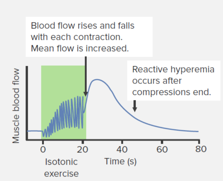
Effects of isotonic Isotonic Solutions having the same osmotic pressure as blood serum, or another solution with which they are compared. Renal Sodium and Water Regulation muscle contraction

  • Contractions are intermittent but repetitive.
  • During the brief contraction, effects are the same as those with isometric contractions:
  • Between each contraction:
    • Blood flow Blood flow Blood flow refers to the movement of a certain volume of blood through the vasculature over a given unit of time (e.g., mL per minute). Vascular Resistance, Flow, and Mean Arterial Pressure
    • Mean flow Flow Blood flows through the heart, arteries, capillaries, and veins in a closed, continuous circuit. Flow is the movement of volume per unit of time. Flow is affected by the pressure gradient and the resistance fluid encounters between 2 points. Vascular resistance is the opposition to flow, which is caused primarily by blood friction against vessel walls. Vascular Resistance, Flow, and Mean Arterial Pressure increases (to a point) with each successive contraction.
    • Peak flow Flow Blood flows through the heart, arteries, capillaries, and veins in a closed, continuous circuit. Flow is the movement of volume per unit of time. Flow is affected by the pressure gradient and the resistance fluid encounters between 2 points. Vascular resistance is the opposition to flow, which is caused primarily by blood friction against vessel walls. Vascular Resistance, Flow, and Mean Arterial Pressure increases (to a point) with each successive contraction.
  • Active hyperemia occurs after contractions end.
Changes in blood flow to the muscle during and after isotonic resistance exercise

Changes in blood flow to the muscle during and after isotonic resistance exercise

Image by Lecturio.
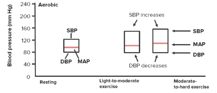
Effects of resistance Resistance Physiologically, the opposition to flow of air caused by the forces of friction. As a part of pulmonary function testing, it is the ratio of driving pressure to the rate of air flow. Ventilation: Mechanics of Breathing exercise on arterial blood pressure

  • With increasing muscle activity, perfusion must increase to meet metabolic demands.
  • The body ↑ blood pressure to increase perfusion via:
    • ↑ Systolic blood pressure
    • ↑ Diastolic blood pressure (DBP)
    • Mean arterial pressure Mean Arterial Pressure Mean arterial pressure (MAP) is the average systemic arterial pressure and is directly related to cardiac output (CO) and systemic vascular resistance (SVR). The SVR and MAP are affected by the vascular anatomy as well as a number of local and neurohumoral factors. Vascular Resistance, Flow, and Mean Arterial Pressure (MAP)
  • Mechanisms of increase in blood pressure:
    • Central command: Higher centers in the CNS initiate changes in anticipation Anticipation The apparent tendency of certain diseases to appear at earlier age of onset and with increasing severity in successive generations. Huntington Disease of exercise, providing the drive for movement.
    • Sensory Sensory Neurons which conduct nerve impulses to the central nervous system. Nervous System: Histology feedback:
      • Baroreceptors Baroreceptors Receptors in the vascular system, particularly the aorta and carotid sinus, which are sensitive to stretch of the vessel walls. Diabetes Insipidus: Note changes in blood pressure.
      • Chemoreceptors: Note changes in PCO2.
      • Muscle afferent Afferent Neurons which conduct nerve impulses to the central nervous system. Nervous System: Histology nerves: Note changes in metabolites (e.g., H+ ions).
    • Cardiorespiratory control center in the brainstem integrates central and sensory Sensory Neurons which conduct nerve impulses to the central nervous system. Nervous System: Histology signals to coordinate a sympathetic response.
Changes in systolic, diastolic, and mean arterial pressures

Changes in systolic, diastolic, and mean arterial pressures with increasing physical exertion
SBP = systolic blood pressure
DBP = diastolic blood pressure
MAP = mean arterial pressure

Image by Lecturio.

Effects of Valsalva maneuver Valsalva maneuver Forced expiratory effort against a closed glottis. Rectal Prolapse

Valsalva: forced expiration Forced expiration Ventilation: Mechanics of Breathing against a closed glottis Glottis The vocal apparatus of the larynx, situated in the middle section of the larynx. Glottis consists of the vocal folds and an opening (rima glottidis) between the folds. Larynx: Anatomy (occurs frequently during exercise). Valsalva causes 4 different changes in blood pressure over time:
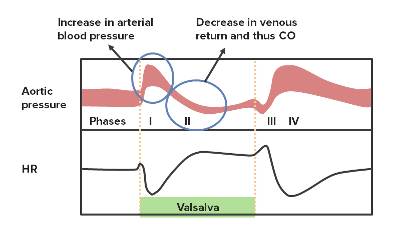
  • Phase 1 Phase 1 Skin: Structure and Functions: ↑ in blood pressure and ↓ in HR:
    • Forced expiration Forced expiration Ventilation: Mechanics of Breathing without significant expulsion of air → significantly ↑ intrathoracic pressure
    • Compression Compression Blunt Chest Trauma of the thoracic aorta Aorta The main trunk of the systemic arteries. Mediastinum and Great Vessels: Anatomy → ↑ arterial blood pressure
    • Baroreflex Baroreflex A response by the baroreceptors to increased blood pressure. Increased pressure stretches blood vessels which activates the baroreceptors in the vessel walls. The net response of the central nervous system is a reduction of central sympathetic outflow. This reduces blood pressure both by decreasing peripheral vascular resistance and by lowering cardiac output. Because the baroreceptors are tonically active, the baroreflex can compensate rapidly for both increases and decreases in blood pressure. Vascular Resistance, Flow, and Mean Arterial Pressure senses ↑ in blood pressure → ↓ HR in an attempt to maintain homeostasis Homeostasis The processes whereby the internal environment of an organism tends to remain balanced and stable. Cell Injury and Death of perfusion
  • Phase 2 Phase 2 Skin: Structure and Functions: ↓ blood pressure and ↑ HR:
    • ↑ Intrathoracic pressure → impedes venous return to the thorax
    • → ↓ Cardiac filling and ↓ preload Preload Cardiac Mechanics → ↓ CO → ↓ blood pressure
    • Baroreflex Baroreflex A response by the baroreceptors to increased blood pressure. Increased pressure stretches blood vessels which activates the baroreceptors in the vessel walls. The net response of the central nervous system is a reduction of central sympathetic outflow. This reduces blood pressure both by decreasing peripheral vascular resistance and by lowering cardiac output. Because the baroreceptors are tonically active, the baroreflex can compensate rapidly for both increases and decreases in blood pressure. Vascular Resistance, Flow, and Mean Arterial Pressure senses ↓ in blood pressure → ↑ HR in an attempt to maintain perfusion homeostasis Homeostasis The processes whereby the internal environment of an organism tends to remain balanced and stable. Cell Injury and Death
    • SNS activated → ↑ SVR attempting to stabilize blood pressure/perfusion ( plateau Plateau Cardiac Physiology portion of phase 2 Phase 2 Skin: Structure and Functions)
  • Phase 3 Phase 3 Skin: Structure and Functions (release of Valsalva):
    • External thoracic compression Compression Blunt Chest Trauma is removed → aortic pressure Aortic pressure Cardiac Mechanics briefly drops again
    • Baroreflex Baroreflex A response by the baroreceptors to increased blood pressure. Increased pressure stretches blood vessels which activates the baroreceptors in the vessel walls. The net response of the central nervous system is a reduction of central sympathetic outflow. This reduces blood pressure both by decreasing peripheral vascular resistance and by lowering cardiac output. Because the baroreceptors are tonically active, the baroreflex can compensate rapidly for both increases and decreases in blood pressure. Vascular Resistance, Flow, and Mean Arterial Pressure ↑ HR
  • Phase 4:
    • External thoracic compression Compression Blunt Chest Trauma is removed → resumption of venous return to the thorax
    • Rapid ventricular filling Ventricular filling Cardiac Cycle → ↑ preload Preload Cardiac Mechanics → ↑ CO → ↑ blood pressure
    • Baroreflex Baroreflex A response by the baroreceptors to increased blood pressure. Increased pressure stretches blood vessels which activates the baroreceptors in the vessel walls. The net response of the central nervous system is a reduction of central sympathetic outflow. This reduces blood pressure both by decreasing peripheral vascular resistance and by lowering cardiac output. Because the baroreceptors are tonically active, the baroreflex can compensate rapidly for both increases and decreases in blood pressure. Vascular Resistance, Flow, and Mean Arterial Pressure ↓ in HR
Phases of the valsalva maneuver with their corresponding changes in hr

Phases of the Valsalva maneuver with their corresponding changes in HR
CO: cardiac output
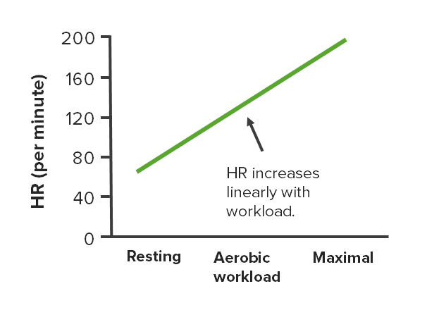
Image by Lecturio.

Cardiovascular Changes That Occur During Endurance (Aerobic) Exercises

Overview

  • Aerobic exercise ↑ total body O2 consumption
  • Body responds by ↑ perfusion to meet metabolic demands
  • Body achieves the response by:
    • ↑ CO through:
      • ↑ HR
      • Stroke volume Stroke volume The amount of blood pumped out of the heart per beat, not to be confused with cardiac output (volume/time). It is calculated as the difference between the end-diastolic volume and the end-systolic volume. Cardiac Cycle
      • Remember: CO = HR x stroke volume Stroke volume The amount of blood pumped out of the heart per beat, not to be confused with cardiac output (volume/time). It is calculated as the difference between the end-diastolic volume and the end-systolic volume. Cardiac Cycle
    • ↑ Systolic blood pressure
    • Redirecting blood flow Blood flow Blood flow refers to the movement of a certain volume of blood through the vasculature over a given unit of time (e.g., mL per minute). Vascular Resistance, Flow, and Mean Arterial Pressure to actively contracting muscles

Increases in HR

  • As workload ↑, the HR ↑
  • The relationship is linear.
  • Due to ↑ sympathetic stimulation at the sinoatrial node Sinoatrial node The small mass of modified cardiac muscle fibers located at the junction of the superior vena cava and right atrium. Contraction impulses probably start in this node, spread over the atrium (heart atrium) and are then transmitted by the atrioventricular bundle (bundle of His) to the ventricle (heart ventricle). Heart: Anatomy
Changes in hr at different intensities of aerobic exercise

Changes in HR at different intensities of aerobic exercise

Image by Lecturio.
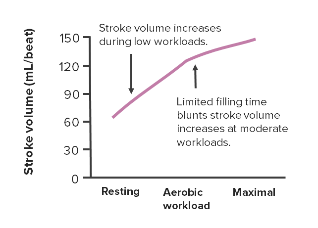
Increases in stroke volume Stroke volume The amount of blood pumped out of the heart per beat, not to be confused with cardiac output (volume/time). It is calculated as the difference between the end-diastolic volume and the end-systolic volume. Cardiac Cycle

  • As workload ↑, the stroke volume Stroke volume The amount of blood pumped out of the heart per beat, not to be confused with cardiac output (volume/time). It is calculated as the difference between the end-diastolic volume and the end-systolic volume. Cardiac Cycle
  • Relationship is linear to a point and then the curve flattens out.
  • Initial steep ↑ in stroke volume Stroke volume The amount of blood pumped out of the heart per beat, not to be confused with cardiac output (volume/time). It is calculated as the difference between the end-diastolic volume and the end-systolic volume. Cardiac Cycle is due to:
    • ↑ Sympathetic stimulation causing ↑ contractile strength of the heart (i.e., ↑ inotropy)
    • ↑ Venous return to the heart → ↑ preload Preload Cardiac Mechanics → ↑ stroke volume Stroke volume The amount of blood pumped out of the heart per beat, not to be confused with cardiac output (volume/time). It is calculated as the difference between the end-diastolic volume and the end-systolic volume. Cardiac Cycle
  • Flattening of the curve: as HR ↑, there is less time for ventricular filling Ventricular filling Cardiac Cycle → difficult to achieve higher end-diastolic volumes → less preload Preload Cardiac Mechanics → less ↑ in stroke volume Stroke volume The amount of blood pumped out of the heart per beat, not to be confused with cardiac output (volume/time). It is calculated as the difference between the end-diastolic volume and the end-systolic volume. Cardiac Cycle
Changes in stroke volume at different intensities of aerobic exercise

Changes in stroke volume at different intensities of aerobic exercise

Image by Lecturio.
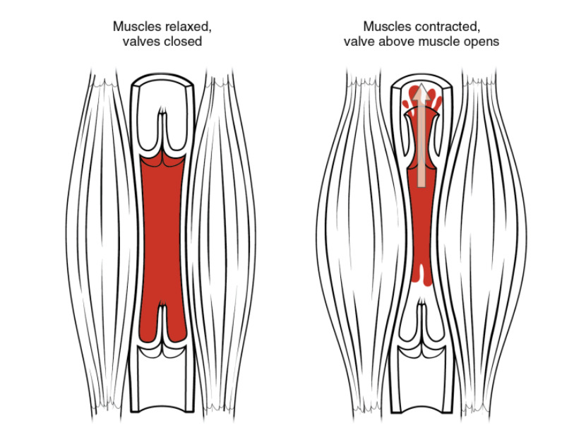
Effects of the skeletal muscle pump Pump ACES and RUSH: Resuscitation Ultrasound Protocols

Skeletal muscle pump

Skeletal muscle pump: As skeletal muscles surrounding a vein contract, the vessel is compressed, forcing blood to move forward. The 1-way valves in the veins prevent backflow and ensure that blood flows only in 1 direction.

Image by Lecturio.

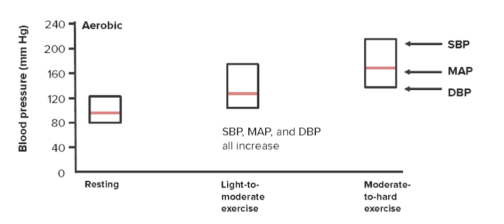
Changes in blood pressure

With aerobic exercise:

  • Systolic blood pressure increases:
    • Due to a sympathetic response
    • Via α-adrenergic receptors Receptors Receptors are proteins located either on the surface of or within a cell that can bind to signaling molecules known as ligands (e.g., hormones) and cause some type of response within the cell. Receptors
  • DBP remains constant or decreases slightly:
  • MAP increases slightly:
    • MAP = (CO x SVR) + central venous pressure Central venous pressure The blood pressure in the central large veins of the body. It is distinguished from peripheral venous pressure which occurs in an extremity. Central Venous Catheter ( CVP CVP The blood pressure in the central large veins of the body. It is distinguished from peripheral venous pressure which occurs in an extremity. Central Venous Catheter) (note: CVP CVP The blood pressure in the central large veins of the body. It is distinguished from peripheral venous pressure which occurs in an extremity. Central Venous Catheter is close to 0 and often disregarded.)
    • MAP can be approximated using systolic blood pressure and DBP:
      • As the heart spends more time in diastole Diastole Post-systolic relaxation of the heart, especially the heart ventricles. Cardiac Cycle than systole Systole Period of contraction of the heart, especially of the heart ventricles. Cardiac Cycle, DBP contributes more to MAP than systolic blood pressure.
      • MAP ≅ [⅓ (systolic blood pressure ‒ DBP) ] + DBP
    • ↑ In systolic blood pressure with minimal change in DBP = slight increase in MAP
Changes to cardiovascular parameters

Changes to cardiovascular parameters at different intensities of resistance exercise
SBP: systolic blood pressure
MAP: mean arterial pressure
DBP: diastolic blood pressure
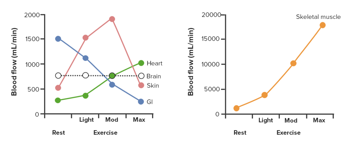
Image by Lecturio.

Redistribution of blood flow Blood flow Blood flow refers to the movement of a certain volume of blood through the vasculature over a given unit of time (e.g., mL per minute). Vascular Resistance, Flow, and Mean Arterial Pressure

  • Different vascular beds Vascular beds Gas Exchange are able to vasoconstrict and/or vasodilate to redistribute blood flow Blood flow Blood flow refers to the movement of a certain volume of blood through the vasculature over a given unit of time (e.g., mL per minute). Vascular Resistance, Flow, and Mean Arterial Pressure to actively contracting skeletal muscles Skeletal muscles A subtype of striated muscle, attached by tendons to the skeleton. Skeletal muscles are innervated and their movement can be consciously controlled. They are also called voluntary muscles. Muscle Tissue: Histology during aerobic exercise.
  • Effects may change in a given tissue depending on the intensity of exercise.
  • Effects on major vascular beds Vascular beds Gas Exchange:
    • Flow Flow Blood flows through the heart, arteries, capillaries, and veins in a closed, continuous circuit. Flow is the movement of volume per unit of time. Flow is affected by the pressure gradient and the resistance fluid encounters between 2 points. Vascular resistance is the opposition to flow, which is caused primarily by blood friction against vessel walls. Vascular Resistance, Flow, and Mean Arterial Pressure to the skeletal muscle
    • Flow Flow Blood flows through the heart, arteries, capillaries, and veins in a closed, continuous circuit. Flow is the movement of volume per unit of time. Flow is affected by the pressure gradient and the resistance fluid encounters between 2 points. Vascular resistance is the opposition to flow, which is caused primarily by blood friction against vessel walls. Vascular Resistance, Flow, and Mean Arterial Pressure to the heart (though to a lesser extent than to the skeletal muscle)
    • Flow Flow Blood flows through the heart, arteries, capillaries, and veins in a closed, continuous circuit. Flow is the movement of volume per unit of time. Flow is affected by the pressure gradient and the resistance fluid encounters between 2 points. Vascular resistance is the opposition to flow, which is caused primarily by blood friction against vessel walls. Vascular Resistance, Flow, and Mean Arterial Pressure to the brain Brain The part of central nervous system that is contained within the skull (cranium). Arising from the neural tube, the embryonic brain is comprised of three major parts including prosencephalon (the forebrain); mesencephalon (the midbrain); and rhombencephalon (the hindbrain). The developed brain consists of cerebrum; cerebellum; and other structures in the brain stem. Nervous System: Anatomy, Structure, and Classification remains constant.
    • Flow Flow Blood flows through the heart, arteries, capillaries, and veins in a closed, continuous circuit. Flow is the movement of volume per unit of time. Flow is affected by the pressure gradient and the resistance fluid encounters between 2 points. Vascular resistance is the opposition to flow, which is caused primarily by blood friction against vessel walls. Vascular Resistance, Flow, and Mean Arterial Pressure to the kidneys Kidneys The kidneys are a pair of bean-shaped organs located retroperitoneally against the posterior wall of the abdomen on either side of the spine. As part of the urinary tract, the kidneys are responsible for blood filtration and excretion of water-soluble waste in the urine. Kidneys: Anatomy and GI tract
    • Flow Flow Blood flows through the heart, arteries, capillaries, and veins in a closed, continuous circuit. Flow is the movement of volume per unit of time. Flow is affected by the pressure gradient and the resistance fluid encounters between 2 points. Vascular resistance is the opposition to flow, which is caused primarily by blood friction against vessel walls. Vascular Resistance, Flow, and Mean Arterial Pressure to skin Skin The skin, also referred to as the integumentary system, is the largest organ of the body. The skin is primarily composed of the epidermis (outer layer) and dermis (deep layer). The epidermis is primarily composed of keratinocytes that undergo rapid turnover, while the dermis contains dense layers of connective tissue. Skin: Structure and Functions:
      • ↑ Initially to help dissipate heat Heat Inflammation generated during exercise (part of thermoregulation Thermoregulation Body temperature can be divided into external temperature, which involves the skin, and core temperature, which involves the CNS and viscera. While external temperature can be variable, the core temperature is maintained within a narrow range of 36.5-37.5ºC (97.7-99.5ºF). Body Temperature Regulation)
      • At maximal exercise, the body prioritizes perfusing the skeletal muscles Skeletal muscles A subtype of striated muscle, attached by tendons to the skeleton. Skeletal muscles are innervated and their movement can be consciously controlled. They are also called voluntary muscles. Muscle Tissue: Histology and heart over thermoregulation Thermoregulation Body temperature can be divided into external temperature, which involves the skin, and core temperature, which involves the CNS and viscera. While external temperature can be variable, the core temperature is maintained within a narrow range of 36.5-37.5ºC (97.7-99.5ºF). Body Temperature Regulation → ↓ perfusion to the skin Skin The skin, also referred to as the integumentary system, is the largest organ of the body. The skin is primarily composed of the epidermis (outer layer) and dermis (deep layer). The epidermis is primarily composed of keratinocytes that undergo rapid turnover, while the dermis contains dense layers of connective tissue. Skin: Structure and Functions
Changes in blood flow distribution during light, moderate (mod), and maximal (max) exercise

Changes in blood flow distribution during light, moderate (mod), and maximal (max) exercise

Image by Lecturio.

Effects of body posture on cardiovascular parameters

Table: Relative effects of body posture on cardiovascular parameters: supine versus upright
Posture Supine (e.g., swimming) Upright
Effect on preload Preload Cardiac Mechanics and stroke volume Stroke volume The amount of blood pumped out of the heart per beat, not to be confused with cardiac output (volume/time). It is calculated as the difference between the end-diastolic volume and the end-systolic volume. Cardiac Cycle Higher Lower
Effect on resting HR Lower Higher

Effects of Chronic Endurance Training (Over Time)

Effects on HR, stroke volume Stroke volume The amount of blood pumped out of the heart per beat, not to be confused with cardiac output (volume/time). It is calculated as the difference between the end-diastolic volume and the end-systolic volume. Cardiac Cycle, and CO at rest and during maximal workload

Regular Regular Insulin aerobic exercise improves cardiovascular health by ↓ HR, ↑ stroke volume Stroke volume The amount of blood pumped out of the heart per beat, not to be confused with cardiac output (volume/time). It is calculated as the difference between the end-diastolic volume and the end-systolic volume. Cardiac Cycle, and ↑ CO (during exercise)

Table: Effects of chronic endurance exercise on HR, stroke volume Stroke volume The amount of blood pumped out of the heart per beat, not to be confused with cardiac output (volume/time). It is calculated as the difference between the end-diastolic volume and the end-systolic volume. Cardiac Cycle, and CO
At rest During maximal workload
HR Decreases Minimal change or slightly reduced to allow for longer filling time
Stroke volume Stroke volume The amount of blood pumped out of the heart per beat, not to be confused with cardiac output (volume/time). It is calculated as the difference between the end-diastolic volume and the end-systolic volume. Cardiac Cycle Increases Increases:
  • Growth of the ventricular wall → stronger contractions
  • ↑ Filling → ↑ preload Preload Cardiac Mechanics → ↑ stroke volume Stroke volume The amount of blood pumped out of the heart per beat, not to be confused with cardiac output (volume/time). It is calculated as the difference between the end-diastolic volume and the end-systolic volume. Cardiac Cycle
Cardiac output Cardiac output The volume of blood passing through the heart per unit of time. It is usually expressed as liters (volume) per minute so as not to be confused with stroke volume (volume per beat). Cardiac Mechanics Minimal change Increased due to ↑ stroke volume Stroke volume The amount of blood pumped out of the heart per beat, not to be confused with cardiac output (volume/time). It is calculated as the difference between the end-diastolic volume and the end-systolic volume. Cardiac Cycle

Other vascular adaptations

Other vascular adaptations that occur with chronic endurance training:

  • ↑ Myocardial vascularization via:
    • ↑ Cross-sectional area of coronary vessels through remodeling
    • ↑ Collateral circulation Circulation The movement of the blood as it is pumped through the cardiovascular system. ABCDE Assessment
  • ↑ Capillary number and density in skeletal muscle
  • Afterload Afterload Afterload is the resistance in the aorta that prevents blood from leaving the heart. Afterload represents the pressure the LV needs to overcome to eject blood into the aorta. Cardiac Mechanics (↓ SVR)
    • Systemic decrease in blood pressure
    • Better vasodilation Vasodilation The physiological widening of blood vessels by relaxing the underlying vascular smooth muscle. Pulmonary Hypertension Drugs of active muscles
    • Better shunting of blood away from nonactive regions

References

  1. Nystoriak, M., Bhatnagar, A. (2018). Cardiovascular effects and benefits of exercise. Front. Cardiovasc. Med. Retrieved Nov 16, 2021, from https://doi.org/10.3389/fcvm.2018.00135
  2. Klabunde, R. (2014). Hemodynamics of a Valsalva Maneuver. Cardiovascular Physiology Concepts. Retrieved Nov 16, 2021, from https://www.cvphysiology.com/Hemodynamics/H014
  3. Klabunde, R. (2020). Skeletal muscle blood flow. Cardiovascular Physiology Concepts. Retrieved Nov 16, 2021, from https://www.cvphysiology.com/Blood%20Flow/BF015
  4. Klabunde, R. (2007). Active hyperemia. Cardiovascular Physiology Concepts. Retrieved Nov 16, 2021, from https://www.cvphysiology.com/Blood%20Flow/BF015
  5. Cornelissen, V., Fagard, R. (2005). Effects of endurance training on blood pressure, blood pressure-regulating mechanisms, and cardiovascular risk factors. Hypertension. 46, 667–675. https://www.ahajournals.org/doi/full/10.1161/01.hyp.0000184225.05629.51
Day
Hrs
Min
Sec
<div class="expire-message">Extended</div>
Last Chance
for the
Back to School Deal!

Create your free account or log in to continue reading!

Sign up now and get free access to Lecturio with concept pages, medical videos, and questions for your medical education.

User Reviews

Details