The primary goals of antepartum testing and monitoring are to assess fetal well-being, identify treatable situations that may cause complications, and evaluate for chromosomal abnormalities. Antepartum tests for aneuploidy are typically performed in the 1st and 2nd trimesters. These tests are divided into screeningScreeningPreoperative Care tests (which include cell-free DNADNAA deoxyribonucleotide polymer that is the primary genetic material of all cells. Eukaryotic and prokaryotic organisms normally contain DNA in a double-stranded state, yet several important biological processes transiently involve single-stranded regions. DNA, which consists of a polysugar-phosphate backbone possessing projections of purines (adenine and guanine) and pyrimidines (thymine and cytosine), forms a double helix that is held together by hydrogen bonds between these purines and pyrimidines (adenine to thymine and guanine to cytosine).DNA Types and Structure testing, serum analyteAnalyteThe molecule of interest (antigen)Immunoassays testing, and nuchal translucencyNuchal translucencyA prenatal ultrasonography measurement of the soft tissue behind the fetal neck. Either the translucent area below the skin in the back of the fetal neck (nuchal translucency) or the distance between occipital bone to the outer skin line (nuchal fold) is measured.Obstetric Imaging measurements), and diagnostic testsDiagnostic testsDiagnostic tests are important aspects in making a diagnosis. Some of the most important epidemiological values of diagnostic tests include sensitivity and specificity, false positives and false negatives, positive and negative predictive values, likelihood ratios, and pre-test and post-test probabilities. Epidemiological Values of Diagnostic Tests, which provide a definitive diagnosis of aneuploidy and include chorionic villus sampling (CVS) and amniocentesisAmniocentesisPercutaneous transabdominal puncture of the uterus during pregnancy to obtain amniotic fluid. It is commonly used for fetal karyotype determination in order to diagnose abnormal fetal conditions.Polyhydramnios. Antepartum monitoring is done in the 2nd and 3rd trimesters to evaluate fetal well-being and assess the risk of fetal death. Antepartum monitoring tests include continuous cardiotocography (i.e., fetal heart rateHeart rateThe number of times the heart ventricles contract per unit of time, usually per minute.Cardiac Physiology tracings), the non-stress and contraction stress tests (NST and CST, respectively), the fetal biophysical profileBiophysical ProfileObstetric Imaging (BPP), and umbilical arteryUmbilical arterySpecialized arterial vessels in the umbilical cord. They carry waste and deoxygenated blood from the fetus to the mother via the placenta. In humans, there are usually two umbilical arteries but sometimes one.Prenatal and Postnatal Physiology of the NeonateDopplerDopplerUltrasonography applying the doppler effect, with frequency-shifted ultrasound reflections produced by moving targets (usually red blood cells) in the bloodstream along the ultrasound axis in direct proportion to the velocity of movement of the targets, to determine both direction and velocity of blood flow.Ultrasound (Sonography) studies.
Diagnostic testsDiagnostic testsDiagnostic tests are important aspects in making a diagnosis. Some of the most important epidemiological values of diagnostic tests include sensitivity and specificity, false positives and false negatives, positive and negative predictive values, likelihood ratios, and pre-test and post-test probabilities. Epidemiological Values of Diagnostic Tests: invasive tests that involve obtaining a sample of material from inside the womb that contains fetal DNADNAA deoxyribonucleotide polymer that is the primary genetic material of all cells. Eukaryotic and prokaryotic organisms normally contain DNA in a double-stranded state, yet several important biological processes transiently involve single-stranded regions. DNA, which consists of a polysugar-phosphate backbone possessing projections of purines (adenine and guanine) and pyrimidines (thymine and cytosine), forms a double helix that is held together by hydrogen bonds between these purines and pyrimidines (adenine to thymine and guanine to cytosine).DNA Types and Structure
Should be offered to all pregnant women to assess the risk for genetic conditions (some may decline)
Antepartum monitoring
Refers to a variety of tests used to evaluate fetal well-being and assess the risk of intrauterine fetal hypoxic injury or death
The most common (and most clinically important) monitoring tests include:
Continuous cardiotocography (also known simply as “continuous fetal monitoring”)
Umbilical arteryUmbilical arterySpecialized arterial vessels in the umbilical cord. They carry waste and deoxygenated blood from the fetus to the mother via the placenta. In humans, there are usually two umbilical arteries but sometimes one.Prenatal and Postnatal Physiology of the NeonateDopplerDopplerUltrasonography applying the doppler effect, with frequency-shifted ultrasound reflections produced by moving targets (usually red blood cells) in the bloodstream along the ultrasound axis in direct proportion to the velocity of movement of the targets, to determine both direction and velocity of blood flow.Ultrasound (Sonography)
Preterm laborPreterm laborPreterm labor refers to regular uterine contractions leading to cervical change prior to 37 weeks of gestation; preterm birth refers to birth prior to 37 weeks of gestation. Preterm birth may be spontaneous due to preterm labor, preterm prelabor rupture of membranes (PPROM), or cervical insufficiency. Preterm Labor and Birth and/or preterm prematurePrematureChildbirth before 37 weeks of pregnancy (259 days from the first day of the mother’s last menstrual period, or 245 days after fertilization).Necrotizing Enterocolitis rupture of membranes (PPROM)
Prior fetal death
Preexisting or gestational diabetesDiabetesDiabetes mellitus (DM) is a metabolic disease characterized by hyperglycemia and dysfunction of the regulation of glucose metabolism by insulin. Type 1 DM is diagnosed mostly in children and young adults as the result of autoimmune destruction of β cells in the pancreas and the resulting lack of insulin. Type 2 DM has a significant association with obesity and is characterized by insulin resistance.Diabetes Mellitus
Chronic or pregnancy-related hypertensionHypertensionHypertension, or high blood pressure, is a common disease that manifests as elevated systemic arterial pressures. Hypertension is most often asymptomatic and is found incidentally as part of a routine physical examination or during triage for an unrelated medical encounter. Hypertension (e.g., preeclampsiaPreeclampsiaA complication of pregnancy, characterized by a complex of symptoms including maternal hypertension and proteinuria with or without pathological edema. Symptoms may range between mild and severe. Pre-eclampsia usually occurs after the 20th week of gestation, but may develop before this time in the presence of trophoblastic disease.Hypertensive Pregnancy Disorders)
Alloimmunization to RBC antigens
Intrahepatic cholestasis of pregnancyPregnancyThe status during which female mammals carry their developing young (embryos or fetuses) in utero before birth, beginning from fertilization to birth.Pregnancy: Diagnosis, Physiology, and Care (ICPICPNormal intracranial pressure (ICP) is defined as < 15 mm Hg, whereas pathologically increased ICP is any pressure ≥ 20 mm Hg. Increased ICP may result from several etiologies, including trauma, intracranial hemorrhage, mass lesions, cerebral edema, increased CSF production, and decreased CSF absorption.Increased Intracranial Pressure (ICP))
Other preexisting medical conditions, including (but not limited to):
Systemic lupus erythematosusSystemic lupus erythematosusSystemic lupus erythematosus (SLE) is a chronic autoimmune, inflammatory condition that causes immune-complex deposition in organs, resulting in systemic manifestations. Women, particularly those of African American descent, are more commonly affected.Systemic Lupus Erythematosus (SLESLESystemic lupus erythematosus (SLE) is a chronic autoimmune, inflammatory condition that causes immune-complex deposition in organs, resulting in systemic manifestations. Women, particularly those of African American descent, are more commonly affected.Systemic Lupus Erythematosus) and/or chronic renal disease
Antiphospholipid syndromeAntiphospholipid syndromeAntiphospholipid syndrome (APLS) is an acquired autoimmune disorder characterized by the persistent presence of antiphospholipid antibodies, which create a hypercoagulable state. These antibodies are most commonly discovered during a workup for a thrombotic event or recurrent pregnancy loss, which are the 2 most common clinical manifestations.Antiphospholipid Syndrome
Maternal cyanotic heart disease
HemoglobinopathiesHemoglobinopathiesA group of inherited disorders characterized by structural alterations within the hemoglobin molecule.Anemia: Overview and Types (e.g., sickle cell diseaseSickle cell diseaseSickle cell disease (SCD) is a group of genetic disorders in which an abnormal Hb molecule (HbS) transforms RBCs into sickle-shaped cells, resulting in chronic anemia, vasoocclusive episodes, pain, and organ damage.Sickle Cell Disease)
Fetal indications
Multiple gestations (e.g., twin pregnancies)
Postterm pregnancyPregnancyThe status during which female mammals carry their developing young (embryos or fetuses) in utero before birth, beginning from fertilization to birth.Pregnancy: Diagnosis, Physiology, and Care
Fetal growth restrictionFetal growth restrictionFetal growth restriction (FGR), also known as intrauterine fetal growth restriction (IUGR), is an estimated fetal weight (EFW) or abdominal circumference < 10th percentile for gestational age. The term small for gestational age (SGA) is sometimes erroneously used interchangeably with FGR. Fetal Growth Restriction
OligohydramniosOligohydramniosOligohydramnios refers to amniotic fluid volume less than expected for the current gestational age. Oligohydramnios is diagnosed by ultrasound and defined as an amniotic fluid index (AFI) of ‰¤ 5 cm or a single deep pocket (SDP) of < 2 cm in the 2nd or 3rd trimester. Oligohydramnios
Decreased fetal movement (detected by mother)
Screening Tests
Routine pregnancyPregnancyThe status during which female mammals carry their developing young (embryos or fetuses) in utero before birth, beginning from fertilization to birth.Pregnancy: Diagnosis, Physiology, and Care laboratory and imaging studies
All pregnant women should have certain screeningScreeningPreoperative Care laboratory tests and imaging studies done at different points during the pregnancyPregnancyThe status during which female mammals carry their developing young (embryos or fetuses) in utero before birth, beginning from fertilization to birth.Pregnancy: Diagnosis, Physiology, and Care, including:
At the 1st OB appointment:
ScreeningScreeningPreoperative Care for anemiaAnemiaAnemia is a condition in which individuals have low Hb levels, which can arise from various causes. Anemia is accompanied by a reduced number of RBCs and may manifest with fatigue, shortness of breath, pallor, and weakness. Subtypes are classified by the size of RBCs, chronicity, and etiology. Anemia: Overview and Types: CBC
UrinalysisUrinalysisExamination of urine by chemical, physical, or microscopic means. Routine urinalysis usually includes performing chemical screening tests, determining specific gravity, observing any unusual color or odor, screening for bacteriuria, and examining the sediment microscopically.Urinary Tract Infections (UTIs) in Children
RubellaRubellaAn acute infectious disease caused by the rubella virus. The virus enters the respiratory tract via airborne droplet and spreads to the lymphatic system.Rubella Virus immunity status
Hepatitis B surface antigenHepatitis B surface antigenThose hepatitis B antigens found on the surface of the dane particle and on the 20 nm spherical and tubular particles. Several subspecificities of the surface antigen are known. These were formerly called the Australia antigen.Hepatitis B Virus
Hepatitis CHepatitis CHepatitis C is an infection of the liver caused by the hepatitis C virus (HCV). The infection can be transmitted through infectious blood or body fluids and may be transmitted during childbirth or through IV drug use or sexual intercourse. Hepatitis C virus can cause both acute and chronic hepatitis, ranging from a mild to a serious, lifelong illness including liver cirrhosis and hepatocellular carcinoma (HCC).Hepatitis C Virus antibody
Rapid plasma reagin testRapid plasma reagin testTreponema for syphilisSyphilisSyphilis is a bacterial infection caused by the spirochete Treponema pallidum pallidum (T. p. pallidum), which is usually spread through sexual contact. Syphilis has 4 clinical stages: primary, secondary, latent, and tertiary. Syphilis
GonorrheaGonorrheaGonorrhea is a sexually transmitted infection (STI) caused by the gram-negative bacteria Neisseria gonorrhoeae (N. gonorrhoeae). Gonorrhea may be asymptomatic but commonly manifests as cervicitis or urethritis with less common presentations such as proctitis, conjunctivitis, or pharyngitis. Gonorrhea and chlamydiaChlamydiaChlamydiae are obligate intracellular gram-negative bacteria. They lack a peptidoglycan layer and are best visualized using Giemsa stain. The family of Chlamydiaceae comprises 3 pathogens that can infect humans: Chlamydia trachomatis, Chlamydia psittaci, and Chlamydia pneumoniae.Chlamydia testing
ScreeningScreeningPreoperative Care for aneuploidy (e.g., trisomy 21Trisomy 21Down syndrome, or trisomy 21, is the most common chromosomal aberration and the most frequent genetic cause of developmental delay. Both boys and girls are affected and have characteristic craniofacial and musculoskeletal features, as well as multiple medical anomalies involving the cardiac, gastrointestinal, ocular, and auditory systems.Down syndrome (Trisomy 21)) based on woman’s desire
Other testing
ScreeningScreeningPreoperative Care for anatomic issues: anatomic ultrasonography at 18‒22 weeks of gestational ageGestational ageThe age of the conceptus, beginning from the time of fertilization. In clinical obstetrics, the gestational age is often estimated as the time from the last day of the last menstruation which is about 2 weeks before ovulation and fertilization.Pregnancy: Diagnosis, Physiology, and Care (wga), which includes assessment of the fetus, placentaPlacentaA highly vascularized mammalian fetal-maternal organ and major site of transport of oxygen, nutrients, and fetal waste products. It includes a fetal portion (chorionic villi) derived from trophoblasts and a maternal portion (decidua) derived from the uterine endometrium. The placenta produces an array of steroid, protein and peptide hormones (placental hormones).Placenta, Umbilical Cord, and Amniotic Cavity, and uterusUterusThe uterus, cervix, and fallopian tubes are part of the internal female reproductive system. The uterus has a thick wall made of smooth muscle (the myometrium) and an inner mucosal layer (the endometrium). The most inferior portion of the uterus is the cervix, which connects the uterine cavity to the vagina.Uterus, Cervix, and Fallopian Tubes: Anatomy/cervixCervixThe uterus, cervix, and fallopian tubes are part of the internal female reproductive system. The most inferior portion of the uterus is the cervix, which connects the uterine cavity to the vagina. Externally, the cervix is lined by stratified squamous cells; however, the cervical canal is lined by columnar epithelium.Uterus, Cervix, and Fallopian Tubes: Anatomy
ScreeningScreeningPreoperative Care for gestational diabetesDiabetesDiabetes mellitus (DM) is a metabolic disease characterized by hyperglycemia and dysfunction of the regulation of glucose metabolism by insulin. Type 1 DM is diagnosed mostly in children and young adults as the result of autoimmune destruction of β cells in the pancreas and the resulting lack of insulin. Type 2 DM has a significant association with obesity and is characterized by insulin resistance.Diabetes Mellitus: 1-hour glucoseGlucoseA primary source of energy for living organisms. It is naturally occurring and is found in fruits and other parts of plants in its free state. It is used therapeutically in fluid and nutrient replacement.Lactose IntolerancetoleranceTolerancePharmacokinetics and Pharmacodynamics test (GTT) at 24‒28 wga
Repeat screeningScreeningPreoperative Care for anemiaAnemiaAnemia is a condition in which individuals have low Hb levels, which can arise from various causes. Anemia is accompanied by a reduced number of RBCs and may manifest with fatigue, shortness of breath, pallor, and weakness. Subtypes are classified by the size of RBCs, chronicity, and etiology. Anemia: Overview and Types: CBC is usually repeated with the GTT.
ScreeningScreeningPreoperative Care for group B streptococcusGroup B StreptococcusMeningitis in Children (GBSGBSAn acute inflammatory autoimmune neuritis caused by t cell- mediated cellular immune response directed towards peripheral myelin. Demyelination occurs in peripheral nerves and nerve roots. The process is often preceded by a viral or bacterial infection, surgery, immunization, lymphoma, or exposure to toxins. Common clinical manifestations include progressive weakness, loss of sensation, and loss of deep tendon reflexes. Weakness of respiratory muscles and autonomic dysfunction may occur.Polyneuropathy) colonizationColonizationBacteriology: GBSGBSAn acute inflammatory autoimmune neuritis caused by t cell- mediated cellular immune response directed towards peripheral myelin. Demyelination occurs in peripheral nerves and nerve roots. The process is often preceded by a viral or bacterial infection, surgery, immunization, lymphoma, or exposure to toxins. Common clinical manifestations include progressive weakness, loss of sensation, and loss of deep tendon reflexes. Weakness of respiratory muscles and autonomic dysfunction may occur.Polyneuropathy rectovaginal culture at 35‒37 wga
ScreeningScreeningPreoperative Care (noninvasive tests) indirectlyassesses the risk of fetal aneuploidy by testing maternal blood and/or by performing ultrasonography.
Multiple options available: combinations of different serum analytes and/or ultrasonography
Some tests can be performed only at specific gestational ages.
Chromosomal abnormality detection rates vary.
Results:
Need to be confirmed by diagnostic testing for definitive diagnosis
Cell-free fetal DNADNAA deoxyribonucleotide polymer that is the primary genetic material of all cells. Eukaryotic and prokaryotic organisms normally contain DNA in a double-stranded state, yet several important biological processes transiently involve single-stranded regions. DNA, which consists of a polysugar-phosphate backbone possessing projections of purines (adenine and guanine) and pyrimidines (thymine and cytosine), forms a double helix that is held together by hydrogen bonds between these purines and pyrimidines (adenine to thymine and guanine to cytosine).DNA Types and Structure testing (cf-DNA)
1st-trimester testing
Quadruple test
Integrated testing
Cell-free fetal DNADNAA deoxyribonucleotide polymer that is the primary genetic material of all cells. Eukaryotic and prokaryotic organisms normally contain DNA in a double-stranded state, yet several important biological processes transiently involve single-stranded regions. DNA, which consists of a polysugar-phosphate backbone possessing projections of purines (adenine and guanine) and pyrimidines (thymine and cytosine), forms a double helix that is held together by hydrogen bonds between these purines and pyrimidines (adenine to thymine and guanine to cytosine).DNA Types and Structure testing
Also known as noninvasive prenatal screeningScreeningPreoperative Care (NIPS) or noninvasive prenatal testing (NIPT)
Performed anytime after 10 wga
How it works:
ApoptosisApoptosisA regulated cell death mechanism characterized by distinctive morphologic changes in the nucleus and cytoplasm, including the endonucleolytic cleavage of genomic DNA, at regularly spaced, internucleosomal sites, I.e., DNA fragmentation. It is genetically-programmed and serves as a balance to mitosis in regulating the size of animal tissues and in mediating pathologic processes associated with tumor growth.Ischemic Cell Damage of placental cells (which contain fetal DNADNAA deoxyribonucleotide polymer that is the primary genetic material of all cells. Eukaryotic and prokaryotic organisms normally contain DNA in a double-stranded state, yet several important biological processes transiently involve single-stranded regions. DNA, which consists of a polysugar-phosphate backbone possessing projections of purines (adenine and guanine) and pyrimidines (thymine and cytosine), forms a double helix that is held together by hydrogen bonds between these purines and pyrimidines (adenine to thymine and guanine to cytosine).DNA Types and Structure) release fragments of fetal DNADNAA deoxyribonucleotide polymer that is the primary genetic material of all cells. Eukaryotic and prokaryotic organisms normally contain DNA in a double-stranded state, yet several important biological processes transiently involve single-stranded regions. DNA, which consists of a polysugar-phosphate backbone possessing projections of purines (adenine and guanine) and pyrimidines (thymine and cytosine), forms a double helix that is held together by hydrogen bonds between these purines and pyrimidines (adenine to thymine and guanine to cytosine).DNA Types and Structure into maternal serum.
These cell-free fetal DNADNAA deoxyribonucleotide polymer that is the primary genetic material of all cells. Eukaryotic and prokaryotic organisms normally contain DNA in a double-stranded state, yet several important biological processes transiently involve single-stranded regions. DNA, which consists of a polysugar-phosphate backbone possessing projections of purines (adenine and guanine) and pyrimidines (thymine and cytosine), forms a double helix that is held together by hydrogen bonds between these purines and pyrimidines (adenine to thymine and guanine to cytosine).DNA Types and Structure fragments (usually about 50‒200 base pairs) can be isolated from maternalblood for testing.
Amount of DNADNAA deoxyribonucleotide polymer that is the primary genetic material of all cells. Eukaryotic and prokaryotic organisms normally contain DNA in a double-stranded state, yet several important biological processes transiently involve single-stranded regions. DNA, which consists of a polysugar-phosphate backbone possessing projections of purines (adenine and guanine) and pyrimidines (thymine and cytosine), forms a double helix that is held together by hydrogen bonds between these purines and pyrimidines (adenine to thymine and guanine to cytosine).DNA Types and Structure from each fetal chromosomeChromosomeIn a prokaryotic cell or in the nucleus of a eukaryotic cell, a structure consisting of or containing DNA which carries the genetic information essential to the cell.Basic Terms of Genetics is determined → elevated proportions of genetic material from 1 chromosomeChromosomeIn a prokaryotic cell or in the nucleus of a eukaryotic cell, a structure consisting of or containing DNA which carries the genetic information essential to the cell.Basic Terms of Genetics strongly suggests triploidy
Has the highest sensitivity for detecting chromosomal abnormalities of all the screeningScreeningPreoperative Care tests:
Trisomy 21Trisomy 21Down syndrome, or trisomy 21, is the most common chromosomal aberration and the most frequent genetic cause of developmental delay. Both boys and girls are affected and have characteristic craniofacial and musculoskeletal features, as well as multiple medical anomalies involving the cardiac, gastrointestinal, ocular, and auditory systems.Down syndrome (Trisomy 21): > 99%
TrisomyTrisomyThe possession of a third chromosome of any one type in an otherwise diploid cell.Types of Mutations 18: 98%
TrisomyTrisomyThe possession of a third chromosome of any one type in an otherwise diploid cell.Types of Mutations 13: 96%
Still considered a screeningScreeningPreoperative Care test owing to lower positive predictive values, especially in younger women, in whom aneuploidies are much less common:
Positive predictive valuePositive predictive valueThe positive predictive value is the percentage of people with a positive test result who actually have the disease among all people with a positive result, regardless of whether or not they have the disease.Epidemiological Values of Diagnostic Tests for trisomy 21Trisomy 21Down syndrome, or trisomy 21, is the most common chromosomal aberration and the most frequent genetic cause of developmental delay. Both boys and girls are affected and have characteristic craniofacial and musculoskeletal features, as well as multiple medical anomalies involving the cardiac, gastrointestinal, ocular, and auditory systems.Down syndrome (Trisomy 21) in a 20-year-old: 48%
Positive predictive valuePositive predictive valueThe positive predictive value is the percentage of people with a positive test result who actually have the disease among all people with a positive result, regardless of whether or not they have the disease.Epidemiological Values of Diagnostic Tests for trisomy 21Trisomy 21Down syndrome, or trisomy 21, is the most common chromosomal aberration and the most frequent genetic cause of developmental delay. Both boys and girls are affected and have characteristic craniofacial and musculoskeletal features, as well as multiple medical anomalies involving the cardiac, gastrointestinal, ocular, and auditory systems.Down syndrome (Trisomy 21) in a 40-year-old: 93%
Diagnostic testing should be used to confirm diagnosis, especially in younger patientsPatientsIndividuals participating in the health care system for the purpose of receiving therapeutic, diagnostic, or preventive procedures.Clinician–Patient Relationship.
Note that fetal sexSexThe totality of characteristics of reproductive structure, functions, phenotype, and genotype, differentiating the male from the female organism.Gender Dysphoria can be determined using cell-free fetal DNADNAA deoxyribonucleotide polymer that is the primary genetic material of all cells. Eukaryotic and prokaryotic organisms normally contain DNA in a double-stranded state, yet several important biological processes transiently involve single-stranded regions. DNA, which consists of a polysugar-phosphate backbone possessing projections of purines (adenine and guanine) and pyrimidines (thymine and cytosine), forms a double helix that is held together by hydrogen bonds between these purines and pyrimidines (adenine to thymine and guanine to cytosine).DNA Types and Structure testing.
Trisomy 21Trisomy 21Down syndrome, or trisomy 21, is the most common chromosomal aberration and the most frequent genetic cause of developmental delay. Both boys and girls are affected and have characteristic craniofacial and musculoskeletal features, as well as multiple medical anomalies involving the cardiac, gastrointestinal, ocular, and auditory systems.Down syndrome (Trisomy 21) detection rate: 85%
Measures 3 markers:
Maternal serum β-hCG
Maternal serum pregnancy-associated plasmaPlasmaThe residual portion of blood that is left after removal of blood cells by centrifugation without prior blood coagulation.Transfusion Products protein A (PAPP-A)
Ultrasound measurement of the nuchal translucencyNuchal translucencyA prenatal ultrasonography measurement of the soft tissue behind the fetal neck. Either the translucent area below the skin in the back of the fetal neck (nuchal translucency) or the distance between occipital bone to the outer skin line (nuchal fold) is measured.Obstetric Imaging, a hypoechoicHypoechoicA structure that produces a low-amplitude echo (darker grays)Ultrasound (Sonography) space in the posterior aspect of the fetal neckNeckThe part of a human or animal body connecting the head to the rest of the body.Peritonsillar Abscess
Table: 1st-trimester screeningScreeningPreoperative Care test results associated with common aneuploidies
Condition
hCG
PAPP-A
Nuchal translucencyNuchal translucencyA prenatal ultrasonography measurement of the soft tissue behind the fetal neck. Either the translucent area below the skin in the back of the fetal neck (nuchal translucency) or the distance between occipital bone to the outer skin line (nuchal fold) is measured.Obstetric Imaging
Trisomy 21Trisomy 21Down syndrome, or trisomy 21, is the most common chromosomal aberration and the most frequent genetic cause of developmental delay. Both boys and girls are affected and have characteristic craniofacial and musculoskeletal features, as well as multiple medical anomalies involving the cardiac, gastrointestinal, ocular, and auditory systems.Down syndrome (Trisomy 21)
↑
↓↓
↑↑
TrisomyTrisomyThe possession of a third chromosome of any one type in an otherwise diploid cell.Types of Mutations 18
↓↓
↓↓
↑↑
TrisomyTrisomyThe possession of a third chromosome of any one type in an otherwise diploid cell.Types of Mutations 13
Done in the 2nd trimester between 15 and 22 ⁶⁄₇ wga
Screens for trisomies 21 and 18
Trisomy 21Trisomy 21Down syndrome, or trisomy 21, is the most common chromosomal aberration and the most frequent genetic cause of developmental delay. Both boys and girls are affected and have characteristic craniofacial and musculoskeletal features, as well as multiple medical anomalies involving the cardiac, gastrointestinal, ocular, and auditory systems.Down syndrome (Trisomy 21) detection rate: 80%
Uses serum markers, maternal age, weight, race, and presence of pregestational diabetesDiabetesDiabetes mellitus (DM) is a metabolic disease characterized by hyperglycemia and dysfunction of the regulation of glucose metabolism by insulin. Type 1 DM is diagnosed mostly in children and young adults as the result of autoimmune destruction of β cells in the pancreas and the resulting lack of insulin. Type 2 DM has a significant association with obesity and is characterized by insulin resistance.Diabetes Mellitus to calculate a risk estimate
Measures 4 serum markers:
Maternal serum β-hCG
Maternal serum alpha fetoproteinAlpha fetoproteinThe first alpha-globulins to appear in mammalian sera during fetal development and the dominant serum proteins in early embryonic life.Testicular Cancer (MS-AFP)
Unconjugated estriolEstriolA hydroxylated metabolite of estradiol or estrogen that has a hydroxyl group at C3, 16-alpha, and 17-beta position. Estriol is a major urinary estrogen. During pregnancy, a large amount of estriol is produced by the placenta. Isomers with inversion of the hydroxyl group or groups are called epiestriol.Noncontraceptive Estrogen and Progestins (uE3)
Inhibin AInhibin AGlycoproteins that inhibit pituitary follicle stimulating hormone secretion. Inhibins are secreted by the sertoli cells of the testes, the granulosa cells of the ovarian follicles, the placenta, and other tissues. Inhibins and activins are modulators of follicle stimulating hormone secretions; both groups belong to the TGF-beta superfamily, as the transforming growth factor beta. Inhibins consist of a disulfide-linked heterodimer with a unique alpha linked to either a beta a or a beta B subunit to form inhibin a or inhibin b, respectively.Menstrual Cycle
Does not require ultrasonography or nuchal translucencyNuchal translucencyA prenatal ultrasonography measurement of the soft tissue behind the fetal neck. Either the translucent area below the skin in the back of the fetal neck (nuchal translucency) or the distance between occipital bone to the outer skin line (nuchal fold) is measured.Obstetric Imaging measurement
EstriolEstriolA hydroxylated metabolite of estradiol or estrogen that has a hydroxyl group at C3, 16-alpha, and 17-beta position. Estriol is a major urinary estrogen. During pregnancy, a large amount of estriol is produced by the placenta. Isomers with inversion of the hydroxyl group or groups are called epiestriol.Noncontraceptive Estrogen and Progestins
Inhibin AInhibin AGlycoproteins that inhibit pituitary follicle stimulating hormone secretion. Inhibins are secreted by the sertoli cells of the testes, the granulosa cells of the ovarian follicles, the placenta, and other tissues. Inhibins and activins are modulators of follicle stimulating hormone secretions; both groups belong to the TGF-beta superfamily, as the transforming growth factor beta. Inhibins consist of a disulfide-linked heterodimer with a unique alpha linked to either a beta a or a beta B subunit to form inhibin a or inhibin b, respectively.Menstrual Cycle
Trisomy 21Trisomy 21Down syndrome, or trisomy 21, is the most common chromosomal aberration and the most frequent genetic cause of developmental delay. Both boys and girls are affected and have characteristic craniofacial and musculoskeletal features, as well as multiple medical anomalies involving the cardiac, gastrointestinal, ocular, and auditory systems.Down syndrome (Trisomy 21)
↑
↓
↓
↑
TrisomyTrisomyThe possession of a third chromosome of any one type in an otherwise diploid cell.Types of Mutations 18
↓↓
↓
↓↓
↔
Integrated testing:
Combines results from 1st- and 2nd-trimester testing
Results are used to calculate a single patient-specific risk for aneuploidies.
Detection rate for trisomy 21Trisomy 21Down syndrome, or trisomy 21, is the most common chromosomal aberration and the most frequent genetic cause of developmental delay. Both boys and girls are affected and have characteristic craniofacial and musculoskeletal features, as well as multiple medical anomalies involving the cardiac, gastrointestinal, ocular, and auditory systems.Down syndrome (Trisomy 21): 96%
Variations:
Full integrated testing: All serum analytes and nuchal translucencyNuchal translucencyA prenatal ultrasonography measurement of the soft tissue behind the fetal neck. Either the translucent area below the skin in the back of the fetal neck (nuchal translucency) or the distance between occipital bone to the outer skin line (nuchal fold) is measured.Obstetric Imaging are assessed.
Serum integrated screeningScreeningPreoperative Care: Only serum is analyzed (no nuchal translucencyNuchal translucencyA prenatal ultrasonography measurement of the soft tissue behind the fetal neck. Either the translucent area below the skin in the back of the fetal neck (nuchal translucency) or the distance between occipital bone to the outer skin line (nuchal fold) is measured.Obstetric Imaging assessment).
SequentialSequentialComputed Tomography (CT)screeningScreeningPreoperative Care: Initial risk assessmentRisk assessmentThe qualitative or quantitative estimation of the likelihood of adverse effects that may result from exposure to specified health hazards or from the absence of beneficial influences.Preoperative Care is provided after 1st-trimester tests and then revised after 2nd-trimester tests.
Diagnostic Tests
Diagnostic testing is invasive and involves sampling the chorionic villiChorionic villiThreadlike vascular projections of the chorion. Chorionic villi may be free or embedded within the decidua forming the site for exchange of substances between fetal and maternal blood (placenta).Placenta, Umbilical Cord, and Amniotic Cavity or amniotic fluidAmniotic fluidA clear, yellowish liquid that envelopes the fetus inside the sac of amnion. In the first trimester, it is likely a transudate of maternal or fetal plasma. In the second trimester, amniotic fluid derives primarily from fetal lung and kidney. Cells or substances in this fluid can be removed for prenatal diagnostic tests (amniocentesis).Placenta, Umbilical Cord, and Amniotic Cavity for fetal tissue that can be used directly for genetic testingGenetic TestingDetection of a mutation; genotype; karyotype; or specific alleles associated with genetic traits, heritable diseases, or predisposition to a disease, or that may lead to the disease in descendants. It includes prenatal genetic testing.Myotonic Dystrophies.
Chorionic villus sampling (CVS)
Procedure:
PlacentaPlacentaA highly vascularized mammalian fetal-maternal organ and major site of transport of oxygen, nutrients, and fetal waste products. It includes a fetal portion (chorionic villi) derived from trophoblasts and a maternal portion (decidua) derived from the uterine endometrium. The placenta produces an array of steroid, protein and peptide hormones (placental hormones).Placenta, Umbilical Cord, and Amniotic Cavity is biopsied under real-time ultrasound guidance.
Sample is tested for genetic anomalies (e.g., karyotypeKaryotypeThe full set of chromosomes presented as a systematized array of metaphase chromosomes from a photomicrograph of a single cell nucleus arranged in pairs in descending order of size and according to the position of the centromere.Congenital Malformations of the Female Reproductive System).
Can be performed transabdominally or transcervically
Timing: done between 10 and 13 wga
Indications:
Confirming abnormal screen results
Congenital anomaly seen on 1st-trimester ultrasound examination
Previous pregnancyPregnancyThe status during which female mammals carry their developing young (embryos or fetuses) in utero before birth, beginning from fertilization to birth.Pregnancy: Diagnosis, Physiology, and Care with a chromosomeChromosomeIn a prokaryotic cell or in the nucleus of a eukaryotic cell, a structure consisting of or containing DNA which carries the genetic information essential to the cell.Basic Terms of Genetics abnormality or genetic disorder
Known genetic anomalies (e.g., balanced translocation) in one of the parents
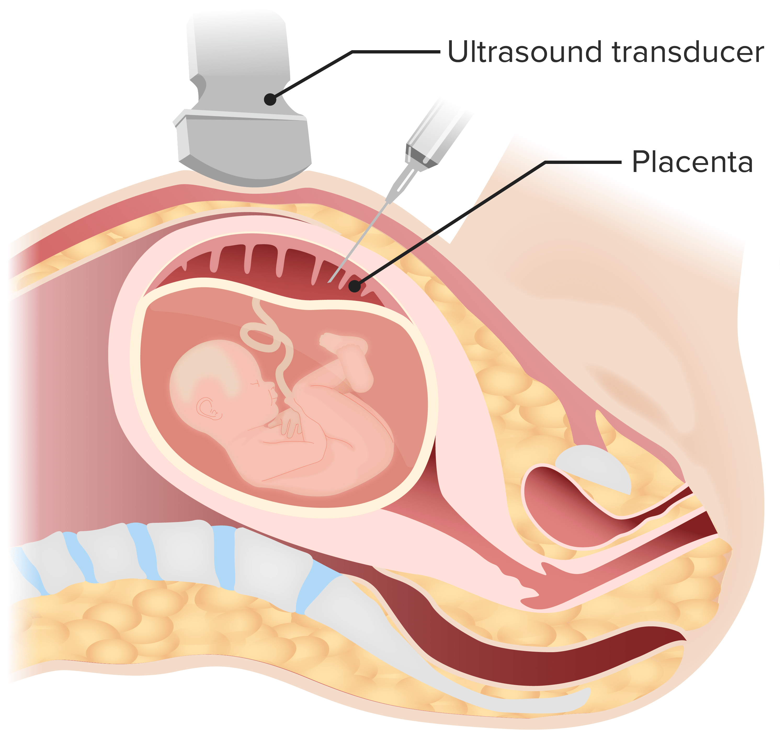
Illustration representing the chorionic villus sampling (CVS) procedure
Image by Lecturio.
AmniocentesisAmniocentesisPercutaneous transabdominal puncture of the uterus during pregnancy to obtain amniotic fluid. It is commonly used for fetal karyotype determination in order to diagnose abnormal fetal conditions.Polyhydramnios
Procedure:
Under ultrasound guidance, a long needle is used to obtain a sample of amniotic fluidAmniotic fluidA clear, yellowish liquid that envelopes the fetus inside the sac of amnion. In the first trimester, it is likely a transudate of maternal or fetal plasma. In the second trimester, amniotic fluid derives primarily from fetal lung and kidney. Cells or substances in this fluid can be removed for prenatal diagnostic tests (amniocentesis).Placenta, Umbilical Cord, and Amniotic Cavity for testing.
Done transabdominally
Timing: any time after 15 wga
Amniotic fluidAmniotic fluidA clear, yellowish liquid that envelopes the fetus inside the sac of amnion. In the first trimester, it is likely a transudate of maternal or fetal plasma. In the second trimester, amniotic fluid derives primarily from fetal lung and kidney. Cells or substances in this fluid can be removed for prenatal diagnostic tests (amniocentesis).Placenta, Umbilical Cord, and Amniotic Cavity obtained can also be used for tests other than geneticsGeneticsGenetics is the study of genes and their functions and behaviors.Basic Terms of Genetics.
Indications:
Confirming abnormal screen results
Fetal evaluation for infection
Fetal structural anomalies
Concern for maternal–fetal blood-type incompatibility
Assessment of fetal lung maturity (previously a common indication, now rarely done)
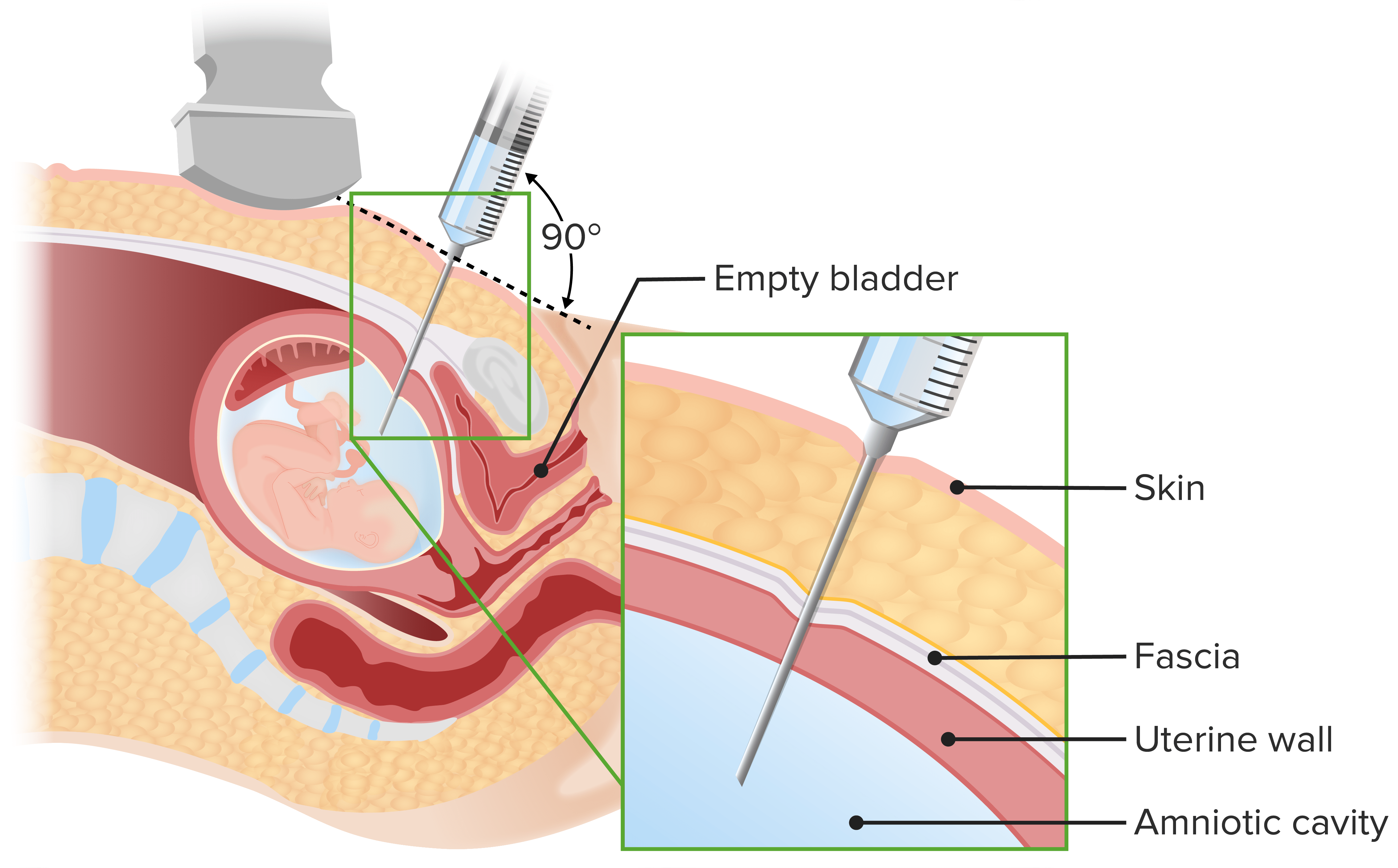
Illustration representing the amniocentesis procedure
Image by Lecturio.
Relative contraindicationsContraindicationsA condition or factor associated with a recipient that makes the use of a drug, procedure, or physical agent improper or inadvisable. Contraindications may be absolute (life threatening) or relative (higher risk of complications in which benefits may outweigh risks).Noninvasive Ventilation
The only relative contraindicationsContraindicationsA condition or factor associated with a recipient that makes the use of a drug, procedure, or physical agent improper or inadvisable. Contraindications may be absolute (life threatening) or relative (higher risk of complications in which benefits may outweigh risks).Noninvasive Ventilation for both procedures include:
Maternal alloimmunization
Poorly controlled maternal infectionsInfectionsInvasion of the host organism by microorganisms or their toxins or by parasites that can cause pathological conditions or diseases.Chronic Granulomatous Disease with risk for vertical transmissionVertical transmissionThe transmission of infectious disease or pathogens from one generation to another. It includes transmission in utero or intrapartum by exposure to blood and secretions, and postpartum exposure via breastfeeding.Congenital TORCH Infections (e.g., HIVHIVAnti-HIV Drugs and hepatitis BHepatitis BHepatitis B virus (HBV) is a partially double-stranded DNA virus, which belongs to the Orthohepadnavirus genus and the Hepadnaviridae family. Most individuals with acute HBV infection are asymptomatic or have mild, self-limiting symptoms. Chronic infection can be asymptomatic or create hepatic inflammation, leading to liver cirrhosis and hepatocellular carcinoma (HCC). Hepatitis B Virus)
Complications
Potential complications are similar for both CVS and amniocentesisAmniocentesisPercutaneous transabdominal puncture of the uterus during pregnancy to obtain amniotic fluid. It is commonly used for fetal karyotype determination in order to diagnose abnormal fetal conditions.Polyhydramnios and include:
Procedure-related fetal loss risk is low (~0.2–0.3% for CVS and ~0.1–0.3% for amniocentesisAmniocentesisPercutaneous transabdominal puncture of the uterus during pregnancy to obtain amniotic fluid. It is commonly used for fetal karyotype determination in order to diagnose abnormal fetal conditions.Polyhydramnios, in experienced hands).
Fetal injury
Infection
Vertical transmissionVertical transmissionThe transmission of infectious disease or pathogens from one generation to another. It includes transmission in utero or intrapartum by exposure to blood and secretions, and postpartum exposure via breastfeeding.Congenital TORCH Infections of maternal infectionsInfectionsInvasion of the host organism by microorganisms or their toxins or by parasites that can cause pathological conditions or diseases.Chronic Granulomatous Disease
PPROM in amniocentesisAmniocentesisPercutaneous transabdominal puncture of the uterus during pregnancy to obtain amniotic fluid. It is commonly used for fetal karyotype determination in order to diagnose abnormal fetal conditions.Polyhydramnios done later in pregnancyPregnancyThe status during which female mammals carry their developing young (embryos or fetuses) in utero before birth, beginning from fertilization to birth.Pregnancy: Diagnosis, Physiology, and Care
Cardiotocography (i.e., Fetal Monitoring) Tests
Cardiotocography is used to monitor the fetal heart rateHeart rateThe number of times the heart ventricles contract per unit of time, usually per minute.Cardiac Physiology (FHR) and contractions plotted over real time, usually via abdominal ultrasonography and pressure transducers, respectively. Fetal heart rateHeart rateThe number of times the heart ventricles contract per unit of time, usually per minute.Cardiac Physiology is recorded in the top panel and contractions in the lower panel of the tracing.
Cardiotocography tracing characteristics
There are 5 major components that should be evaluated when interpreting a cardiotocography tracing:
FHR baseline: the average number of beats per minute
Normal: 110‒160/min
TachycardiaTachycardiaAbnormally rapid heartbeat, usually with a heart rate above 100 beats per minute for adults. Tachycardia accompanied by disturbance in the cardiac depolarization (cardiac arrhythmia) is called tachyarrhythmia.Sepsis in Children: > 160/min when sustained ≥ 10 minutes
BradycardiaBradycardiaBradyarrhythmia is a rhythm in which the heart rate is less than 60/min. Bradyarrhythmia can be physiologic, without symptoms or hemodynamic change. Pathologic bradyarrhythmia results in reduced cardiac output and hemodynamic instability causing syncope, dizziness, or dyspnea.Bradyarrhythmias: < 110/min when sustained ≥ 10 minutes
FHR variability: how much the FHR changes from beat to beat above or below baseline (i.e., how “bumpy” the line is)
Moderate variability (normal): 6‒25/min
The fetal heart has minimal ability to adjust stroke volumeStroke volumeThe amount of blood pumped out of the heart per beat, not to be confused with cardiac output (volume/time). It is calculated as the difference between the end-diastolic volume and the end-systolic volume.Cardiac Cycle, so must adjust HR in order to adjust cardiac outputCardiac outputThe volume of blood passing through the heart per unit of time. It is usually expressed as liters (volume) per minute so as not to be confused with stroke volume (volume per beat).Cardiac Mechanics.
Rapid adjustments to FHR represent a functioning CNS → reliably predicts the absence of hypoxic damage
Represents a fairly constant FHR owing to CNS depression
May be seen normally during fetal sleepSleepA readily reversible suspension of sensorimotor interaction with the environment, usually associated with recumbency and immobility.Physiology of Sleep cycles or after maternal opiate use (often prescribed in early labor)
VariableVariableVariables represent information about something that can change. The design of the measurement scales, or of the methods for obtaining information, will determine the data gathered and the characteristics of that data. As a result, a variable can be qualitative or quantitative, and may be further classified into subgroups.Types of Variables (sometimes a concern):
Sharp decline with rapid return to baseline (V-shaped)
Due to uteroplacental insufficiencyUteroplacental InsufficiencyUteroplacental insufficiency may be acute or chronic and refers to the inability of the placenta to deliver a sufficient supply of O2 and nutrients to the fetusPlacental Abnormalities
Always requires intervention
Contractions:
Normal: 2‒5 contractions in 10 minutes during labor
Tachysystole: > 5 contractions in 10 minutes
External pressure transducers versus intrauterine pressure catheters (IUPCs):
External pressure transducers: only show the relative pressure within the uterusUterusThe uterus, cervix, and fallopian tubes are part of the internal female reproductive system. The uterus has a thick wall made of smooth muscle (the myometrium) and an inner mucosal layer (the endometrium). The most inferior portion of the uterus is the cervix, which connects the uterine cavity to the vagina.Uterus, Cervix, and Fallopian Tubes: Anatomy at a given time
IUPC: a catheter inserted into the uterusUterusThe uterus, cervix, and fallopian tubes are part of the internal female reproductive system. The uterus has a thick wall made of smooth muscle (the myometrium) and an inner mucosal layer (the endometrium). The most inferior portion of the uterus is the cervix, which connects the uterine cavity to the vagina.Uterus, Cervix, and Fallopian Tubes: Anatomy during labor (after rupture of membranes) to measure the actual numeric pressure generated by the contractions; measured in Montevideo units (MVUs)
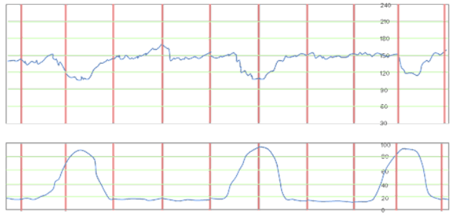
Early decelerations: Note how the nadir of the deceleration is aligned with the peak of the contractions.
Image by Lecturio.
Variable decelerations: These 1st 2 variable decelerations are a concern because of how low they go (down to < 60/min) and how long they last (nearly a full minute). The 3rd deceleration is shallower and shorter, and, if seen without any other decelerations and in combination with moderate variability and/or accelerations, would be less worrisome.
Image by Lecturio.
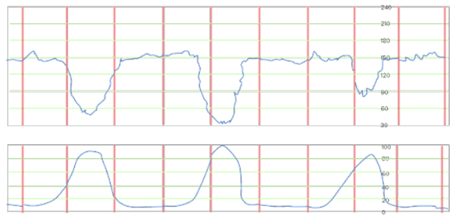
Late decelerations: Note how the nadir of these decelerations follows the peak of the contraction, which indicates uteroplacental insufficiency, which requires intervention.
Image by Lecturio.
Interpreting cardiotocography tracings
Characteristics of a continuousFHR tracing are assessed periodically and used to classify tracings into 1 of 3 categories. These categories provide some prognostic information on the current status of fetal well-being and can help guide management.
Category I: normal
All of the following characteristics must be present:
FHR: 110‒160/min
Moderate variability
No variableVariableVariables represent information about something that can change. The design of the measurement scales, or of the methods for obtaining information, will determine the data gathered and the characteristics of that data. As a result, a variable can be qualitative or quantitative, and may be further classified into subgroups.Types of Variables or late decelerations
Early decelerations may be present or absent
Interpretation: minimal chance of fetal acidemiaAcidemiaRespiratory Acidosis/hypoxic injury at this point in time
Management: observation/continue current course of action
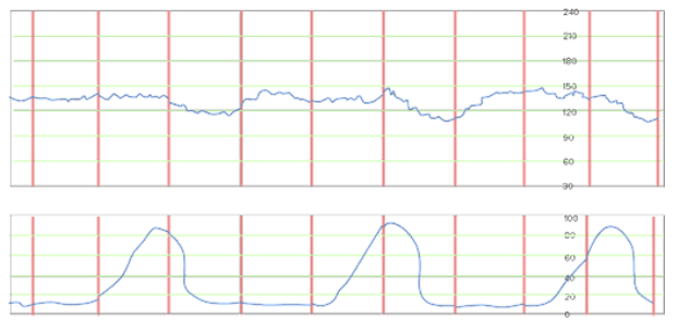
Category I (normal) fetal heart rate tracing An FHR baseline of 150/min, moderate variability, several accelerations, and no decelerations are seen. Three contractions are shown; if these contractions were measured using an intrauterine pressure catheter, they each reach approximately 60 Montevideo units (MVUs), for a total of 180 MVUs in the 8 minutes shown. Based on this pattern, there would likely be a 4th contraction near the 10-minute mark, making this contraction pattern adequate (defined as > 200 MVUs per 10 minutes) to induce cervical change.
Image by Lecturio.
Category II: indeterminate
Everything that is not category I or III
This category includes:
A normal FHR (110‒160/min) with variableVariableVariables represent information about something that can change. The design of the measurement scales, or of the methods for obtaining information, will determine the data gathered and the characteristics of that data. As a result, a variable can be qualitative or quantitative, and may be further classified into subgroups.Types of Variables or late decelerations
A normal FHR (110‒160/min) with minimal or absent variability withoutany decelerations
Baseline FHR < 110/min or > 160/min with minimal or moderate variability
Interpretation: requires ongoing observation to confirm fetal well-being
Management:
Depends on clinical assessment (Are there known reasons to explain the category II tracing? For example, maternal feverFeverFever is defined as a measured body temperature of at least 38°C (100.4°F). Fever is caused by circulating endogenous and/or exogenous pyrogens that increase levels of prostaglandin E2 in the hypothalamus. Fever is commonly associated with chills, rigors, sweating, and flushing of the skin. Fever usually causes fetal tachycardiaTachycardiaAbnormally rapid heartbeat, usually with a heart rate above 100 beats per minute for adults. Tachycardia accompanied by disturbance in the cardiac depolarization (cardiac arrhythmia) is called tachyarrhythmia.Sepsis in Children.)
Antibiotics for diagnosed maternal infectionsInfectionsInvasion of the host organism by microorganisms or their toxins or by parasites that can cause pathological conditions or diseases.Chronic Granulomatous Disease
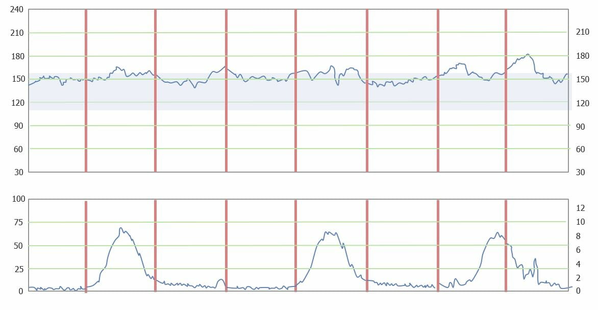
Category II fetal heart rate tracing, which is indeterminate: The baseline is normal, but there is minimal variability. There are no decelerations or accelerations in this tracing.
Image by Lecturio.
Category III: abnormal
Sinusoidal pattern, or absent variability with recurrent late decelerations, recurrent variableVariableVariables represent information about something that can change. The design of the measurement scales, or of the methods for obtaining information, will determine the data gathered and the characteristics of that data. As a result, a variable can be qualitative or quantitative, and may be further classified into subgroups.Types of Variables decelerations, or bradycardiaBradycardiaBradyarrhythmia is a rhythm in which the heart rate is less than 60/min. Bradyarrhythmia can be physiologic, without symptoms or hemodynamic change. Pathologic bradyarrhythmia results in reduced cardiac output and hemodynamic instability causing syncope, dizziness, or dyspnea.Bradyarrhythmias.
Interpretation: indicates that fetus is acidotic and in distress
Management: usually requires immediate delivery (e.g., urgent cesarean deliveryCesarean DeliveryCesarean delivery (CD) is the operative delivery of ≥ 1 infants through a surgical incision in the maternal abdomen and uterus. Cesarean deliveries may be indicated for a number of either maternal or fetal reasons, most commonly including fetal intolerance to labor, arrest of labor, a history of prior uterine surgery, fetal malpresentation, and placental abnormalities. Cesarean Delivery)
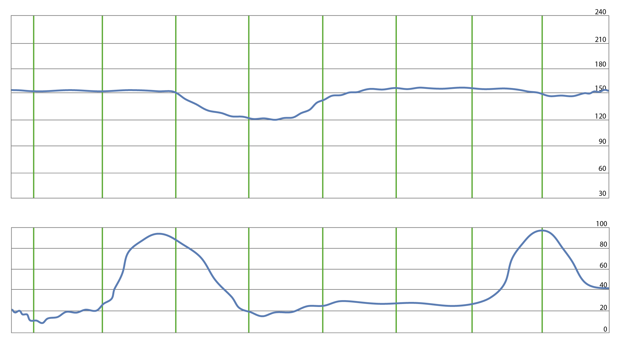
Fetal heart rate tracing demonstrating a prolonged late deceleration: Variability appears moderate, however, and there is a spontaneous return to baseline. If this pattern continues, this is a worrisome tracing, demonstrating fetal distress.
Image by Lecturio.
Fetal non-stress test (NST)
An NST is cardiotocometry performed for 20 minutes (or longer if needed) to assess antepartumfetal well-being, usually after 28 weeks. Non-stress tests are classified as either reactive or nonreactive rather than by categories used to describe continuous fetal monitoring.
Reactive NST
≥ 2 FHR accelerations in 20 minutes
Moderate variability is almost always present with ≥ 2 accelerations, though not required to be considered reactive
VariableVariableVariables represent information about something that can change. The design of the measurement scales, or of the methods for obtaining information, will determine the data gathered and the characteristics of that data. As a result, a variable can be qualitative or quantitative, and may be further classified into subgroups.Types of Variables decelerations may be present.
A reassuring pattern: predicts a low likelihood of fetal death due to hypoxic injury within the next few days
Management: observation
Nonreactive NST
< 2 FHR accelerations in 20 minutes
VariableVariableVariables represent information about something that can change. The design of the measurement scales, or of the methods for obtaining information, will determine the data gathered and the characteristics of that data. As a result, a variable can be qualitative or quantitative, and may be further classified into subgroups.Types of Variables decelerations may be present
Nonreassuring patterns: unable to rule out fetal death due to hypoxic injury within the next few days
Management:
Indicates that further monitoring is required (e.g., prolonged/continuous fetal monitoring, BPP). Example: normal FHR, moderate variability, no decelerations, but no accelerations (nonreactive) → continue to monitor with cardiotocography (infant may be in a sleepSleepA readily reversible suspension of sensorimotor interaction with the environment, usually associated with recumbency and immobility.Physiology of Sleep cycle and become reactive over the next hour or 2)
Depending on the gestational ageGestational ageThe age of the conceptus, beginning from the time of fertilization. In clinical obstetrics, the gestational age is often estimated as the time from the last day of the last menstruation which is about 2 weeks before ovulation and fertilization.Pregnancy: Diagnosis, Physiology, and Care and severity of the nonreassuring NST, immediate delivery may be considered. Example: bradycardiaBradycardiaBradyarrhythmia is a rhythm in which the heart rate is less than 60/min. Bradyarrhythmia can be physiologic, without symptoms or hemodynamic change. Pathologic bradyarrhythmia results in reduced cardiac output and hemodynamic instability causing syncope, dizziness, or dyspnea.Bradyarrhythmias with absent variability in a full-term infant → urgent cesarean deliveryCesarean DeliveryCesarean delivery (CD) is the operative delivery of ≥ 1 infants through a surgical incision in the maternal abdomen and uterus. Cesarean deliveries may be indicated for a number of either maternal or fetal reasons, most commonly including fetal intolerance to labor, arrest of labor, a history of prior uterine surgery, fetal malpresentation, and placental abnormalities. Cesarean Delivery
Contraction stress test (CST)
A CST is similar to an NST but is performed with induced (e.g., with pitocin) or spontaneous contractions, which allows providers to assess the impacts of the contractions on the FHR pattern. Interpretations include:
Negative:
No significant decelerations
A reassuring result
Positive:
Presence of late decelerations > 50% of the time
A nonreassuring result → intervention is indicated
Equivocal: presence of late decelerations < 50% of the time or inadequate contractions to make an assessment
Amniotic fluidAmniotic fluidA clear, yellowish liquid that envelopes the fetus inside the sac of amnion. In the first trimester, it is likely a transudate of maternal or fetal plasma. In the second trimester, amniotic fluid derives primarily from fetal lung and kidney. Cells or substances in this fluid can be removed for prenatal diagnostic tests (amniocentesis).Placenta, Umbilical Cord, and Amniotic Cavity volume
NST to assess the FHR reactivity.
Each component receives a score of either 0 (abnormal) or 2 (normal) points.
No partial credit (cannot earn a score of 1)
E.g., to get credit for fetal movement, 3 distinct movements must be visualized; if only 2 movements are seen, the infant gets 0 points for movement.
The BPP is scored out of 10 points (or 8 when an NST is not performed).
Interpretation:
Total score ≥ 8 points: no signs of fetal compromise
Total score 6 points: unclear risk of fetal compromise
Total score ≤ 4 points: potential fetal compromise, intervention indicated
≥ 1 episode(s) of a fetal extremity or fetal spineSpineThe human spine, or vertebral column, is the most important anatomical and functional axis of the human body. It consists of 7 cervical vertebrae, 12 thoracic vertebrae, and 5 lumbar vertebrae and is limited cranially by the skull and caudally by the sacrum.Vertebral Column: AnatomyextensionExtensionExamination of the Upper Limbs with return to flexionFlexionExamination of the Upper Limbs
2
Amniotic fluidAmniotic fluidA clear, yellowish liquid that envelopes the fetus inside the sac of amnion. In the first trimester, it is likely a transudate of maternal or fetal plasma. In the second trimester, amniotic fluid derives primarily from fetal lung and kidney. Cells or substances in this fluid can be removed for prenatal diagnostic tests (amniocentesis).Placenta, Umbilical Cord, and Amniotic Cavity volume
A single deepest vertical pocket ≥ 2 cm with a horizontal dimension ≥ 1 cm
2
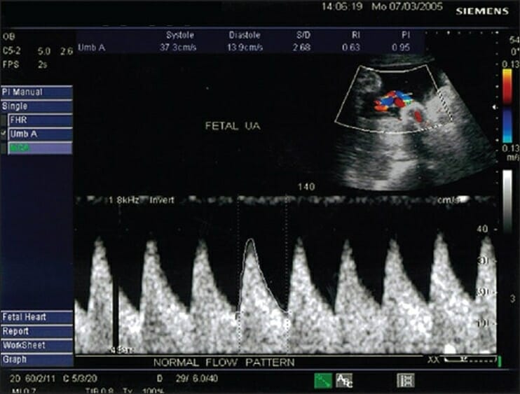
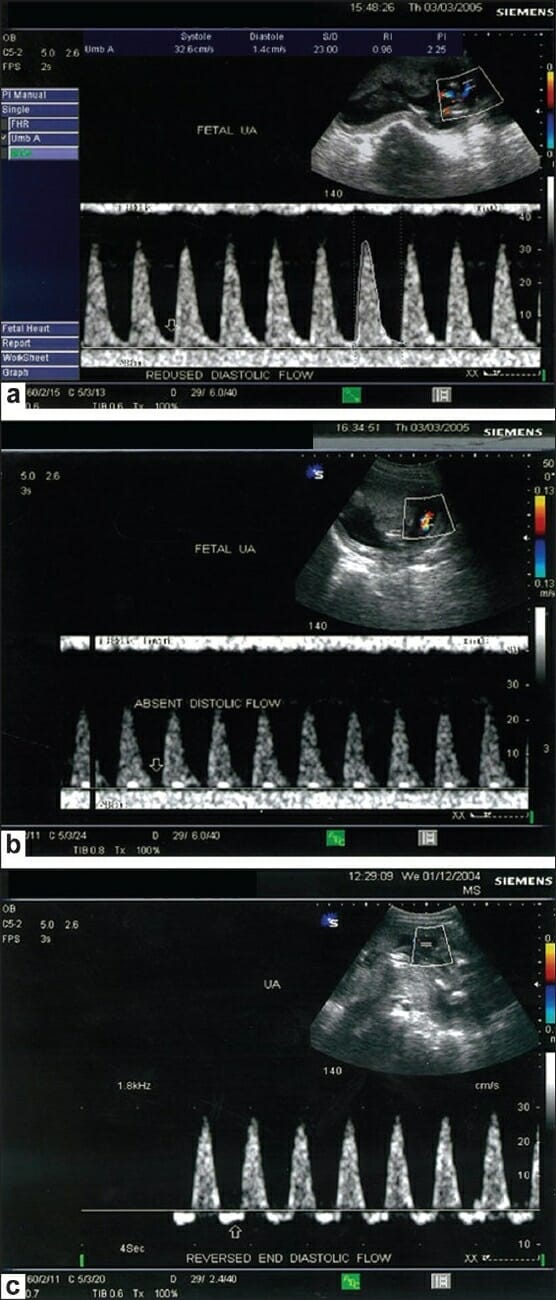
Umbilical arteryUmbilical arterySpecialized arterial vessels in the umbilical cord. They carry waste and deoxygenated blood from the fetus to the mother via the placenta. In humans, there are usually two umbilical arteries but sometimes one.Prenatal and Postnatal Physiology of the NeonateDopplerDopplerUltrasonography applying the doppler effect, with frequency-shifted ultrasound reflections produced by moving targets (usually red blood cells) in the bloodstream along the ultrasound axis in direct proportion to the velocity of movement of the targets, to determine both direction and velocity of blood flow.Ultrasound (Sonography)
Umbilical arteryUmbilical arterySpecialized arterial vessels in the umbilical cord. They carry waste and deoxygenated blood from the fetus to the mother via the placenta. In humans, there are usually two umbilical arteries but sometimes one.Prenatal and Postnatal Physiology of the Neonate (UA) DopplerDopplerUltrasonography applying the doppler effect, with frequency-shifted ultrasound reflections produced by moving targets (usually red blood cells) in the bloodstream along the ultrasound axis in direct proportion to the velocity of movement of the targets, to determine both direction and velocity of blood flow.Ultrasound (Sonography) studies (done on ultrasound machines) are used to help assess fetal well-being in pregnancies affected by fetal growth restrictionFetal growth restrictionFetal growth restriction (FGR), also known as intrauterine fetal growth restriction (IUGR), is an estimated fetal weight (EFW) or abdominal circumference < 10th percentile for gestational age. The term small for gestational age (SGA) is sometimes erroneously used interchangeably with FGR. Fetal Growth Restriction (FGRFGRFetal growth restriction (FGR), also known as intrauterine fetal growth restriction (IUGR), is an estimated fetal weight (EFW) or abdominal circumference < 10th percentile for gestational age. The term small for gestational age (sga) is sometimes erroneously used interchangeably with FGR.Fetal Growth Restriction).
Measures the direction and velocity of blood flowBlood flowBlood flow refers to the movement of a certain volume of blood through the vasculature over a given unit of time (e.g., mL per minute).Vascular Resistance, Flow, and Mean Arterial Pressure through the UA, which is directly related to blood flowBlood flowBlood flow refers to the movement of a certain volume of blood through the vasculature over a given unit of time (e.g., mL per minute).Vascular Resistance, Flow, and Mean Arterial Pressure through the placentaPlacentaA highly vascularized mammalian fetal-maternal organ and major site of transport of oxygen, nutrients, and fetal waste products. It includes a fetal portion (chorionic villi) derived from trophoblasts and a maternal portion (decidua) derived from the uterine endometrium. The placenta produces an array of steroid, protein and peptide hormones (placental hormones).Placenta, Umbilical Cord, and Amniotic Cavity
UA DopplerDopplerUltrasonography applying the doppler effect, with frequency-shifted ultrasound reflections produced by moving targets (usually red blood cells) in the bloodstream along the ultrasound axis in direct proportion to the velocity of movement of the targets, to determine both direction and velocity of blood flow.Ultrasound (Sonography) studies provide assessment of placental function:
Diseased vessels in the placentaPlacentaA highly vascularized mammalian fetal-maternal organ and major site of transport of oxygen, nutrients, and fetal waste products. It includes a fetal portion (chorionic villi) derived from trophoblasts and a maternal portion (decidua) derived from the uterine endometrium. The placenta produces an array of steroid, protein and peptide hormones (placental hormones).Placenta, Umbilical Cord, and Amniotic Cavity cause an increase in resistanceResistancePhysiologically, the opposition to flow of air caused by the forces of friction. As a part of pulmonary function testing, it is the ratio of driving pressure to the rate of air flow.Ventilation: Mechanics of Breathing to fetal blood flowBlood flowBlood flow refers to the movement of a certain volume of blood through the vasculature over a given unit of time (e.g., mL per minute).Vascular Resistance, Flow, and Mean Arterial Pressure (commonly due to maternal hypertensive and diabetic disorders)
↑ ResistanceResistancePhysiologically, the opposition to flow of air caused by the forces of friction. As a part of pulmonary function testing, it is the ratio of driving pressure to the rate of air flow.Ventilation: Mechanics of Breathing in the placentaPlacentaA highly vascularized mammalian fetal-maternal organ and major site of transport of oxygen, nutrients, and fetal waste products. It includes a fetal portion (chorionic villi) derived from trophoblasts and a maternal portion (decidua) derived from the uterine endometrium. The placenta produces an array of steroid, protein and peptide hormones (placental hormones).Placenta, Umbilical Cord, and Amniotic Cavity → slower fetal blood flowBlood flowBlood flow refers to the movement of a certain volume of blood through the vasculature over a given unit of time (e.g., mL per minute).Vascular Resistance, Flow, and Mean Arterial Pressure through the placentaPlacentaA highly vascularized mammalian fetal-maternal organ and major site of transport of oxygen, nutrients, and fetal waste products. It includes a fetal portion (chorionic villi) derived from trophoblasts and a maternal portion (decidua) derived from the uterine endometrium. The placenta produces an array of steroid, protein and peptide hormones (placental hormones).Placenta, Umbilical Cord, and Amniotic Cavity → less effective O2 and nutrient exchange → poor growth → FGRFGRFetal growth restriction (FGR), also known as intrauterine fetal growth restriction (IUGR), is an estimated fetal weight (EFW) or abdominal circumference < 10th percentile for gestational age. The term small for gestational age (sga) is sometimes erroneously used interchangeably with FGR.Fetal Growth Restriction
The UA waveform usually has a sawtooth pattern:
Higher/faster forward flowFlowBlood flows through the heart, arteries, capillaries, and veins in a closed, continuous circuit. Flow is the movement of volume per unit of time. Flow is affected by the pressure gradient and the resistance fluid encounters between 2 points. Vascular resistance is the opposition to flow, which is caused primarily by blood friction against vessel walls.Vascular Resistance, Flow, and Mean Arterial Pressure during fetal systoleSystolePeriod of contraction of the heart, especially of the heart ventricles.Cardiac Cycle
Lower/slower forward flowFlowBlood flows through the heart, arteries, capillaries, and veins in a closed, continuous circuit. Flow is the movement of volume per unit of time. Flow is affected by the pressure gradient and the resistance fluid encounters between 2 points. Vascular resistance is the opposition to flow, which is caused primarily by blood friction against vessel walls.Vascular Resistance, Flow, and Mean Arterial Pressure during fetal diastoleDiastolePost-systolic relaxation of the heart, especially the heart ventricles.Cardiac Cycle
The systolic-to-diastolic (S:D) ratio is calculated.
As resistanceResistancePhysiologically, the opposition to flow of air caused by the forces of friction. As a part of pulmonary function testing, it is the ratio of driving pressure to the rate of air flow.Ventilation: Mechanics of Breathing in the placentaPlacentaA highly vascularized mammalian fetal-maternal organ and major site of transport of oxygen, nutrients, and fetal waste products. It includes a fetal portion (chorionic villi) derived from trophoblasts and a maternal portion (decidua) derived from the uterine endometrium. The placenta produces an array of steroid, protein and peptide hormones (placental hormones).Placenta, Umbilical Cord, and Amniotic Cavity increases, forward flowFlowBlood flows through the heart, arteries, capillaries, and veins in a closed, continuous circuit. Flow is the movement of volume per unit of time. Flow is affected by the pressure gradient and the resistance fluid encounters between 2 points. Vascular resistance is the opposition to flow, which is caused primarily by blood friction against vessel walls.Vascular Resistance, Flow, and Mean Arterial Pressure during fetal diastoleDiastolePost-systolic relaxation of the heart, especially the heart ventricles.Cardiac Cycle decreases to a much greater extent than flowFlowBlood flows through the heart, arteries, capillaries, and veins in a closed, continuous circuit. Flow is the movement of volume per unit of time. Flow is affected by the pressure gradient and the resistance fluid encounters between 2 points. Vascular resistance is the opposition to flow, which is caused primarily by blood friction against vessel walls.Vascular Resistance, Flow, and Mean Arterial Pressure during systoleSystolePeriod of contraction of the heart, especially of the heart ventricles.Cardiac Cycle → S:D ratio increases
In severe cases, diastolic flowFlowBlood flows through the heart, arteries, capillaries, and veins in a closed, continuous circuit. Flow is the movement of volume per unit of time. Flow is affected by the pressure gradient and the resistance fluid encounters between 2 points. Vascular resistance is the opposition to flow, which is caused primarily by blood friction against vessel walls.Vascular Resistance, Flow, and Mean Arterial Pressure may be completely absent or even reversed.
Results are used to help guide decisions surrounding when to deliver an infant with FGRFGRFetal growth restriction (FGR), also known as intrauterine fetal growth restriction (IUGR), is an estimated fetal weight (EFW) or abdominal circumference < 10th percentile for gestational age. The term small for gestational age (sga) is sometimes erroneously used interchangeably with FGR.Fetal Growth Restriction.
Color Doppler of the umbilical artery: The scan shows a normal umbilical artery waveform pattern in the bottom portion of the image. The peak of the waveforms represent systolic flow, while the trough of the waveforms represent end diastolic flow.
Image: “Color Doppler of the umbilical artery” by Bano S, Chaudhary V, Pande S, Mehta V, Sharma A. License: CC BY 2.0
American College of Obstetricians and Gynecologists. (2020). Screening for fetal chromosomal abnormalities. ACOG Practice Bulletin Summary, no. 226. Obstetrics & Gynecology 136(4):859–867. https://doi.org/10.1097/aog.0000000000004107
Create your free account or log in to continue reading!