The cubital fossa is the region anterior to the elbow joint Elbow joint The elbow is the synovial hinge joint between the humerus in the upper arm and the radius and ulna in the forearm. The elbow consists of 3 joints, which form a functional unit enclosed within a single articular capsule. The elbow is the link between the powerful motions of the shoulder and the intricate fine-motor function of the hand. Elbow Joint: Anatomy. The cubital fossa is seen as the triangular depression between the brachioradialis Brachioradialis Forearm: Anatomy and pronator teres Pronator teres Forearm: Anatomy muscles. Except for the ulnar nerve Ulnar Nerve A major nerve of the upper extremity. In humans, the fibers of the ulnar nerve originate in the lower cervical and upper thoracic spinal cord (usually C7 to T1), travel via the medial cord of the brachial plexus, and supply sensory and motor innervation to parts of the hand and forearm. Axilla and Brachial Plexus: Anatomy, which runs posteriorly, most of the major neurovascular structures transition from the arm Arm The arm, or "upper arm" in common usage, is the region of the upper limb that extends from the shoulder to the elbow joint and connects inferiorly to the forearm through the cubital fossa. It is divided into 2 fascial compartments (anterior and posterior). Arm: Anatomy to the forearm Forearm The forearm is the region of the upper limb between the elbow and the wrist. The term "forearm" is used in anatomy to distinguish this area from the arm, a term that is commonly used to describe the entire upper limb. The forearm consists of 2 long bones (the radius and the ulna), the interosseous membrane, and multiple arteries, nerves, and muscles. Forearm: Anatomy via the cubital fossa. The 4 important structures of the cubital fossa (from lateral to medial) are the radial nerve Radial Nerve A major nerve of the upper extremity. In humans the fibers of the radial nerve originate in the lower cervical and upper thoracic spinal cord (usually C5 to T1), travel via the posterior cord of the brachial plexus, and supply motor innervation to extensor muscles of the arm and cutaneous sensory fibers to extensor regions of the arm and hand. Axilla and Brachial Plexus: Anatomy, tendon of the biceps brachii muscle Biceps brachii muscle Elbow Joint: Anatomy, brachial artery, and median nerve.
Last updated: Dec 15, 2025
The cubital fossa is the transitional area between the upper arm Upper Arm The arm, or “upper arm” in common usage, is the region of the upper limb that extends from the shoulder to the elbow joint and connects inferiorly to the forearm through the cubital fossa. It is divided into 2 fascial compartments (anterior and posterior). Arm: Anatomy and the forearm Forearm The forearm is the region of the upper limb between the elbow and the wrist. The term “forearm” is used in anatomy to distinguish this area from the arm, a term that is commonly used to describe the entire upper limb. The forearm consists of 2 long bones (the radius and the ulna), the interosseous membrane, and multiple arteries, nerves, and muscles. Forearm: Anatomy anterior to the elbow joint Elbow joint The elbow is the synovial hinge joint between the humerus in the upper arm and the radius and ulna in the forearm. The elbow consists of 3 joints, which form a functional unit enclosed within a single articular capsule. The elbow is the link between the powerful motions of the shoulder and the intricate fine-motor function of the hand. Elbow Joint: Anatomy. The cubital fossa is defined by the following anatomical boundaries:
| Boundary | Structure |
|---|---|
| Superior | Horizontal line joining medial and lateral epicondyles of the humerus Humerus Bone in humans and primates extending from the shoulder joint to the elbow joint. Arm: Anatomy |
| Lateral | Medial border of the brachioradialis Brachioradialis Forearm: Anatomy muscle |
| Medial | Lateral border of the pronator teres Pronator teres Forearm: Anatomy muscle |
| Apex | Directed inferiorly, the meeting point of the lateral and medial boundaries |
| Floor | Brachialis muscle Brachialis muscle Elbow Joint: Anatomy (proximally) and supinator Supinator Forearm: Anatomy muscle (distally) |
| Roof | Joining of brachial and forearm Forearm The forearm is the region of the upper limb between the elbow and the wrist. The term “forearm” is used in anatomy to distinguish this area from the arm, a term that is commonly used to describe the entire upper limb. The forearm consists of 2 long bones (the radius and the ulna), the interosseous membrane, and multiple arteries, nerves, and muscles. Forearm: Anatomy fascia Fascia Layers of connective tissue of variable thickness. The superficial fascia is found immediately below the skin; the deep fascia invests muscles, nerves, and other organs. Cellulitis plus bicipital aponeurosis |
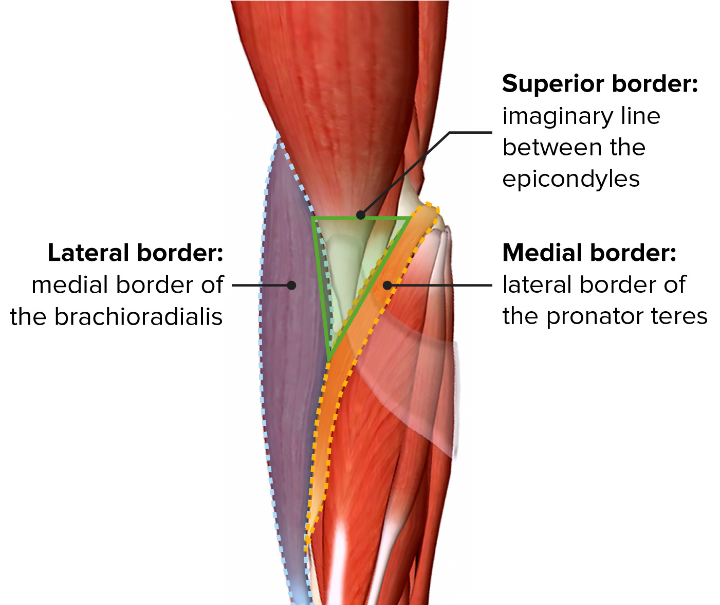
Boundaries of the cubital fossa
Image by BioDigital, edited by Lecturio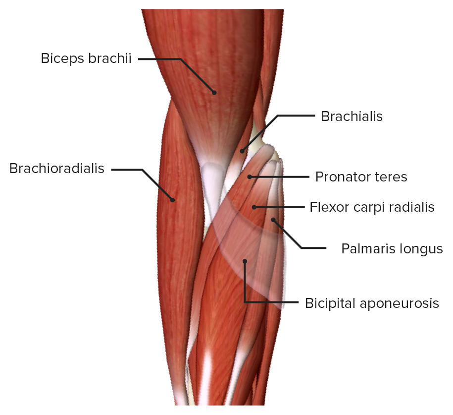
Superficial muscles of the right cubital fossa
Image by BioDigital, edited by Lecturio
Deep muscles of the right cubital fossa
Image by BioDigital, edited by LecturioThe important structures of the cubital fossa can be organized from lateral to medial and include all the important neurovascular structures except for the ulnar nerve Ulnar Nerve A major nerve of the upper extremity. In humans, the fibers of the ulnar nerve originate in the lower cervical and upper thoracic spinal cord (usually C7 to T1), travel via the medial cord of the brachial plexus, and supply sensory and motor innervation to parts of the hand and forearm. Axilla and Brachial Plexus: Anatomy, which runs posterior to the medial epicondyle Medial epicondyle Arm: Anatomy.
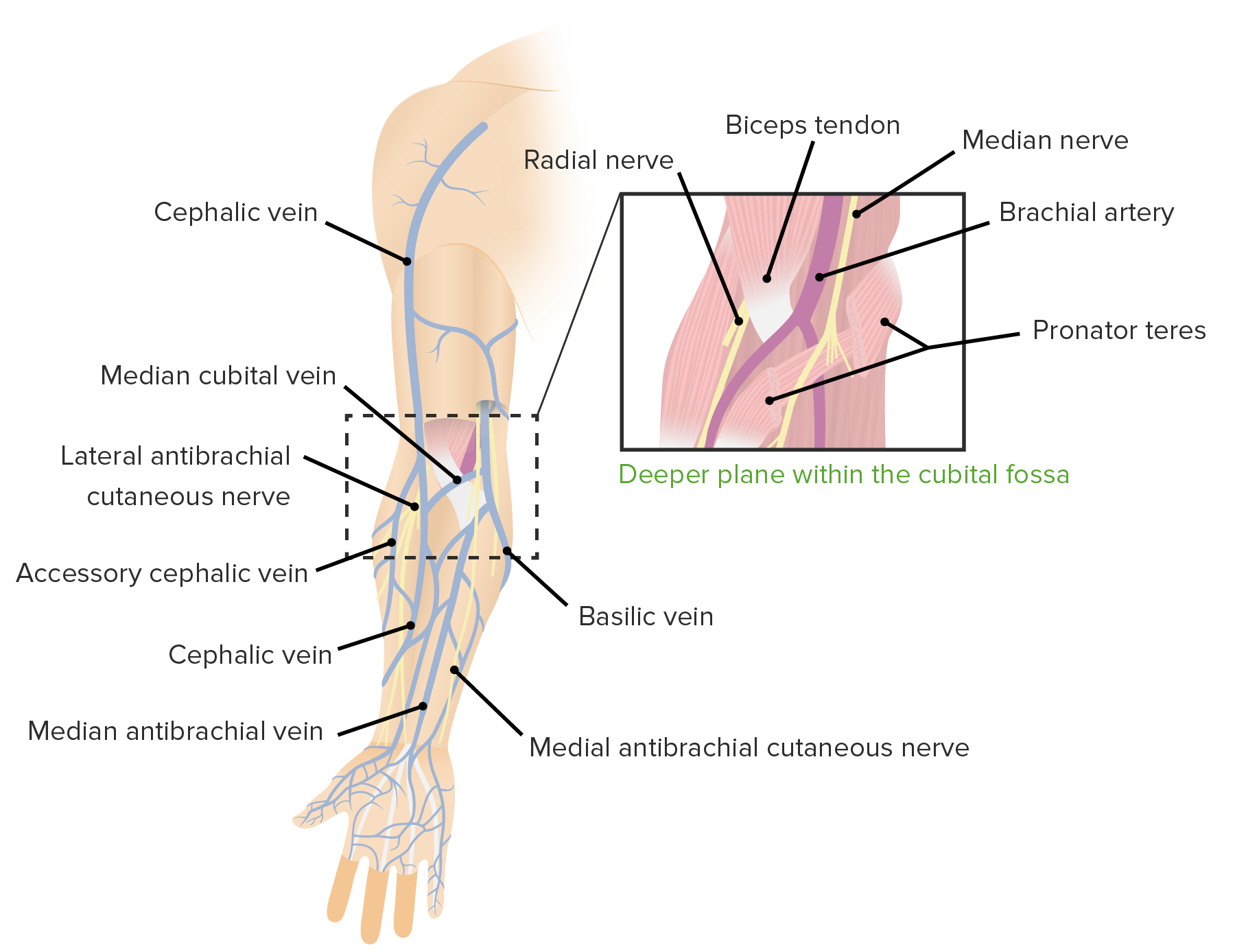
Contents of the cubital fossa:
Deep within the fossa (right box, lateral to medial): radial nerve, biceps tendon, bifurcation of the brachial artery into radial and ulnar arteries, and median nerve
Superficial to the biceps aponeurosis (left side): cephalic vein, median cubital vein, basilic vein, lateral cutaneous nerve, and medial cutaneous nerve of the forearm
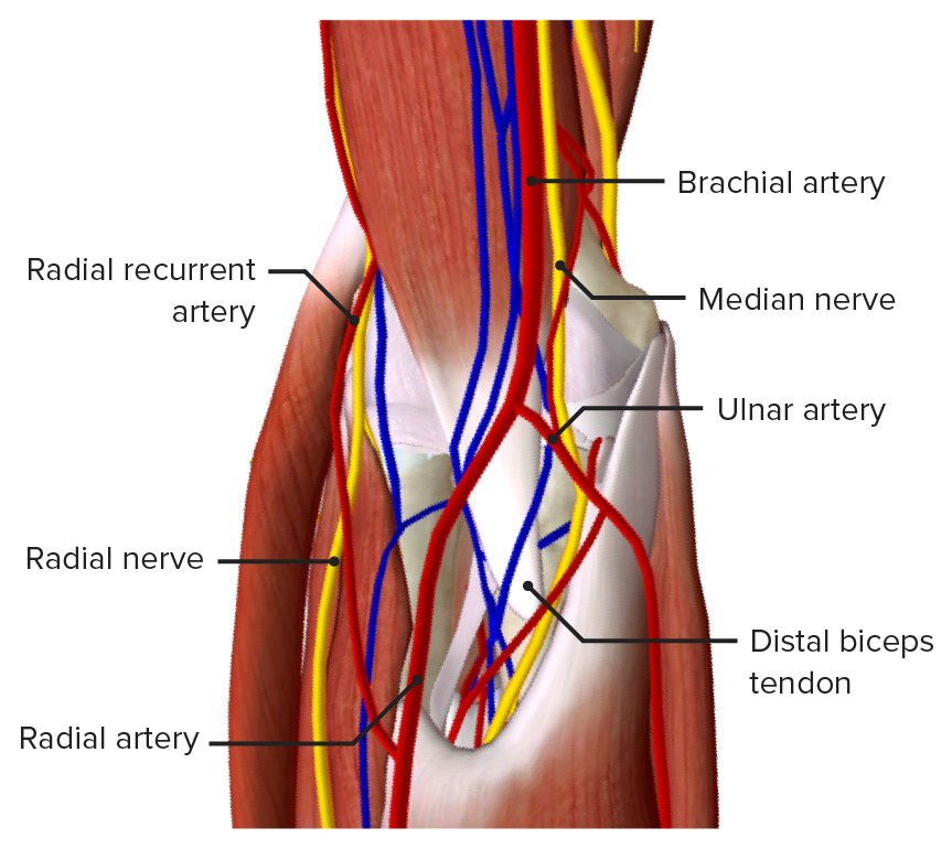
Deep view of the cubital fossa
Image by BioDigital, edited by Lecturio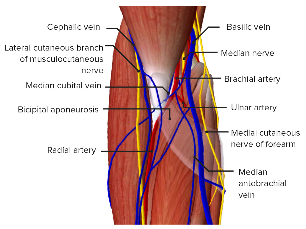
Superficial view of the cubital fossa
Image by BioDigital, edited by LecturioThe following are important clinical concepts related to the structures found within the cubital fossa: