The wrist connects the forearm Forearm The forearm is the region of the upper limb between the elbow and the wrist. The term "forearm" is used in anatomy to distinguish this area from the arm, a term that is commonly used to describe the entire upper limb. The forearm consists of 2 long bones (the radius and the ulna), the interosseous membrane, and multiple arteries, nerves, and muscles. Forearm: Anatomy to the hand Hand The hand constitutes the distal part of the upper limb and provides the fine, precise movements needed in activities of daily living. It consists of 5 metacarpal bones and 14 phalanges, as well as numerous muscles innervated by the median and ulnar nerves. Hand: Anatomy. It consists of 8 carpal bones Carpal bones The eight bones of the wrist: scaphoid bone; lunate bone; triquetrum bone; pisiform bone; trapezium bone; trapezoid bone; capitate bone; and hamate bone. Carpal Tunnel Syndrome, multiple joints, and various supporting ligaments, as well as the distal bones of the forearm Forearm The forearm is the region of the upper limb between the elbow and the wrist. The term "forearm" is used in anatomy to distinguish this area from the arm, a term that is commonly used to describe the entire upper limb. The forearm consists of 2 long bones (the radius and the ulna), the interosseous membrane, and multiple arteries, nerves, and muscles. Forearm: Anatomy and the proximal portion of the 5 metacarpal bones of the hand Hand The hand constitutes the distal part of the upper limb and provides the fine, precise movements needed in activities of daily living. It consists of 5 metacarpal bones and 14 phalanges, as well as numerous muscles innervated by the median and ulnar nerves. Hand: Anatomy. The wrist is crucial for the functioning of the upper limb, and it provides stability while positioning the hand Hand The hand constitutes the distal part of the upper limb and provides the fine, precise movements needed in activities of daily living. It consists of 5 metacarpal bones and 14 phalanges, as well as numerous muscles innervated by the median and ulnar nerves. Hand: Anatomy for intricate motions.
Last updated: Dec 15, 2025
The bones of the wrist consist of:
There are many mnemonics to help memorize the order and location of the carpal bones Carpal bones The eight bones of the wrist: scaphoid bone; lunate bone; triquetrum bone; pisiform bone; trapezium bone; trapezoid bone; capitate bone; and hamate bone. Carpal Tunnel Syndrome. They are learned from lateral to medial and proximal to distal: scaphoid, lunate, triquetrum, pisiform, trapezium, trapezoid, capitate, hamate.
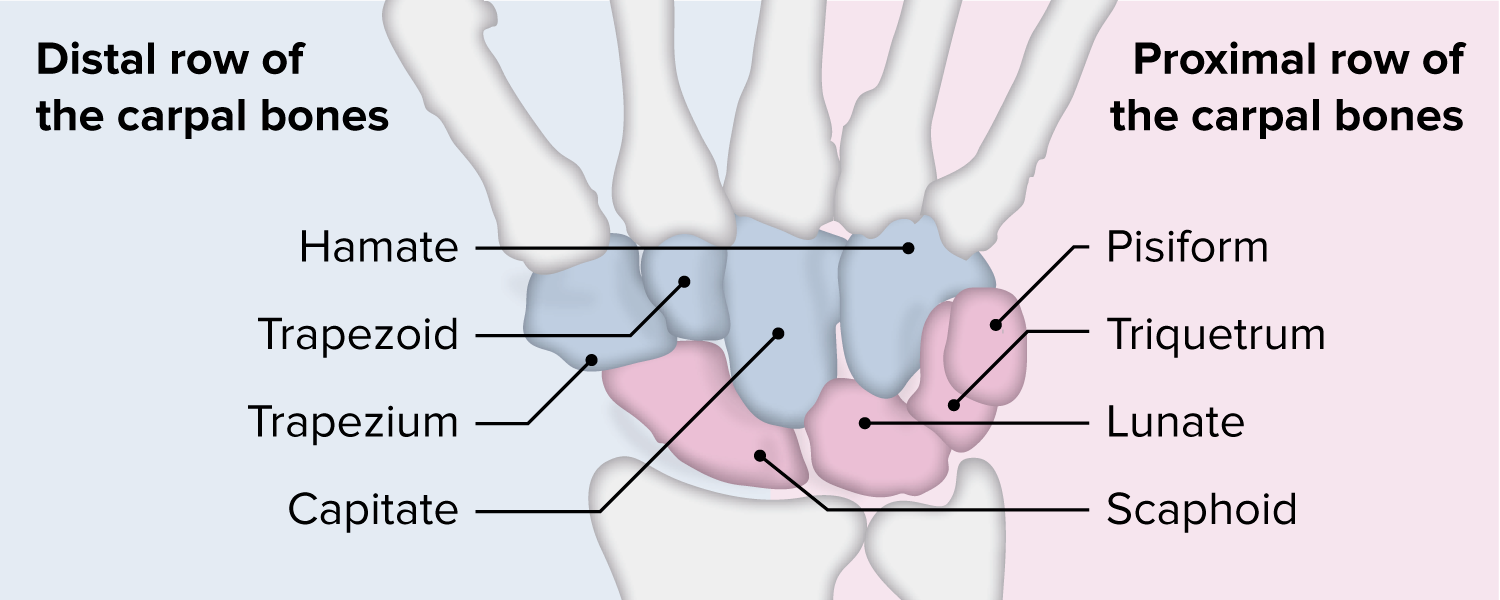
The proximal and distal rows of the carpal bones
Image by Lecturio.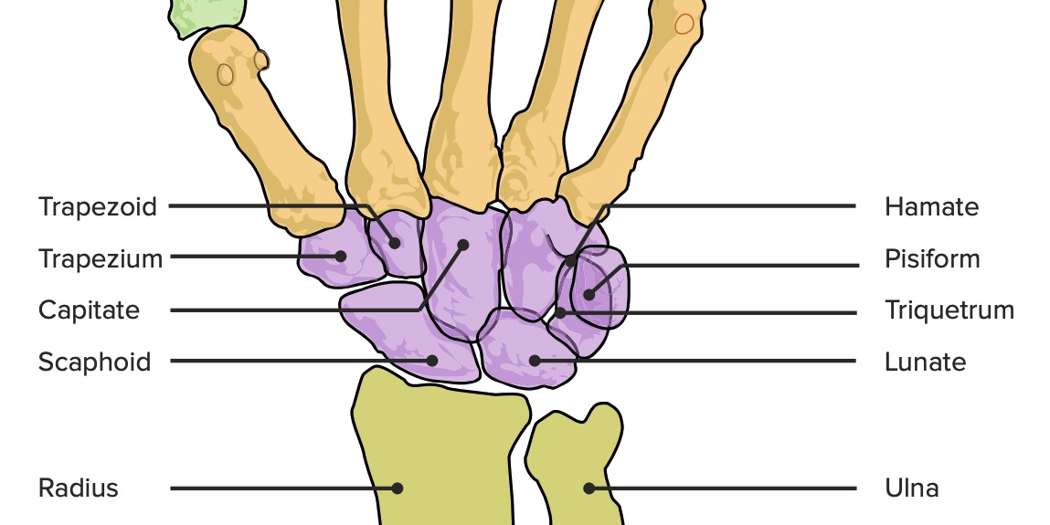
Wrist joint, featuring the 8 carpal bones plus the radius and ulna
Image by Lecturio.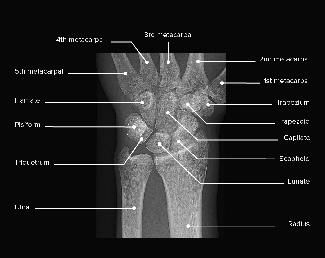
X-ray of a left wrist, showing normal anatomy and no injuries
Image: “X-ray of normal wrist” by Mikael Häggström, M.D. License: CC0 1.0, edited by Lecturio.The wrist is able to move in multiple directions, as it functions as a stable platform for the hand Hand The hand constitutes the distal part of the upper limb and provides the fine, precise movements needed in activities of daily living. It consists of 5 metacarpal bones and 14 phalanges, as well as numerous muscles innervated by the median and ulnar nerves. Hand: Anatomy.
| Joint | Type | Components | Ligaments | Function |
|---|---|---|---|---|
| Distal radioulnar | Pivot joint | Head of ulna Ulna The inner and longer bone of the forearm. Forearm: Anatomy and ulnar notch on radius Radius The outer shorter of the two bones of the forearm, lying parallel to the ulna and partially revolving around it. Forearm: Anatomy |
|
Supination-pronation |
| Radiocarpal | Synovial ellipsoid joint | Distal radius Radius The outer shorter of the two bones of the forearm, lying parallel to the ulna and partially revolving around it. Forearm: Anatomy and proximal row of carpal bones Carpal bones The eight bones of the wrist: scaphoid bone; lunate bone; triquetrum bone; pisiform bone; trapezium bone; trapezoid bone; capitate bone; and hamate bone. Carpal Tunnel Syndrome (except pisiform) |
|
|
| Midcarpal | Compound synovial | Proximal and distal row of carpal bones Carpal bones The eight bones of the wrist: scaphoid bone; lunate bone; triquetrum bone; pisiform bone; trapezium bone; trapezoid bone; capitate bone; and hamate bone. Carpal Tunnel Syndrome | Intercarpal, volar, dorsal, radial, and ulnar ligaments | Sliding and gliding |
| Intercarpal | Amphiarthroses | Among the carpal bones Carpal bones The eight bones of the wrist: scaphoid bone; lunate bone; triquetrum bone; pisiform bone; trapezium bone; trapezoid bone; capitate bone; and hamate bone. Carpal Tunnel Syndrome of each row | Intercarpal, dorsal, and volar ligaments | Stabilize the wrist |
| Carpometacarpal | Amphiarthroses (2nd–5th) | Distal carpal and 2nd–5th metacarpals | Dorsal and palmar carpometacarpal ligaments | Limited gliding |
| Saddle (thumb) | Trapezium and 1st metacarpal | Intermetacarpal, dorsoradial, collateral, and volar ligaments |
|
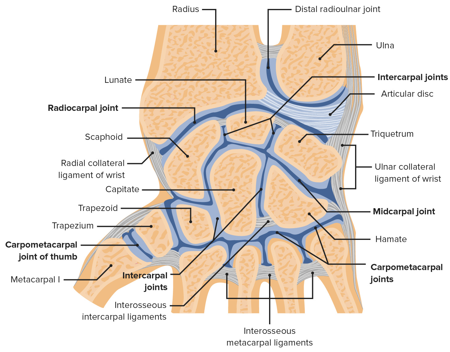
Coronal cross-section of the right wrist featuring the distal radioulnar, radiocarpal (labeled wrist joint), mid carpal, intercarpal, and carpometacarpal joints of the wrist
Image by Lecturio.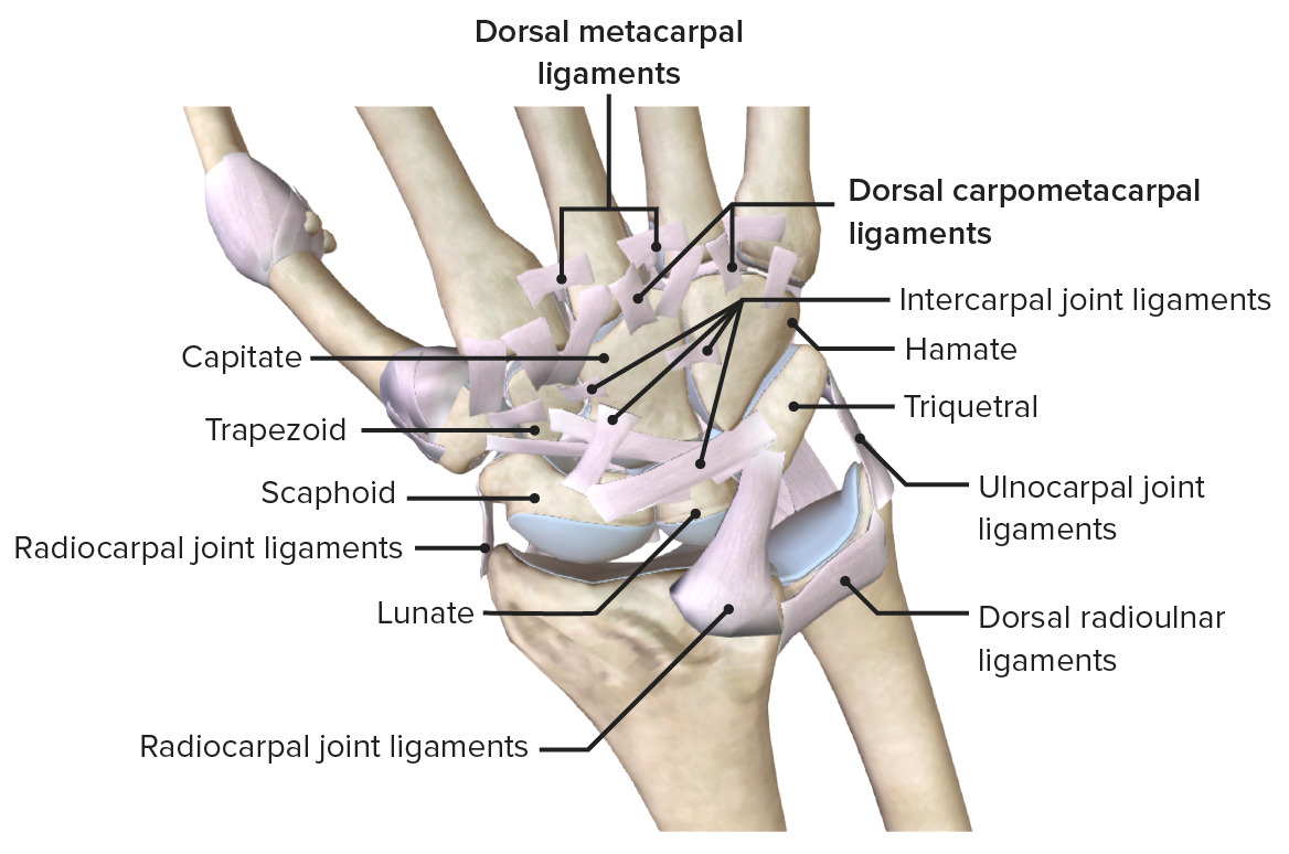
Posterior view of the wrist, featuring its supporting ligaments
Image by BioDigital, edited by Lecturio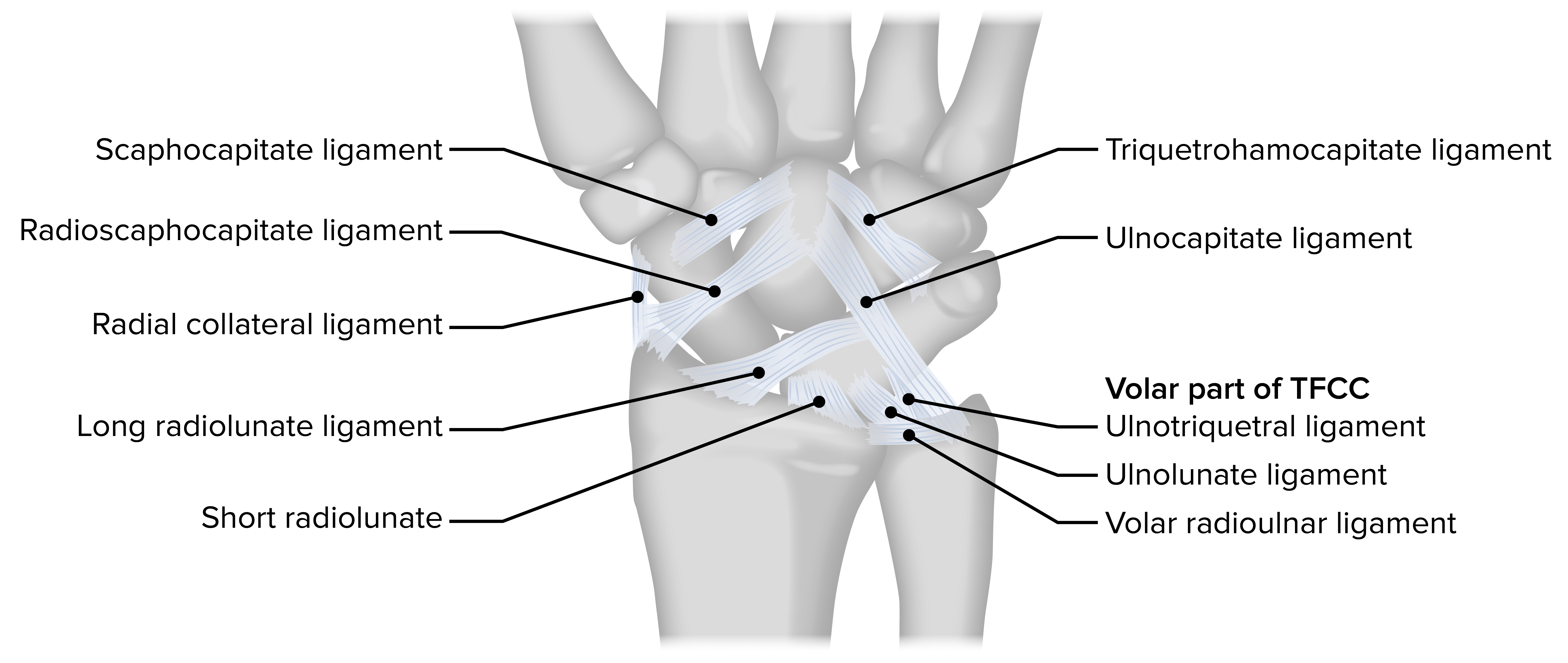
Extrinsic palmar carpal ligaments of the wrist:
TFCC: triangular fibrocartilage complex
The wrist acts as a transition between the forearm Forearm The forearm is the region of the upper limb between the elbow and the wrist. The term “forearm” is used in anatomy to distinguish this area from the arm, a term that is commonly used to describe the entire upper limb. The forearm consists of 2 long bones (the radius and the ulna), the interosseous membrane, and multiple arteries, nerves, and muscles. Forearm: Anatomy and hand Hand The hand constitutes the distal part of the upper limb and provides the fine, precise movements needed in activities of daily living. It consists of 5 metacarpal bones and 14 phalanges, as well as numerous muscles innervated by the median and ulnar nerves. Hand: Anatomy. The table below summarizes the forearm Forearm The forearm is the region of the upper limb between the elbow and the wrist. The term “forearm” is used in anatomy to distinguish this area from the arm, a term that is commonly used to describe the entire upper limb. The forearm consists of 2 long bones (the radius and the ulna), the interosseous membrane, and multiple arteries, nerves, and muscles. Forearm: Anatomy muscles and their associated movements. The name of each muscle is a reflection of the action of the muscle on the wrist and hand Hand The hand constitutes the distal part of the upper limb and provides the fine, precise movements needed in activities of daily living. It consists of 5 metacarpal bones and 14 phalanges, as well as numerous muscles innervated by the median and ulnar nerves. Hand: Anatomy.
| Movement | Muscle |
|---|---|
| Flexion Flexion Examination of the Upper Limbs | Main:
|
| Extension Extension Examination of the Upper Limbs | Main:
|
| Abduction Abduction Examination of the Upper Limbs | The following extensor and flexor muscles work in tandem to cause
abduction
Abduction
Examination of the Upper Limbs:
|
| Adduction Adduction Examination of the Upper Limbs | The following extensor and flexor muscles work in tandem to cause
adduction
Adduction
Examination of the Upper Limbs:
|
The carpal tunnel Carpal Tunnel The carpal tunnel is formed by the transverse carpal ligament (flexor retinaculum) superiorly and the carpal bones inferiorly. Carpal Tunnel Syndrome is formed by the following:
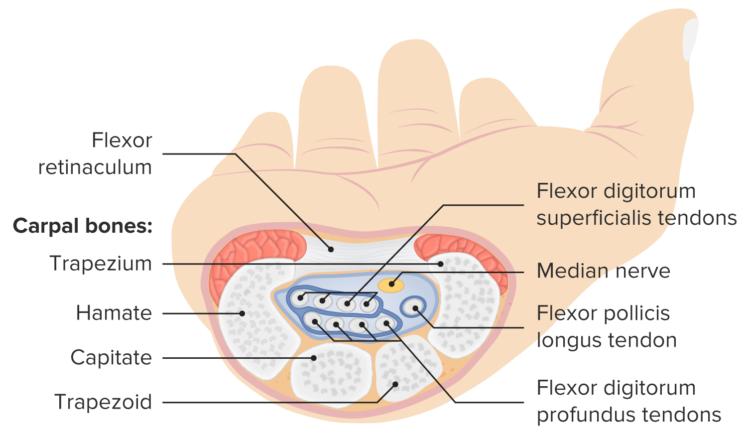
Structure and contents of the carpal tunnel
Image by Lecturio.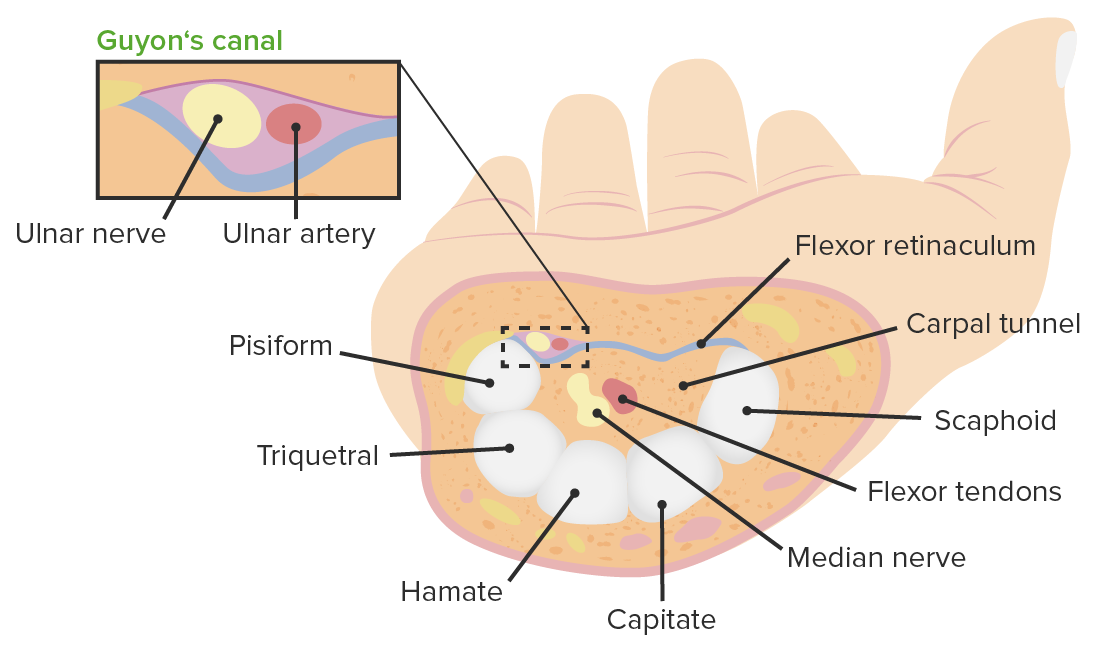
Cross-section of the wrist showcasing Guyon’s canal containing the ulnar artery and nerve, superior to the flexor retinaculum and carpal tunnel
Image by Lecturio.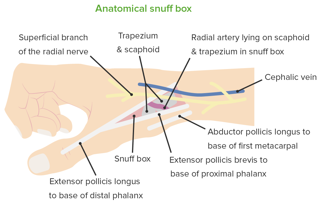
Medial view of the hand and wrist, featuring the borders and contents of the anatomical snuff box
Image by Lecturio.