Achieve Mastery of Medical Concepts

Study for medical school and boards with Lecturio

Hypersensitivity Pneumonitis

Hypersensitivity pneumonitis Pneumonitis Human Herpesvirus 6 and 7 (HP), previously called extrinsic allergic alveolitis, is an immunologically induced inflammatory disease affecting the alveoli Alveoli Small polyhedral outpouchings along the walls of the alveolar sacs, alveolar ducts and terminal bronchioles through the walls of which gas exchange between alveolar air and pulmonary capillary blood takes place. Acute Respiratory Distress Syndrome (ARDS), bronchioles Bronchioles The small airways branching off the tertiary bronchi. Terminal bronchioles lead into several orders of respiratory bronchioles which in turn lead into alveolar ducts and then into pulmonary alveoli. Bronchial Tree: Anatomy, and lung parenchyma. It is caused by repeated inhalation of an inciting agent in a susceptible host that triggers first a type III (immune complex-mediated) hypersensitivity reaction in the acute phase Acute phase Short Bowel Syndrome and then a type IV (delayed) reaction in the subacute and chronic phases. The clinical presentation of acute HP includes cough, fever Fever Fever is defined as a measured body temperature of at least 38°C (100.4°F). Fever is caused by circulating endogenous and/or exogenous pyrogens that increase levels of prostaglandin E2 in the hypothalamus. Fever is commonly associated with chills, rigors, sweating, and flushing of the skin. Fever, and malaise Malaise Tick-borne Encephalitis Virus, while subacute and chronic forms present as insidious onset of a cough and dyspnea Dyspnea Dyspnea is the subjective sensation of breathing discomfort. Dyspnea is a normal manifestation of heavy physical or psychological exertion, but also may be caused by underlying conditions (both pulmonary and extrapulmonary). Dyspnea over weeks to months. Diagnosis is aided by high-resolution CT High-resolution CT Imaging of the Lungs and Pleura scans and bronchoalveolar lavage Bronchoalveolar lavage Washing out of the lungs with saline or mucolytic agents for diagnostic or therapeutic purposes. It is very useful in the diagnosis of diffuse pulmonary infiltrates in immunosuppressed patients. Pulmonary Fibrosis. Management includes avoiding the inciting agent and administration of steroids Steroids A group of polycyclic compounds closely related biochemically to terpenes. They include cholesterol, numerous hormones, precursors of certain vitamins, bile acids, alcohols (sterols), and certain natural drugs and poisons. Steroids have a common nucleus, a fused, reduced 17-carbon atom ring system, cyclopentanoperhydrophenanthrene. Most steroids also have two methyl groups and an aliphatic side-chain attached to the nucleus. Benign Liver Tumors in subacute and chronic cases. Early treatment has a good prognosis Prognosis A prediction of the probable outcome of a disease based on a individual's condition and the usual course of the disease as seen in similar situations. Non-Hodgkin Lymphomas, but long-term exposure can cause permanent scarring Scarring Inflammation and fibrosis Fibrosis Any pathological condition where fibrous connective tissue invades any organ, usually as a consequence of inflammation or other injury. Bronchiolitis Obliterans.

Last updated: Mar 28, 2023

Editorial responsibility: Stanley Oiseth, Lindsay Jones, Evelin Maza

Overview

Definition

Hypersensitivity pneumonitis Pneumonitis Human Herpesvirus 6 and 7 (HP), also called extrinsic allergic alveolitis, is an immunologically induced inflammatory disease affecting the lung parenchyma, alveoli Alveoli Small polyhedral outpouchings along the walls of the alveolar sacs, alveolar ducts and terminal bronchioles through the walls of which gas exchange between alveolar air and pulmonary capillary blood takes place. Acute Respiratory Distress Syndrome (ARDS), and bronchioles Bronchioles The small airways branching off the tertiary bronchi. Terminal bronchioles lead into several orders of respiratory bronchioles which in turn lead into alveolar ducts and then into pulmonary alveoli. Bronchial Tree: Anatomy. This disorder is caused by repeated inhalation of inciting agents in a susceptible host.

Interstitial lung disease caused by hypersensitivity reactions:

  • Acute phase Acute phase Short Bowel Syndrome: type III (immune-complex–mediated)
  • Subacute and chronic phases: type IV (delayed hypersensitivity/T-cell–mediated)
Ild classification

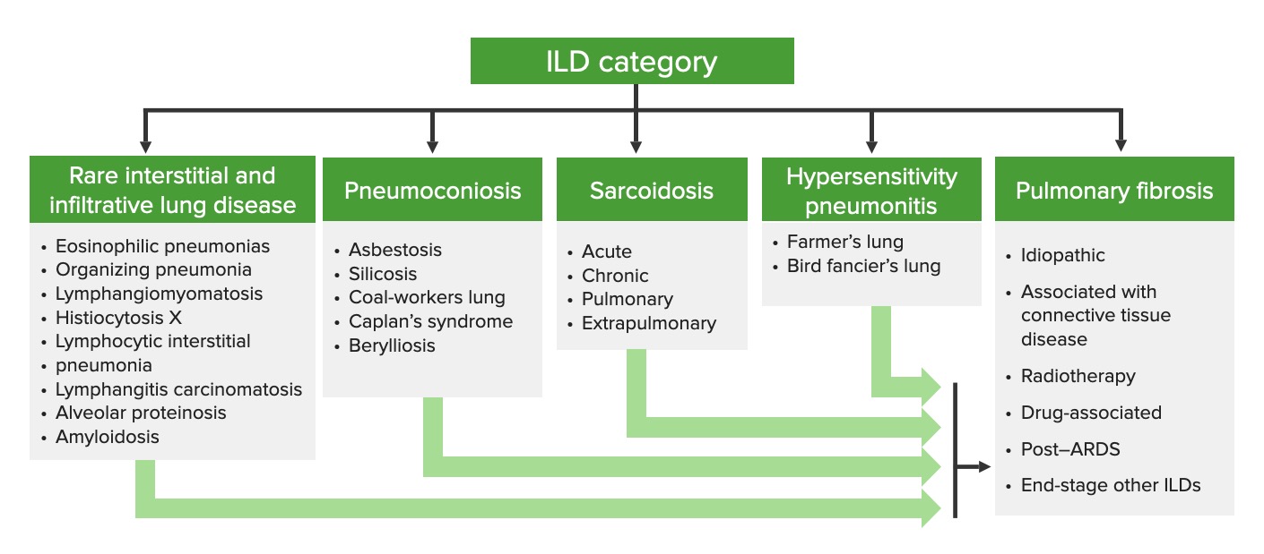
Categorization of interstitial lung diseases
ILD: interstitial lung disease

Image by Lecturio.

Classification

Classification is based on duration of illness and frequency, duration, and intensity of exposure. There is considerable variability in presentation and course.

  • Acute HP:
    • Acute onset of symptoms following a massive exposure to the inciting agent
    • Duration < 6 months
    • Disease is reversible.
  • Subacute HP:  mild, intermittent, or progressive symptoms developing over weeks due to repeated and continued exposure to the inciting antigen Antigen Substances that are recognized by the immune system and induce an immune reaction. Vaccination
  • Chronic HP: 
    • Repeated and continuous low-level exposure to the inciting antigenic agent 
    • Duration > 6 months
    • Disease is less reversible with ↑ chance of progression.

Epidemiology

  • Exact prevalence Prevalence The total number of cases of a given disease in a specified population at a designated time. It is differentiated from incidence, which refers to the number of new cases in the population at a given time. Measures of Disease Frequency unknown
  • Approximate prevalence Prevalence The total number of cases of a given disease in a specified population at a designated time. It is differentiated from incidence, which refers to the number of new cases in the population at a given time. Measures of Disease Frequency:
    • 1–3 per 100,000 persons
    • 11 per 100,000 persons age ≥ 65 years
  • Age at onset: usually 30–60 years
  • Gender Gender Gender Dysphoria:
    • Men > women due to gender Gender Gender Dysphoria bias Bias Epidemiological studies are designed to evaluate a hypothesized relationship between an exposure and an outcome; however, the existence and/or magnitude of these relationships may be erroneously affected by the design and execution of the study itself or by conscious or unconscious errors perpetrated by the investigators or the subjects. These systematic errors are called biases. Types of Biases in affected occupations 
    • Women are equally as susceptible as men when exposed.
  • Most common types:
    • Farmer’s lung
    • Bird fancier’s lung
    • Chemical worker’s lung

Etiology

More than 300 etiologies of HP have been reported across a wide range of exposures involving airborne antigens.

Selected etiologic agents for HP
Disease Exposure Antigens
Farmer’s lung
  • Forking moldy hay
  • Moldy vegetable material
  • Thermophilic Thermophilic Campylobacter actinomycetes
  • Fungi Fungi A kingdom of eukaryotic, heterotrophic organisms that live parasitically as saprobes, including mushrooms; yeasts; smuts, molds, etc. They reproduce either sexually or asexually, and have life cycles that range from simple to complex. Filamentous fungi, commonly known as molds, refer to those that grow as multicellular colonies. Mycology (e.g., Aspergillus Aspergillus A genus of mitosporic fungi containing about 100 species and eleven different teleomorphs in the family trichocomaceae. Echinocandins umbrasus)
Bird fancier’s lung
  • Pigeons
  • Parakeets
  • Chickens
  • Turkeys
Proteins Proteins Linear polypeptides that are synthesized on ribosomes and may be further modified, crosslinked, cleaved, or assembled into complex proteins with several subunits. The specific sequence of amino acids determines the shape the polypeptide will take, during protein folding, and the function of the protein. Energy Homeostasis present on feathers and in excreta
Chemical worker’s lung Many different chemicals Isocyanates
Byssinosis Working in the textile industry
  • Cotton
  • Linen
  • Hemp fibers
Bagassosis Moldy sugar cane Thermophilic Thermophilic Campylobacter actinomycetes
Silo filler’s lung Fermentation in silos Nitrogen Nitrogen An element with the atomic symbol n, atomic number 7, and atomic weight [14. 00643; 14. 00728]. Nitrogen exists as a diatomic gas and makes up about 78% of the earth’s atmosphere by volume. It is a constituent of proteins and nucleic acids and found in all living cells. Urea Cycle dioxide gas
Maltworker’s lung Turning germinating barley
  • A. clavatus
  • A. fumigatus A. fumigatus A species of imperfect fungi from which the antibiotic fumigatin is obtained. Its spores may cause respiratory infection in birds and mammals. Aspergillus/Aspergillosis
Humidifier fever Fever Fever is defined as a measured body temperature of at least 38°C (100.4°F). Fever is caused by circulating endogenous and/or exogenous pyrogens that increase levels of prostaglandin E2 in the hypothalamus. Fever is commonly associated with chills, rigors, sweating, and flushing of the skin. Fever Contaminated humidifying systems in air conditioners
  • Thermophilic Thermophilic Campylobacter actinomycetes
  • Mycobacterium Mycobacterium Mycobacterium is a genus of the family Mycobacteriaceae in the phylum Actinobacteria. Mycobacteria comprise more than 150 species of facultative intracellular bacilli that are mostly obligate aerobes. Mycobacteria are responsible for multiple human infections including serious diseases, such as tuberculosis (M. tuberculosis), leprosy (M. leprae), and M. avium complex infections. Mycobacterium
  • Candida albicans Candida albicans A unicellular budding fungus which is the principal pathogenic species causing candidiasis (moniliasis). Candida/Candidiasis
  • Aureobasidium pullulans
Mushroom workers lung Turning mushroom compost Thermophilic Thermophilic Campylobacter actinomycetes
Cheese washer’s lung Moldy cheese Penicillin Penicillin Rheumatic Fever casei
Winemaker’s lung Mold Mold Mycology on grapes Botrytis

Pathophysiology

Initially during acute HP, the disease process is driven by a type III (immune-complex–mediated) hypersensitivity reaction. Continued exposure shifts the process to a type IV delayed hypersensitivity reaction Delayed Hypersensitivity Reaction Glycopeptides mediated by T cells T cells Lymphocytes responsible for cell-mediated immunity. Two types have been identified – cytotoxic (t-lymphocytes, cytotoxic) and helper T-lymphocytes (t-lymphocytes, helper-inducer). They are formed when lymphocytes circulate through the thymus gland and differentiate to thymocytes. When exposed to an antigen, they divide rapidly and produce large numbers of new T cells sensitized to that antigen. T cells: Types and Functions.

Pathogenesis is directly related to antibodies Antibodies Immunoglobulins (Igs), also known as antibodies, are glycoprotein molecules produced by plasma cells that act in immune responses by recognizing and binding particular antigens. The various Ig classes are IgG (the most abundant), IgM, IgE, IgD, and IgA, which differ in their biologic features, structure, target specificity, and distribution. Immunoglobulins: Types and Functions targeting specific protein antigens:

  • Proteins Proteins Linear polypeptides that are synthesized on ribosomes and may be further modified, crosslinked, cleaved, or assembled into complex proteins with several subunits. The specific sequence of amino acids determines the shape the polypeptide will take, during protein folding, and the function of the protein. Energy Homeostasis are often on microbes, but disease is not caused by direct infection by these microbial agents.
  • Inciting proteins Proteins Linear polypeptides that are synthesized on ribosomes and may be further modified, crosslinked, cleaved, or assembled into complex proteins with several subunits. The specific sequence of amino acids determines the shape the polypeptide will take, during protein folding, and the function of the protein. Energy Homeostasis may be organic or nonorganic.

Acute HP (type III immune-complex–mediated hypersensitivity reaction):

  • Immune-complex formation:
    • Specific antigens trigger Trigger The type of signal that initiates the inspiratory phase by the ventilator Invasive Mechanical Ventilation IgG antibody formation.
    • Antigens bind BIND Hyperbilirubinemia of the Newborn antibodies Antibodies Immunoglobulins (Igs), also known as antibodies, are glycoprotein molecules produced by plasma cells that act in immune responses by recognizing and binding particular antigens. The various Ig classes are IgG (the most abundant), IgM, IgE, IgD, and IgA, which differ in their biologic features, structure, target specificity, and distribution. Immunoglobulins: Types and Functions and form circulating immune complexes Immune complexes The complex formed by the binding of antigen and antibody molecules. The deposition of large antigen-antibody complexes leading to tissue damage causes immune complex diseases. C3 Deficiency.
  • Immune-complex deposition within the lung parenchyma, alveoli Alveoli Small polyhedral outpouchings along the walls of the alveolar sacs, alveolar ducts and terminal bronchioles through the walls of which gas exchange between alveolar air and pulmonary capillary blood takes place. Acute Respiratory Distress Syndrome (ARDS), and bronchioles Bronchioles The small airways branching off the tertiary bronchi. Terminal bronchioles lead into several orders of respiratory bronchioles which in turn lead into alveolar ducts and then into pulmonary alveoli. Bronchial Tree: Anatomy
  • Activation of complement via the classical pathway Classical pathway Complement activation initiated by the binding of complement C1 to antigen-antibody complexes at the complement C1q subunit. This leads to the sequential activation of complement C1r and complement C1s subunits. Activated C1s cleaves complement C4 and complement C2 forming the membrane-bound classical C3 convertase (c4b2a) and the subsequent C5 convertase (c4b2a3b) leading to cleavage of complement C5 and the assembly of complement membrane attack complex. Innate Immunity: Barriers, Complement, and Cytokines:
    • Leads to infiltration of PMNs (primarily neutrophils Neutrophils Granular leukocytes having a nucleus with three to five lobes connected by slender threads of chromatin, and cytoplasm containing fine inconspicuous granules and stainable by neutral dyes. Innate Immunity: Phagocytes and Antigen Presentation) and macrophages Macrophages The relatively long-lived phagocytic cell of mammalian tissues that are derived from blood monocytes. Main types are peritoneal macrophages; alveolar macrophages; histiocytes; kupffer cells of the liver; and osteoclasts. They may further differentiate within chronic inflammatory lesions to epithelioid cells or may fuse to form foreign body giant cells or langhans giant cells. Innate Immunity: Phagocytes and Antigen Presentation
    • Inflammatory tissue damage

Subacute and chronic HP (type IV delayed hypersensitivity reaction Delayed Hypersensitivity Reaction Glycopeptides mediated by T cells T cells Lymphocytes responsible for cell-mediated immunity. Two types have been identified – cytotoxic (t-lymphocytes, cytotoxic) and helper T-lymphocytes (t-lymphocytes, helper-inducer). They are formed when lymphocytes circulate through the thymus gland and differentiate to thymocytes. When exposed to an antigen, they divide rapidly and produce large numbers of new T cells sensitized to that antigen. T cells: Types and Functions):

  • Macrophages Macrophages The relatively long-lived phagocytic cell of mammalian tissues that are derived from blood monocytes. Main types are peritoneal macrophages; alveolar macrophages; histiocytes; kupffer cells of the liver; and osteoclasts. They may further differentiate within chronic inflammatory lesions to epithelioid cells or may fuse to form foreign body giant cells or langhans giant cells. Innate Immunity: Phagocytes and Antigen Presentation and monocytes Monocytes Large, phagocytic mononuclear leukocytes produced in the vertebrate bone marrow and released into the blood; contain a large, oval or somewhat indented nucleus surrounded by voluminous cytoplasm and numerous organelles. Innate Immunity: Phagocytes and Antigen Presentation engulf and present antigens to T cells T cells Lymphocytes responsible for cell-mediated immunity. Two types have been identified – cytotoxic (t-lymphocytes, cytotoxic) and helper T-lymphocytes (t-lymphocytes, helper-inducer). They are formed when lymphocytes circulate through the thymus gland and differentiate to thymocytes. When exposed to an antigen, they divide rapidly and produce large numbers of new T cells sensitized to that antigen. T cells: Types and Functions.
  • T cells T cells Lymphocytes responsible for cell-mediated immunity. Two types have been identified – cytotoxic (t-lymphocytes, cytotoxic) and helper T-lymphocytes (t-lymphocytes, helper-inducer). They are formed when lymphocytes circulate through the thymus gland and differentiate to thymocytes. When exposed to an antigen, they divide rapidly and produce large numbers of new T cells sensitized to that antigen. T cells: Types and Functions release inflammatory mediators (e.g., cytokines Cytokines Non-antibody proteins secreted by inflammatory leukocytes and some non-leukocytic cells, that act as intercellular mediators. They differ from classical hormones in that they are produced by a number of tissue or cell types rather than by specialized glands. They generally act locally in a paracrine or autocrine rather than endocrine manner. Adaptive Immune Response, chemokines Chemokines Class of pro-inflammatory cytokines that have the ability to attract and activate leukocytes. They can be divided into at least three structural branches: c; cc; and cxc; according to variations in a shared cysteine motif. Adaptive Cell-mediated Immunity).
  • Result in tissue damage and interstitial pneumonitis Pneumonitis Human Herpesvirus 6 and 7 
  • T cells T cells Lymphocytes responsible for cell-mediated immunity. Two types have been identified – cytotoxic (t-lymphocytes, cytotoxic) and helper T-lymphocytes (t-lymphocytes, helper-inducer). They are formed when lymphocytes circulate through the thymus gland and differentiate to thymocytes. When exposed to an antigen, they divide rapidly and produce large numbers of new T cells sensitized to that antigen. T cells: Types and Functions are not able to fully destroy antigens engulfed by macrophages Macrophages The relatively long-lived phagocytic cell of mammalian tissues that are derived from blood monocytes. Main types are peritoneal macrophages; alveolar macrophages; histiocytes; kupffer cells of the liver; and osteoclasts. They may further differentiate within chronic inflammatory lesions to epithelioid cells or may fuse to form foreign body giant cells or langhans giant cells. Innate Immunity: Phagocytes and Antigen Presentation noncaseating granuloma Noncaseating Granuloma Inflammation formation
  • May progress to fibrosis Fibrosis Any pathological condition where fibrous connective tissue invades any organ, usually as a consequence of inflammation or other injury. Bronchiolitis Obliterans (mechanism poorly understood)

Clinical Presentation

Acute hypersensitivity pneumonitis Pneumonitis Human Herpesvirus 6 and 7

Acute HP usually follows massive exposure to an inciting agent:

  • Abrupt onset of symptoms (4–8 hours) after exposure
  • Symptom resolution within 12–48 hours after removal of inciting antigen Antigen Substances that are recognized by the immune system and induce an immune reaction. Vaccination
  • Aside from history of exposure, may appear clinically similar to a viral or bacterial infection
  • Symptoms:
    • Abrupt onset of a cough
    • Chest tightness
    • Dyspnea Dyspnea Dyspnea is the subjective sensation of breathing discomfort. Dyspnea is a normal manifestation of heavy physical or psychological exertion, but also may be caused by underlying conditions (both pulmonary and extrapulmonary). Dyspnea without wheezing Wheezing Wheezing is an abnormal breath sound characterized by a whistling noise that can be relatively high-pitched and shrill (more common) or coarse. Wheezing is produced by the movement of air through narrowed or compressed small (intrathoracic) airways. Wheezing
    • Fever Fever Fever is defined as a measured body temperature of at least 38°C (100.4°F). Fever is caused by circulating endogenous and/or exogenous pyrogens that increase levels of prostaglandin E2 in the hypothalamus. Fever is commonly associated with chills, rigors, sweating, and flushing of the skin. Fever
    • Chills Chills The sudden sensation of being cold. It may be accompanied by shivering. Fever
    • Malaise Malaise Tick-borne Encephalitis Virus
    • Nausea Nausea An unpleasant sensation in the stomach usually accompanied by the urge to vomit. Common causes are early pregnancy, sea and motion sickness, emotional stress, intense pain, food poisoning, and various enteroviruses. Antiemetics
    • Headache Headache The symptom of pain in the cranial region. It may be an isolated benign occurrence or manifestation of a wide variety of headache disorders. Brain Abscess
  • Clinical exam:

Subacute and chronic hypersensitivity pneumonitis Pneumonitis Human Herpesvirus 6 and 7

  • Insidious, gradual onset over weeks to months
  • The patient may not have a history of acute HP or may not be able to identify a trigger Trigger The type of signal that initiates the inspiratory phase by the ventilator Invasive Mechanical Ventilation.
  • Symptoms:
    • Progressive cough 
    • Dyspnea Dyspnea Dyspnea is the subjective sensation of breathing discomfort. Dyspnea is a normal manifestation of heavy physical or psychological exertion, but also may be caused by underlying conditions (both pulmonary and extrapulmonary). Dyspnea
    • Fatigue Fatigue The state of weariness following a period of exertion, mental or physical, characterized by a decreased capacity for work and reduced efficiency to respond to stimuli. Fibromyalgia
    • Anorexia Anorexia The lack or loss of appetite accompanied by an aversion to food and the inability to eat. It is the defining characteristic of the disorder anorexia nervosa. Anorexia Nervosa and weight loss Weight loss Decrease in existing body weight. Bariatric Surgery
  • Clinical exam:

Diagnosis

Laboratory and pathology

  • Serum:
  • Bronchoalveolar lavage Bronchoalveolar lavage Washing out of the lungs with saline or mucolytic agents for diagnostic or therapeutic purposes. It is very useful in the diagnosis of diffuse pulmonary infiltrates in immunosuppressed patients. Pulmonary Fibrosis:
    • Most sensitive test for HP
    • Shows marked lymphocytosis Lymphocytosis WBCs develop from stem cells in the bone marrow and are called leukocytes when circulating in the bloodstream. Lymphocytes are 1 of the 5 subclasses of WBCs. Lymphocytosis is an increase in the number or proportion of the lymphocyte subclass of WBCs, often as a result of an immune response to infection (known as reactive lymphocytosis). Lymphocytosis (due to ↑ T lymphocytes T lymphocytes Lymphocytes responsible for cell-mediated immunity. Two types have been identified – cytotoxic (t-lymphocytes, cytotoxic) and helper T-lymphocytes (t-lymphocytes, helper-inducer). They are formed when lymphocytes circulate through the thymus gland and differentiate to thymocytes. When exposed to an antigen, they divide rapidly and produce large numbers of new T cells sensitized to that antigen. T cells: Types and Functions present in type IV hypersensitivity reactions)
    • ↓ CD4 (helper T cells T cells Lymphocytes responsible for cell-mediated immunity. Two types have been identified – cytotoxic (t-lymphocytes, cytotoxic) and helper T-lymphocytes (t-lymphocytes, helper-inducer). They are formed when lymphocytes circulate through the thymus gland and differentiate to thymocytes. When exposed to an antigen, they divide rapidly and produce large numbers of new T cells sensitized to that antigen. T cells: Types and Functions) to CD8 ( cytotoxic Cytotoxic Parvovirus B19 T cells T cells Lymphocytes responsible for cell-mediated immunity. Two types have been identified – cytotoxic (t-lymphocytes, cytotoxic) and helper T-lymphocytes (t-lymphocytes, helper-inducer). They are formed when lymphocytes circulate through the thymus gland and differentiate to thymocytes. When exposed to an antigen, they divide rapidly and produce large numbers of new T cells sensitized to that antigen. T cells: Types and Functions) ratio
    • ↑ PMNs ( neutrophils Neutrophils Granular leukocytes having a nucleus with three to five lobes connected by slender threads of chromatin, and cytoplasm containing fine inconspicuous granules and stainable by neutral dyes. Innate Immunity: Phagocytes and Antigen Presentation) in acute disease
  • Biopsy Biopsy Removal and pathologic examination of specimens from the living body. Ewing Sarcoma:
    • Transbronchial (lower diagnostic yield) or surgical specimens may be obtained.
    • Lymphocyte infiltration into the:
      • Peribronchial tissue
      • Alveolar walls
    • Poorly formed granulomas Granulomas A relatively small nodular inflammatory lesion containing grouped mononuclear phagocytes, caused by infectious and noninfectious agents. Sarcoidosis located near respiratory or terminal bronchioles Bronchioles The small airways branching off the tertiary bronchi. Terminal bronchioles lead into several orders of respiratory bronchioles which in turn lead into alveolar ducts and then into pulmonary alveoli. Bronchial Tree: Anatomy
    • Fibrosis Fibrosis Any pathological condition where fibrous connective tissue invades any organ, usually as a consequence of inflammation or other injury. Bronchiolitis Obliterans in chronic disease
Hypersensitivity pneumonitis biopsy

Hypersensitivity pneumonitis (HP):
The cellular interstitium of subacute HP (A) is dominated by plasma cells (magnification in B). A typical poorly formed granuloma of HP is present in (B).
(A,B) H&E stain; (A) 40× original magnification; (B) 400× original magnification

Image: “My approach to interstitial lung disease using clinical, radiological and histopathological patterns” by Leslie KO. License: CC BY 2.0

Spirometry Spirometry Measurement of volume of air inhaled or exhaled by the lung. Pulmonary Function Tests

  • Restrictive pattern:
    • ↓ Forced vital capacity Vital capacity The volume of air that is exhaled by a maximal expiration following a maximal inspiration. Ventilation: Mechanics of Breathing (FVC; total forced exhaled volume)
    • Total lung capacity Total lung capacity The volume of air contained in the lungs at the end of a maximal inspiration. It is the equivalent to each of the following sums: vital capacity plus residual volume; inspiratory capacity plus functional residual capacity; tidal volume plus inspiratory reserve volume plus functional residual capacity; or tidal volume plus inspiratory reserve volume plus expiratory reserve volume plus residual volume. Ventilation: Mechanics of Breathing (TLC; total volume in lungs Lungs Lungs are the main organs of the respiratory system. Lungs are paired viscera located in the thoracic cavity and are composed of spongy tissue. The primary function of the lungs is to oxygenate blood and eliminate CO2. Lungs: Anatomy at the end of a maximal inspiration Inspiration Ventilation: Mechanics of Breathing)
    • Compliance Compliance Distensibility measure of a chamber such as the lungs (lung compliance) or bladder. Compliance is expressed as a change in volume per unit change in pressure. Veins: Histology ( lungs Lungs Lungs are the main organs of the respiratory system. Lungs are paired viscera located in the thoracic cavity and are composed of spongy tissue. The primary function of the lungs is to oxygenate blood and eliminate CO2. Lungs: Anatomy have difficulty expanding)
  • Obstructive (or mixed) patterns are also possible; more common in severe disease:
    • ↓ Forced expiratory volume in 1 second (FEV1):FVC ratio
    • ↓ Airflow ( bronchioles Bronchioles The small airways branching off the tertiary bronchi. Terminal bronchioles lead into several orders of respiratory bronchioles which in turn lead into alveolar ducts and then into pulmonary alveoli. Bronchial Tree: Anatomy are partially obstructed by inflammation Inflammation Inflammation is a complex set of responses to infection and injury involving leukocytes as the principal cellular mediators in the body’s defense against pathogenic organisms. Inflammation is also seen as a response to tissue injury in the process of wound healing. The 5 cardinal signs of inflammation are pain, heat, redness, swelling, and loss of function. Inflammation and fibrosis Fibrosis Any pathological condition where fibrous connective tissue invades any organ, usually as a consequence of inflammation or other injury. Bronchiolitis Obliterans)
  • Impaired gas diffusion Diffusion The tendency of a gas or solute to pass from a point of higher pressure or concentration to a point of lower pressure or concentration and to distribute itself throughout the available space. Diffusion, especially facilitated diffusion, is a major mechanism of biological transport. Peritoneal Dialysis and Hemodialysis across the alveolar membrane:
    • ↓ Diffusing capacity of the lung for carbon monoxide Carbon monoxide Carbon monoxide (CO). A poisonous colorless, odorless, tasteless gas. It combines with hemoglobin to form carboxyhemoglobin, which has no oxygen carrying capacity. The resultant oxygen deprivation causes headache, dizziness, decreased pulse and respiratory rates, unconsciousness, and death. Carbon Monoxide Poisoning ( DLCO DLCO Pulmonary Function Tests)
    • Always seen in chronic HP, usually in subacute HP, and occasionally in acute HP
  • Pulmonary function test Pulmonary function test Pulmonary function tests are a group of diagnostic procedures yielding useful, quantifiable information about the rate of the flow of air through the individual’s airways, lung capacity, and the efficiency of gas exchange in relation to time. The most commonly utilized tests include spirometry (before and after bronchodilator use), lung volumes, and quantitation of diffusing capacity for carbon monoxide (CO). The tests can be influenced by the individual’s effort/fatigue, disease state, or anatomical malformation. Pulmonary Function Tests results may revert to normal in acute/subacute forms with no further exposure.

Imaging

  • High-resolution CT High-resolution CT Imaging of the Lungs and Pleura of the chest:
    • More sensitive for HP than chest X-ray X-ray Penetrating electromagnetic radiation emitted when the inner orbital electrons of an atom are excited and release radiant energy. X-ray wavelengths range from 1 pm to 10 nm. Hard x-rays are the higher energy, shorter wavelength x-rays. Soft x-rays or grenz rays are less energetic and longer in wavelength. The short wavelength end of the x-ray spectrum overlaps the gamma rays wavelength range. The distinction between gamma rays and x-rays is based on their radiation source. Pulmonary Function Tests
    • Findings develop with worsening disease:
      • Mid-to-upper zone predominance of centrilobular ground-glass or nodular opacities
      • Signs of air trapping (compare inspiratory and expiratory images)
    • As disease progresses:
      • ↑ Reticulation (indicates fibrosis Fibrosis Any pathological condition where fibrous connective tissue invades any organ, usually as a consequence of inflammation or other injury. Bronchiolitis Obliterans)
      • Traction bronchiectasis Bronchiectasis Bronchiectasis is a chronic disease of the airways that results from permanent bronchial distortion. This results from a continuous cycle of inflammation, bronchial damage and dilation, impaired clearance of secretions, and recurrent infections. Bronchiectasis
      • Honeycombing
      • Signs of emphysema Emphysema Enlargement of air spaces distal to the terminal bronchioles where gas-exchange normally takes place. This is usually due to destruction of the alveolar wall. Pulmonary emphysema can be classified by the location and distribution of the lesions. Chronic Obstructive Pulmonary Disease (COPD)
  • Chest X-ray X-ray Penetrating electromagnetic radiation emitted when the inner orbital electrons of an atom are excited and release radiant energy. X-ray wavelengths range from 1 pm to 10 nm. Hard x-rays are the higher energy, shorter wavelength x-rays. Soft x-rays or grenz rays are less energetic and longer in wavelength. The short wavelength end of the x-ray spectrum overlaps the gamma rays wavelength range. The distinction between gamma rays and x-rays is based on their radiation source. Pulmonary Function Tests:
    • No specific or distinctive changes in HP
    • Often normal and/or revert to normal following an acute episode
    • Typical findings:
      • Poorly defined, patchy, or diffuse infiltrates 
      • Micronodular or reticular opacities 
      • Progressive fibrotic changes in more severe disease
      • Findings are classically in the upper lobes (different from other pulmonary fibrosis Fibrosis Any pathological condition where fibrous connective tissue invades any organ, usually as a consequence of inflammation or other injury. Bronchiolitis Obliterans).
Radiological and pathological assessment hypersensitivity pneumonitis

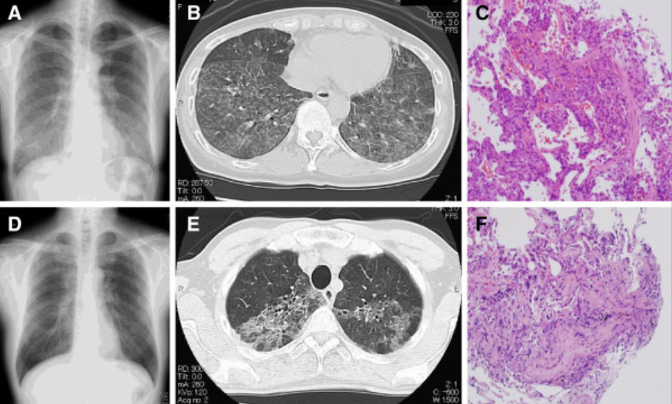
Radiological and pathological assessment for a husband and wife in Japan who both developed hypersensitivity pneumonitis caused by inhalation of Trichosporon asahii which was growing in the straw mats of their home.
Wife: Chest x-ray on the day of admission shows mild infiltrates predominantly in the middle to lower lung fields with multiple scattered nodular lesions (Panel A).
Thoracic CT (computed tomography) taken on the same day confirms that these lesions correspond to ground-glass opacities and abundant centrilobular nodules, predominantly located in the bilateral lower lobes (Panel B).
Specimens obtained from transbronchial lung biopsy (TBLB) demonstrate organization within the peribronchial area with alveolitis, suggesting transbronchial spread (Panel C).
Husband: Chest x-ray (Panel D) and thoracic CT (Panel E) show ground glass opacities in the upper lungs on both sides.
The specimens obtained from TBLB at his local hospital reveals organizing tissue within the peribronchial area with alveolitis, suggesting transbronchial spread (Panel F).

Image: “Familial summer-type hypersensitivity pneumonitis in Japan: two case reports and review of the literature” by Nakajima A, Saraya T, Mori T, Ikeda R, Sugita T, Watanabe T, Fujiwara M, Takizawa H, Goto H. License: CC BY 2.0

Inhalation challenge testing

  • Reexpose patient to the suspected environmental trigger Trigger The type of signal that initiates the inspiratory phase by the ventilator Invasive Mechanical Ventilation.
  • Should be done in a hospital setting with appropriate support
  • Look for development of symptoms and for spirometry Spirometry Measurement of volume of air inhaled or exhaled by the lung. Pulmonary Function Tests and radiographic findings consistent with HP.

Diagnostic criteria

Exact diagnostic criteria are still being debated but generally include the following:

  1. Known exposure to an offending antigen Antigen Substances that are recognized by the immune system and induce an immune reaction. Vaccination identified by:
    • History of exposure
    • IgG serum antibodies Antibodies Immunoglobulins (Igs), also known as antibodies, are glycoprotein molecules produced by plasma cells that act in immune responses by recognizing and binding particular antigens. The various Ig classes are IgG (the most abundant), IgM, IgE, IgD, and IgA, which differ in their biologic features, structure, target specificity, and distribution. Immunoglobulins: Types and Functions against the antigen Antigen Substances that are recognized by the immune system and induce an immune reaction. Vaccination
  2. Compatible clinical, radiographic, or physiologic findings:
  3. Bronchoalveolar lavage Bronchoalveolar lavage Washing out of the lungs with saline or mucolytic agents for diagnostic or therapeutic purposes. It is very useful in the diagnosis of diffuse pulmonary infiltrates in immunosuppressed patients. Pulmonary Fibrosis (BAL) with lymphocytosis Lymphocytosis WBCs develop from stem cells in the bone marrow and are called leukocytes when circulating in the bloodstream. Lymphocytes are 1 of the 5 subclasses of WBCs. Lymphocytosis is an increase in the number or proportion of the lymphocyte subclass of WBCs, often as a result of an immune response to infection (known as reactive lymphocytosis). Lymphocytosis > 20% (often > 50%)
  4. Positive inhalation challenge test:
    • Reexposure to the environment
    • Inhalation challenge in a hospital setting
  5. Histopathology consistent with HP:
    • Poorly formed, noncaseating granulomas Noncaseating granulomas Crohn’s Disease
    • Mononuclear-cell infiltrates (mostly lymphocytes Lymphocytes Lymphocytes are heterogeneous WBCs involved in immune response. Lymphocytes develop from the bone marrow, starting from hematopoietic stem cells (HSCs) and progressing to common lymphoid progenitors (CLPs). B and T lymphocytes and natural killer (NK) cells arise from the lineage. Lymphocytes: Histology)

Management

  • Eliminate (preferred) or reduce exposure to the inciting antigen Antigen Substances that are recognized by the immune system and induce an immune reaction. Vaccination:
    • Remove the patient from the environment where the antigen Antigen Substances that are recognized by the immune system and induce an immune reaction. Vaccination exists.
    • Remove the antigen Antigen Substances that are recognized by the immune system and induce an immune reaction. Vaccination from the environment.
    • Purify the inhaled air:
      • Pollen masks
      • Airstream helmets
      • Supply fresh air 
  • Glucocorticoids Glucocorticoids Glucocorticoids are a class within the corticosteroid family. Glucocorticoids are chemically and functionally similar to endogenous cortisol. There are a wide array of indications, which primarily benefit from the antiinflammatory and immunosuppressive effects of this class of drugs. Glucocorticoids:
    • Prednisone Prednisone A synthetic anti-inflammatory glucocorticoid derived from cortisone. It is biologically inert and converted to prednisolone in the liver. Immunosuppressants 
    • Rarely needed in acute cases
    • Short trial (7–14 days) helpful in subacute and chronic cases
    • More likely to help patients Patients Individuals participating in the health care system for the purpose of receiving therapeutic, diagnostic, or preventive procedures. Clinician–Patient Relationship with inflammatory features:
      • Ground-glass opacities on HRCT HRCT Pulmonary Function Tests
      • Lymphocytosis Lymphocytosis WBCs develop from stem cells in the bone marrow and are called leukocytes when circulating in the bloodstream. Lymphocytes are 1 of the 5 subclasses of WBCs. Lymphocytosis is an increase in the number or proportion of the lymphocyte subclass of WBCs, often as a result of an immune response to infection (known as reactive lymphocytosis). Lymphocytosis on BAL
      • Granulomas Granulomas A relatively small nodular inflammatory lesion containing grouped mononuclear phagocytes, caused by infectious and noninfectious agents. Sarcoidosis on pathology
  • Immunosuppressive agents:
    • Options:
      • Azathioprine Azathioprine An immunosuppressive agent used in combination with cyclophosphamide and hydroxychloroquine in the treatment of rheumatoid arthritis. According to the fourth annual report on carcinogens, this substance has been listed as a known carcinogen. Immunosuppressants
      • Mycophenolate Mycophenolate Immunosuppressants mofetil
    • Poorly studied in HP
    • May be tried in patients Patients Individuals participating in the health care system for the purpose of receiving therapeutic, diagnostic, or preventive procedures. Clinician–Patient Relationship with chronic HP who have not responded to steroids Steroids A group of polycyclic compounds closely related biochemically to terpenes. They include cholesterol, numerous hormones, precursors of certain vitamins, bile acids, alcohols (sterols), and certain natural drugs and poisons. Steroids have a common nucleus, a fused, reduced 17-carbon atom ring system, cyclopentanoperhydrophenanthrene. Most steroids also have two methyl groups and an aliphatic side-chain attached to the nucleus. Benign Liver Tumors
  • Lung transplantation Lung transplantation The transference of either one or both of the lungs from one human or animal to another. Organ Transplantation

Differential Diagnosis

  • Inhalation fevers: a group of conditions (e.g., metal fume fever Fever Fever is defined as a measured body temperature of at least 38°C (100.4°F). Fever is caused by circulating endogenous and/or exogenous pyrogens that increase levels of prostaglandin E2 in the hypothalamus. Fever is commonly associated with chills, rigors, sweating, and flushing of the skin. Fever) caused by inhalation of chemicals and organic agents that directly damage the lungs Lungs Lungs are the main organs of the respiratory system. Lungs are paired viscera located in the thoracic cavity and are composed of spongy tissue. The primary function of the lungs is to oxygenate blood and eliminate CO2. Lungs: Anatomy, but are not true forms of HP (which are hypersensitivity reactions): Inhalation fevers are characterized by fevers, chills Chills The sudden sensation of being cold. It may be accompanied by shivering. Fever, malaise Malaise Tick-borne Encephalitis Virus, headaches, and myalgias Myalgias Painful sensation in the muscles. Tick-borne Encephalitis Virus without prominent pulmonary findings. Symptom onset is usually 4–12 hours after exposure. Management is supportive.
  • Organic dust toxic syndrome: occurs after exposure to bioaerosols contaminated with toxin-producing fungi Fungi A kingdom of eukaryotic, heterotrophic organisms that live parasitically as saprobes, including mushrooms; yeasts; smuts, molds, etc. They reproduce either sexually or asexually, and have life cycles that range from simple to complex. Filamentous fungi, commonly known as molds, refer to those that grow as multicellular colonies. Mycology: Symptoms start after exposure to contaminated dust and include fever Fever Fever is defined as a measured body temperature of at least 38°C (100.4°F). Fever is caused by circulating endogenous and/or exogenous pyrogens that increase levels of prostaglandin E2 in the hypothalamus. Fever is commonly associated with chills, rigors, sweating, and flushing of the skin. Fever, chills Chills The sudden sensation of being cold. It may be accompanied by shivering. Fever, dyspnea Dyspnea Dyspnea is the subjective sensation of breathing discomfort. Dyspnea is a normal manifestation of heavy physical or psychological exertion, but also may be caused by underlying conditions (both pulmonary and extrapulmonary). Dyspnea, myalgias Myalgias Painful sensation in the muscles. Tick-borne Encephalitis Virus, and cough. Workup may reveal leukocytosis Leukocytosis A transient increase in the number of leukocytes in a body fluid. West Nile Virus, diffuse opacities on chest radiography, and restrictive pattern on spirometry Spirometry Measurement of volume of air inhaled or exhaled by the lung. Pulmonary Function Tests, and biopsies show obliterative bronchiolitis Bronchiolitis Inflammation of the bronchioles. Pediatric Chest Abnormalities without granulomas Granulomas A relatively small nodular inflammatory lesion containing grouped mononuclear phagocytes, caused by infectious and noninfectious agents. Sarcoidosis. Cases can arise without prior sensitization, which is why they are not true forms of HP.
  • Chronic obstructive pulmonary disease Pulmonary disease Diseases involving the respiratory system. Blastomyces/Blastomycosis ( COPD COPD Chronic obstructive pulmonary disease (COPD) is a lung disease characterized by progressive, largely irreversible airflow obstruction. The condition usually presents in middle-aged or elderly persons with a history of cigarette smoking. Signs and symptoms include prolonged expiration, wheezing, diminished breath sounds, progressive dyspnea, and chronic cough. Chronic Obstructive Pulmonary Disease (COPD)): an inflammatory respiratory disease characterized by airflow obstruction: This disease includes emphysema Emphysema Enlargement of air spaces distal to the terminal bronchioles where gas-exchange normally takes place. This is usually due to destruction of the alveolar wall. Pulmonary emphysema can be classified by the location and distribution of the lesions. Chronic Obstructive Pulmonary Disease (COPD), chronic bronchitis Chronic bronchitis A subcategory of chronic obstructive pulmonary disease. The disease is characterized by hypersecretion of mucus accompanied by a chronic (more than 3 months in 2 consecutive years) productive cough. Infectious agents are a major cause of chronic bronchitis. Rhinovirus, and chronic obstructive asthma Asthma Asthma is a chronic inflammatory respiratory condition characterized by bronchial hyperresponsiveness and airflow obstruction. The disease is believed to result from the complex interaction of host and environmental factors that increase disease predisposition, with inflammation causing symptoms and structural changes. Patients typically present with wheezing, cough, and dyspnea. Asthma. Chronic bronchitis Chronic bronchitis A subcategory of chronic obstructive pulmonary disease. The disease is characterized by hypersecretion of mucus accompanied by a chronic (more than 3 months in 2 consecutive years) productive cough. Infectious agents are a major cause of chronic bronchitis. Rhinovirus is the most common cause of lung disease in agricultural workers, even in those without a history of cigarette smoking Smoking Willful or deliberate act of inhaling and exhaling smoke from burning substances or agents held by hand. Interstitial Lung Diseases. Diagnosis involves spirometry Spirometry Measurement of volume of air inhaled or exhaled by the lung. Pulmonary Function Tests demonstrating an obstructive pattern. Management is with beta-agonists, muscarinic antagonists Muscarinic antagonists Drugs that bind to but do not activate muscarinic receptors, thereby blocking the actions of endogenous acetylcholine or exogenous agonists. Muscarinic antagonists have widespread effects including actions on the iris and ciliary muscle of the eye, the heart and blood vessels, secretions of the respiratory tract, GI system, and salivary glands, GI motility, urinary bladder tone, and the central nervous system. Anticholinergic Drugs, and steroids Steroids A group of polycyclic compounds closely related biochemically to terpenes. They include cholesterol, numerous hormones, precursors of certain vitamins, bile acids, alcohols (sterols), and certain natural drugs and poisons. Steroids have a common nucleus, a fused, reduced 17-carbon atom ring system, cyclopentanoperhydrophenanthrene. Most steroids also have two methyl groups and an aliphatic side-chain attached to the nucleus. Benign Liver Tumors.
  • Idiopathic Idiopathic Dermatomyositis pulmonary fibrosis Fibrosis Any pathological condition where fibrous connective tissue invades any organ, usually as a consequence of inflammation or other injury. Bronchiolitis Obliterans: spontaneously occurring, diffuse, progressive parenchymal lung disease resulting in chronic fibrosis Fibrosis Any pathological condition where fibrous connective tissue invades any organ, usually as a consequence of inflammation or other injury. Bronchiolitis Obliterans without an identifiable cause: Patients Patients Individuals participating in the health care system for the purpose of receiving therapeutic, diagnostic, or preventive procedures. Clinician–Patient Relationship typically present at an older age with gradual onset of cough and dyspnea Dyspnea Dyspnea is the subjective sensation of breathing discomfort. Dyspnea is a normal manifestation of heavy physical or psychological exertion, but also may be caused by underlying conditions (both pulmonary and extrapulmonary). Dyspnea over months. The condition can be difficult to distinguish from HP and often requires a lung biopsy Lung Biopsy Antineutrophil Cytoplasmic Antibody (ANCA)-Associated Vasculitis in addition to spirometry Spirometry Measurement of volume of air inhaled or exhaled by the lung. Pulmonary Function Tests and HRCT HRCT Pulmonary Function Tests for diagnosis. Management is mostly supportive, and the prognosis Prognosis A prediction of the probable outcome of a disease based on a individual’s condition and the usual course of the disease as seen in similar situations. Non-Hodgkin Lymphomas is poor.
  • Sarcoidosis Sarcoidosis Sarcoidosis is a multisystem inflammatory disease that causes noncaseating granulomas. The exact etiology is unknown. Sarcoidosis usually affects the lungs and thoracic lymph nodes, but it can also affect almost every system in the body, including the skin, heart, and eyes, most commonly. Sarcoidosis: a granulomatous disorder affecting multiple organ systems without a known etiology: The most common presenting findings include pulmonary reticular opacities, bilateral hilar adenopathy, and skin Skin The skin, also referred to as the integumentary system, is the largest organ of the body. The skin is primarily composed of the epidermis (outer layer) and dermis (deep layer). The epidermis is primarily composed of keratinocytes that undergo rapid turnover, while the dermis contains dense layers of connective tissue. Skin: Structure and Functions, joint, or eye lesions. Patients Patients Individuals participating in the health care system for the purpose of receiving therapeutic, diagnostic, or preventive procedures. Clinician–Patient Relationship are often asymptomatic, though they may present with cough, dyspnea Dyspnea Dyspnea is the subjective sensation of breathing discomfort. Dyspnea is a normal manifestation of heavy physical or psychological exertion, but also may be caused by underlying conditions (both pulmonary and extrapulmonary). Dyspnea, fever Fever Fever is defined as a measured body temperature of at least 38°C (100.4°F). Fever is caused by circulating endogenous and/or exogenous pyrogens that increase levels of prostaglandin E2 in the hypothalamus. Fever is commonly associated with chills, rigors, sweating, and flushing of the skin. Fever, and malaise Malaise Tick-borne Encephalitis Virus. Diagnosis involves imaging, spirometry Spirometry Measurement of volume of air inhaled or exhaled by the lung. Pulmonary Function Tests, and BAL and often requires a biopsy Biopsy Removal and pathologic examination of specimens from the living body. Ewing Sarcoma. Management is usually with glucocorticoids Glucocorticoids Glucocorticoids are a class within the corticosteroid family. Glucocorticoids are chemically and functionally similar to endogenous cortisol. There are a wide array of indications, which primarily benefit from the antiinflammatory and immunosuppressive effects of this class of drugs. Glucocorticoids.

References

  1. King TE (2020). Approach to the adult with interstitial lung disease: Diagnostic testing. In Hollingsworth H (Ed.), UpToDate. Retrieved January 26, 2021, from https://www.uptodate.com/contents/approach-to-the-adult-with-interstitial-lung-disease-diagnostic-testing
  2. King TE (2019). Hypersensitivity pneumonitis (extrinsic allergic alveolitis): Clinical manifestations and diagnosis. In Hollingsworth H (Ed.), UpToDate. Retrieved February 26, 2021, from https://www.uptodate.com/contents/hypersensitivity-pneumonitis-extrinsic-allergic-alveolitis-clinical-manifestations-and-diagnosis
  3. King TE (2019). Hypersensitivity pneumonitis (extrinsic allergic alveolitis): Epidemiology, causes, and pathogenesis. In Hollingsworth H (Ed.), UpToDate. Retrieved March 2, 2021, from https://www.uptodate.com/contents/hypersensitivity-pneumonitis-extrinsic-allergic-alveolitis-epidemiology-causes-and-pathogenesis
  4. King TE (2019). Hypersensitivity pneumonitis (extrinsic allergic alveolitis): Treatment, prognosis, and prevention. In Hollingsworth H (Ed.), UpToDate. Retrieved March 2, 2021, from https://www.uptodate.com/contents/hypersensitivity-pneumonitis-extrinsic-allergic-alveolitis-treatment-prognosis-and-prevention 
  5. Johannson KA, Elicker BM, Vittinghoff E, et al. (2016). A diagnostic model for chronic hypersensitivity pneumonitis. Thorax 71:951.
  6. Hsieh C (2020). Hypersensitivity pneumonitis. Emedicine. Retrieved January 26, 2021, from https://emedicine.medscape.com/article/299174-overview#a7
  7. Kline JN, Hunninghake GW (2008). Hypersensitivity pneumonitis and pulmonary infiltrates with eosinophilia. In Fauci AS, Braunwald E, Kasper DL, et al. (Eds.) Harrison’s Internal Medicine (17th ed., pp. 1607-1610).

Create your free account or log in to continue reading!

Sign up now and get free access to Lecturio with concept pages, medical videos, and questions for your medical education.

User Reviews

Details