Ovarian torsion is a clinical emergency in which an ovary with or without the fallopian tubes twists along its axis, leading to partial or complete obstruction of its blood supply. It is also called an adnexal or tubo-ovarian torsion, especially if the fallopian tube is also involved. The ovaries twist along the suspensory ligament of the ovary and the utero-ovarian ligament, which support the ovaries and secure them to the inner pelvic walls and uterus, respectively. It most commonly occurs in women of reproductive age and when the ovary is larger than 5 cm. The torsion cuts off the ovarian blood supply, leading to pooling of blood, edema, and severe pain. As a clinical emergency, it needs to be treated promptly with surgical intervention in order to prevent necrosis of the ovaries and other complications.
Last updated: Jun 19, 2025
Ovarian torsion is the twisting of the ovaries Ovaries Ovaries are the paired gonads of the female reproductive system that contain haploid gametes known as oocytes. The ovaries are located intraperitoneally in the pelvis, just posterior to the broad ligament, and are connected to the pelvic sidewall and to the uterus by ligaments. These organs function to secrete hormones (estrogen and progesterone) and to produce the female germ cells (oocytes). Ovaries: Anatomy along their axis. Ovarian torsion may or may not include the fallopian tubes Fallopian tubes The uterus, cervix, and fallopian tubes are part of the internal female reproductive system. The fallopian tubes receive an ovum after ovulation and help move it and/or a fertilized embryo toward the uterus via ciliated cells lining the tubes and peristaltic movements of its smooth muscle. Uterus, Cervix, and Fallopian Tubes: Anatomy and if it does, it is termed adnexal torsion.
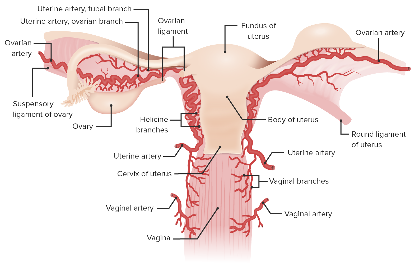
Posterior view of the uterus showing the blood supply of the uterus and ovaries:
Note the ovarian artery traveling along the ovarian suspensory ligament, supplying both the ovaries and the lateral portion of the fallopian tube. The ovarian artery continues in the mesosalpinx to anastomose with branches of the uterine artery. (Note: the round ligament comes off the anterior surface of the uterus.)
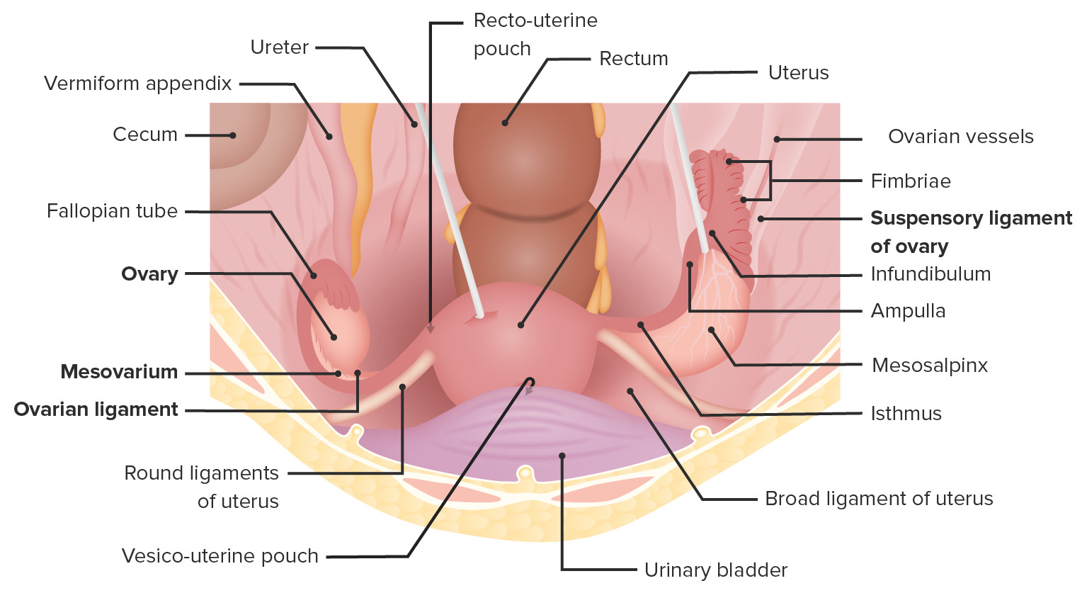
Superior view of the female pelvis depicting the ovaries in situ and their supporting ligaments and relation to the uterus
Image by Lecturio. License: CC BY-NC-SA 4.0Ovarian or adnexal torsion involves the following sequence of events:
Adnexal torsion is suspected based on typical symptoms, supported by imaging with transvaginal ultrasonography or color Doppler Doppler Ultrasonography applying the doppler effect, with frequency-shifted ultrasound reflections produced by moving targets (usually red blood cells) in the bloodstream along the ultrasound axis in direct proportion to the velocity of movement of the targets, to determine both direction and velocity of blood flow. Ultrasound (Sonography) ultrasonography, and confirmed during immediate exploratory surgery.
Laparoscopy Laparoscopy Laparoscopy is surgical exploration and interventions performed through small incisions with a camera and long instruments. Laparotomy and Laparoscopy/ laparotomy Laparotomy Incision into the side of the abdomen between the ribs and pelvis. Laparotomy and Laparoscopy is the gold standard for the diagnosis of ovarian torsion, as the twisted ovary can be directly visualized by the surgeon.