Achieve Mastery of Medical Concepts

Study for medical school and boards with Lecturio

Thrombolytics

Thrombolytics, also known as fibrinolytics, include recombinant tissue plasminogen activator Tissue plasminogen activator A proteolytic enzyme in the serine protease family found in many tissues which converts plasminogen to fibrinolysin. It has fibrin-binding activity and is immunologically different from urokinase-type plasminogen activator. The primary sequence, composed of 527 amino acids, is identical in both the naturally occurring and synthetic proteases. Hemostasis ( TPa tPA Ischemic Stroke) (i.e., alteplase, reteplase, and tenecteplase), urokinase, and streptokinase. The agents promote the breakdown of a blood clot by converting plasminogen Plasminogen Precursor of plasmin (fibrinolysin). It is a single-chain beta-globulin of molecular weight 80-90, 000 found mostly in association with fibrinogen in plasma; plasminogen activators change it to fibrinolysin. It is used in wound debriding and has been investigated as a thrombolytic agent. Hemostasis to plasmin Plasmin A product of the lysis of plasminogen (profibrinolysin) by plasminogen activators. It is composed of two polypeptide chains, light (b) and heavy (a), with a molecular weight of 75, 000. It is the major proteolytic enzyme involved in blood clot retraction or the lysis of fibrin and quickly inactivated by antiplasmins. Hemostasis, which then degrades fibrin Fibrin A protein derived from fibrinogen in the presence of thrombin, which forms part of the blood clot. Rapidly Progressive Glomerulonephritis. Thrombolytics are particularly helpful for conditions related to vascular obstruction by a blood clot (e.g., acute STEMI, pulmonary embolism Pulmonary Embolism Pulmonary embolism (PE) is a potentially fatal condition that occurs as a result of intraluminal obstruction of the main pulmonary artery or its branches. The causative factors include thrombi, air, amniotic fluid, and fat. In PE, gas exchange is impaired due to the decreased return of deoxygenated blood to the lungs. Pulmonary Embolism (PE), deep venous thrombosis Venous thrombosis The formation or presence of a blood clot (thrombus) within a vein. Budd-Chiari Syndrome ( DVT DVT Deep vein thrombosis (DVT) usually occurs in the deep veins of the lower extremities. The affected veins include the femoral, popliteal, iliofemoral, and pelvic veins. Proximal DVT is more likely to cause a pulmonary embolism (PE) and is generally considered more serious. Deep Vein Thrombosis), and acute ischemic stroke Ischemic Stroke An ischemic stroke (also known as cerebrovascular accident) is an acute neurologic injury that occurs as a result of brain ischemia; this condition may be due to cerebral blood vessel occlusion by thrombosis or embolism, or rarely due to systemic hypoperfusion. Ischemic Stroke). Efficacy declines the longer tissue ischemia Ischemia A hypoperfusion of the blood through an organ or tissue caused by a pathologic constriction or obstruction of its blood vessels, or an absence of blood circulation. Ischemic Cell Damage persists; therefore, timing of therapy is particularly important in myocardial infarction Myocardial infarction MI is ischemia and death of an area of myocardial tissue due to insufficient blood flow and oxygenation, usually from thrombus formation on a ruptured atherosclerotic plaque in the epicardial arteries. Clinical presentation is most commonly with chest pain, but women and patients with diabetes may have atypical symptoms. Myocardial Infarction and stroke. Life-threatening bleeding and allergic reactions Allergic Reactions Type I hypersensitivity reaction against plasma proteins in donor blood Transfusion Reactions are potentially serious complications. Weighing the risks and benefits of thrombolytics and evaluating individuals for contraindications Contraindications A condition or factor associated with a recipient that makes the use of a drug, procedure, or physical agent improper or inadvisable. Contraindications may be absolute (life threatening) or relative (higher risk of complications in which benefits may outweigh risks). Noninvasive Ventilation/bleeding risk before initiating therapy is important.

Last updated: Aug 19, 2022

Editorial responsibility: Stanley Oiseth, Lindsay Jones, Evelin Maza

Chemistry and Pharmacodynamics

Chemistry

  • Recombinant tissue plasminogen activator Tissue plasminogen activator A proteolytic enzyme in the serine protease family found in many tissues which converts plasminogen to fibrinolysin. It has fibrin-binding activity and is immunologically different from urokinase-type plasminogen activator. The primary sequence, composed of 527 amino acids, is identical in both the naturally occurring and synthetic proteases. Hemostasis ( TPa tPA Ischemic Stroke):
    • Alteplase
    • Reteplase
    • Tenecteplase
  • Urokinase: a physiologic enzyme produced in renal parenchymal cells 
  • Streptokinase: a protein (not enzyme) synthesized by beta-hemolytic streptococcus Streptococcus Streptococcus is one of the two medically important genera of gram-positive cocci, the other being Staphylococcus. Streptococci are identified as different species on blood agar on the basis of their hemolytic pattern and sensitivity to optochin and bacitracin. There are many pathogenic species of streptococci, including S. pyogenes, S. agalactiae, S. pneumoniae, and the viridans streptococci. Streptococcus

Mechanism of action

Normal physiology:

  • The function of the fibrinolytic system is to remove clots. 
  • Plasminogen Plasminogen Precursor of plasmin (fibrinolysin). It is a single-chain beta-globulin of molecular weight 80-90, 000 found mostly in association with fibrinogen in plasma; plasminogen activators change it to fibrinolysin. It is used in wound debriding and has been investigated as a thrombolytic agent. Hemostasis is activated and cleaved to plasmin Plasmin A product of the lysis of plasminogen (profibrinolysin) by plasminogen activators. It is composed of two polypeptide chains, light (b) and heavy (a), with a molecular weight of 75, 000. It is the major proteolytic enzyme involved in blood clot retraction or the lysis of fibrin and quickly inactivated by antiplasmins. Hemostasis by:
    • TPa tPA Ischemic Stroke
    • Urine plasminogen activator Urine plasminogen activator A proteolytic enzyme that converts plasminogen to fibrinolysin where the preferential cleavage is between arginine and valine. It was isolated originally from human urine, but is found in most tissues of most vertebrates. Hemostasis (UPa), also known as urokinase
  • Plasmin Plasmin A product of the lysis of plasminogen (profibrinolysin) by plasminogen activators. It is composed of two polypeptide chains, light (b) and heavy (a), with a molecular weight of 75, 000. It is the major proteolytic enzyme involved in blood clot retraction or the lysis of fibrin and quickly inactivated by antiplasmins. Hemostasis cleaves fibrin Fibrin A protein derived from fibrinogen in the presence of thrombin, which forms part of the blood clot. Rapidly Progressive Glomerulonephritis polymers (fibrinolysis): 

Thrombolytics (also known as fibrinolytics):

  • Recombinant TPa tPA Ischemic Stroke
    • Catalyzes the cleavage of plasminogen Plasminogen Precursor of plasmin (fibrinolysin). It is a single-chain beta-globulin of molecular weight 80-90, 000 found mostly in association with fibrinogen in plasma; plasminogen activators change it to fibrinolysin. It is used in wound debriding and has been investigated as a thrombolytic agent. Hemostasis plasmin Plasmin A product of the lysis of plasminogen (profibrinolysin) by plasminogen activators. It is composed of two polypeptide chains, light (b) and heavy (a), with a molecular weight of 75, 000. It is the major proteolytic enzyme involved in blood clot retraction or the lysis of fibrin and quickly inactivated by antiplasmins. Hemostasis
    • Fibrin Fibrin A protein derived from fibrinogen in the presence of thrombin, which forms part of the blood clot. Rapidly Progressive Glomerulonephritis specific → activates clot-bound plasminogen Plasminogen Precursor of plasmin (fibrinolysin). It is a single-chain beta-globulin of molecular weight 80-90, 000 found mostly in association with fibrinogen in plasma; plasminogen activators change it to fibrinolysin. It is used in wound debriding and has been investigated as a thrombolytic agent. Hemostasis (tenecteplase is the most specific agent)
  • Urokinase: 
    • Directly cleaves and converts plasminogen Plasminogen Precursor of plasmin (fibrinolysin). It is a single-chain beta-globulin of molecular weight 80-90, 000 found mostly in association with fibrinogen in plasma; plasminogen activators change it to fibrinolysin. It is used in wound debriding and has been investigated as a thrombolytic agent. Hemostasis plasmin Plasmin A product of the lysis of plasminogen (profibrinolysin) by plasminogen activators. It is composed of two polypeptide chains, light (b) and heavy (a), with a molecular weight of 75, 000. It is the major proteolytic enzyme involved in blood clot retraction or the lysis of fibrin and quickly inactivated by antiplasmins. Hemostasis
    • Not fibrin Fibrin A protein derived from fibrinogen in the presence of thrombin, which forms part of the blood clot. Rapidly Progressive Glomerulonephritis specific → activates free circulating and clot-bound plasminogen Plasminogen Precursor of plasmin (fibrinolysin). It is a single-chain beta-globulin of molecular weight 80-90, 000 found mostly in association with fibrinogen in plasma; plasminogen activators change it to fibrinolysin. It is used in wound debriding and has been investigated as a thrombolytic agent. Hemostasis
  • Streptokinase:
    • Binds plasminogen Plasminogen Precursor of plasmin (fibrinolysin). It is a single-chain beta-globulin of molecular weight 80-90, 000 found mostly in association with fibrinogen in plasma; plasminogen activators change it to fibrinolysin. It is used in wound debriding and has been investigated as a thrombolytic agent. Hemostasis → forms a complex 
    • Conformational change of bound plasminogen Plasminogen Precursor of plasmin (fibrinolysin). It is a single-chain beta-globulin of molecular weight 80-90, 000 found mostly in association with fibrinogen in plasma; plasminogen activators change it to fibrinolysin. It is used in wound debriding and has been investigated as a thrombolytic agent. Hemostasis allows cleavage of other plasminogen Plasminogen Precursor of plasmin (fibrinolysin). It is a single-chain beta-globulin of molecular weight 80-90, 000 found mostly in association with fibrinogen in plasma; plasminogen activators change it to fibrinolysin. It is used in wound debriding and has been investigated as a thrombolytic agent. Hemostasis plasmin Plasmin A product of the lysis of plasminogen (profibrinolysin) by plasminogen activators. It is composed of two polypeptide chains, light (b) and heavy (a), with a molecular weight of 75, 000. It is the major proteolytic enzyme involved in blood clot retraction or the lysis of fibrin and quickly inactivated by antiplasmins. Hemostasis
    • Not fibrin Fibrin A protein derived from fibrinogen in the presence of thrombin, which forms part of the blood clot. Rapidly Progressive Glomerulonephritis specific → binds and activates free and clot-bound plasminogen Plasminogen Precursor of plasmin (fibrinolysin). It is a single-chain beta-globulin of molecular weight 80-90, 000 found mostly in association with fibrinogen in plasma; plasminogen activators change it to fibrinolysin. It is used in wound debriding and has been investigated as a thrombolytic agent. Hemostasis
Normal physiology relating to thrombolysis

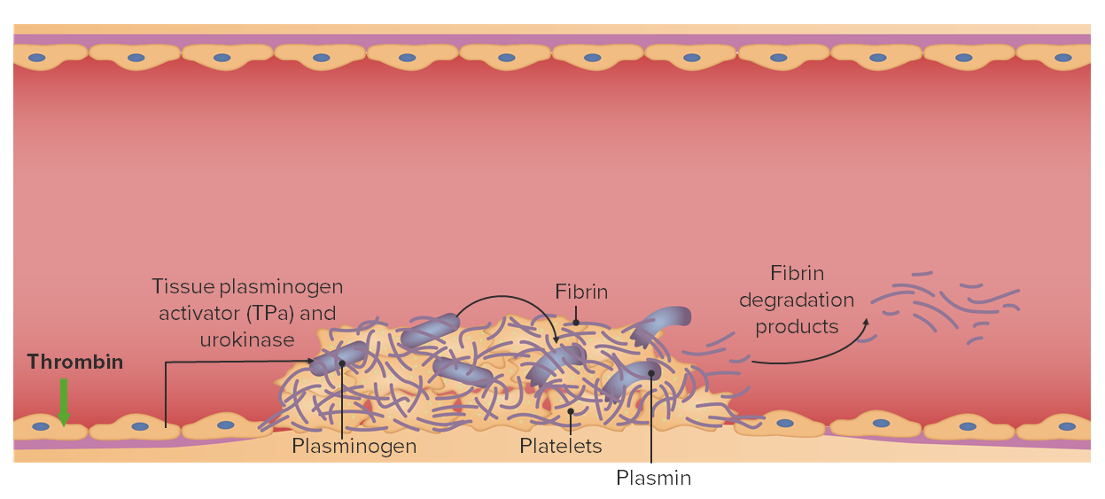
Normal physiology relating to thrombolysis:
Endogenous tissue plasminogen activator (TPa) and urokinase cleave plasminogen into plasmin, resulting in the degradation of fibrin.
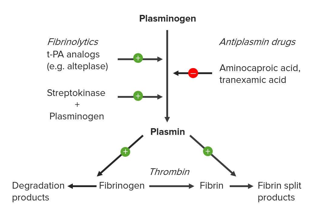
Image by Lecturio.

Indications

Administration

  • Formulation: bolus and/or infusion IV
  • Half-life Half-Life The time it takes for a substance (drug, radioactive nuclide, or other) to lose half of its pharmacologic, physiologic, or radiologic activity. Pharmacokinetics and Pharmacodynamics:
    • Alteplase: approximately 5 minutes
    • Reteplase: 13–16 minutes
    • Tenecteplase: 20–24 minutes (final clearance approximately 90–130 minutes)
    • Urokinase: approximately 20 minutes
    • Streptokinase: approximately 18 minutes

Indications

ST elevation myocardial infarction ST elevation myocardial infarction A clinical syndrome defined by myocardial ischemia symptoms; persistent elevation in the ST segments of the electrocardiogram; and release of biomarkers of myocardial necrosis (e.g., elevated troponin levels). ST segment elevation in the ECG is often used in determining the treatment protocol. Myocardial Infarction (STEMI):

  • Used when percutaneous coronary intervention Percutaneous coronary intervention A family of percutaneous techniques that are used to manage coronary occlusion, including standard balloon angioplasty (percutaneous transluminal coronary angioplasty), the placement of intracoronary stents, and atheroablative technologies (e.g., atherectomy; endarterectomy; thrombectomy; percutaneous transluminal laser angioplasty). Ptca was the dominant form of pci, before the widespread use of stenting. Cardiac Surgery (PCI) is not readily available
  • Timing:
    • Ideally given within 30 minutes of presentation
    • Can be given up to 12 hours after symptom onset, but efficacy declines with time

Acute ischemic stroke Ischemic Stroke An ischemic stroke (also known as cerebrovascular accident) is an acute neurologic injury that occurs as a result of brain ischemia; this condition may be due to cerebral blood vessel occlusion by thrombosis or embolism, or rarely due to systemic hypoperfusion. Ischemic Stroke:

  • For individuals with persistent neurologic deficits Neurologic Deficits High-Risk Headaches
  • Given within 4.5 hours of symptoms (ideally < 3 hours)
  • Rule out intracranial hemorrhage Intracranial hemorrhage Subarachnoid hemorrhage (SAH) is a type of cerebrovascular accident (stroke) resulting from intracranial hemorrhage into the subarachnoid space between the arachnoid and the pia mater layers of the meninges surrounding the brain. Most sahs originate from a saccular aneurysm in the circle of willis but may also occur as a result of trauma, uncontrolled hypertension, vasculitis, anticoagulant use, or stimulant use. Subarachnoid Hemorrhage with CT prior to administration.

Pulmonary embolism Pulmonary Embolism Pulmonary embolism (PE) is a potentially fatal condition that occurs as a result of intraluminal obstruction of the main pulmonary artery or its branches. The causative factors include thrombi, air, amniotic fluid, and fat. In PE, gas exchange is impaired due to the decreased return of deoxygenated blood to the lungs. Pulmonary Embolism (PE):

  • Indicated for:
    • Hemodynamically unstable PE
    • Select individuals with hemodynamically stable PE and right ventricular strain
  • Can be given as:
    • Systemic therapy
    • Catheter-directed therapy

Deep vein thrombosis Thrombosis Formation and development of a thrombus or blood clot in the blood vessel. Epidemic Typhus ( DVT DVT Deep vein thrombosis (DVT) usually occurs in the deep veins of the lower extremities. The affected veins include the femoral, popliteal, iliofemoral, and pelvic veins. Proximal DVT is more likely to cause a pulmonary embolism (PE) and is generally considered more serious. Deep Vein Thrombosis):

  • May be indicated for:
    • Large, proximal DVT DVT Deep vein thrombosis (DVT) usually occurs in the deep veins of the lower extremities. The affected veins include the femoral, popliteal, iliofemoral, and pelvic veins. Proximal DVT is more likely to cause a pulmonary embolism (PE) and is generally considered more serious. Deep Vein Thrombosis
    • Individuals at risk of limb ischemia Ischemia A hypoperfusion of the blood through an organ or tissue caused by a pathologic constriction or obstruction of its blood vessels, or an absence of blood circulation. Ischemic Cell Damage
  • Catheter-directed therapy is preferred.

Additional indications:

  • Acute limb ischemia Ischemia A hypoperfusion of the blood through an organ or tissue caused by a pathologic constriction or obstruction of its blood vessels, or an absence of blood circulation. Ischemic Cell Damage
  • Indwelling catheter Indwelling catheter Catheters designed to be left within an organ or passage for an extended period of time. Urinary tract infections (UTIs) occlusion:
    • Central venous catheters Central Venous Catheters Catheters that are inserted into a large central vein such as a subclavian vein or femoral vein. Central Venous Catheter
    • Dialysis Dialysis Renal replacement therapy refers to dialysis and/or kidney transplantation. Dialysis is a procedure by which toxins and excess water are removed from the circulation. Hemodialysis and peritoneal dialysis (PD) are the two types of dialysis, and their primary difference is the location of the filtration process (external to the body in hemodialysis versus inside the body for PD). Peritoneal Dialysis and Hemodialysis catheters

Adverse Effects and Contraindications

Use thrombolytics cautiously. The medications have a significant risk of bleeding, which can be life threatening or fatal.

Adverse effects

  • Bleeding/hemorrhage, including:
  • Cholesterol Cholesterol The principal sterol of all higher animals, distributed in body tissues, especially the brain and spinal cord, and in animal fats and oils. Cholesterol Metabolism embolization Embolization A method of hemostasis utilizing various agents such as gelfoam, silastic, metal, glass, or plastic pellets, autologous clot, fat, and muscle as emboli. It has been used in the treatment of spinal cord and intracranial arteriovenous malformations, renal arteriovenous fistulas, gastrointestinal bleeding, epistaxis, hypersplenism, certain highly vascular tumors, traumatic rupture of blood vessels, and control of operative hemorrhage. Gastrointestinal Bleeding
  • Allergic reactions Allergic Reactions Type I hypersensitivity reaction against plasma proteins in donor blood Transfusion Reactions:
    • Urticaria Urticaria Urticaria is raised, well-circumscribed areas (wheals) of edema (swelling) and erythema (redness) involving the dermis and epidermis with associated pruritus (itch). Urticaria is not a single disease but rather is a reaction pattern representing cutaneous mast cell degranulation. Urticaria (Hives)
    • Angioedema Angioedema Angioedema is a localized, self-limited (but potentially life-threatening), nonpitting, asymmetrical edema occurring in the deep layers of the skin and mucosal tissue. The common underlying pathophysiology involves inflammatory mediators triggering significant vasodilation and increased capillary permeability. Angioedema
    • Anaphylaxis Anaphylaxis An acute hypersensitivity reaction due to exposure to a previously encountered antigen. The reaction may include rapidly progressing urticaria, respiratory distress, vascular collapse, systemic shock, and death. Type I Hypersensitivity Reaction
  • Reperfusion arrhythmias (from treatment of STEMI)

Contraindications Contraindications A condition or factor associated with a recipient that makes the use of a drug, procedure, or physical agent improper or inadvisable. Contraindications may be absolute (life threatening) or relative (higher risk of complications in which benefits may outweigh risks). Noninvasive Ventilation

Contraindications Contraindications A condition or factor associated with a recipient that makes the use of a drug, procedure, or physical agent improper or inadvisable. Contraindications may be absolute (life threatening) or relative (higher risk of complications in which benefits may outweigh risks). Noninvasive Ventilation depend on the medication, dosing, and pathology and may include:

  • Active or recent internal bleeding:
    • Intracranial hemorrhage Intracranial hemorrhage Subarachnoid hemorrhage (SAH) is a type of cerebrovascular accident (stroke) resulting from intracranial hemorrhage into the subarachnoid space between the arachnoid and the pia mater layers of the meninges surrounding the brain. Most sahs originate from a saccular aneurysm in the circle of willis but may also occur as a result of trauma, uncontrolled hypertension, vasculitis, anticoagulant use, or stimulant use. Subarachnoid Hemorrhage 
    • GI bleed
  • Bleeding diathesis Bleeding diathesis Wiskott-Aldrich Syndrome
  • Intracranial pathology:
    • Cerebral vascular lesions (e.g., arteriovenous malformation Arteriovenous malformation Abnormal formation of blood vessels that shunt arterial blood directly into veins without passing through the capillaries. They usually are crooked, dilated, and with thick vessel walls. A common type is the congenital arteriovenous fistula. The lack of blood flow and oxygen in the capillaries can lead to tissue damage in the affected areas. Erysipelas or aneurysm Aneurysm An aneurysm is a bulging, weakened area of a blood vessel that causes an abnormal widening of its diameter > 1.5 times the size of the native vessel. Aneurysms occur more often in arteries than in veins and are at risk of dissection and rupture, which can be life-threatening. Thoracic Aortic Aneurysms)
    • Neoplasm 
    • Recent ischemic stroke Ischemic Stroke An ischemic stroke (also known as cerebrovascular accident) is an acute neurologic injury that occurs as a result of brain ischemia; this condition may be due to cerebral blood vessel occlusion by thrombosis or embolism, or rarely due to systemic hypoperfusion. Ischemic Stroke
    • Recent significant head injury or facial trauma
  • Possible aortic dissection Aortic dissection Aortic dissection occurs due to shearing stress from pulsatile pressure causing a tear in the tunica intima of the aortic wall. This tear allows blood to flow into the media, creating a “false lumen.” Aortic dissection is most commonly caused by uncontrolled hypertension. Aortic Dissection
  • Recent intracranial or spinal surgery
  • Severe, uncontrolled hypertension Uncontrolled hypertension Although hypertension is defined as a blood pressure of > 130/80 mm Hg, individuals can present with comorbidities of severe asymptomatic or “uncontrolled” hypertension (≥ 180 mm Hg systolic and/or ≥ 120 mm Hg diastolic) that carries with it a significant risk of morbidity and mortality. Uncontrolled Hypertension

Drug interactions

An increased risk of bleeding may occur in individuals taking:

  • Antiplatelet agents Antiplatelet agents Antiplatelet agents are medications that inhibit platelet aggregation, a critical step in the formation of the initial platelet plug. Abnormal, or inappropriate, platelet aggregation is a key step in the pathophysiology of arterial ischemic events. The primary categories of antiplatelet agents include aspirin, ADP inhibitors, phosphodiesterase/adenosine uptake inhibitors, and glycoprotein IIb/IIIa inhibitors. Antiplatelet Drugs
  • Anticoagulants Anticoagulants Anticoagulants are drugs that retard or interrupt the coagulation cascade. The primary classes of available anticoagulants include heparins, vitamin K-dependent antagonists (e.g., warfarin), direct thrombin inhibitors, and factor Xa inhibitors. Anticoagulants
  • Salicylates
  • Prostacyclin Prostacyclin A prostaglandin that is a powerful vasodilator and inhibits platelet aggregation. It is biosynthesized enzymatically from prostaglandin endoperoxides in human vascular tissue. The sodium salt has been also used to treat primary pulmonary hypertension. Eicosanoids analogs

References

  1. Baig, M. U., Bodle, J. (2021). Thrombolytic Therapy. StatPearls. Treasure Island (FL): StatPearls Publishing. https://www.ncbi.nlm.nih.gov/books/NBK557411/
  2. Medscape. (n.a.). Reteplase. Retrieved September 11, 2021, from https://reference.medscape.com/drug/retavase-reteplase-342289
  3. Medscape. (n.a.). Tenecteplase. Retrieved September 11, 2021, from https://reference.medscape.com/drug/tnk-tpa-tnkase-tenecteplase-342291
  4. Medscape. (n.a.). Alteplase. Retrieved September 11, 2021, from https://reference.medscape.com/drug/activase-tpa-alteplase-342287
  5. Rivera-Bou, W. L. (2021). Thrombolytic therapy. In Schraga, E.D., et al. (Ed.), Medscape. Retrieved September 11, 2021, from https://emedicine.medscape.com/article/811234-overview#a2
  6. Kumar, A., et al. (2011). Evolutionary trend of thrombolytics: Their significance. ResearchGate. https://www.researchgate.net/publication/266286698_Evolutionary_Trend_of_Thrombolytic_-_Their_Significance
  7. Weinberg, A. S., & Tapson, V. F. (2021). Catheter-directed thrombolytic therapy in deep venous thrombosis of the lower extremity: Patient selection and administration. In Finlay, G. (Ed.), UpToDate. Retrieved September 29, 2021, from https://www.uptodate.com/contents/catheter-directed-thrombolytic-therapy-in-deep-venous-thrombosis-of-the-lower-extremity-patient-selection-and-administration
  8. Tapson, V. F., & Weinberg, A. S. (2021). Approach to thrombolytic (fibrinolytic) therapy in acute pulmonary embolism: Patient selection and administration. In Finlay, G. (Ed.), UpToDate. Retrieved September 29, 2021, from https://www.uptodate.com/contents/approach-to-thrombolytic-fibrinolytic-therapy-in-acute-pulmonary-embolism-patient-selection-and-administration
  9. Gibson, C. M., Corbalan, R., & Alexander, T. (2020). Acute ST-elevation myocardial infarction: The use of fibrinolytic therapy. In Dardas, T. F. (Ed.), UpToDate. Retrieved September 29, 2021, from https://www.uptodate.com/contents/acute-st-elevation-myocardial-infarction-the-use-of-fibrinolytic-therapy
  10. Oliveira-Filho, J., & Samuels, O. B. (2020). Intravenous thrombolytic therapy for acute ischemic stroke: Therapeutic use. In Dashe, J. F. (2020). UpToDate. Retrieved September 29, 2021, from https://www.uptodate.com/contents/intravenous-thrombolytic-therapy-for-acute-ischemic-stroke-therapeutic-use
  11. Runge, M. S., Quertermous, T., & Haber, E. (1989). Plasminogen activators: The old and the new. Circulation. 79(2), pp. 217–224. https://www.ahajournals.org/doi/pdf/10.1161/01.CIR.79.2.217

Create your free account or log in to continue reading!

Sign up now and get free access to Lecturio with concept pages, medical videos, and questions for your medical education.

User Reviews

Details