Achieve Mastery of Medical Concepts

Study for medical school and boards with Lecturio

MALT Lymphoma

Extranodal marginal zone lymphoma Lymphoma A general term for various neoplastic diseases of the lymphoid tissue. Imaging of the Mediastinum (EMZL) of mucosa-associated lymphoid tissue Mucosa-associated lymphoid tissue Colon, Cecum, and Appendix: Anatomy (also called MALToma, MALT MALT Colon, Cecum, and Appendix: Anatomy lymphoma Lymphoma A general term for various neoplastic diseases of the lymphoid tissue. Imaging of the Mediastinum, and pseudolymphoma) is a group of non-Hodgkin’s lymphomas that have historically been grouped together because they appear to arise from postgerminal center marginal zone B cells B cells Lymphoid cells concerned with humoral immunity. They are short-lived cells resembling bursa-derived lymphocytes of birds in their production of immunoglobulin upon appropriate stimulation. B cells: Types and Functions and share a similar immunophenotype. MALT MALT Colon, Cecum, and Appendix: Anatomy lymphoma Lymphoma A general term for various neoplastic diseases of the lymphoid tissue. Imaging of the Mediastinum is thought to arise in the setting of chronic immune stimulation, which is usually due to bacterial, viral, or autoimmune stimuli. MALT MALT Colon, Cecum, and Appendix: Anatomy lymphomas present with symptoms due to localized involvement of glandular epithelial tissues in the specific site where they develop. Diagnosis of MALT MALT Colon, Cecum, and Appendix: Anatomy lymphoma Lymphoma A general term for various neoplastic diseases of the lymphoid tissue. Imaging of the Mediastinum is made by morphologic, immunophenotypic, and genetic analysis of biopsy Biopsy Removal and pathologic examination of specimens from the living body. Ewing Sarcoma samples. Helicobacter pylori Helicobacter pylori A spiral bacterium active as a human gastric pathogen. It is a gram-negative, urease-positive, curved or slightly spiral organism initially isolated in 1982 from patients with lesions of gastritis or peptic ulcers in Western Australia. Helicobacter pylori was originally classified in the genus campylobacter, but RNA sequencing, cellular fatty acid profiles, growth patterns, and other taxonomic characteristics indicate that the micro-organism should be included in the genus Helicobacter. It has been officially transferred to Helicobacter gen. Helicobacterpositive gastric MALT MALT Colon, Cecum, and Appendix: Anatomy lymphoma Lymphoma A general term for various neoplastic diseases of the lymphoid tissue. Imaging of the Mediastinum is treated with H. pylori H. pylori A spiral bacterium active as a human gastric pathogen. It is a gram-negative, urease-positive, curved or slightly spiral organism initially isolated in 1982 from patients with lesions of gastritis or peptic ulcers in Western Australia. Helicobacter pylori was originally classified in the genus campylobacter, but RNA sequencing, cellular fatty acid profiles, growth patterns, and other taxonomic characteristics indicate that the micro-organism should be included in the genus Helicobacter. It has been officially transferred to Helicobacter gen. Helicobacter eradication therapy, and H. pylori H. pylori A spiral bacterium active as a human gastric pathogen. It is a gram-negative, urease-positive, curved or slightly spiral organism initially isolated in 1982 from patients with lesions of gastritis or peptic ulcers in Western Australia. Helicobacter pylori was originally classified in the genus campylobacter, but RNA sequencing, cellular fatty acid profiles, growth patterns, and other taxonomic characteristics indicate that the micro-organism should be included in the genus Helicobacter. It has been officially transferred to Helicobacter gen. Helicobacter–negative gastric MALT MALT Colon, Cecum, and Appendix: Anatomy lymphoma Lymphoma A general term for various neoplastic diseases of the lymphoid tissue. Imaging of the Mediastinum is treated with radiation Radiation Emission or propagation of acoustic waves (sound), electromagnetic energy waves (such as light; radio waves; gamma rays; or x-rays), or a stream of subatomic particles (such as electrons; neutrons; protons; or alpha particles). Osteosarcoma therapy. Nongastric MALT MALT Colon, Cecum, and Appendix: Anatomy lymphoma Lymphoma A general term for various neoplastic diseases of the lymphoid tissue. Imaging of the Mediastinum is treated based on the involved area and extent of disease. MALT MALT Colon, Cecum, and Appendix: Anatomy lymphoma Lymphoma A general term for various neoplastic diseases of the lymphoid tissue. Imaging of the Mediastinum patients Patients Individuals participating in the health care system for the purpose of receiving therapeutic, diagnostic, or preventive procedures. Clinician–Patient Relationship have a good prognosis Prognosis A prediction of the probable outcome of a disease based on a individual's condition and the usual course of the disease as seen in similar situations. Non-Hodgkin Lymphomas, with a median survival of > 10 years.

Last updated: Oct 31, 2022

Editorial responsibility: Stanley Oiseth, Lindsay Jones, Evelin Maza

Overview

Definition

MALT MALT Colon, Cecum, and Appendix: Anatomy lymphoma Lymphoma A general term for various neoplastic diseases of the lymphoid tissue. Imaging of the Mediastinum is a clinically indolent non-Hodgkin’s lymphoma Lymphoma A general term for various neoplastic diseases of the lymphoid tissue. Imaging of the Mediastinum postulated to arise from postgerminal center memory Memory Complex mental function having four distinct phases: (1) memorizing or learning, (2) retention, (3) recall, and (4) recognition. Clinically, it is usually subdivided into immediate, recent, and remote memory. Psychiatric Assessment B cells B cells Lymphoid cells concerned with humoral immunity. They are short-lived cells resembling bursa-derived lymphocytes of birds in their production of immunoglobulin upon appropriate stimulation. B cells: Types and Functions with the capacity to differentiate into marginal zone cells and plasma Plasma The residual portion of blood that is left after removal of blood cells by centrifugation without prior blood coagulation. Transfusion Products cells.

Epidemiology

  • Relatively uncommon subtype of non-Hodgkin’s lymphoma Lymphoma A general term for various neoplastic diseases of the lymphoid tissue. Imaging of the Mediastinum:
    • 5%–10% of all non-Hodgkin’s lymphomas
    • Approximately half of non-Hodgkin’s lymphomas in stomach Stomach The stomach is a muscular sac in the upper left portion of the abdomen that plays a critical role in digestion. The stomach develops from the foregut and connects the esophagus with the duodenum. Structurally, the stomach is C-shaped and forms a greater and lesser curvature and is divided grossly into regions: the cardia, fundus, body, and pylorus. Stomach: Anatomy, ocular adnexa, and lung
  • Incidence Incidence The number of new cases of a given disease during a given period in a specified population. It also is used for the rate at which new events occur in a defined population. It is differentiated from prevalence, which refers to all cases in the population at a given time. Measures of Disease Frequency in the United States: 18.3 cases per 1 million person-years
  • Primarily seen in adults (median age at diagnosis: 66 years)
  • Highest incidence Incidence The number of new cases of a given disease during a given period in a specified population. It also is used for the rate at which new events occur in a defined population. It is differentiated from prevalence, which refers to all cases in the population at a given time. Measures of Disease Frequency in non-Hispanic whites
  • Lowest incidence Incidence The number of new cases of a given disease during a given period in a specified population. It also is used for the rate at which new events occur in a defined population. It is differentiated from prevalence, which refers to all cases in the population at a given time. Measures of Disease Frequency in African Americans
  • In males, more common involvement in:
    • Stomach Stomach The stomach is a muscular sac in the upper left portion of the abdomen that plays a critical role in digestion. The stomach develops from the foregut and connects the esophagus with the duodenum. Structurally, the stomach is C-shaped and forms a greater and lesser curvature and is divided grossly into regions: the cardia, fundus, body, and pylorus. Stomach: Anatomy
    • Small intestine Small intestine The small intestine is the longest part of the GI tract, extending from the pyloric orifice of the stomach to the ileocecal junction. The small intestine is the major organ responsible for chemical digestion and absorption of nutrients. It is divided into 3 segments: the duodenum, the jejunum, and the ileum. Small Intestine: Anatomy
    • Skin Skin The skin, also referred to as the integumentary system, is the largest organ of the body. The skin is primarily composed of the epidermis (outer layer) and dermis (deep layer). The epidermis is primarily composed of keratinocytes that undergo rapid turnover, while the dermis contains dense layers of connective tissue. Skin: Structure and Functions
    • Kidney
  • In females, more common involvement in:
    • Salivary glands Salivary glands The salivary glands are exocrine glands positioned in and around the oral cavity. These glands are responsible for secreting saliva into the mouth, which aids in digestion. There are 3 major paired salivary glands: the sublingual, submandibular, and parotid glands. Salivary Glands: Anatomy
    • Soft tissue Soft Tissue Soft Tissue Abscess
    • Thyroid Thyroid The thyroid gland is one of the largest endocrine glands in the human body. The thyroid gland is a highly vascular, brownish-red gland located in the visceral compartment of the anterior region of the neck. Thyroid Gland: Anatomy

Etiology

  • Generally present in the setting of chronic immune stimulation due to bacterial, viral, or autoimmune stimuli:
    • Implicated autoimmune conditions:
      • Sjögren’s syndrome (parotid extranodal marginal zone lymphoma Lymphoma A general term for various neoplastic diseases of the lymphoid tissue. Imaging of the Mediastinum (EMZL))
      • Systemic lupus erythematosus Systemic lupus erythematosus Systemic lupus erythematosus (SLE) is a chronic autoimmune, inflammatory condition that causes immune-complex deposition in organs, resulting in systemic manifestations. Women, particularly those of African American descent, are more commonly affected. Systemic Lupus Erythematosus
      • Relapsing polychondritis Polychondritis An acquired disease of unknown etiology, chronic course, and tendency to recur. It is characterized by inflammation and degeneration of cartilage and can result in deformities such as floppy ear and saddle nose. Loss of cartilage in the respiratory tract can lead to respiratory obstruction. Antineutrophil Cytoplasmic Antibody (ANCA)-Associated Vasculitis
      • Hashimoto’s thyroiditis Thyroiditis Thyroiditis is a catchall term used to describe a variety of conditions that have inflammation of the thyroid gland in common. It includes pathologies that cause an acute illness with severe thyroid pain (e.g., subacute thyroiditis and infectious thyroiditis) as well as conditions in which there is no clinically evident inflammation and the manifestations primarily reflect thyroid dysfunction or a goiter (e.g., painless thyroiditis and fibrous Riedel’s thyroiditis). Thyroiditis ( thyroid Thyroid The thyroid gland is one of the largest endocrine glands in the human body. The thyroid gland is a highly vascular, brownish-red gland located in the visceral compartment of the anterior region of the neck. Thyroid Gland: Anatomy EMZL)
    • Implicated bacteria Bacteria Bacteria are prokaryotic single-celled microorganisms that are metabolically active and divide by binary fission. Some of these organisms play a significant role in the pathogenesis of diseases. Bacteriology:
      • Helicobacter pylori Helicobacter pylori A spiral bacterium active as a human gastric pathogen. It is a gram-negative, urease-positive, curved or slightly spiral organism initially isolated in 1982 from patients with lesions of gastritis or peptic ulcers in Western Australia. Helicobacter pylori was originally classified in the genus campylobacter, but RNA sequencing, cellular fatty acid profiles, growth patterns, and other taxonomic characteristics indicate that the micro-organism should be included in the genus Helicobacter. It has been officially transferred to Helicobacter gen. Helicobacter gastritis Gastritis Gastritis refers to inflammation of the gastric mucosa. Gastritis may occur suddenly (acute gastritis) or slowly over time (chronic gastritis). Gastritis may be asymptomatic or with symptoms, including burning abdominal pain (which either worsens or improves with eating), dyspepsia, nausea, and vomiting. Gastritis: best-established relationship Relationship A connection, association, or involvement between 2 or more parties. Clinician–Patient Relationship
      • Chlamydia psittaci Chlamydia psittaci A genus of chlamydophila infecting primarily birds. It contains eight known serovars, some of which infect more than one type of host, including humans. Chlamydia (ocular adnexa)
      • Campylobacter jejuni Campylobacter jejuni A species of bacteria that resemble small tightly coiled spirals. Its organisms are known to cause abortion in sheep and fever and enteritis in man and may be associated with enteric diseases of calves, lambs, and other animals. Campylobacter ( small intestine Small intestine The small intestine is the longest part of the GI tract, extending from the pyloric orifice of the stomach to the ileocecal junction. The small intestine is the major organ responsible for chemical digestion and absorption of nutrients. It is divided into 3 segments: the duodenum, the jejunum, and the ileum. Small Intestine: Anatomy)
      • Borrelia afzelii Borrelia afzelii Borrelia ( skin Skin The skin, also referred to as the integumentary system, is the largest organ of the body. The skin is primarily composed of the epidermis (outer layer) and dermis (deep layer). The epidermis is primarily composed of keratinocytes that undergo rapid turnover, while the dermis contains dense layers of connective tissue. Skin: Structure and Functions)
      • Achromobacter xylosoxidans (lung)
  • Several translocations resulting in increased activation of nuclear factor kappa-B with and without immune stimulation have been implicated as a potential etiology.

Pathophysiology

  • MALT MALT Colon, Cecum, and Appendix: Anatomy lymphoma Lymphoma A general term for various neoplastic diseases of the lymphoid tissue. Imaging of the Mediastinum develops because of chronic stimulation of lymphoid tissue: 
    • Leads to local accumulation and proliferation of antigen-dependent B cells B cells Lymphoid cells concerned with humoral immunity. They are short-lived cells resembling bursa-derived lymphocytes of birds in their production of immunoglobulin upon appropriate stimulation. B cells: Types and Functions and T cells T cells Lymphocytes responsible for cell-mediated immunity. Two types have been identified – cytotoxic (t-lymphocytes, cytotoxic) and helper T-lymphocytes (t-lymphocytes, helper-inducer). They are formed when lymphocytes circulate through the thymus gland and differentiate to thymocytes. When exposed to an antigen, they divide rapidly and produce large numbers of new T cells sensitized to that antigen. T cells: Types and Functions
    • Over time, the B cells B cells Lymphoid cells concerned with humoral immunity. They are short-lived cells resembling bursa-derived lymphocytes of birds in their production of immunoglobulin upon appropriate stimulation. B cells: Types and Functions, which are still antigen-dependent, develop mutations.
    • Monoclonal B cells B cells Lymphoid cells concerned with humoral immunity. They are short-lived cells resembling bursa-derived lymphocytes of birds in their production of immunoglobulin upon appropriate stimulation. B cells: Types and Functions then develop additional mutations and become antigen-independent and capable of systemic spread.
  • Proliferation of MALT MALT Colon, Cecum, and Appendix: Anatomy cells at certain sites depends on activated, antigen-driven T cells T cells Lymphocytes responsible for cell-mediated immunity. Two types have been identified – cytotoxic (t-lymphocytes, cytotoxic) and helper T-lymphocytes (t-lymphocytes, helper-inducer). They are formed when lymphocytes circulate through the thymus gland and differentiate to thymocytes. When exposed to an antigen, they divide rapidly and produce large numbers of new T cells sensitized to that antigen. T cells: Types and Functions.
  • MALT MALT Colon, Cecum, and Appendix: Anatomy lymphoma Lymphoma A general term for various neoplastic diseases of the lymphoid tissue. Imaging of the Mediastinum reproduces the morphologic features of normal MALT MALT Colon, Cecum, and Appendix: Anatomy:
    • Reactive follicles are usually present with neoplastic cells occupying the marginal zone and/or the interfollicular region.
    • Neoplastic cells may occasionally colonize follicles.
  • Common chromosome Chromosome In a prokaryotic cell or in the nucleus of a eukaryotic cell, a structure consisting of or containing DNA which carries the genetic information essential to the cell. Basic Terms of Genetics translocations leading to increased expression of nuclear factor kappa-B (which provides a survival advantage to the neoplastic B cells B cells Lymphoid cells concerned with humoral immunity. They are short-lived cells resembling bursa-derived lymphocytes of birds in their production of immunoglobulin upon appropriate stimulation. B cells: Types and Functions):
    • t(11;18)(q21;q21)
    • t(14;18)(q32;q21)
    • t(1;14)(p22;q32)
    • t(3;14)(p13;q32)
  • Monoclonal gammopathy:
    • Present in 27%–36% of patients Patients Individuals participating in the health care system for the purpose of receiving therapeutic, diagnostic, or preventive procedures. Clinician–Patient Relationship
    • Correlates with:
      •  Plasmacytic differentiation
      •  Advanced disease
      •  Involvement of lymph nodes Lymph Nodes They are oval or bean shaped bodies (1 – 30 mm in diameter) located along the lymphatic system. Lymphatic Drainage System: Anatomy and bone marrow Bone marrow The soft tissue filling the cavities of bones. Bone marrow exists in two types, yellow and red. Yellow marrow is found in the large cavities of large bones and consists mostly of fat cells and a few primitive blood cells. Red marrow is a hematopoietic tissue and is the site of production of erythrocytes and granular leukocytes. Bone marrow is made up of a framework of connective tissue containing branching fibers with the frame being filled with marrow cells. Bone Marrow: Composition and Hematopoiesis
  • Prototypical example: 
    • H. pylori H. pylori A spiral bacterium active as a human gastric pathogen. It is a gram-negative, urease-positive, curved or slightly spiral organism initially isolated in 1982 from patients with lesions of gastritis or peptic ulcers in Western Australia. Helicobacter pylori was originally classified in the genus campylobacter, but RNA sequencing, cellular fatty acid profiles, growth patterns, and other taxonomic characteristics indicate that the micro-organism should be included in the genus Helicobacter. It has been officially transferred to Helicobacter gen. Helicobacter infection with chronic gastritis Gastritis Gastritis refers to inflammation of the gastric mucosa. Gastritis may occur suddenly (acute gastritis) or slowly over time (chronic gastritis). Gastritis may be asymptomatic or with symptoms, including burning abdominal pain (which either worsens or improves with eating), dyspepsia, nausea, and vomiting. Gastritis
    • May lead to development of gastric MALT MALT Colon, Cecum, and Appendix: Anatomy
    • Remains localized for significant periods of time (but is a clonal B-cell neoplasm)
    • Frequently recurs
    • Potential for systemic spread
    • Can transform into an aggressive B-cell lymphoma Lymphoma A general term for various neoplastic diseases of the lymphoid tissue. Imaging of the Mediastinum

Clinical Presentation

Clinical presentation varies depending on the location where the MALT MALT Colon, Cecum, and Appendix: Anatomy lymphoma Lymphoma A general term for various neoplastic diseases of the lymphoid tissue. Imaging of the Mediastinum develops.

  • Stomach Stomach The stomach is a muscular sac in the upper left portion of the abdomen that plays a critical role in digestion. The stomach develops from the foregut and connects the esophagus with the duodenum. Structurally, the stomach is C-shaped and forms a greater and lesser curvature and is divided grossly into regions: the cardia, fundus, body, and pylorus. Stomach: Anatomy:
  • Ocular adnexa:
  • Salivary gland Salivary gland Glands that secrete saliva in the mouth. There are three pairs of salivary glands (parotid gland; sublingual gland; submandibular gland). Diseases of the Salivary Glands:
    • Slow-growing mass Mass Three-dimensional lesion that occupies a space within the breast Imaging of the Breast
    • Minority of patients Patients Individuals participating in the health care system for the purpose of receiving therapeutic, diagnostic, or preventive procedures. Clinician–Patient Relationship: bilateral involvement
  • Skin Skin The skin, also referred to as the integumentary system, is the largest organ of the body. The skin is primarily composed of the epidermis (outer layer) and dermis (deep layer). The epidermis is primarily composed of keratinocytes that undergo rapid turnover, while the dermis contains dense layers of connective tissue. Skin: Structure and Functions
    • Red to violaceous papules, plaques, or nodules
    • Commonly localized to trunk and upper extremities
  • Lung:
  • Small intestine Small intestine The small intestine is the longest part of the GI tract, extending from the pyloric orifice of the stomach to the ileocecal junction. The small intestine is the major organ responsible for chemical digestion and absorption of nutrients. It is divided into 3 segments: the duodenum, the jejunum, and the ileum. Small Intestine: Anatomy:
    • Intermittent diarrhea Diarrhea Diarrhea is defined as ≥ 3 watery or loose stools in a 24-hour period. There are a multitude of etiologies, which can be classified based on the underlying mechanism of disease. The duration of symptoms (acute or chronic) and characteristics of the stools (e.g., watery, bloody, steatorrheic, mucoid) can help guide further diagnostic evaluation. Diarrhea
    • Colicky abdominal pain Abdominal Pain Acute Abdomen
    • Symptoms related to malabsorption Malabsorption General term for a group of malnutrition syndromes caused by failure of normal intestinal absorption of nutrients. Malabsorption and Maldigestion

Dissemination to other sites of MALT MALT Colon, Cecum, and Appendix: Anatomy tissue:

Peripheral blood is usually not involved.

Systemic B symptoms are uncommon:

Diagnosis

History

  • Constitutional:
    • Anorexia Anorexia The lack or loss of appetite accompanied by an aversion to food and the inability to eat. It is the defining characteristic of the disorder anorexia nervosa. Anorexia Nervosa
    • Weight loss Weight loss Decrease in existing body weight. Bariatric Surgery
  • Head, eyes, ears, nose Nose The nose is the human body’s primary organ of smell and functions as part of the upper respiratory system. The nose may be best known for inhaling oxygen and exhaling carbon dioxide, but it also contributes to other important functions, such as tasting. The anatomy of the nose can be divided into the external nose and the nasal cavity. Nose Anatomy (External & Internal), throat Throat The pharynx is a component of the digestive system that lies posterior to the nasal cavity, oral cavity, and larynx. The pharynx can be divided into the oropharynx, nasopharynx, and laryngopharynx. Pharyngeal muscles play an integral role in vital processes such as breathing, swallowing, and speaking. Pharynx: Anatomy (HEENT):
  • GI:
    • GERD GERD Gastroesophageal reflux disease (GERD) occurs when the stomach acid frequently flows back into the esophagus. This backwash (acid reflux) can irritate the lining of the esophagus, causing symptoms such as retrosternal burning pain (heartburn). Gastroesophageal Reflux Disease (GERD)
    • Epigastric pain Epigastric pain Mallory-Weiss Syndrome (Mallory-Weiss Tear) and discomfort
    • GI bleeding
    • Diarrhea Diarrhea Diarrhea is defined as ≥ 3 watery or loose stools in a 24-hour period. There are a multitude of etiologies, which can be classified based on the underlying mechanism of disease. The duration of symptoms (acute or chronic) and characteristics of the stools (e.g., watery, bloody, steatorrheic, mucoid) can help guide further diagnostic evaluation. Diarrhea
    • Colicky abdominal pain Abdominal Pain Acute Abdomen
  • Skin Skin The skin, also referred to as the integumentary system, is the largest organ of the body. The skin is primarily composed of the epidermis (outer layer) and dermis (deep layer). The epidermis is primarily composed of keratinocytes that undergo rapid turnover, while the dermis contains dense layers of connective tissue. Skin: Structure and Functions: trunk and upper extremity changes
  • Nutritional and performance evaluation using the following performance scales Scales Dry or greasy masses of keratin that represent thickened stratum corneum. Secondary Skin Lesions:
    • Eastern Cooperative Oncology Group (ECOG)
    • Karnofsky

Physical exam

  • HEENT:
  • Enlarged lymph nodes Lymph Nodes They are oval or bean shaped bodies (1 – 30 mm in diameter) located along the lymphatic system. Lymphatic Drainage System: Anatomy
  • Hepatosplenomegaly Hepatosplenomegaly Cytomegalovirus (if liver Liver The liver is the largest gland in the human body. The liver is found in the superior right quadrant of the abdomen and weighs approximately 1.5 kilograms. Its main functions are detoxification, metabolism, nutrient storage (e.g., iron and vitamins), synthesis of coagulation factors, formation of bile, filtration, and storage of blood. Liver: Anatomy or spleen Spleen The spleen is the largest lymphoid organ in the body, located in the LUQ of the abdomen, superior to the left kidney and posterior to the stomach at the level of the 9th-11th ribs just below the diaphragm. The spleen is highly vascular and acts as an important blood filter, cleansing the blood of pathogens and damaged erythrocytes. Spleen: Anatomy involved)
  • Skin Skin The skin, also referred to as the integumentary system, is the largest organ of the body. The skin is primarily composed of the epidermis (outer layer) and dermis (deep layer). The epidermis is primarily composed of keratinocytes that undergo rapid turnover, while the dermis contains dense layers of connective tissue. Skin: Structure and Functions:
    • Reddish papules, plaques, or nodules
    • More common on:
      • Trunk
      • Upper extremities

Laboratory

  • Biopsy Biopsy Removal and pathologic examination of specimens from the living body. Ewing Sarcoma of affected site:
    • Morphologic, immunophenotypic, and genetic analysis of the biopsy Biopsy Removal and pathologic examination of specimens from the living body. Ewing Sarcoma sample should be analyzed within clinical context:
      • Morphology: polymorphous infiltrate of small cells with reactive-appearing follicles
      • Immunophenotype: B cells B cells Lymphoid cells concerned with humoral immunity. They are short-lived cells resembling bursa-derived lymphocytes of birds in their production of immunoglobulin upon appropriate stimulation. B cells: Types and Functions positive for CD19, CD20, and CD22 and negative for CD5, CD10, and CD23
      • Genetic analysis: PCR PCR Polymerase chain reaction (PCR) is a technique that amplifies DNA fragments exponentially for analysis. The process is highly specific, allowing for the targeting of specific genomic sequences, even with minuscule sample amounts. The PCR cycles multiple times through 3 phases: denaturation of the template DNA, annealing of a specific primer to the individual DNA strands, and synthesis/elongation of new DNA molecules. Polymerase Chain Reaction (PCR) analysis of IgH rearrangement is helpful in differentiating MALT MALT Colon, Cecum, and Appendix: Anatomy lymphoma Lymphoma A general term for various neoplastic diseases of the lymphoid tissue. Imaging of the Mediastinum from reactive proliferations.
    • Obtain large sample (so cancer is not missed in biopsy Biopsy Removal and pathologic examination of specimens from the living body. Ewing Sarcoma sample).
    • Esophagogastroduodenoscopy:
    • Biopsies for H. pylori H. pylori A spiral bacterium active as a human gastric pathogen. It is a gram-negative, urease-positive, curved or slightly spiral organism initially isolated in 1982 from patients with lesions of gastritis or peptic ulcers in Western Australia. Helicobacter pylori was originally classified in the genus campylobacter, but RNA sequencing, cellular fatty acid profiles, growth patterns, and other taxonomic characteristics indicate that the micro-organism should be included in the genus Helicobacter. It has been officially transferred to Helicobacter gen. Helicobacter:
      • If negative: perform stool antigen Antigen Substances that are recognized by the immune system and induce an immune reaction. Vaccination test or urea breath test Urea breath test Gastritis
      • FISH FISH A type of in situ hybridization in which target sequences are stained with fluorescent dye so their location and size can be determined using fluorescence microscopy. This staining is sufficiently distinct that the hybridization signal can be seen both in metaphase spreads and in interphase nuclei. Chromosome Testing or PCR PCR Polymerase chain reaction (PCR) is a technique that amplifies DNA fragments exponentially for analysis. The process is highly specific, allowing for the targeting of specific genomic sequences, even with minuscule sample amounts. The PCR cycles multiple times through 3 phases: denaturation of the template DNA, annealing of a specific primer to the individual DNA strands, and synthesis/elongation of new DNA molecules. Polymerase Chain Reaction (PCR) testing for t(11;18) to identify tumors unlikely to respond to H. pylori H. pylori A spiral bacterium active as a human gastric pathogen. It is a gram-negative, urease-positive, curved or slightly spiral organism initially isolated in 1982 from patients with lesions of gastritis or peptic ulcers in Western Australia. Helicobacter pylori was originally classified in the genus campylobacter, but RNA sequencing, cellular fatty acid profiles, growth patterns, and other taxonomic characteristics indicate that the micro-organism should be included in the genus Helicobacter. It has been officially transferred to Helicobacter gen. Helicobacter eradication
  • Blood tests (may provide information about organs involved/associated diseases):
    • CBC/DIFF
    • Serum electrolytes Electrolytes Electrolytes are mineral salts that dissolve in water and dissociate into charged particles called ions, which can be either be positively (cations) or negatively (anions) charged. Electrolytes are distributed in the extracellular and intracellular compartments in different concentrations. Electrolytes are essential for various basic life-sustaining functions. Electrolytes
    • Liver function tests Liver function tests Liver function tests, also known as hepatic function panels, are one of the most commonly performed screening blood tests. Such tests are also used to detect, evaluate, and monitor acute and chronic liver diseases. Liver Function Tests
    • Renal function tests
    • LDH LDH Osteosarcoma
    • Serum protein electrophoresis Electrophoresis An electrochemical process in which macromolecules or colloidal particles with a net electric charge migrate in a solution under the influence of an electric current. Blotting Techniques (to evaluate for monoclonal gammopathy)
    • Serologic testing:
      • HIV HIV Anti-HIV Drugs
      • Hepatitis C Hepatitis C Hepatitis C is an infection of the liver caused by the hepatitis C virus (HCV). The infection can be transmitted through infectious blood or body fluids and may be transmitted during childbirth or through IV drug use or sexual intercourse. Hepatitis C virus can cause both acute and chronic hepatitis, ranging from a mild to a serious, lifelong illness including liver cirrhosis and hepatocellular carcinoma (HCC). Hepatitis C Virus or B
  • Imaging:
    • CT and MRI:
      • To evaluate for distant disease
      • Extent of primary lesion
      • Do not differentiate malignant MALT MALT Colon, Cecum, and Appendix: Anatomy lesions from benign Benign Fibroadenoma
      • Can image chest, abdomen, pelvis Pelvis The pelvis consists of the bony pelvic girdle, the muscular and ligamentous pelvic floor, and the pelvic cavity, which contains viscera, vessels, and multiple nerves and muscles. The pelvic girdle, composed of 2 “hip” bones and the sacrum, is a ring-like bony structure of the axial skeleton that links the vertebral column with the lower extremities. Pelvis: Anatomy, orbits, and salivary glands Salivary glands The salivary glands are exocrine glands positioned in and around the oral cavity. These glands are responsible for secreting saliva into the mouth, which aids in digestion. There are 3 major paired salivary glands: the sublingual, submandibular, and parotid glands. Salivary Glands: Anatomy
    • Whole-body combined fluorodeoxyglucose (FDG) PET PET An imaging technique that combines a positron-emission tomography (PET) scanner and a ct X ray scanner. This establishes a precise anatomic localization in the same session. Nuclear Imaging/CT to evaluate for:
      • Radiation Radiation Emission or propagation of acoustic waves (sound), electromagnetic energy waves (such as light; radio waves; gamma rays; or x-rays), or a stream of subatomic particles (such as electrons; neutrons; protons; or alpha particles). Osteosarcoma therapy
      • Suspected large-cell transformation Transformation Change brought about to an organism’s genetic composition by unidirectional transfer (transfection; transduction, genetic; conjugation, genetic, etc.) and incorporation of foreign DNA into prokaryotic or eukaryotic cells by recombination of part or all of that DNA into the cell’s genome. Bacteriology
  • Bone marrow Bone marrow The soft tissue filling the cavities of bones. Bone marrow exists in two types, yellow and red. Yellow marrow is found in the large cavities of large bones and consists mostly of fat cells and a few primitive blood cells. Red marrow is a hematopoietic tissue and is the site of production of erythrocytes and granular leukocytes. Bone marrow is made up of a framework of connective tissue containing branching fibers with the frame being filled with marrow cells. Bone Marrow: Composition and Hematopoiesis biopsy Biopsy Removal and pathologic examination of specimens from the living body. Ewing Sarcoma to confirm suspected early (stage I/II) disease
Mucosa-associated lymphoid tissue lymphoma of the esophagus

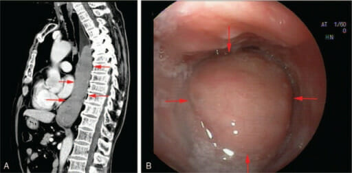
Mucosa-associated lymphoid tissue lymphoma of the esophagus:
Computed tomography (image A) and endoscopy (image B) show a neoplasm of the middle and lower esophagus.

Image: “F1” by Medicine. License: CC BY 4.0
Gastric malt lymphoma associated with h. Pylori

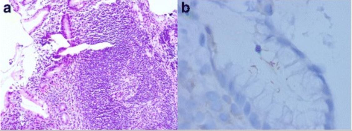
Gastric MALT lymphoma associated with H. pylori:
a: Lymphoid follicle with a reactive germinal center and a slightly expanded marginal zone (H&E, original magnification, 100X)
b: Immunostaining of H. pylori shows intra-foveolar bacteria with curving configuration, characteristic of H. pylori.

Image: “Gastric mucosa-associated lymphoid tissue lymphoma and Helicobacter pylori infection: a review of current diagnosis and management” by Hu Q, Zhang Y, Zhang X, Fu K. License: CC BY 4.0
Malt lymphoma of the colon

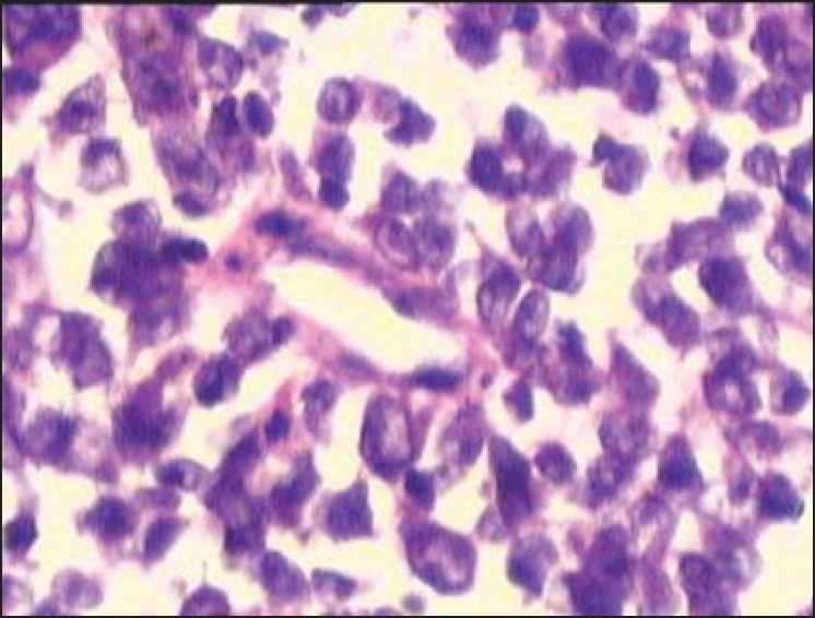
MALT lymphoma of the colon:
Infiltration of malignant-looking lymphoid cells in the lamina propria suggestive of malt lymphoma

Image: “Synchronous adenocarcinoma and mucosa-associated lymphoid tissue lymphoma of the colon” by Devi P, Pattanayak L, Samantaray S. License: CC BY 2.0

Staging Staging Methods which attempt to express in replicable terms the extent of the neoplasm in the patient. Grading, Staging, and Metastasis

  • Lugano staging Staging Methods which attempt to express in replicable terms the extent of the neoplasm in the patient. Grading, Staging, and Metastasis system (for gastric involvement):
    • Stage I/II:
      • Single primary lesion
      • Multiple noncontiguous lesions confined to the GI tract that may have nodal involvement
    • No stage III defined
    • Stage IV:
      • Disseminated extranodal involvement or
      • Concomitant supradiaphragmatic nodal involvement
  • Ann Arbor staging Staging Methods which attempt to express in replicable terms the extent of the neoplasm in the patient. Grading, Staging, and Metastasis system for non-gastric MALT MALT Colon, Cecum, and Appendix: Anatomy:
    • Stage 1 Stage 1 Trypanosoma brucei/African trypanosomiasis: single lymph Lymph The interstitial fluid that is in the lymphatic system. Secondary Lymphatic Organs node region
    • Stage 2: one side of diaphragm Diaphragm The diaphragm is a large, dome-shaped muscle that separates the thoracic cavity from the abdominal cavity. The diaphragm consists of muscle fibers and a large central tendon, which is divided into right and left parts. As the primary muscle of inspiration, the diaphragm contributes 75% of the total inspiratory muscle force. Diaphragm: Anatomy
    • Stage 3: both sides of diaphragm Diaphragm The diaphragm is a large, dome-shaped muscle that separates the thoracic cavity from the abdominal cavity. The diaphragm consists of muscle fibers and a large central tendon, which is divided into right and left parts. As the primary muscle of inspiration, the diaphragm contributes 75% of the total inspiratory muscle force. Diaphragm: Anatomy
    • Stage 4: disseminated

Management and Prognosis

Management

  • Gastric MALT MALT Colon, Cecum, and Appendix: Anatomy lymphoma Lymphoma A general term for various neoplastic diseases of the lymphoid tissue. Imaging of the Mediastinum:
    • H. pylori H. pylori A spiral bacterium active as a human gastric pathogen. It is a gram-negative, urease-positive, curved or slightly spiral organism initially isolated in 1982 from patients with lesions of gastritis or peptic ulcers in Western Australia. Helicobacter pylori was originally classified in the genus campylobacter, but RNA sequencing, cellular fatty acid profiles, growth patterns, and other taxonomic characteristics indicate that the micro-organism should be included in the genus Helicobacter. It has been officially transferred to Helicobacter gen. Helicobacter–positive:
      • H. pylori H. pylori A spiral bacterium active as a human gastric pathogen. It is a gram-negative, urease-positive, curved or slightly spiral organism initially isolated in 1982 from patients with lesions of gastritis or peptic ulcers in Western Australia. Helicobacter pylori was originally classified in the genus campylobacter, but RNA sequencing, cellular fatty acid profiles, growth patterns, and other taxonomic characteristics indicate that the micro-organism should be included in the genus Helicobacter. It has been officially transferred to Helicobacter gen. Helicobacter eradication therapy
      • Follow up with serial endoscopies
      • Treatment failures: treat with radiation Radiation Emission or propagation of acoustic waves (sound), electromagnetic energy waves (such as light; radio waves; gamma rays; or x-rays), or a stream of subatomic particles (such as electrons; neutrons; protons; or alpha particles). Osteosarcoma therapy (RT)
      • Gastric resection for perforation Perforation A pathological hole in an organ, blood vessel or other soft part of the body, occurring in the absence of external force. Esophagitis, uncontrolled bleeding, or obstruction
    • H. pylori H. pylori A spiral bacterium active as a human gastric pathogen. It is a gram-negative, urease-positive, curved or slightly spiral organism initially isolated in 1982 from patients with lesions of gastritis or peptic ulcers in Western Australia. Helicobacter pylori was originally classified in the genus campylobacter, but RNA sequencing, cellular fatty acid profiles, growth patterns, and other taxonomic characteristics indicate that the micro-organism should be included in the genus Helicobacter. It has been officially transferred to Helicobacter gen. Helicobacter–negative:
  • Nongastric MALT MALT Colon, Cecum, and Appendix: Anatomy lymphoma Lymphoma A general term for various neoplastic diseases of the lymphoid tissue. Imaging of the Mediastinum: treatment is dependent on location and stage of the disease:
    • Involved-site RT (ISRT): primary option
    • Surgery: for diagnostic purposes or for tumors in areas not conducive to RT, such as lung
    • Rituximab Rituximab A murine-derived monoclonal antibody and antineoplastic agent that binds specifically to the cd20 antigen and is used in the treatment of leukemia; lymphoma and rheumatoid arthritis. Immunosuppressants if patient cannot receive ISRT or if advanced disease
  • Coexistent large-cell lymphoma Lymphoma A general term for various neoplastic diseases of the lymphoid tissue. Imaging of the Mediastinum (in any site or stage) treated as diffuse large B-cell lymphoma Lymphoma A general term for various neoplastic diseases of the lymphoid tissue. Imaging of the Mediastinum

Prognosis Prognosis A prediction of the probable outcome of a disease based on a individual’s condition and the usual course of the disease as seen in similar situations. Non-Hodgkin Lymphomas

Differential Diagnosis

  • Reactive lesions: painless hyperplastic Hyperplastic Colon Polyps tissue resulting from a repair response: Reactive lesions develop in response to chronic inflammation Chronic Inflammation Inflammation. Reactive lesions are usually limited to the follicle. MALT MALT Colon, Cecum, and Appendix: Anatomy lymphoma Lymphoma A general term for various neoplastic diseases of the lymphoid tissue. Imaging of the Mediastinum can usually be distinguished from reactive lesions by demonstration of immunoglobulin light-chain restriction or clonal IgH rearrangements by molecular techniques. Management is highly variable Variable Variables represent information about something that can change. The design of the measurement scales, or of the methods for obtaining information, will determine the data gathered and the characteristics of that data. As a result, a variable can be qualitative or quantitative, and may be further classified into subgroups. Types of Variables and depends on the etiology and site.
  • Nodal and splenic marginal zone lymphoma Lymphoma A general term for various neoplastic diseases of the lymphoid tissue. Imaging of the Mediastinum (NMZL): a primary nodal lymphoma Lymphoma A general term for various neoplastic diseases of the lymphoid tissue. Imaging of the Mediastinum with histologic features identical to those of lymph nodes Lymph Nodes They are oval or bean shaped bodies (1 – 30 mm in diameter) located along the lymphatic system. Lymphatic Drainage System: Anatomy involved by MALT MALT Colon, Cecum, and Appendix: Anatomy lymphoma Lymphoma A general term for various neoplastic diseases of the lymphoid tissue. Imaging of the Mediastinum or splenic marginal zone lymphoma Lymphoma A general term for various neoplastic diseases of the lymphoid tissue. Imaging of the Mediastinum, but without prominent extranodal involvement: A diagnosis of NMZL is more likely where there is widespread nodal involvement, even if there is splenic enlargement and/or minimal extranodal disease. Management focuses on alleviation of symptoms, reversal of cytopenias Cytopenias IPEX Syndrome, and improvement of quality Quality Activities and programs intended to assure or improve the quality of care in either a defined medical setting or a program. The concept includes the assessment or evaluation of the quality of care; identification of problems or shortcomings in the delivery of care; designing activities to overcome these deficiencies; and follow-up monitoring to ensure effectiveness of corrective steps. Quality Measurement and Improvement of life.
  • Other B-cell neoplasms Neoplasms New abnormal growth of tissue. Malignant neoplasms show a greater degree of anaplasia and have the properties of invasion and metastasis, compared to benign neoplasms. Benign Bone Tumors:
    • Diffuse large B-cell lymphoma Lymphoma A general term for various neoplastic diseases of the lymphoid tissue. Imaging of the Mediastinum ( DLBCL DLBCL Malignant lymphoma composed of large B lymphoid cells whose nuclear size can exceed normal macrophage nuclei, or more than twice the size of a normal lymphocyte. The pattern is predominantly diffuse. Most of these lymphomas represent the malignant counterpart of B-lymphocytes at midstage in the process of differentiation. Non-Hodgkin Lymphomas): the most common subtype of non-Hodgkin’s lymphoma Lymphoma A general term for various neoplastic diseases of the lymphoid tissue. Imaging of the Mediastinum with a primarily extranodal presentation: Diffuse large B-cell lymphoma Lymphoma A general term for various neoplastic diseases of the lymphoid tissue. Imaging of the Mediastinum is very aggressive clinically and has rapid growth. Characterized by large clusters of cells or sheets of large cells, DLBCL DLBCL Malignant lymphoma composed of large B lymphoid cells whose nuclear size can exceed normal macrophage nuclei, or more than twice the size of a normal lymphocyte. The pattern is predominantly diffuse. Most of these lymphomas represent the malignant counterpart of B-lymphocytes at midstage in the process of differentiation. Non-Hodgkin Lymphomas is cytogenetically, biologically, and clinically different from MALT MALT Colon, Cecum, and Appendix: Anatomy lymphoma Lymphoma A general term for various neoplastic diseases of the lymphoid tissue. Imaging of the Mediastinum and has a much worse prognosis Prognosis A prediction of the probable outcome of a disease based on a individual’s condition and the usual course of the disease as seen in similar situations. Non-Hodgkin Lymphomas. Treatment is with rituximab Rituximab A murine-derived monoclonal antibody and antineoplastic agent that binds specifically to the cd20 antigen and is used in the treatment of leukemia; lymphoma and rheumatoid arthritis. Immunosuppressants and/or chemotherapy Chemotherapy Osteosarcoma.
    • Small lymphocytic lymphoma Small Lymphocytic Lymphoma Chronic Lymphocytic Leukemia ( SLL SLL Chronic Lymphocytic Leukemia): a solid-tumor variant of the more common CLL CLL Chronic lymphocytic leukemia (CLL) is a hematologic malignancy characterized by excess production of monoclonal B lymphocytes in the peripheral blood. When the involvement is primarily nodal, the condition is called small lymphocytic lymphoma (SLL). The disease usually presents in older adults, with a median age of 70 years. Chronic Lymphocytic Leukemia: Small lymphocytic lymphoma Small Lymphocytic Lymphoma Chronic Lymphocytic Leukemia is primarily node-based, and extranodal disease develops only late in the disease course. Immunophenotypically, SLL SLL Chronic Lymphocytic Leukemia co-expresses CD5 and CD23, while MALT MALT Colon, Cecum, and Appendix: Anatomy lymphomas do not. Management is primarily observation; there is no commonly agreed-on treatment algorithm.
    • Mantle cell lymphoma Lymphoma A general term for various neoplastic diseases of the lymphoid tissue. Imaging of the Mediastinum ( MCL MCL Knee Joint: Anatomy): a mature B-cell non-Hodgkin’s lymphoma Lymphoma A general term for various neoplastic diseases of the lymphoid tissue. Imaging of the Mediastinum composed of small- to medium-sized lymphocytes Lymphocytes Lymphocytes are heterogeneous WBCs involved in immune response. Lymphocytes develop from the bone marrow, starting from hematopoietic stem cells (HSCs) and progressing to common lymphoid progenitors (CLPs). B and T lymphocytes and natural killer (NK) cells arise from the lineage. Lymphocytes: Histology that can involve GI tract: On immunophenotyping Immunophenotyping Process of classifying cells of the immune system based on structural and functional differences. The process is commonly used to analyze and sort T-lymphocytes into subsets based on CD antigens by the technique of flow cytometry. Non-Hodgkin Lymphomas, MCL MCL Knee Joint: Anatomy expresses CD5 and cyclin D1 Cyclin D1 Protein encoded by the bcl-1 gene which plays a critical role in regulating the cell cycle. Overexpression of cyclin D1 is the result of bcl-1 rearrangement, a t(11; 14) translocation, and is implicated in various neoplasms. Non-Hodgkin Lymphomas due to t(11;14) translocation. MALT MALT Colon, Cecum, and Appendix: Anatomy lymphoma Lymphoma A general term for various neoplastic diseases of the lymphoid tissue. Imaging of the Mediastinum does not express these markers or have this translocation. Management is with combination chemotherapy Chemotherapy Osteosarcoma plus immunotherapy with autologous hematopoietic cell transplantation (HCT).
    • Follicular lymphoma Lymphoma A general term for various neoplastic diseases of the lymphoid tissue. Imaging of the Mediastinum (FL): lymphoma Lymphoma A general term for various neoplastic diseases of the lymphoid tissue. Imaging of the Mediastinum derived from germinal-center B cells B cells Lymphoid cells concerned with humoral immunity. They are short-lived cells resembling bursa-derived lymphocytes of birds in their production of immunoglobulin upon appropriate stimulation. B cells: Types and Functions with prominent and/or reactive follicles that are sometimes colonized with marginal zone or monocytoid cells: Presentation is usually painless peripheral adenopathy. Management is observation, with treatment directed at alleviating symptoms, including RT and CD20 monoclonal antibody therapy. 

References

  1. Freedman AS, Aster JC. (2020). Clinical manifestations, pathologic features, and diagnosis of extranodal marginal zone lymphoma of mucosa associated lymphoid tissue (MALT). UpToDate. Retrieved March 20, 2021, from https://www.uptodate.com/contents/clinical-manifestations-pathologic-features-and-diagnosis-of-extranodal-marginal-zone-lymphoma-of-mucosa-associated-lymphoid-tissue-malt
  2. Freedman AS, Friedberg JW, Ng AK. (2021). Treatment of extranodal marginal node lymphoma of mucosa associated lymphoid tissue (MALT). UpToDate. Retrieved March 20, 2021, from https://www.uptodate.com/contents/treatment-of-extranodal-marginal-zone-lymphoma-of-mucosa-associated-lymphoid-tissue-malt-lymphoma
  3. Kumar V, Abbas AK, Aster JC. (2015). Robbins & Cotran Pathologic Basis of Disease. Philadelphia: Elsevier Saunders.
  4. Novak U, et al. (2011). Genomic analysis of non-splenic marginal zone lymphomas (MZL) indicates similarities between nodal and extranodal MZL and supports their derivation from memory B-cells. Br J Haematol 155(3):362–365. https://pubmed.ncbi.nlm.nih.gov/21883140/
  5. Sagaert X, et al. (2007). The pathogenesis of MALT lymphomas: where do we stand? Leukemia 21(3):89–396. https://pubmed.ncbi.nlm.nih.gov/17230229/
  6. Zucca E, et al. (2020). Marginal zone lymphomas: ESMO Clinical Practice Guidelines for diagnosis, treatment and follow-up. Ann Oncol 31(1):17–29. https://pubmed.ncbi.nlm.nih.gov/31912792/

Create your free account or log in to continue reading!

Sign up now and get free access to Lecturio with concept pages, medical videos, and questions for your medical education.

User Reviews

Details