The facial muscles (also called mimetic muscles) control facial expression and are supplied by the facial nerve Facial nerve The 7th cranial nerve. The facial nerve has two parts, the larger motor root which may be called the facial nerve proper, and the smaller intermediate or sensory root. Together they provide efferent innervation to the muscles of facial expression and to the lacrimal and salivary glands, and convey afferent information for taste from the anterior two-thirds of the tongue and for touch from the external ear. The 12 Cranial Nerves: Overview and Functions. Most of them originate from the skull Skull The skull (cranium) is the skeletal structure of the head supporting the face and forming a protective cavity for the brain. The skull consists of 22 bones divided into the viscerocranium (facial skeleton) and the neurocranium. Skull: Anatomy and attach to the skin Skin The skin, also referred to as the integumentary system, is the largest organ of the body. The skin is primarily composed of the epidermis (outer layer) and dermis (deep layer). The epidermis is primarily composed of keratinocytes that undergo rapid turnover, while the dermis contains dense layers of connective tissue. Skin: Structure and Functions around the facial openings, which serve as a method to group or classify them. Also located within the face are the masticatory muscles, which move the temporomandibular joint, allowing for mastication Mastication The act and process of chewing and grinding food in the mouth. Jaw and Temporomandibular Joint: Anatomy and the initial stages of digestion Digestion Digestion refers to the process of the mechanical and chemical breakdown of food into smaller particles, which can then be absorbed and utilized by the body. Digestion and Absorption.
Last updated: Dec 15, 2025
The muscles of the cranium Cranium The skull (cranium) is the skeletal structure of the head supporting the face and forming a protective cavity for the brain. The skull consists of 22 bones divided into the viscerocranium (facial skeleton) and the neurocranium. Skull: Anatomy assist with actions of facial expression. These muscles receive nerve supply from the facial nerve Facial nerve The 7th cranial nerve. The facial nerve has two parts, the larger motor root which may be called the facial nerve proper, and the smaller intermediate or sensory root. Together they provide efferent innervation to the muscles of facial expression and to the lacrimal and salivary glands, and convey afferent information for taste from the anterior two-thirds of the tongue and for touch from the external ear. The 12 Cranial Nerves: Overview and Functions (cranial nerve (CN) VII).
| Muscle | Origin | Insertion | Nerve supply | Function |
|---|---|---|---|---|
| Occipitofrontalis | Frontal Frontal The bone that forms the frontal aspect of the skull. Its flat part forms the forehead, articulating inferiorly with the nasal bone and the cheek bone on each side of the face. Skull: Anatomy belly (frontalis): galea aponeurotica (epicranial aponeurosis) | Skin Skin The skin, also referred to as the integumentary system, is the largest organ of the body. The skin is primarily composed of the epidermis (outer layer) and dermis (deep layer). The epidermis is primarily composed of keratinocytes that undergo rapid turnover, while the dermis contains dense layers of connective tissue. Skin: Structure and Functions of the eyebrow | Temporal branch of the facial nerve Facial nerve The 7th cranial nerve. The facial nerve has two parts, the larger motor root which may be called the facial nerve proper, and the smaller intermediate or sensory root. Together they provide efferent innervation to the muscles of facial expression and to the lacrimal and salivary glands, and convey afferent information for taste from the anterior two-thirds of the tongue and for touch from the external ear. The 12 Cranial Nerves: Overview and Functions | Lifts eyebrows, wrinkling the forehead Forehead The part of the face above the eyes. Melasma |
| Occipital Occipital Part of the back and base of the cranium that encloses the foramen magnum. Skull: Anatomy belly (occipitalis): superior nuchal line | Galea aponeurotica | Occipital Occipital Part of the back and base of the cranium that encloses the foramen magnum. Skull: Anatomy belly: posterior auricular nerve from facial nerve Facial nerve The 7th cranial nerve. The facial nerve has two parts, the larger motor root which may be called the facial nerve proper, and the smaller intermediate or sensory root. Together they provide efferent innervation to the muscles of facial expression and to the lacrimal and salivary glands, and convey afferent information for taste from the anterior two-thirds of the tongue and for touch from the external ear. The 12 Cranial Nerves: Overview and Functions | Moves the scalp posteriorly | |
| Temporoparietalis | Aponeurosis above auriculares muscles | Galea aponeurosis | Temporal branch of the facial nerve Facial nerve The 7th cranial nerve. The facial nerve has two parts, the larger motor root which may be called the facial nerve proper, and the smaller intermediate or sensory root. Together they provide efferent innervation to the muscles of facial expression and to the lacrimal and salivary glands, and convey afferent information for taste from the anterior two-thirds of the tongue and for touch from the external ear. The 12 Cranial Nerves: Overview and Functions | Pulls the ears cranially and dorsally |
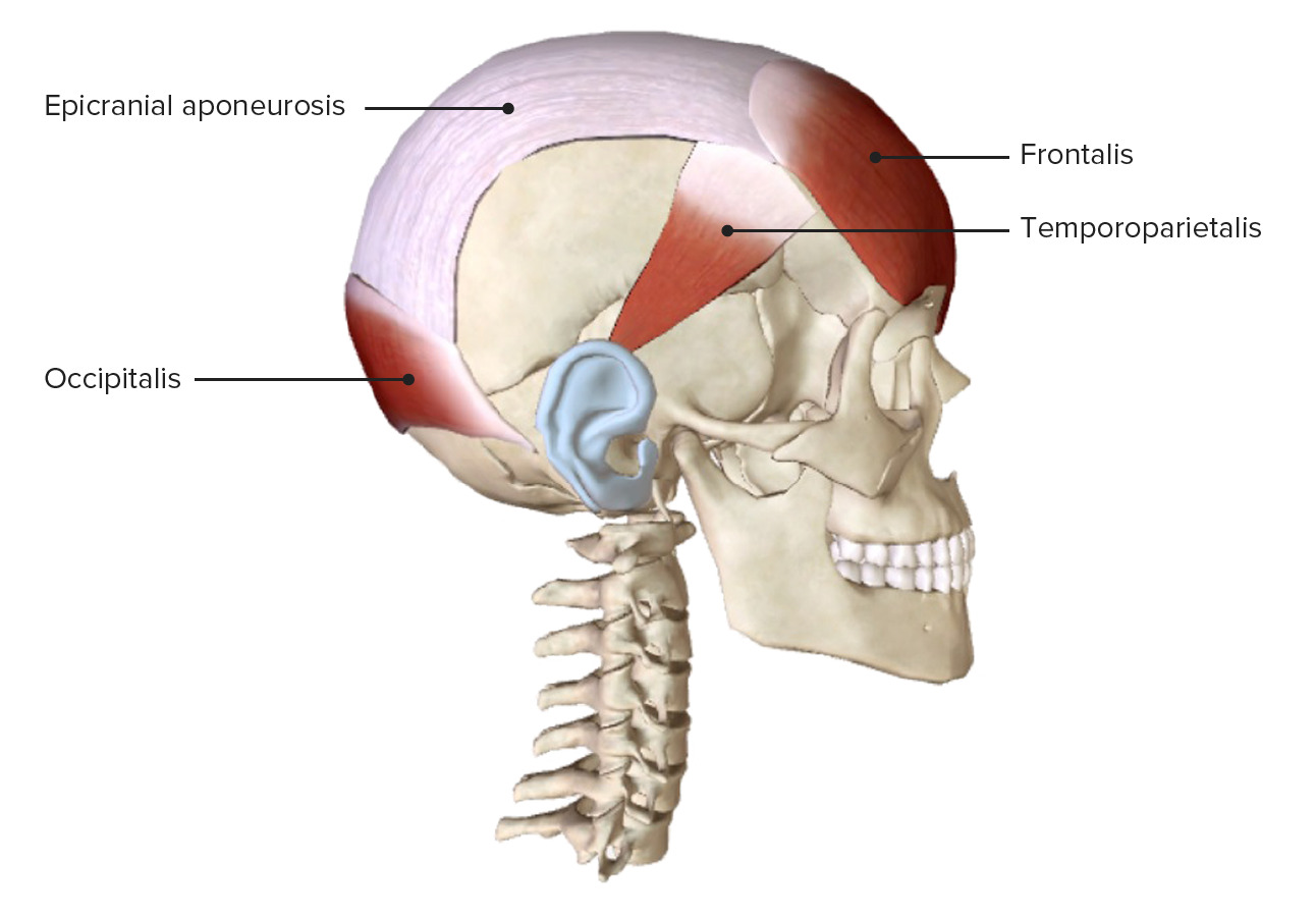
Lateral view of the face featuring both bellies of the occipitofrontalis muscle and the temporoparietalis muscle
Image by BioDigital, edited by LecturioThe muscles of the mouth assist with facial expression, chewing, and communication Communication The exchange or transmission of ideas, attitudes, or beliefs between individuals or groups. Decision-making Capacity and Legal Competence. These muscles are supplied by the facial nerve Facial nerve The 7th cranial nerve. The facial nerve has two parts, the larger motor root which may be called the facial nerve proper, and the smaller intermediate or sensory root. Together they provide efferent innervation to the muscles of facial expression and to the lacrimal and salivary glands, and convey afferent information for taste from the anterior two-thirds of the tongue and for touch from the external ear. The 12 Cranial Nerves: Overview and Functions (CN VII).
| Muscle | Origin | Insertion | Nerve supply | Function |
|---|---|---|---|---|
| Orbicularis oris | Deep surface of the perioral skin Skin The skin, also referred to as the integumentary system, is the largest organ of the body. The skin is primarily composed of the epidermis (outer layer) and dermis (deep layer). The epidermis is primarily composed of keratinocytes that undergo rapid turnover, while the dermis contains dense layers of connective tissue. Skin: Structure and Functions, angle of the mouth (modiolus) | Mucous membrane Mucous membrane An epithelium with mucus-secreting cells, such as goblet cells. It forms the lining of many body cavities, such as the digestive tract, the respiratory tract, and the reproductive tract. Mucosa, rich in blood and lymph vessels, comprises an inner epithelium, a middle layer (lamina propria) of loose connective tissue, and an outer layer (muscularis mucosae) of smooth muscle cells that separates the mucosa from submucosa. Barrett Esophagus of the lips Lips The lips are the soft and movable most external parts of the oral cavity. The blood supply of the lips originates from the external carotid artery, and the innervation is through cranial nerves. Lips and Tongue: Anatomy | Buccal branch of the facial nerve Facial nerve The 7th cranial nerve. The facial nerve has two parts, the larger motor root which may be called the facial nerve proper, and the smaller intermediate or sensory root. Together they provide efferent innervation to the muscles of facial expression and to the lacrimal and salivary glands, and convey afferent information for taste from the anterior two-thirds of the tongue and for touch from the external ear. The 12 Cranial Nerves: Overview and Functions | Closes mouth, purses lips Lips The lips are the soft and movable most external parts of the oral cavity. The blood supply of the lips originates from the external carotid artery, and the innervation is through cranial nerves. Lips and Tongue: Anatomy |
| Buccinator | Alveolar processes of the maxilla Maxilla One of a pair of irregularly shaped bones that form the upper jaw. A maxillary bone provides tooth sockets for the superior teeth, forms part of the orbit, and contains the maxillary sinus. Skull: Anatomy and mandible Mandible The largest and strongest bone of the face constituting the lower jaw. It supports the lower teeth. Jaw and Temporomandibular Joint: Anatomy (at the 1st–2nd molars Molars The most posterior teeth on either side of the jaw, totaling eight in the deciduous dentition (2 on each side, upper and lower), and usually 12 in the permanent dentition (three on each side, upper and lower). They are grinding teeth, having large crowns and broad chewing surfaces. Teeth: Anatomy) | Angle of the mouth radiating into the orbicularis oris | Buccal branch of the facial nerve Facial nerve The 7th cranial nerve. The facial nerve has two parts, the larger motor root which may be called the facial nerve proper, and the smaller intermediate or sensory root. Together they provide efferent innervation to the muscles of facial expression and to the lacrimal and salivary glands, and convey afferent information for taste from the anterior two-thirds of the tongue and for touch from the external ear. The 12 Cranial Nerves: Overview and Functions |
|
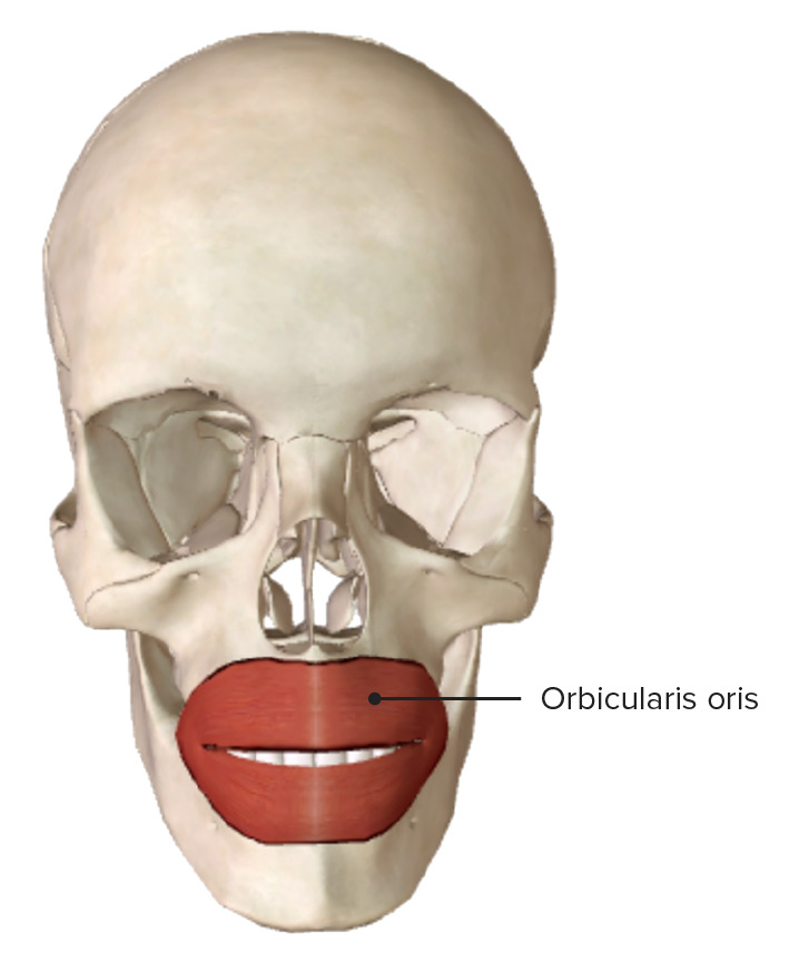
Anterior view of the orbicularis oris muscle fibers
Image by BioDigital, edited by Lecturio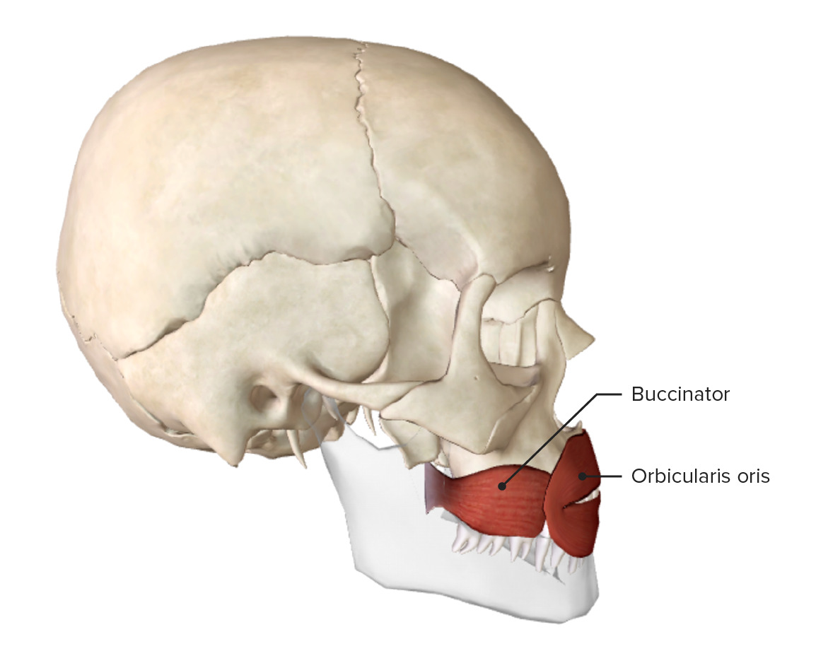
Lateral view of the buccinator and orbicularis oris muscles
Image by BioDigital, edited by Lecturio| Muscle | Origin | Insertion | Nerve supply | Function |
|---|---|---|---|---|
| Risorius | Masseteric fascia Fascia Layers of connective tissue of variable thickness. The superficial fascia is found immediately below the skin; the deep fascia invests muscles, nerves, and other organs. Cellulitis | Skin Skin The skin, also referred to as the integumentary system, is the largest organ of the body. The skin is primarily composed of the epidermis (outer layer) and dermis (deep layer). The epidermis is primarily composed of keratinocytes that undergo rapid turnover, while the dermis contains dense layers of connective tissue. Skin: Structure and Functions of angle of the mouth | Buccal branch of the facial nerve Facial nerve The 7th cranial nerve. The facial nerve has two parts, the larger motor root which may be called the facial nerve proper, and the smaller intermediate or sensory root. Together they provide efferent innervation to the muscles of facial expression and to the lacrimal and salivary glands, and convey afferent information for taste from the anterior two-thirds of the tongue and for touch from the external ear. The 12 Cranial Nerves: Overview and Functions | Pulls angle of the mouth laterally |
| Zygomaticus major | Lateral surface of the anterior aspect of the zygomatic bone Zygomatic bone Either of a pair of bones that form the prominent part of the cheek and contribute to the orbit on each side of the skull. Orbit and Extraocular Muscles: Anatomy | Skin Skin The skin, also referred to as the integumentary system, is the largest organ of the body. The skin is primarily composed of the epidermis (outer layer) and dermis (deep layer). The epidermis is primarily composed of keratinocytes that undergo rapid turnover, while the dermis contains dense layers of connective tissue. Skin: Structure and Functions of angle of the mouth, blending with levator anguli/orbicularis oris | Zygomatic Zygomatic Either of a pair of bones that form the prominent part of the cheek and contribute to the orbit on each side of the skull. Skull: Anatomy and buccal branches of the facial nerve Facial nerve The 7th cranial nerve. The facial nerve has two parts, the larger motor root which may be called the facial nerve proper, and the smaller intermediate or sensory root. Together they provide efferent innervation to the muscles of facial expression and to the lacrimal and salivary glands, and convey afferent information for taste from the anterior two-thirds of the tongue and for touch from the external ear. The 12 Cranial Nerves: Overview and Functions |
|
| Zygomaticus minor | Front of the zygomatic Zygomatic Either of a pair of bones that form the prominent part of the cheek and contribute to the orbit on each side of the skull. Skull: Anatomy arch | Skin Skin The skin, also referred to as the integumentary system, is the largest organ of the body. The skin is primarily composed of the epidermis (outer layer) and dermis (deep layer). The epidermis is primarily composed of keratinocytes that undergo rapid turnover, while the dermis contains dense layers of connective tissue. Skin: Structure and Functions of lateral part of upper lip Upper Lip Melasma, extends to nasolabial sulcus | Buccal branch of the facial nerve Facial nerve The 7th cranial nerve. The facial nerve has two parts, the larger motor root which may be called the facial nerve proper, and the smaller intermediate or sensory root. Together they provide efferent innervation to the muscles of facial expression and to the lacrimal and salivary glands, and convey afferent information for taste from the anterior two-thirds of the tongue and for touch from the external ear. The 12 Cranial Nerves: Overview and Functions | |
| Levator labii superioris | Infraorbital margin of the maxilla Maxilla One of a pair of irregularly shaped bones that form the upper jaw. A maxillary bone provides tooth sockets for the superior teeth, forms part of the orbit, and contains the maxillary sinus. Skull: Anatomy | Skin Skin The skin, also referred to as the integumentary system, is the largest organ of the body. The skin is primarily composed of the epidermis (outer layer) and dermis (deep layer). The epidermis is primarily composed of keratinocytes that undergo rapid turnover, while the dermis contains dense layers of connective tissue. Skin: Structure and Functions of the upper lip Upper Lip Melasma | Buccal branch of the facial nerve Facial nerve The 7th cranial nerve. The facial nerve has two parts, the larger motor root which may be called the facial nerve proper, and the smaller intermediate or sensory root. Together they provide efferent innervation to the muscles of facial expression and to the lacrimal and salivary glands, and convey afferent information for taste from the anterior two-thirds of the tongue and for touch from the external ear. The 12 Cranial Nerves: Overview and Functions | |
| Levator labii superioris alaeque nasi | Frontal Frontal The bone that forms the frontal aspect of the skull. Its flat part forms the forehead, articulating inferiorly with the nasal bone and the cheek bone on each side of the face. Skull: Anatomy process of the maxilla Maxilla One of a pair of irregularly shaped bones that form the upper jaw. A maxillary bone provides tooth sockets for the superior teeth, forms part of the orbit, and contains the maxillary sinus. Skull: Anatomy | Skin Skin The skin, also referred to as the integumentary system, is the largest organ of the body. The skin is primarily composed of the epidermis (outer layer) and dermis (deep layer). The epidermis is primarily composed of keratinocytes that undergo rapid turnover, while the dermis contains dense layers of connective tissue. Skin: Structure and Functions of lateral part of the nostril and skin Skin The skin, also referred to as the integumentary system, is the largest organ of the body. The skin is primarily composed of the epidermis (outer layer) and dermis (deep layer). The epidermis is primarily composed of keratinocytes that undergo rapid turnover, while the dermis contains dense layers of connective tissue. Skin: Structure and Functions of upper lip Upper Lip Melasma | Zygomatic Zygomatic Either of a pair of bones that form the prominent part of the cheek and contribute to the orbit on each side of the skull. Skull: Anatomy branches of the facial nerve Facial nerve The 7th cranial nerve. The facial nerve has two parts, the larger motor root which may be called the facial nerve proper, and the smaller intermediate or sensory root. Together they provide efferent innervation to the muscles of facial expression and to the lacrimal and salivary glands, and convey afferent information for taste from the anterior two-thirds of the tongue and for touch from the external ear. The 12 Cranial Nerves: Overview and Functions |
|
| Levator anguli oris | Canine fossa (maxillary fossa) on anterior surface of the maxilla Maxilla One of a pair of irregularly shaped bones that form the upper jaw. A maxillary bone provides tooth sockets for the superior teeth, forms part of the orbit, and contains the maxillary sinus. Skull: Anatomy | Skin Skin The skin, also referred to as the integumentary system, is the largest organ of the body. The skin is primarily composed of the epidermis (outer layer) and dermis (deep layer). The epidermis is primarily composed of keratinocytes that undergo rapid turnover, while the dermis contains dense layers of connective tissue. Skin: Structure and Functions of angle of the mouth | Buccal branch of the facial nerve Facial nerve The 7th cranial nerve. The facial nerve has two parts, the larger motor root which may be called the facial nerve proper, and the smaller intermediate or sensory root. Together they provide efferent innervation to the muscles of facial expression and to the lacrimal and salivary glands, and convey afferent information for taste from the anterior two-thirds of the tongue and for touch from the external ear. The 12 Cranial Nerves: Overview and Functions | Raises the angle of the mouth while smiling |
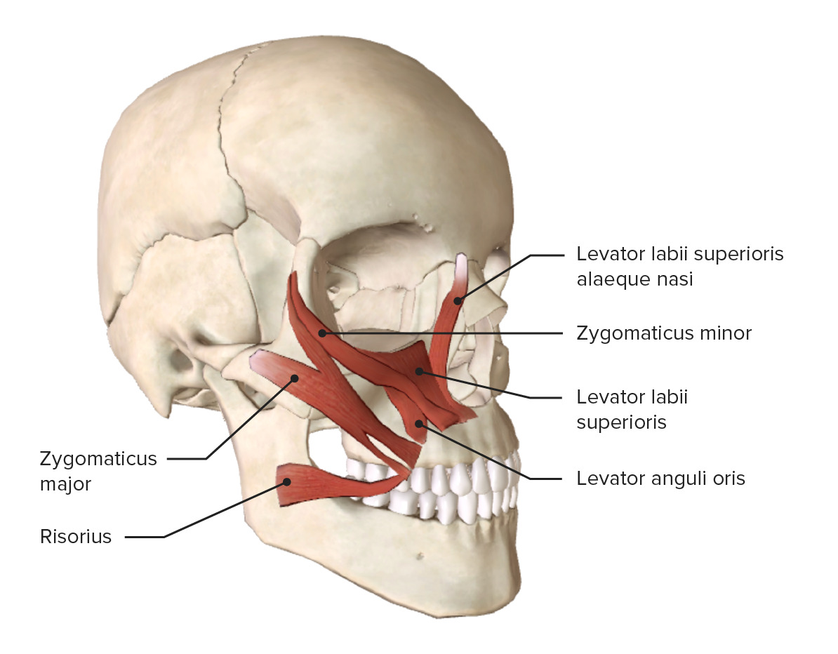
Oblique view of the muscles of the mouth (upper group)
Image by BioDigital, edited by Lecturio| Muscle | Origin | Insertion | Nerve supply | Function |
|---|---|---|---|---|
| Depressor anguli oris | Oblique line of the base of the mandible Mandible The largest and strongest bone of the face constituting the lower jaw. It supports the lower teeth. Jaw and Temporomandibular Joint: Anatomy | Skin Skin The skin, also referred to as the integumentary system, is the largest organ of the body. The skin is primarily composed of the epidermis (outer layer) and dermis (deep layer). The epidermis is primarily composed of keratinocytes that undergo rapid turnover, while the dermis contains dense layers of connective tissue. Skin: Structure and Functions of angle of the mouth | Marginal mandibular branch of the facial nerve Facial nerve The 7th cranial nerve. The facial nerve has two parts, the larger motor root which may be called the facial nerve proper, and the smaller intermediate or sensory root. Together they provide efferent innervation to the muscles of facial expression and to the lacrimal and salivary glands, and convey afferent information for taste from the anterior two-thirds of the tongue and for touch from the external ear. The 12 Cranial Nerves: Overview and Functions | Pulls angle of the mouth downward |
| Depressor labii inferioris | Oblique line of the base of the mandible Mandible The largest and strongest bone of the face constituting the lower jaw. It supports the lower teeth. Jaw and Temporomandibular Joint: Anatomy | Skin Skin The skin, also referred to as the integumentary system, is the largest organ of the body. The skin is primarily composed of the epidermis (outer layer) and dermis (deep layer). The epidermis is primarily composed of keratinocytes that undergo rapid turnover, while the dermis contains dense layers of connective tissue. Skin: Structure and Functions of lower lip | Retracts (depresses) and everts lower lip (pouting) | |
| Mentalis | Incisive fossa on alveolar process of the mandible Mandible The largest and strongest bone of the face constituting the lower jaw. It supports the lower teeth. Jaw and Temporomandibular Joint: Anatomy | Skin Skin The skin, also referred to as the integumentary system, is the largest organ of the body. The skin is primarily composed of the epidermis (outer layer) and dermis (deep layer). The epidermis is primarily composed of keratinocytes that undergo rapid turnover, while the dermis contains dense layers of connective tissue. Skin: Structure and Functions of the chin Chin The anatomical frontal portion of the mandible, also known as the mentum, that contains the line of fusion of the two separate halves of the mandible (symphysis menti). This line of fusion divides inferiorly to enclose a triangular area called the mental protuberance. On each side, inferior to the second premolar tooth, is the mental foramen for the passage of blood vessels and a nerve. Melasma |
|
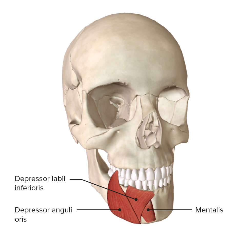
Oblique view of the muscles of the mouth (lower group)
Image by BioDigital, edited by LecturioThe muscles of the extraorbital eye assist with actions of blinking and facial expression. These muscles are primarily supplied by branches of the facial nerve Facial nerve The 7th cranial nerve. The facial nerve has two parts, the larger motor root which may be called the facial nerve proper, and the smaller intermediate or sensory root. Together they provide efferent innervation to the muscles of facial expression and to the lacrimal and salivary glands, and convey afferent information for taste from the anterior two-thirds of the tongue and for touch from the external ear. The 12 Cranial Nerves: Overview and Functions.
| Muscle | Origin | Insertion | Nerve supply | Function |
|---|---|---|---|---|
| Orbicularis oculi | Orbital part:
|
Temporal and zygomatic Zygomatic Either of a pair of bones that form the prominent part of the cheek and contribute to the orbit on each side of the skull. Skull: Anatomy branches of the facial nerve Facial nerve The 7th cranial nerve. The facial nerve has two parts, the larger motor root which may be called the facial nerve proper, and the smaller intermediate or sensory root. Together they provide efferent innervation to the muscles of facial expression and to the lacrimal and salivary glands, and convey afferent information for taste from the anterior two-thirds of the tongue and for touch from the external ear. The 12 Cranial Nerves: Overview and Functions |
|
|
| Depressor supercilii | Medial orbital rim Orbital Rim Orbital Fractures | Skin Skin The skin, also referred to as the integumentary system, is the largest organ of the body. The skin is primarily composed of the epidermis (outer layer) and dermis (deep layer). The epidermis is primarily composed of keratinocytes that undergo rapid turnover, while the dermis contains dense layers of connective tissue. Skin: Structure and Functions below the eyebrow and intercanthal region | Temporal branch of the facial nerve Facial nerve The 7th cranial nerve. The facial nerve has two parts, the larger motor root which may be called the facial nerve proper, and the smaller intermediate or sensory root. Together they provide efferent innervation to the muscles of facial expression and to the lacrimal and salivary glands, and convey afferent information for taste from the anterior two-thirds of the tongue and for touch from the external ear. The 12 Cranial Nerves: Overview and Functions | Depresses the eyebrow |
| Corrugator supercilii | Medial part of superciliary arch | Skin Skin The skin, also referred to as the integumentary system, is the largest organ of the body. The skin is primarily composed of the epidermis (outer layer) and dermis (deep layer). The epidermis is primarily composed of keratinocytes that undergo rapid turnover, while the dermis contains dense layers of connective tissue. Skin: Structure and Functions over middle of the eyebrow (penetrates frontalis and orbicularis oculi) | Temporal branch of the facial nerve Facial nerve The 7th cranial nerve. The facial nerve has two parts, the larger motor root which may be called the facial nerve proper, and the smaller intermediate or sensory root. Together they provide efferent innervation to the muscles of facial expression and to the lacrimal and salivary glands, and convey afferent information for taste from the anterior two-thirds of the tongue and for touch from the external ear. The 12 Cranial Nerves: Overview and Functions | Pulls skin Skin The skin, also referred to as the integumentary system, is the largest organ of the body. The skin is primarily composed of the epidermis (outer layer) and dermis (deep layer). The epidermis is primarily composed of keratinocytes that undergo rapid turnover, while the dermis contains dense layers of connective tissue. Skin: Structure and Functions of eyebrow downward and medially |

Oblique view of the skull, showing the superomedial extraorbital muscles
Image by BioDigital, edited by Lecturio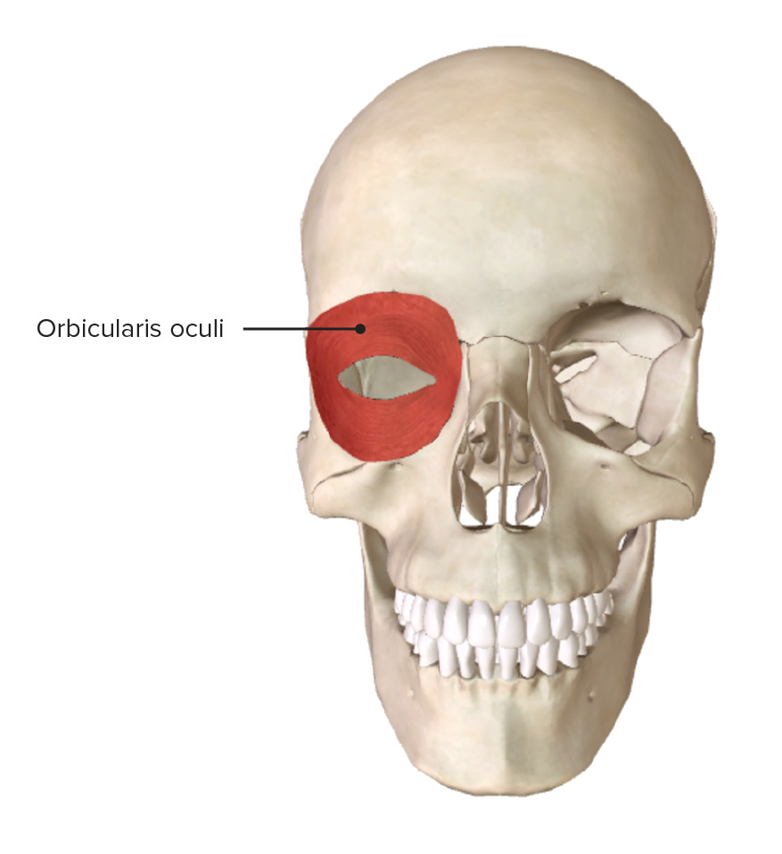
Anterior view of the skull, showcasing the origin and insertion of the orbicularis oculi muscle
Image by BioDigital, edited by LecturioThe muscles of the nose Nose The nose is the human body’s primary organ of smell and functions as part of the upper respiratory system. The nose may be best known for inhaling oxygen and exhaling carbon dioxide, but it also contributes to other important functions, such as tasting. The anatomy of the nose can be divided into the external nose and the nasal cavity. Nose Anatomy (External & Internal) assist in respiration Respiration The act of breathing with the lungs, consisting of inhalation, or the taking into the lungs of the ambient air, and of exhalation, or the expelling of the modified air which contains more carbon dioxide than the air taken in. Nose Anatomy (External & Internal) and facial expression. These muscles are supplied by branches of the facial nerve Facial nerve The 7th cranial nerve. The facial nerve has two parts, the larger motor root which may be called the facial nerve proper, and the smaller intermediate or sensory root. Together they provide efferent innervation to the muscles of facial expression and to the lacrimal and salivary glands, and convey afferent information for taste from the anterior two-thirds of the tongue and for touch from the external ear. The 12 Cranial Nerves: Overview and Functions.
| Muscle | Origin | Insertion | Nerve supply | Function |
|---|---|---|---|---|
| Nasalis | Transverse nasalis (compressor naris): maxilla Maxilla One of a pair of irregularly shaped bones that form the upper jaw. A maxillary bone provides tooth sockets for the superior teeth, forms part of the orbit, and contains the maxillary sinus. Skull: Anatomy, lateral to incisive fossa | Aponeurosis of bridge of the nose Nose The nose is the human body’s primary organ of smell and functions as part of the upper respiratory system. The nose may be best known for inhaling oxygen and exhaling carbon dioxide, but it also contributes to other important functions, such as tasting. The anatomy of the nose can be divided into the external nose and the nasal cavity. Nose Anatomy (External & Internal) | Buccal branch of the facial nerve Facial nerve The 7th cranial nerve. The facial nerve has two parts, the larger motor root which may be called the facial nerve proper, and the smaller intermediate or sensory root. Together they provide efferent innervation to the muscles of facial expression and to the lacrimal and salivary glands, and convey afferent information for taste from the anterior two-thirds of the tongue and for touch from the external ear. The 12 Cranial Nerves: Overview and Functions | Compresses nostril |
| Alar nasalis (dilator naris): outer surface of maxilla Maxilla One of a pair of irregularly shaped bones that form the upper jaw. A maxillary bone provides tooth sockets for the superior teeth, forms part of the orbit, and contains the maxillary sinus. Skull: Anatomy, above lateral incisor tooth | Skin Skin The skin, also referred to as the integumentary system, is the largest organ of the body. The skin is primarily composed of the epidermis (outer layer) and dermis (deep layer). The epidermis is primarily composed of keratinocytes that undergo rapid turnover, while the dermis contains dense layers of connective tissue. Skin: Structure and Functions of ala, superior to lateral crus of major alar cartilage Cartilage Cartilage is a type of connective tissue derived from embryonic mesenchyme that is responsible for structural support, resilience, and the smoothness of physical actions. Perichondrium (connective tissue membrane surrounding cartilage) compensates for the absence of vasculature in cartilage by providing nutrition and support. Cartilage: Histology | Dilates nostril | ||
| Procerus | Midline of nasal bone Bone Bone is a compact type of hardened connective tissue composed of bone cells, membranes, an extracellular mineralized matrix, and central bone marrow. The 2 primary types of bone are compact and spongy. Bones: Structure and Types and lateral nasal cartilage Cartilage Cartilage is a type of connective tissue derived from embryonic mesenchyme that is responsible for structural support, resilience, and the smoothness of physical actions. Perichondrium (connective tissue membrane surrounding cartilage) compensates for the absence of vasculature in cartilage by providing nutrition and support. Cartilage: Histology | Skin Skin The skin, also referred to as the integumentary system, is the largest organ of the body. The skin is primarily composed of the epidermis (outer layer) and dermis (deep layer). The epidermis is primarily composed of keratinocytes that undergo rapid turnover, while the dermis contains dense layers of connective tissue. Skin: Structure and Functions of lower part of forehead Forehead The part of the face above the eyes. Melasma between eyebrows | Draws the medial border of the eyebrows downward to produce transverse wrinkles over the bridge of the nose Nose The nose is the human body’s primary organ of smell and functions as part of the upper respiratory system. The nose may be best known for inhaling oxygen and exhaling carbon dioxide, but it also contributes to other important functions, such as tasting. The anatomy of the nose can be divided into the external nose and the nasal cavity. Nose Anatomy (External & Internal) | |
| Depressor septi nasi | Incisive fossa of the maxilla Maxilla One of a pair of irregularly shaped bones that form the upper jaw. A maxillary bone provides tooth sockets for the superior teeth, forms part of the orbit, and contains the maxillary sinus. Skull: Anatomy | Nasal septum Nasal septum The partition separating the two nasal cavities in the midplane. It is formed by the septal nasal cartilage, parts of skull bones, and membranous parts. Nose Anatomy (External & Internal) and posterior aspect of alar nasalis | Depresses nasal septum Nasal septum The partition separating the two nasal cavities in the midplane. It is formed by the septal nasal cartilage, parts of skull bones, and membranous parts. Nose Anatomy (External & Internal) and pulls wings of the nose Nose The nose is the human body’s primary organ of smell and functions as part of the upper respiratory system. The nose may be best known for inhaling oxygen and exhaling carbon dioxide, but it also contributes to other important functions, such as tasting. The anatomy of the nose can be divided into the external nose and the nasal cavity. Nose Anatomy (External & Internal) downward |

Oblique view of the skull showing the facial muscles of the nose
Image by BioDigital, edited by LecturioThe muscles of the ear are more simple than the other muscle groups of the face. These muscles assist with moving the ear and are innervated by fibers of the facial nerve Facial nerve The 7th cranial nerve. The facial nerve has two parts, the larger motor root which may be called the facial nerve proper, and the smaller intermediate or sensory root. Together they provide efferent innervation to the muscles of facial expression and to the lacrimal and salivary glands, and convey afferent information for taste from the anterior two-thirds of the tongue and for touch from the external ear. The 12 Cranial Nerves: Overview and Functions (CN VII).
| Muscle | Origin | Insertion | Nerve supply | Function |
|---|---|---|---|---|
| Anterior auricular | Lateral edge of the galea aponeurotica | Projection on the front of the helix | Temporal branch of the facial nerve Facial nerve The 7th cranial nerve. The facial nerve has two parts, the larger motor root which may be called the facial nerve proper, and the smaller intermediate or sensory root. Together they provide efferent innervation to the muscles of facial expression and to the lacrimal and salivary glands, and convey afferent information for taste from the anterior two-thirds of the tongue and for touch from the external ear. The 12 Cranial Nerves: Overview and Functions | Pulls ear upward and forward |
| Posterior auricular | Mastoid portion of temporal bone Temporal bone Either of a pair of compound bones forming the lateral (left and right) surfaces and base of the skull which contains the organs of hearing. It is a large bone formed by the fusion of parts: the squamous (the flattened anterior-superior part), the tympanic (the curved anterior-inferior part), the mastoid (the irregular posterior portion), and the petrous (the part at the base of the skull). Jaw and Temporomandibular Joint: Anatomy | Lower part of the cranial surface of the concha | Posterior auricular nerve of facial nerve Facial nerve The 7th cranial nerve. The facial nerve has two parts, the larger motor root which may be called the facial nerve proper, and the smaller intermediate or sensory root. Together they provide efferent innervation to the muscles of facial expression and to the lacrimal and salivary glands, and convey afferent information for taste from the anterior two-thirds of the tongue and for touch from the external ear. The 12 Cranial Nerves: Overview and Functions | Retracts and elevates the ear |
| Superior auricular | Galea aponeurotica | Cranial surface of the auricula | Pulls ear upward |
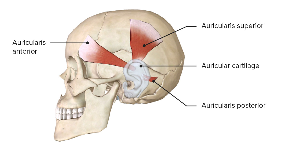
Lateral view of the head, featuring the facial muscles around the ear:
Note the posterior, superior, and anterior auricular muscles.
The muscles of mastication Mastication The act and process of chewing and grinding food in the mouth. Jaw and Temporomandibular Joint: Anatomy assist in the action of chewing via movement of the mandible Mandible The largest and strongest bone of the face constituting the lower jaw. It supports the lower teeth. Jaw and Temporomandibular Joint: Anatomy. They are supplied by the mandibular branches (V3) of the trigeminal nerve Trigeminal nerve The 5th and largest cranial nerve. The trigeminal nerve is a mixed motor and sensory nerve. The larger sensory part forms the ophthalmic, mandibular, and maxillary nerves which carry afferents sensitive to external or internal stimuli from the skin, muscles, and joints of the face and mouth and from the teeth. Most of these fibers originate from cells of the trigeminal ganglion and project to the trigeminal nucleus of the brain stem. The smaller motor part arises from the brain stem trigeminal motor nucleus and innervates the muscles of mastication. The 12 Cranial Nerves: Overview and Functions (CN V).
| Muscle | Origin | Insertion | Nerve supply | Function |
|---|---|---|---|---|
| Temporalis Temporalis A masticatory muscle whose action is closing the jaws; its posterior portion retracts the mandible. Jaw and Temporomandibular Joint: Anatomy | Temporal fossa Temporal fossa Skull: Anatomy | Coronoid process of the mandible Mandible The largest and strongest bone of the face constituting the lower jaw. It supports the lower teeth. Jaw and Temporomandibular Joint: Anatomy | Branches of mandibular division of trigeminal nerve Trigeminal nerve The 5th and largest cranial nerve. The trigeminal nerve is a mixed motor and sensory nerve. The larger sensory part forms the ophthalmic, mandibular, and maxillary nerves which carry afferents sensitive to external or internal stimuli from the skin, muscles, and joints of the face and mouth and from the teeth. Most of these fibers originate from cells of the trigeminal ganglion and project to the trigeminal nucleus of the brain stem. The smaller motor part arises from the brain stem trigeminal motor nucleus and innervates the muscles of mastication. The 12 Cranial Nerves: Overview and Functions | Elevates and retracts the mandible Mandible The largest and strongest bone of the face constituting the lower jaw. It supports the lower teeth. Jaw and Temporomandibular Joint: Anatomy |
| Masseter Masseter A masticatory muscle whose action is closing the jaws. Jaw and Temporomandibular Joint: Anatomy | Zygomatic Zygomatic Either of a pair of bones that form the prominent part of the cheek and contribute to the orbit on each side of the skull. Skull: Anatomy arch | Masseteric tuberosity of the mandible Mandible The largest and strongest bone of the face constituting the lower jaw. It supports the lower teeth. Jaw and Temporomandibular Joint: Anatomy | Elevates the mandible Mandible The largest and strongest bone of the face constituting the lower jaw. It supports the lower teeth. Jaw and Temporomandibular Joint: Anatomy | |
| Lateral pterygoid Lateral pterygoid Two of the masticatory muscles: the internal, or medial, pterygoid muscle and external, or lateral, pterygoid muscle. Action of the former is closing the jaws and that of the latter is opening the jaws, protruding the mandible, and moving the mandible from side to side. Jaw and Temporomandibular Joint: Anatomy | Infratemporal surface of the greater wing of sphenoid and lateral plate of pterygoid process | Pterygoid fossa of the mandible Mandible The largest and strongest bone of the face constituting the lower jaw. It supports the lower teeth. Jaw and Temporomandibular Joint: Anatomy |
|
|
| Medial pterygoid Medial pterygoid Two of the masticatory muscles: the internal, or medial, pterygoid muscle and external, or lateral, pterygoid muscle. Action of the former is closing the jaws and that of the latter is opening the jaws, protruding the mandible, and moving the mandible from side to side. Jaw and Temporomandibular Joint: Anatomy | Pterygoid fossa of sphenoid bone Sphenoid bone An irregular unpaired bone situated at the skull base and wedged between the frontal, temporal, and occipital bones (frontal bone; temporal bone; occipital bone). Sphenoid bone consists of a median body and three pairs of processes resembling a bat with spread wings. The body is hollowed out in its inferior to form two large cavities (sphenoid sinus). Orbit and Extraocular Muscles: Anatomy | Pterygoid tuberosity of the mandible Mandible The largest and strongest bone of the face constituting the lower jaw. It supports the lower teeth. Jaw and Temporomandibular Joint: Anatomy |
|
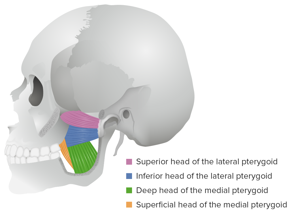
Lateral view of the lateral and medial pterygoid muscles
Image by Lecturio. License: CC BY-NC-SA 4.0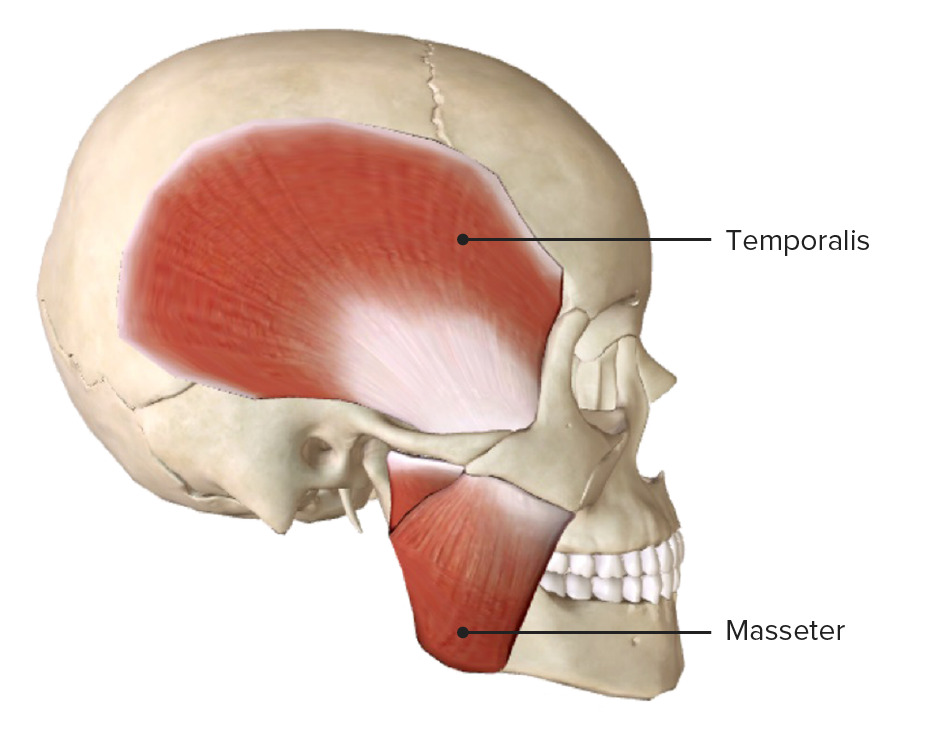
Lateral view of the skull, featuring the temporalis and masseter muscles
Image by BioDigital, edited by Lecturio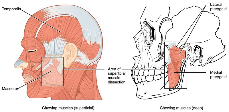
Lateral view of the head, featuring the muscles of mastication:
Note that the masseter and temporalis muscles are superficial, while the lateral and medial pterygoid muscles are on a deeper muscular layer in direct relation with the temporomandibular joint.