Epidemiology and Etiology
Epidemiology
- Incidence: 1 of 7–8 people per year
- Occurs in up to 80% of patients with uncomplicated upper respiratory infections (URIs)
- Bacterial sinusitis is only found in 0.5%–2% of cases
- Peak annual incidence coincides with peak of viral URIs in fall/winter months
- Higher incidence in women and in those aged 45–64 years
Etiology
- Predisposing factors:
- Anatomic abnormalities of the paranasal sinuses or nasal cavity (deviation in nasal septum, concha bullosa, bony spurs, nasal polyp, choanal atresia)
- Impairment of ciliary function (cystic fibrosis, primary ciliary dyskinesia, immunodeficiency)
- Allergic and vasomotor rhinitis
- Recurrent URIs
- Asthma and allergies
- Dental disease
- Immunodeficiency
- Older age
- Smoking
- Cocaine abuse
- Air travel
- Atmospheric pressure change exposure (e.g., deep sea diving)
- Swimming
- Foreign body in the nasal cavity
- Acute: lasts ≤ 4 weeks
- Viral (most common): rhinovirus, coronavirus, influenza virus, adenovirus, parainfluenza virus → rhinovirus, influenza virus, parainfluenza virus, adenovirus, coronavirus
- Bacterial: Streptococcus pneumoniae, Haemophilus influenzae, Moraxella catarrhalis, Staphylococcus aureus, Escherichia coli, Klebsiella
- S. pnuemoniae and H. influenzae are responsible for 75% of cases
- Chronic: lasts over 12 weeks; characterized by prolonged inflammation of the sinuses; occasionally associated with immunodeficiency disorders
- Fungal: Aspergillus, Rhizopus oryzae
- Bacterial: S. aureus (often methicillin-resistant S. aureus) and anaerobic organisms (Prevotella, Porphyromonas, Fusobacterium, and Peptostreptococcus spp.)
Pathophysiology
Viral
- Inoculation via direct contact with the conjunctiva or nasal mucosa
- Viral replication can be detected via viral levels in nasal secretions within 8–10 hours
- Symptoms can occur after the first day of inoculation
Bacterial
- Most important pathophysiological change: obstruction of sinus drainage
- Normal metachronous (sequential) movement of mucous toward the natural ostia of the sinuses is usually disrupted by mucosal inflammation → stasis
- Ciliary function impairment leads to a reduction in mucus drainage → stasis
- Stasis of secretions inside the sinuses leads to a proliferation of microorganisms
- Alterations in the composition and quantity of mucus can also contribute to infection; increased mucus production is seen with asthma, rhinitis, and viral URIs
Clinical Presentation
General manifestations
- Nasal congestion and obstruction
- Mucopurulent or purulent nasal discharge
- Headache
- Facial pain or heaviness over the affected sinus (increases with bending forward)
- Maxillary sinuses: over the cheeks
- Frontal sinuses: lower forehead
- Ethmoidal sinuses: nasal bridge or between/behind the eyes
- Fever
- Maxillary tooth discomfort and other facial pain
- General malaise and myalgia
Viral sinusitis
Viral sinusitis usually presents in a milder form and lasts 7–10 days.
Bacterial sinusitis
- Persists for > 10 days without clinical improvement
- Biphasic pattern: may initially improve, then worsens after 5–6 days (“double worsening”)
- Presents with more severe symptoms:
- High fever
- Intense facial pain
- Hyposmia or anosmia
- Halitosis
- Cough
- Ear pain, pressure, fullness, hearing loss, or tinnitus
Fungal sinusitis
Fungal sinusitis usually presents chronically, with atypical symptoms (epistaxis, dyspnea, and black/brown nasal secretions).
Complications and/or associated conditions
- Acute otitis media
- Pharyngitis
- Meningitis
- Pneumonia
- Subperiosteal/intracranial abscess
- Osteomyelitis (usually of the frontal bone)
- Preseptal/orbital cellulitis
- Septic cavernous sinus thrombosis
Diagnosis
Diagnosis is usually based on clinical symptoms.
- Uncomplicated sinusitis: < 4 weeks of purulent nasal discharge + nasal obstruction or facial pain/pressure/fullness or both
- Complicated sinusitis: When signs and symptoms extend beyond paranasal sinuses and nasal cavity, urgent evaluation is required.
- Signs: severe/persistent headache, vision changes, periorbital edema, abnormal extraocular movements with or without pain, altered mental status, meningeal signs, signs of increased intracranial pressure
To differentiate viral sinusitis from bacterial sinusitis:
- Viral rhinosinusitis is diagnosed clinically when symptoms last 7–10 days and do not worsen.
- Bacterial rhinosinusitis is diagnosed clinically when symptoms last > 10 days or are of a biphasic pattern (see “double worsening,” above) and are more severe in nature. Requires antibiotics for resolution
Laboratory tests
- Indicated if no response to treatment or worsening is observed
- Leukocytosis is rarely seen.
- May help determine the underlying cause:
- Allergic rhinitis confirmed with a radioallergosorbent test, allergen skin test, or smear of nasal secretions showing eosinophilia
- Sweat chloride test for cystic fibrosis
Culture
Presence of more than 104 colony-forming units/mL on bacterial culture is confirmatory for bacterial sinusitis in children.
Imaging
- Not recommended unless complicated sinusitis arises
- Skull X-rays show decreased transparency of sinus and air-fluid levels.
- Computed tomography is the modality of choice and shows mucoperiosteal thickening, air-fluid levels, and soft tissue extension in case of complications (e.g., orbital cellulitis).
- Nasal endoscopy may be used to exclude structural lesions.
Biopsy
Only needed if infection recurs or if there is no response to different empiric therapies.
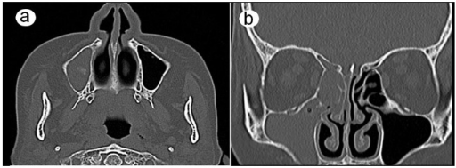
CT scans: (a) the axial plane and (b) the coronal plane show complete opacification in the maxillary, ethmoid, and frontal sinuses on the right side.
Image: “CT scans 6 weeks after the first visit” by Department of Oral and Maxillofacial Surgery, Nara Medical University, Nara, Japan. License: CC BY 4.0
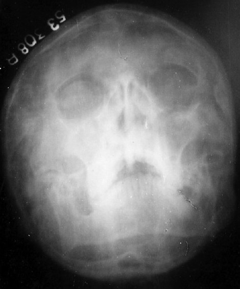
X-ray of paranasal sinuses showing maxillary sinusitis and bilateral hypoplastic frontal sinuses.
Image: “X-ray paranasal sinus showing maxillary sinusitis” by J.L.N. Medical College, Ajmer, India. License: CC BY 2.0
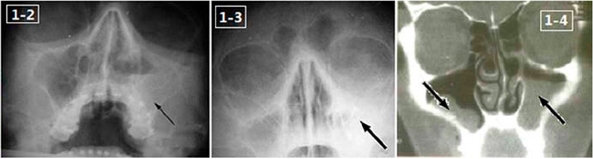
X-ray images in Water’s position (1–2 and 1–3) and magnetic resonance image (1–4) show the opacification of the maxillary sinus and bilateral air-fluid levels.
Image: “Endoscopic image of maxillary sinus mucosa” by Lin Feng, Department of Stomatology, Chinese PLA General Hospital, Beijing 100853, P. R. China. License: CC BY 3.0, edited by Lecturio.
Management
General supportive care
- Acetaminophen or nonsteroidal anti-inflammatory drugs should be given for facial pain and, if present, fever.
Viral sinusitis
- Aims only to provide symptomatic relief, as viral sinusitis is self-limiting
- Nasal lavage with hypertonic NaCl solution
- Decongestants (e.g., xylometazoline nasal spray) or sympathomimetics (e.g., pseudoephedrine)
- Antihistamines (e.g., loratadine) if concurrent allergy symptoms are present
- Intranasal steroids (e.g., fluticasone) to relieve mucosal swelling/facial pain
- Mucolytics (e.g., guaifenesin) can thin secretions and promote drainage
Bacterial sinusitis
- First-line: amoxicillin, with or without clavulanate for 5–10 days
- Second-line:
- IV ceftriaxone is used for children who are unable to tolerate oral medication.
- Oxycycline, levofloxacin, or moxifloxacin are used in case of penicillin allergy.
- Levofloxacin, moxifloxacin, or clindamycin are used if symptoms persist > 14 days or no improvement is seen within the first 3 days of treatment.
- Chronic bacterial sinusitis: broad-spectrum aminopenicillin + beta-lactamase inhibitor
- Decongestants and antihistamines are not recommended.
Fungal sinusitis
- Antifungal therapy (e.g., amphotericin B)
- Management of immunocompromising conditions (e.g., AIDS, HIV, cancer, etc.)
- If the infection is chronic and invasive, it may require surgical debridement of necrotic tissue.
Related videos
Clinical Relevance
The following are potential underlying conditions or differential diagnoses of sinusitis:
- Cystic fibrosis: an autosomal recessive disorder caused by mutation of the CFTR gene, which leads to defective chloride channels and hyperviscosity of exocrine gland secretions
- Primary ciliary dyskinesia: an autosomal recessive disease associated with sinusitis, situs inversus, recurrent respiratory infections, and bronchiectasis, among other abnormalities
- Dental abscess: a collection of pus in the pulp of a tooth that can spread to local or regional structures, including the gums, facial bones, tongue, and facial muscles
- Foreign nasal body: common in children < 5 years old. Often involves food items or small toys. Presents with unilateral rhinorrhea that can become foul-smelling or purulent, signs of nasal obstruction, or epistaxis
- Migraine: a type of headache characterized by recurrent, debilitating episodes that are typically unilateral, throbbing and/or pulsatile in quality, and frequently accompanied by nausea, vomiting, and sensitivity to light and sound
- Rhinitis: an inflammation of the nasal mucosa, classified into allergic, non-allergic, and infectious
- Asthma: a chronic inflammatory disease of the respiratory system characterized by bronchial hyperresponsiveness, episodic exacerbation, and reversible airflow obstruction.
- Otitis media: an infection of the middle ear that typically follows an upper respiratory tract infection in children < 5 years old. Most commonly caused by S. pneumoniae. Presents with otalgia and fever. Examination shows a bulging tympanic membrane.
- Granulomatosis with polyangiitis: an antineutrophil cytoplasmic antibody–associated vasculitis leading to inflammation of small- and medium-sized blood vessels. Results in damage to several organ systems of the body, most often the respiratory tract and kidneys.