Sickle cell disease (SCD) is a group of genetic disorders in which an abnormal Hb molecule (Hb S) transforms RBCs into sickle-shaped cells, resulting in chronic anemia, vasoocclusive episodes, pain, and organ damage. Sickle cell trait, which is the heterozygous condition, is the only 1 of the group that is generally benign and rarely associated with serious SCD-like complications. Triggers such as stress and hypoxia can induce or worsen the sickling of RBCs. Individuals with SCD are susceptible to infection, infarction of various organs, and bone marrow aplasia; lung involvement in acute chest syndrome can be rapidly fatal. Sickle cells can usually be seen on the peripheral blood smear, but Hb electrophoresis is needed for diagnosis. The management of painful episodes consists of IV fluids and analgesics, and in severe episodes, exchange transfusions may be required. Survival is improved by hydroxyurea intake, vaccination against bacterial infections, prophylactic antibiotics, and aggressive treatment of infections. Although trials are still ongoing and the long-term effects are still unclear, promising results from early trials show the potential for gene therapy as a treatment option.
Sickle cell diseaseSickle cell diseaseSickle cell disease (SCD) is a group of genetic disorders in which an abnormal Hb molecule (HbS) transforms RBCs into sickle-shaped cells, resulting in chronic anemia, vasoocclusive episodes, pain, and organ damage.Sickle Cell Disease (SCDSCDSickle cell disease (SCD) is a group of genetic disorders in which an abnormal Hb molecule (HbS) transforms RBCs into sickle-shaped cells, resulting in chronic anemia, vasoocclusive episodes, pain, and organ damage.Sickle Cell Disease) is a group of genetic disorders that cause an abnormal HbHbThe oxygen-carrying proteins of erythrocytes. They are found in all vertebrates and some invertebrates. The number of globin subunits in the hemoglobin quaternary structure differs between species. Structures range from monomeric to a variety of multimeric arrangements.Gas Exchange molecule (HbHbThe oxygen-carrying proteins of erythrocytes. They are found in all vertebrates and some invertebrates. The number of globin subunits in the hemoglobin quaternary structure differs between species. Structures range from monomeric to a variety of multimeric arrangements.Gas Exchange S) that transforms RBCsRBCsErythrocytes, or red blood cells (RBCs), are the most abundant cells in the blood. While erythrocytes in the fetus are initially produced in the yolk sac then the liver, the bone marrow eventually becomes the main site of production.Erythrocytes: Histology into sickle-shaped cells, resulting in chronic anemiaAnemiaAnemia is a condition in which individuals have low Hb levels, which can arise from various causes. Anemia is accompanied by a reduced number of RBCs and may manifest with fatigue, shortness of breath, pallor, and weakness. Subtypes are classified by the size of RBCs, chronicity, and etiology. Anemia: Overview and Types, vasoocclusive episodes, painPainAn unpleasant sensation induced by noxious stimuli which are detected by nerve endings of nociceptive neurons.Pain: Types and Pathways, and organ damage.
IncidenceIncidenceThe number of new cases of a given disease during a given period in a specified population. It also is used for the rate at which new events occur in a defined population. It is differentiated from prevalence, which refers to all cases in the population at a given time.Measures of Disease Frequency of sickle cell traitsickle cell traitThe condition of being heterozygous for hemoglobin S.Sickle Cell Disease:
30% of individuals in sub-Saharan Africa
7.3% of African Americans
Equally present in men and women
Etiology[10]
Sickle cell diseaseSickle cell diseaseSickle cell disease (SCD) is a group of genetic disorders in which an abnormal Hb molecule (HbS) transforms RBCs into sickle-shaped cells, resulting in chronic anemia, vasoocclusive episodes, pain, and organ damage.Sickle Cell Disease is any syndrome with a sickle mutationMutationGenetic mutations are errors in DNA that can cause protein misfolding and dysfunction. There are various types of mutations, including chromosomal, point, frameshift, and expansion mutations. Types of Mutations that alters normal beta globin:
Homozygous genotypeGenotypeThe genetic constitution of the individual, comprising the alleles present at each genetic locus.Basic Terms of Genetics = HbSSHbSSSickle cell disease (SCD) is a group of genetic disorders in which an abnormal Hb molecule (HbS) transforms RBCs into sickle-shaped cells, resulting in chronic anemia, vasoocclusive episodes, pain, and organ damage.Sickle Cell Disease; causes sickle cell anemiaSickle cell anemiaA disease characterized by chronic hemolytic anemia, episodic painful crises, and pathologic involvement of many organs. It is the clinical expression of homozygosity for hemoglobin S.Sickle Cell Disease
Only 1 alleleAlleleVariant forms of the same gene, occupying the same locus on homologous chromosomes, and governing the variants in production of the same gene product.Basic Terms of Genetics has the HbHbThe oxygen-carrying proteins of erythrocytes. They are found in all vertebrates and some invertebrates. The number of globin subunits in the hemoglobin quaternary structure differs between species. Structures range from monomeric to a variety of multimeric arrangements.Gas Exchange S mutationMutationGenetic mutations are errors in DNA that can cause protein misfolding and dysfunction. There are various types of mutations, including chromosomal, point, frameshift, and expansion mutations. Types of Mutations.
Important for counseling about risk of having a child with SCDSCDSickle cell disease (SCD) is a group of genetic disorders in which an abnormal Hb molecule (HbS) transforms RBCs into sickle-shaped cells, resulting in chronic anemia, vasoocclusive episodes, pain, and organ damage.Sickle Cell Disease
Occur as compound heterozygotes with HbHbThe oxygen-carrying proteins of erythrocytes. They are found in all vertebrates and some invertebrates. The number of globin subunits in the hemoglobin quaternary structure differs between species. Structures range from monomeric to a variety of multimeric arrangements.Gas Exchange S from 1 parent
VariableVariableVariables represent information about something that can change. The design of the measurement scales, or of the methods for obtaining information, will determine the data gathered and the characteristics of that data. As a result, a variable can be qualitative or quantitative, and may be further classified into subgroups.Types of Variables clinical severity as compared with homozygous sickle mutationMutationGenetic mutations are errors in DNA that can cause protein misfolding and dysfunction. There are various types of mutations, including chromosomal, point, frameshift, and expansion mutations. Types of Mutations (HbSSHbSSSickle cell disease (SCD) is a group of genetic disorders in which an abnormal Hb molecule (HbS) transforms RBCs into sickle-shaped cells, resulting in chronic anemia, vasoocclusive episodes, pain, and organ damage.Sickle Cell Disease)
Affect the alpha-, beta-, or gamma-globin genesGenesA category of nucleic acid sequences that function as units of heredity and which code for the basic instructions for the development, reproduction, and maintenance of organisms.DNA Types and Structure:
Sickle-beta thalassemiaSickle-Beta ThalassemiaSickle Cell Disease: less severe if HbHbThe oxygen-carrying proteins of erythrocytes. They are found in all vertebrates and some invertebrates. The number of globin subunits in the hemoglobin quaternary structure differs between species. Structures range from monomeric to a variety of multimeric arrangements.Gas Exchange S compounded with beta-plus thalassemiaThalassemiaThalassemia is a hereditary cause of microcytic hypochromic anemia and results from a deficiency in either the α or β globin chains, resulting in hemoglobinopathy. The presentation of thalassemia depends on the number of defective chains present and can range from being asymptomatic to rendering the more severely affected patients to be transfusion dependent. Thalassemia (some beta globin is produced) than with beta-zero thalassemiaThalassemiaThalassemia is a hereditary cause of microcytic hypochromic anemia and results from a deficiency in either the α or β globin chains, resulting in hemoglobinopathy. The presentation of thalassemia depends on the number of defective chains present and can range from being asymptomatic to rendering the more severely affected patients to be transfusion dependent. Thalassemia (no beta globin is produced)
HbSC diseaseHbSC diseaseA disease characterized by chronic hemolytic anemia, episodic painful crises, and pathologic involvement of many organs. It is the clinical expression of homozygosity for hemoglobin s.Sickle Cell Disease: symptoms similar to SCDSCDSickle cell disease (SCD) is a group of genetic disorders in which an abnormal Hb molecule (HbS) transforms RBCs into sickle-shaped cells, resulting in chronic anemia, vasoocclusive episodes, pain, and organ damage.Sickle Cell Disease but less severe
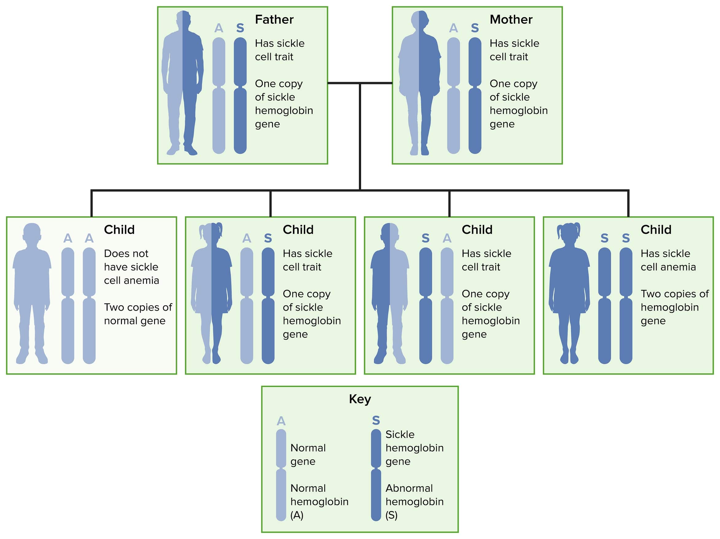
Autosomal recessive inheritance of sickle cell disease and trait
Normal adult hemoglobin molecule (HbA1) consists of 2 pairs of chains called alpha and beta.[10]
HbHbThe oxygen-carrying proteins of erythrocytes. They are found in all vertebrates and some invertebrates. The number of globin subunits in the hemoglobin quaternary structure differs between species. Structures range from monomeric to a variety of multimeric arrangements.Gas Exchange S is produced by a point mutationPoint MutationA mutation caused by the substitution of one nucleotide for another. This results in the DNA molecule having a change in a single base pair.Types of Mutations on chromosomeChromosomeIn a prokaryotic cell or in the nucleus of a eukaryotic cell, a structure consisting of or containing DNA which carries the genetic information essential to the cell.Basic Terms of Genetics 11, causing substitution of valine (amino acidAmino acidAmino acids (AAs) are composed of a central carbon atom attached to a carboxyl group, an amino group, a hydrogen atom, and a side chain (R group). Basics of Amino Acids) for glutamic acidGlutamic acidA non-essential amino acid naturally occurring in the l-form. Glutamic acid is the most common excitatory neurotransmitter in the central nervous system.Urea Cycle at the 6th position in the beta-globin chain.
HbHbThe oxygen-carrying proteins of erythrocytes. They are found in all vertebrates and some invertebrates. The number of globin subunits in the hemoglobin quaternary structure differs between species. Structures range from monomeric to a variety of multimeric arrangements.Gas Exchange S is prone to polymerization with other HbHbThe oxygen-carrying proteins of erythrocytes. They are found in all vertebrates and some invertebrates. The number of globin subunits in the hemoglobin quaternary structure differs between species. Structures range from monomeric to a variety of multimeric arrangements.Gas Exchange molecules under conditions of low oxygen tension.
Polymerization alone does not account for the pathophysiology but leads to:
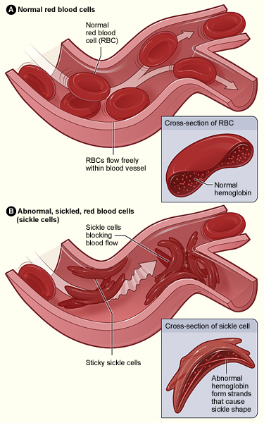
A rigid cell structure that distorts the membrane of the RBC
Membrane damageMembrane DamageCell Injury and Death → influx of calciumCalciumA basic element found in nearly all tissues. It is a member of the alkaline earth family of metals with the atomic symbol ca, atomic number 20, and atomic weight 40. Calcium is the most abundant mineral in the body and combines with phosphorus to form calcium phosphate in the bones and teeth. It is essential for the normal functioning of nerves and muscles and plays a role in blood coagulation (as factor IV) and in many enzymatic processes.Electrolytes, efflux of potassiumPotassiumAn element in the alkali group of metals with an atomic symbol k, atomic number 19, and atomic weight 39. 10. It is the chief cation in the intracellular fluid of muscle and other cells. Potassium ion is a strong electrolyte that plays a significant role in the regulation of fluid volume and maintenance of the water-electrolyte balance.Hyperkalemia and water → dehydrates the RBC → sicklingSicklingSickle Cell Disease
Vasoocclusion(microvascular occlusion): multiple pathophysiologic mechanisms, only partially related to the number of irreversibly sickled cells; other factors include:
The tendency of sickled RBCsRBCsErythrocytes, or red blood cells (RBCs), are the most abundant cells in the blood. While erythrocytes in the fetus are initially produced in the yolk sac then the liver, the bone marrow eventually becomes the main site of production.Erythrocytes: Histology to adhere to and activate vascular endotheliumEndotheliumA layer of epithelium that lines the heart, blood vessels (vascular endothelium), lymph vessels (lymphatic endothelium), and the serous cavities of the body.Arteries: Histology, which exposes subendothelialSubendothelialMembranoproliferative Glomerulonephritis matrix proteinsProteinsLinear polypeptides that are synthesized on ribosomes and may be further modified, crosslinked, cleaved, or assembled into complex proteins with several subunits. The specific sequence of amino acids determines the shape the polypeptide will take, during protein folding, and the function of the protein.Energy Homeostasis (e.g., lamininLamininLarge, noncollagenous glycoprotein with antigenic properties. It is localized in the basement membrane lamina lucida and functions to bind epithelial cells to the basement membrane. Evidence suggests that the protein plays a role in tumor invasion.Connective Tissue: Histology, thrombospondin on plateletsPlateletsPlatelets are small cell fragments involved in hemostasis. Thrombopoiesis takes place primarily in the bone marrow through a series of cell differentiation and is influenced by several cytokines. Platelets are formed after fragmentation of the megakaryocyte cytoplasm. Platelets: Histology, and von Willebrand factorvon Willebrand factorA high-molecular-weight plasma protein, produced by endothelial cells and megakaryocytes, that is part of the factor VIII/von Willebrand factor complex. The von Willebrand factor has receptors for collagen, platelets, and ristocetin activity as well as the immunologically distinct antigenic determinants. It functions in adhesion of platelets to collagen and hemostatic plug formation. The prolonged bleeding time in von Willebrand diseases is due to the deficiency of this factor.Hemostasis) that also adhere to sickle cells
Activation of macrophage tissue factor: can activate endothelial cells and plateletsPlateletsPlatelets are small cell fragments involved in hemostasis. Thrombopoiesis takes place primarily in the bone marrow through a series of cell differentiation and is influenced by several cytokines. Platelets are formed after fragmentation of the megakaryocyte cytoplasm. Platelets: Histology, as well as initiate coagulation
InflammationInflammationInflammation is a complex set of responses to infection and injury involving leukocytes as the principal cellular mediators in the body’s defense against pathogenic organisms. Inflammation is also seen as a response to tissue injury in the process of wound healing. The 5 cardinal signs of inflammation are pain, heat, redness, swelling, and loss of function. Inflammation, with leukocyte adhesionLeukocyte adhesionLeukocyte Adhesion Deficiency Type 1 to endotheliumEndotheliumA layer of epithelium that lines the heart, blood vessels (vascular endothelium), lymph vessels (lymphatic endothelium), and the serous cavities of the body.Arteries: Histology and reduction of blood flow velocityFlow velocityVascular Resistance, Flow, and Mean Arterial Pressure → increases RBC sicklingSicklingSickle Cell Disease
Reduction of nitric acid because it becomes bound to free hemoglobin from lysed cells → vascular smooth muscle cell contraction and platelet aggregationPlatelet aggregationThe attachment of platelets to one another. This clumping together can be induced by a number of agents (e.g., thrombin; collagen) and is part of the mechanism leading to the formation of a thrombus.Hemostasis
Hemolysis itself is a major proinflammatory driver which can elevate levels of thrombospondin and von Willebrand factorvon Willebrand factorA high-molecular-weight plasma protein, produced by endothelial cells and megakaryocytes, that is part of the factor VIII/von Willebrand factor complex. The von Willebrand factor has receptors for collagen, platelets, and ristocetin activity as well as the immunologically distinct antigenic determinants. It functions in adhesion of platelets to collagen and hemostatic plug formation. The prolonged bleeding time in von Willebrand diseases is due to the deficiency of this factor.Hemostasis, which promote adhesionAdhesionThe process whereby platelets adhere to something other than platelets, e.g., collagen; basement membrane; microfibrils; or other ‘foreign’ surfaces.Coagulation Studies of RBCsRBCsErythrocytes, or red blood cells (RBCs), are the most abundant cells in the blood. While erythrocytes in the fetus are initially produced in the yolk sac then the liver, the bone marrow eventually becomes the main site of production.Erythrocytes: Histology to the endotheliumEndotheliumA layer of epithelium that lines the heart, blood vessels (vascular endothelium), lymph vessels (lymphatic endothelium), and the serous cavities of the body.Arteries: Histology
Initiation of a positive (“vicious”) cycle of ischemic tissue damage promoting more vasoocclusion due to the production of reactive oxygen speciesReactive oxygen speciesMolecules or ions formed by the incomplete one-electron reduction of oxygen. These reactive oxygen intermediates include singlet oxygen; superoxides; peroxides; hydroxyl radical; and hypochlorous acid. They contribute to the microbicidal activity of phagocytes, regulation of signal transduction and gene expression, and the oxidative damage to nucleic acids; proteins; and lipids.Metabolic Dysfunction-associated Steatotic Liver Disease (MASLD) causing inflammationInflammationInflammation is a complex set of responses to infection and injury involving leukocytes as the principal cellular mediators in the body’s defense against pathogenic organisms. Inflammation is also seen as a response to tissue injury in the process of wound healing. The 5 cardinal signs of inflammation are pain, heat, redness, swelling, and loss of function. Inflammation and more platelet activationPlatelet activationA series of progressive, overlapping events, triggered by exposure of the platelets to subendothelial tissue. These events include shape change, adhesiveness, aggregation, and release reactions. When carried through to completion, these events lead to the formation of a stable hemostatic plug.Hemostasis, leukocyte recruitmentRecruitmentSkeletal Muscle Contraction, and endothelial activationEndothelial ActivationTumor Necrosis Factor (TNF).
A shortened half-lifeHalf-LifeThe time it takes for a substance (drug, radioactive nuclide, or other) to lose half of its pharmacologic, physiologic, or radiologic activity.Pharmacokinetics and Pharmacodynamics of the HbS-containing RBCsRBCsErythrocytes, or red blood cells (RBCs), are the most abundant cells in the blood. While erythrocytes in the fetus are initially produced in the yolk sac then the liver, the bone marrow eventually becomes the main site of production.Erythrocytes: Histology to 17 days (normally 120 days)
FeverFeverFever is defined as a measured body temperature of at least 38°C (100.4°F). Fever is caused by circulating endogenous and/or exogenous pyrogens that increase levels of prostaglandin E2 in the hypothalamus. Fever is commonly associated with chills, rigors, sweating, and flushing of the skin. Fever
AcidosisAcidosisA pathologic condition of acid accumulation or depletion of base in the body. The two main types are respiratory acidosis and metabolic acidosis, due to metabolic acid build up.Respiratory Acidosis
Abnormal hemoglobin results in RBC sickling and adhesion of the sickled cells to endothelium, which is activated by the adherent RBCs. Occlusion of small vessels occurs by an aggregate of sickled RBCs, with platelets and white blood cells (not shown in figure). However, vasoocclusion (microvascular occlusion) is caused by multiple pathophysiologic mechanisms, not only by blockage due to sickled cells.
Image: “Sickle cell 01” by The National Heart, Lung, and Blood Institute (NHLBI). License: Public Domain
Most symptoms result from the anemiaAnemiaAnemia is a condition in which individuals have low Hb levels, which can arise from various causes. Anemia is accompanied by a reduced number of RBCs and may manifest with fatigue, shortness of breath, pallor, and weakness. Subtypes are classified by the size of RBCs, chronicity, and etiology. Anemia: Overview and Types and vasoocclusive events seen in individuals with SCDSCDSickle cell disease (SCD) is a group of genetic disorders in which an abnormal Hb molecule (HbS) transforms RBCs into sickle-shaped cells, resulting in chronic anemia, vasoocclusive episodes, pain, and organ damage.Sickle Cell Disease or complications including infection.
Sickle cell diseaseSickle cell diseaseSickle cell disease (SCD) is a group of genetic disorders in which an abnormal Hb molecule (HbS) transforms RBCs into sickle-shaped cells, resulting in chronic anemia, vasoocclusive episodes, pain, and organ damage.Sickle Cell Disease (HbSSHbSSSickle cell disease (SCD) is a group of genetic disorders in which an abnormal Hb molecule (HbS) transforms RBCs into sickle-shaped cells, resulting in chronic anemia, vasoocclusive episodes, pain, and organ damage.Sickle Cell Disease)[1]
Infants with SCDSCDSickle cell disease (SCD) is a group of genetic disorders in which an abnormal Hb molecule (HbS) transforms RBCs into sickle-shaped cells, resulting in chronic anemia, vasoocclusive episodes, pain, and organ damage.Sickle Cell Disease are generally healthy at birth. Symptoms develop after about age 6 months, when HbF levels start to diminish (protective in neonates)
Major acute manifestations:
Vasoocclusive events: present suddenly with severe painPainAn unpleasant sensation induced by noxious stimuli which are detected by nerve endings of nociceptive neurons.Pain: Types and Pathways and infarction of the affected tissue
Infection (see complications below for more details):[11]
SepsisSepsisSystemic inflammatory response syndrome with a proven or suspected infectious etiology. When sepsis is associated with organ dysfunction distant from the site of infection, it is called severe sepsis. When sepsis is accompanied by hypotension despite adequate fluid infusion, it is called septic shock.Sepsis and Septic Shock
PneumoniaPneumoniaPneumonia or pulmonary inflammation is an acute or chronic inflammation of lung tissue. Causes include infection with bacteria, viruses, or fungi. In more rare cases, pneumonia can also be caused through toxic triggers through inhalation of toxic substances, immunological processes, or in the course of radiotherapy.Pneumonia
MeningitisMeningitisMeningitis is inflammation of the meninges, the protective membranes of the brain, and spinal cord. The causes of meningitis are varied, with the most common being bacterial or viral infection. The classic presentation of meningitis is a triad of fever, altered mental status, and nuchal rigidity. Meningitis
AnemiaAnemiaAnemia is a condition in which individuals have low Hb levels, which can arise from various causes. Anemia is accompanied by a reduced number of RBCs and may manifest with fatigue, shortness of breath, pallor, and weakness. Subtypes are classified by the size of RBCs, chronicity, and etiology. Anemia: Overview and Types
Can be brought on by infection with human parvovirus B19Parvovirus B19Primate erythroparvovirus 1 (generally referred to as parvovirus B19, B19 virus, or sometimes erythrovirus B19) ranks among the smallest DNA viruses. Parvovirus B19 is of the family Parvoviridae and genus Erythrovirus. In immunocompetent humans, parvovirus B19 classically results in erythema infectiosum (5th disease) or “slapped cheek syndrome.”Parvovirus B19 → transient slowing of bone marrowBone marrowThe soft tissue filling the cavities of bones. Bone marrow exists in two types, yellow and red. Yellow marrow is found in the large cavities of large bones and consists mostly of fat cells and a few primitive blood cells. Red marrow is a hematopoietic tissue and is the site of production of erythrocytes and granular leukocytes. Bone marrow is made up of a framework of connective tissue containing branching fibers with the frame being filled with marrow cells.Bone Marrow: Composition and HematopoiesiserythropoiesisErythropoiesisThe production of red blood cells (erythrocytes). In humans, erythrocytes are produced by the yolk sac in the first trimester; by the liver in the second trimester; by the bone marrow in the third trimester and after birth. In normal individuals, the erythrocyte count in the peripheral blood remains relatively constant implying a balance between the rate of erythrocyte production and rate of destruction.Erythrocytes: Histology (leading to aplastic anemiaAplastic AnemiaAplastic anemia (AA) is a rare, life-threatening condition characterized by pancytopenia and hypocellularity of the bone marrow (in the absence of any abnormal cells) reflecting damage to hematopoietic stem cells. Aplastic anemia can be acquired or inherited, however, most cases of AA are acquired and caused by autoimmune damage to hematopoietic stem cells. Aplastic Anemia)
Acute anemiaAnemiaAnemia is a condition in which individuals have low Hb levels, which can arise from various causes. Anemia is accompanied by a reduced number of RBCs and may manifest with fatigue, shortness of breath, pallor, and weakness. Subtypes are classified by the size of RBCs, chronicity, and etiology. Anemia: Overview and Types also occurs in acute chest syndrome, splenic/hepatic sequestration (see below).
CNS: ischemic strokeIschemic StrokeAn ischemic stroke (also known as cerebrovascular accident) is an acute neurologic injury that occurs as a result of brain ischemia; this condition may be due to cerebral blood vessel occlusion by thrombosis or embolism, or rarely due to systemic hypoperfusion. Ischemic Stroke or TIATIATransient ischemic attack (TIA) is a temporary episode of neurologic dysfunction caused by ischemia without infarction that resolves completely when blood supply is restored. Transient ischemic attack is a neurologic emergency that warrants urgent medical attention. Transient Ischemic Attack (TIA)[12,13,15,16]
Hemorrhagic strokeHemorrhagic strokeStroke due to rupture of a weakened blood vessel in the brain (e.g., cerebral hemispheres; cerebellum; subarachnoid space).Subarachnoid Hemorrhage can occur, and it is more common in adults.
Lung:
Pulmonary embolismPulmonary EmbolismPulmonary embolism (PE) is a potentially fatal condition that occurs as a result of intraluminal obstruction of the main pulmonary artery or its branches. The causative factors include thrombi, air, amniotic fluid, and fat. In PE, gas exchange is impaired due to the decreased return of deoxygenated blood to the lungs. Pulmonary Embolism or microemboli (eventually leading to pulmonary hypertensionPulmonary HypertensionPulmonary hypertension (PH) or pulmonary arterial hypertension (PAH) is characterized by elevated pulmonary arterial pressure, which can lead to chronic progressive right heart failure. Pulmonary hypertension is grouped into 5 categories based on etiology, which include primary PAH, and PH due to cardiac disease, lung or hypoxic disease, chronic thromboembolic disease, and multifactorial or unclear etiologies. Pulmonary Hypertension) and pulmonary fibrosisFibrosisAny pathological condition where fibrous connective tissue invades any organ, usually as a consequence of inflammation or other injury.Bronchiolitis Obliterans
Acute chest syndrome: chest painPainAn unpleasant sensation induced by noxious stimuli which are detected by nerve endings of nociceptive neurons.Pain: Types and Pathways, feverFeverFever is defined as a measured body temperature of at least 38°C (100.4°F). Fever is caused by circulating endogenous and/or exogenous pyrogens that increase levels of prostaglandin E2 in the hypothalamus. Fever is commonly associated with chills, rigors, sweating, and flushing of the skin. Fever, pulmonary infiltrates, hypoxemiaHypoxemiaNeonatal Respiratory Distress Syndrome, acute anemiaAnemiaAnemia is a condition in which individuals have low Hb levels, which can arise from various causes. Anemia is accompanied by a reduced number of RBCs and may manifest with fatigue, shortness of breath, pallor, and weakness. Subtypes are classified by the size of RBCs, chronicity, and etiology. Anemia: Overview and Types
Acute kidney injuryAcute Kidney InjuryAcute kidney injury refers to sudden and often reversible loss of renal function, which develops over days or weeks. Azotemia refers to elevated levels of nitrogen-containing substances in the blood that accompany AKI, which include BUN and creatinine. Acute Kidney Injury (AKIAKIAcute kidney injury refers to sudden and often reversible loss of renal function, which develops over days or weeks. Azotemia refers to elevated levels of nitrogen-containing substances in the blood that accompany AKI, which include BUN and creatinine. Acute Kidney Injury)
PainPainAn unpleasant sensation induced by noxious stimuli which are detected by nerve endings of nociceptive neurons.Pain: Types and Pathways in the long bonesLong bonesLength greater than width.Bones: Structure and Types, hands and feet, back, joints. Triggers include infectionsInfectionsInvasion of the host organism by microorganisms or their toxins or by parasites that can cause pathological conditions or diseases.Chronic Granulomatous Disease, stress, and cold exposure; in many cases, the cause is not identified.
Avascular necrosisAvascular NecrosisHip Fractures (due to ischemiaIschemiaA hypoperfusion of the blood through an organ or tissue caused by a pathologic constriction or obstruction of its blood vessels, or an absence of blood circulation.Ischemic Cell Damage commonly affecting the femoral and humeral headHumeral headThe upper rounded extremity of the humerus fitting into the glenoid cavity of the scapula.Arm: Anatomy; typically asymptomatic until late in the disease)
DactylitisDactylitisAnkylosing Spondylitis (painful inflammationInflammationInflammation is a complex set of responses to infection and injury involving leukocytes as the principal cellular mediators in the body’s defense against pathogenic organisms. Inflammation is also seen as a response to tissue injury in the process of wound healing. The 5 cardinal signs of inflammation are pain, heat, redness, swelling, and loss of function. Inflammation of the fingers and/or toes)
Heart: MIMIMI is ischemia and death of an area of myocardial tissue due to insufficient blood flow and oxygenation, usually from thrombus formation on a ruptured atherosclerotic plaque in the epicardial arteries. Clinical presentation is most commonly with chest pain, but women and patients with diabetes may have atypical symptoms.Myocardial Infarction (from ↑ oxygen demand with limited oxygen-carrying capacity)
LiverLiverThe liver is the largest gland in the human body. The liver is found in the superior right quadrant of the abdomen and weighs approximately 1.5 kilograms. Its main functions are detoxification, metabolism, nutrient storage (e.g., iron and vitamins), synthesis of coagulation factors, formation of bile, filtration, and storage of blood. Liver: Anatomy with changes due to ischemiaIschemiaA hypoperfusion of the blood through an organ or tissue caused by a pathologic constriction or obstruction of its blood vessels, or an absence of blood circulation.Ischemic Cell Damage caused by sinusoidal obstruction:
Acute sickle cell hepatic crisis (ASCHC) if the obstruction is not extensive
Sickle cell intrahepatic cholestasis (SCIC): a severe, often fatal variant of ASCHC that occurs if the obstruction is extensive; associated with severe jaundiceJaundiceJaundice is the abnormal yellowing of the skin and/or sclera caused by the accumulation of bilirubin. Hyperbilirubinemia is caused by either an increase in bilirubin production or a decrease in the hepatic uptake, conjugation, or excretion of bilirubin. Jaundice and renal impairment
LiverLiverThe liver is the largest gland in the human body. The liver is found in the superior right quadrant of the abdomen and weighs approximately 1.5 kilograms. Its main functions are detoxification, metabolism, nutrient storage (e.g., iron and vitamins), synthesis of coagulation factors, formation of bile, filtration, and storage of blood. Liver: Anatomy with changes due to acute sequestration of RBCsRBCsErythrocytes, or red blood cells (RBCs), are the most abundant cells in the blood. While erythrocytes in the fetus are initially produced in the yolk sac then the liver, the bone marrow eventually becomes the main site of production.Erythrocytes: Histology and plateletsPlateletsPlatelets are small cell fragments involved in hemostasis. Thrombopoiesis takes place primarily in the bone marrow through a series of cell differentiation and is influenced by several cytokines. Platelets are formed after fragmentation of the megakaryocyte cytoplasm. Platelets: Histology:
LiverLiverThe liver is the largest gland in the human body. The liver is found in the superior right quadrant of the abdomen and weighs approximately 1.5 kilograms. Its main functions are detoxification, metabolism, nutrient storage (e.g., iron and vitamins), synthesis of coagulation factors, formation of bile, filtration, and storage of blood. Liver: Anatomy increases acutely in size → associated with right upper quadrantRight upper quadrantAnterior Abdominal Wall: Anatomyabdominal painAbdominal PainAcute Abdomen, a > 2 g/dL decrease in hemoglobin level, thrombocytopeniaThrombocytopeniaThrombocytopenia occurs when the platelet count is < 150,000 per microliter. The normal range for platelets is usually 150,000-450,000/µL of whole blood. Thrombocytopenia can be a result of decreased production, increased destruction, or splenic sequestration of platelets. Patients are often asymptomatic until platelet counts are < 50,000/µL. Thrombocytopenia, and liver failureLiver failureSevere inability of the liver to perform its normal metabolic functions, as evidenced by severe jaundice and abnormal serum levels of ammonia; bilirubin; alkaline phosphatase; aspartate aminotransferase; lactate dehydrogenases; and albumin/globulin ratio.Autoimmune Hepatitis
LiverLiverThe liver is the largest gland in the human body. The liver is found in the superior right quadrant of the abdomen and weighs approximately 1.5 kilograms. Its main functions are detoxification, metabolism, nutrient storage (e.g., iron and vitamins), synthesis of coagulation factors, formation of bile, filtration, and storage of blood. Liver: Anatomy size and blood counts usually normalize after 3–4 days if hemodynamic stability is maintained.
Eyes: retinal artery occlusionRetinal Artery OcclusionRetinal Vessel Occlusion, retinal detachmentRetinal detachmentRetinal detachment is the separation of the neurosensory retina from the retinal pigmented epithelium and choroid. Rhegmatogenous retinal detachment, the most common type, stems from a break in the retina, allowing fluid to accumulate in the subretinal space. Retinal Detachment, orbital infarction
SpleenSpleenThe spleen is the largest lymphoid organ in the body, located in the LUQ of the abdomen, superior to the left kidney and posterior to the stomach at the level of the 9th-11th ribs just below the diaphragm. The spleen is highly vascular and acts as an important blood filter, cleansing the blood of pathogens and damaged erythrocytes. Spleen: Anatomy:
Children: enlarged, may have pooling of blood or trapping of red cell massMassThree-dimensional lesion that occupies a space within the breastImaging of the Breast causing LUQ abdominal painAbdominal PainAcute Abdomen, weakness, hypotensionHypotensionHypotension is defined as low blood pressure, specifically < 90/60 mm Hg, and is most commonly a physiologic response. Hypotension may be mild, serious, or life threatening, depending on the cause. Hypotension, and shockShockShock is a life-threatening condition associated with impaired circulation that results in tissue hypoxia. The different types of shock are based on the underlying cause: distributive (↑ cardiac output (CO), ↓ systemic vascular resistance (SVR)), cardiogenic (↓ CO, ↑ SVR), hypovolemic (↓ CO, ↑ SVR), obstructive (↓ CO), and mixed. Types of Shock (splenic sequestrationSplenic sequestrationSevere Congenital Neutropenia)
Adults: AutosplenectomyAutosplenectomyAsplenia occurs with age, leading to infectious complications
PenisPenisThe penis is the male organ of copulation and micturition. The organ is composed of a root, body, and glans. The root is attached to the pubic bone by the crura penis. The body consists of the 2 parallel corpora cavernosa and the corpus spongiosum. The glans is ensheathed by the prepuce or foreskin. Penis: Anatomy: priapismPriapismA prolonged painful erection that may lasts hours and is not associated with sexual activity. It is seen in patients with sickle cell anemia, advanced malignancy, spinal trauma; and certain drug treatments.Penile Anomalies and Conditions (sustained erectionErectionThe state of the penis when the erectile tissue becomes filled or swollen (tumid) with blood and causes the penis to become rigid and elevated. It is a complex process involving central nervous system; peripheral nervous systems; hormones; smooth muscles; and vascular functions.Penis: Anatomy that lasts > 4 hours); can also be seen in children with SCDSCDSickle cell disease (SCD) is a group of genetic disorders in which an abnormal Hb molecule (HbS) transforms RBCs into sickle-shaped cells, resulting in chronic anemia, vasoocclusive episodes, pain, and organ damage.Sickle Cell Disease
Vascular: venous thromboembolismThromboembolismObstruction of a blood vessel (embolism) by a blood clot (thrombus) in the blood stream.Systemic Lupus Erythematosus (SCDSCDSickle cell disease (SCD) is a group of genetic disorders in which an abnormal Hb molecule (HbS) transforms RBCs into sickle-shaped cells, resulting in chronic anemia, vasoocclusive episodes, pain, and organ damage.Sickle Cell Disease is a hypercoagulableHypercoagulableHypercoagulable states (also referred to as thrombophilias) are a group of hematologic diseases defined by an increased risk of clot formation (i.e., thrombosis) due to either an increase in procoagulants, a decrease in anticoagulants, or a decrease in fibrinolysis. Hypercoagulable States state)
Major chronic manifestations:
Chronic painChronic painAching sensation that persists for more than a few months. It may or may not be associated with trauma or disease, and may persist after the initial injury has healed. Its localization, character, and timing are more vague than with acute pain.Pain Management from:
Tissue infarction
Osteonecrosis
Chronic infection:
LegLegThe lower leg, or just “leg” in anatomical terms, is the part of the lower limb between the knee and the ankle joint. The bony structure is composed of the tibia and fibula bones, and the muscles of the leg are grouped into the anterior, lateral, and posterior compartments by extensions of fascia.Leg: Anatomy ulcers (most commonly affects the lateral malleoli)
OsteomyelitisOsteomyelitisOsteomyelitis is an infection of the bone that results from the spread of microorganisms from the blood (hematogenous), nearby infected tissue, or open wounds (non-hematogenous). Infections are most commonly caused by Staphylococcus aureus.Osteomyelitis
Chronic hemolysis (intravascular and extravascular) → anemiaAnemiaAnemia is a condition in which individuals have low Hb levels, which can arise from various causes. Anemia is accompanied by a reduced number of RBCs and may manifest with fatigue, shortness of breath, pallor, and weakness. Subtypes are classified by the size of RBCs, chronicity, and etiology. Anemia: Overview and Types:
FatigueFatigueThe state of weariness following a period of exertion, mental or physical, characterized by a decreased capacity for work and reduced efficiency to respond to stimuli.Fibromyalgia
Pallor
TachycardiaTachycardiaAbnormally rapid heartbeat, usually with a heart rate above 100 beats per minute for adults. Tachycardia accompanied by disturbance in the cardiac depolarization (cardiac arrhythmia) is called tachyarrhythmia.Sepsis in Children
Cognitive delay and delayed growth in children
Folate deficiencyFolate deficiencyA nutritional condition produced by a deficiency of folic acid in the diet. Many plant and animal tissues contain folic acid, abundant in green leafy vegetables, yeast, liver, and mushrooms but destroyed by long-term cooking. Alcohol interferes with its intermediate metabolism and absorption. Folic acid deficiency may develop in long-term anticonvulsant therapy or with use of oral contraceptives. This deficiency causes anemia, macrocytic anemia, and megaloblastic anemia. It is indistinguishable from vitamin B12 deficiency in peripheral blood and bone marrow findings, but the neurologic lesions seen in B12 deficiency do not occur.Megaloblastic Anemia can worsen chronic anemiaAnemiaAnemia is a condition in which individuals have low Hb levels, which can arise from various causes. Anemia is accompanied by a reduced number of RBCs and may manifest with fatigue, shortness of breath, pallor, and weakness. Subtypes are classified by the size of RBCs, chronicity, and etiology. Anemia: Overview and Types.
OsteoporosisOsteoporosisOsteoporosis refers to a decrease in bone mass and density leading to an increased number of fractures. There are 2 forms of osteoporosis: primary, which is commonly postmenopausal or senile; and secondary, which is a manifestation of immobilization, underlying medical disorders, or long-term use of certain medications. Osteoporosis
BlindnessBlindnessThe inability to see or the loss or absence of perception of visual stimuli. This condition may be the result of eye diseases; optic nerve diseases; optic chiasm diseases; or brain diseases affecting the visual pathways or occipital lobe.Retinopathy of Prematurity from chronic retinopathyRetinopathyDegenerative changes to the retina due to hypertension.Alport Syndrome
Hemolysis of RBCsRBCsErythrocytes, or red blood cells (RBCs), are the most abundant cells in the blood. While erythrocytes in the fetus are initially produced in the yolk sac then the liver, the bone marrow eventually becomes the main site of production.Erythrocytes: Histology can cause pigment gallstonesGallstonesCholelithiasis (gallstones) is the presence of stones in the gallbladder. Most gallstones are cholesterol stones, while the rest are composed of bilirubin (pigment stones) and other mixed components. Patients are commonly asymptomatic but may present with biliary colic (intermittent pain in the right upper quadrant).Cholelithiasis.
Complications:
Recurrent vasoocclusive events and tissue infarctions → organ damage
Acute chest syndrome can be life-threatening; it is the most common cause of death in patientsPatientsIndividuals participating in the health care system for the purpose of receiving therapeutic, diagnostic, or preventive procedures.Clinician–Patient Relationship with SCDSCDSickle cell disease (SCD) is a group of genetic disorders in which an abnormal Hb molecule (HbS) transforms RBCs into sickle-shaped cells, resulting in chronic anemia, vasoocclusive episodes, pain, and organ damage.Sickle Cell Disease.
Heart: pulmonary hypertensionPulmonary HypertensionPulmonary hypertension (PH) or pulmonary arterial hypertension (PAH) is characterized by elevated pulmonary arterial pressure, which can lead to chronic progressive right heart failure. Pulmonary hypertension is grouped into 5 categories based on etiology, which include primary PAH, and PH due to cardiac disease, lung or hypoxic disease, chronic thromboembolic disease, and multifactorial or unclear etiologies. Pulmonary Hypertension, cor pulmonaleCor PulmonaleCor pulmonale is right ventricular (RV) dysfunction caused by lung disease that results in pulmonary artery hypertension. The most common cause of cor pulmonale is chronic obstructive pulmonary disease. Dyspnea is the usual presenting symptom. Cor Pulmonale, and heart failureHeart FailureA heterogeneous condition in which the heart is unable to pump out sufficient blood to meet the metabolic need of the body. Heart failure can be caused by structural defects, functional abnormalities (ventricular dysfunction), or a sudden overload beyond its capacity. Chronic heart failure is more common than acute heart failure which results from sudden insult to cardiac function, such as myocardial infarction.Total Anomalous Pulmonary Venous Return (TAPVR)
Kidney: CKDCKDChronic kidney disease (CKD) is kidney impairment that lasts for ≥ 3 months, implying that it is irreversible. Hypertension and diabetes are the most common causes; however, there are a multitude of other etiologies. In the early to moderate stages, CKD is usually asymptomatic and is primarily diagnosed by laboratory abnormalities.Chronic Kidney Disease, hypertensionHypertensionHypertension, or high blood pressure, is a common disease that manifests as elevated systemic arterial pressures. Hypertension is most often asymptomatic and is found incidentally as part of a routine physical examination or during triage for an unrelated medical encounter. Hypertension
Musculoskeletal: osteonecrosis, osteoporosisOsteoporosisOsteoporosis refers to a decrease in bone mass and density leading to an increased number of fractures. There are 2 forms of osteoporosis: primary, which is commonly postmenopausal or senile; and secondary, which is a manifestation of immobilization, underlying medical disorders, or long-term use of certain medications. Osteoporosis, and short stature in childrenShort stature in childrenShort stature in children is defined as a height more than 2 standard deviations below the mean for age and gender or growing below the 3rd percentile when plotting height on standardized growth charts. Short stature can be pathological or due to a normal variant in growth pattern. Short Stature in Children
InfectionsInfectionsInvasion of the host organism by microorganisms or their toxins or by parasites that can cause pathological conditions or diseases.Chronic Granulomatous Disease:
Increased risk of infection with encapsulatedEncapsulatedKlebsiellabacteriaBacteriaBacteria are prokaryotic single-celled microorganisms that are metabolically active and divide by binary fission. Some of these organisms play a significant role in the pathogenesis of diseases. Bacteriology (e.g., StreptococcusStreptococcusStreptococcus is one of the two medically important genera of gram-positive cocci, the other being Staphylococcus. Streptococci are identified as different species on blood agar on the basis of their hemolytic pattern and sensitivity to optochin and bacitracin. There are many pathogenic species of streptococci, including S. pyogenes, S. agalactiae, S. pneumoniae, and the viridans streptococci.Streptococcus pneumoniae, Haemophilus influenzaeHaemophilus InfluenzaeA species of Haemophilus found on the mucous membranes of humans and a variety of animals. The species is further divided into biotypes I through viii.Haemophilus, Neisseria meningitidisNeisseria meningitidisA species of gram-negative, aerobic bacteria. It is a commensal and pathogen only of humans, and can be carried asymptomatically in the nasopharynx. When found in cerebrospinal fluid it is the causative agent of cerebrospinal meningitis. It is also found in venereal discharges and blood. There are at least 13 serogroups based on antigenic differences in the capsular polysaccharides; the ones causing most meningitis infections being a, b, c, y, and w-135. Each serogroup can be further classified by serotype, serosubtype, and immunotype.Neisseria) due to loss of splenic function (hyposplenismHyposplenismAsplenia and/or functional aspleniaFunctional AspleniaAsplenia)
Hepatitis
OsteomyelitisOsteomyelitisOsteomyelitis is an infection of the bone that results from the spread of microorganisms from the blood (hematogenous), nearby infected tissue, or open wounds (non-hematogenous). Infections are most commonly caused by Staphylococcus aureus.Osteomyelitis
PneumoniaPneumoniaPneumonia or pulmonary inflammation is an acute or chronic inflammation of lung tissue. Causes include infection with bacteria, viruses, or fungi. In more rare cases, pneumonia can also be caused through toxic triggers through inhalation of toxic substances, immunological processes, or in the course of radiotherapy.Pneumonia
MeningitisMeningitisMeningitis is inflammation of the meninges, the protective membranes of the brain, and spinal cord. The causes of meningitis are varied, with the most common being bacterial or viral infection. The classic presentation of meningitis is a triad of fever, altered mental status, and nuchal rigidity. Meningitis
SepsisSepsisSystemic inflammatory response syndrome with a proven or suspected infectious etiology. When sepsis is associated with organ dysfunction distant from the site of infection, it is called severe sepsis. When sepsis is accompanied by hypotension despite adequate fluid infusion, it is called septic shock.Sepsis and Septic Shock
Complications associated with transfusions:
Alloimmunization, especially in those receiving chronic transfusions
Iron overloadIron overloadAn excessive accumulation of iron in the body due to a greater than normal absorption of iron from the gastrointestinal tract or from parenteral injection. This may arise from idiopathic hemochromatosis, excessive iron intake, chronic alcoholism, certain types of refractory anemia, or transfusional hemosiderosis.Hereditary Hemochromatosis
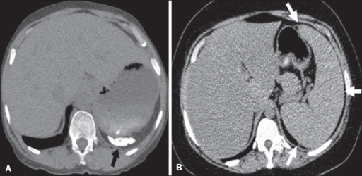
Different patterns of splenic involvement in sickle cell disease:
A: atrophy and calcification of the spleen (arrow)
B: splenomegaly (arrows)
Image: “f6: Different patterns of splenic involvement in sickle cell disease. Sicklecell disease (HbSS) shows atrophy and calcification of the spleen (arrowon A), whereas non-HbSS sickle cell disease can showsplenomegaly (arrows on B).” by Ursula David Alves et al. License: CC BY 4.0
May have painless hematuriaHematuriaPresence of blood in the urine.Renal Cell Carcinoma due to renal papillary necrosisNecrosisThe death of cells in an organ or tissue due to disease, injury or failure of the blood supply.Ischemic Cell Damage
Decreased (hyposthenuria) or loss of (isosthenuria) ability to concentrate the urine
Rarely:
Sickle cell acute episode, rhabdomyolysisRhabdomyolysisRhabdomyolysis is characterized by muscle necrosis and the release of toxic intracellular contents, especially myoglobin, into the circulation.Rhabdomyolysis, and death if exposed to high altitudes or with extreme dehydrationDehydrationThe condition that results from excessive loss of water from a living organism.Volume Depletion and Dehydration or exercise
CKDCKDChronic kidney disease (CKD) is kidney impairment that lasts for ≥ 3 months, implying that it is irreversible. Hypertension and diabetes are the most common causes; however, there are a multitude of other etiologies. In the early to moderate stages, CKD is usually asymptomatic and is primarily diagnosed by laboratory abnormalities.Chronic Kidney Disease
Pulmonary embolismPulmonary EmbolismPulmonary embolism (PE) is a potentially fatal condition that occurs as a result of intraluminal obstruction of the main pulmonary artery or its branches. The causative factors include thrombi, air, amniotic fluid, and fat. In PE, gas exchange is impaired due to the decreased return of deoxygenated blood to the lungs. Pulmonary Embolism
Rare medullary carcinoma of the kidney
Benefit of sickle cell traitsickle cell traitThe condition of being heterozygous for hemoglobin S.Sickle Cell Disease: improved survival with malarial infection
Sickle cell homozygotes (HbSSHbSSSickle cell disease (SCD) is a group of genetic disorders in which an abnormal Hb molecule (HbS) transforms RBCs into sickle-shaped cells, resulting in chronic anemia, vasoocclusive episodes, pain, and organ damage.Sickle Cell Disease) have no benefits from SCDSCDSickle cell disease (SCD) is a group of genetic disorders in which an abnormal Hb molecule (HbS) transforms RBCs into sickle-shaped cells, resulting in chronic anemia, vasoocclusive episodes, pain, and organ damage.Sickle Cell Disease; they are harmed by malarial infectionsInfectionsInvasion of the host organism by microorganisms or their toxins or by parasites that can cause pathological conditions or diseases.Chronic Granulomatous Disease, which worsen the preexisting anemiaAnemiaAnemia is a condition in which individuals have low Hb levels, which can arise from various causes. Anemia is accompanied by a reduced number of RBCs and may manifest with fatigue, shortness of breath, pallor, and weakness. Subtypes are classified by the size of RBCs, chronicity, and etiology. Anemia: Overview and Types.
Sickle cell diseaseSickle cell diseaseSickle cell disease (SCD) is a group of genetic disorders in which an abnormal Hb molecule (HbS) transforms RBCs into sickle-shaped cells, resulting in chronic anemia, vasoocclusive episodes, pain, and organ damage.Sickle Cell Disease is usually diagnosed prenatally or at birth by mandatory neonatal screeningScreeningPreoperative Care. Methods vary from state to state.[2,4,8]
Testing:
Prenatal:
PCRPCRPolymerase chain reaction (PCR) is a technique that amplifies DNA fragments exponentially for analysis. The process is highly specific, allowing for the targeting of specific genomic sequences, even with minuscule sample amounts. The PCR cycles multiple times through 3 phases: denaturation of the template DNA, annealing of a specific primer to the individual DNA strands, and synthesis/elongation of new DNA molecules.Polymerase Chain Reaction (PCR) or direct DNADNAA deoxyribonucleotide polymer that is the primary genetic material of all cells. Eukaryotic and prokaryotic organisms normally contain DNA in a double-stranded state, yet several important biological processes transiently involve single-stranded regions. DNA, which consists of a polysugar-phosphate backbone possessing projections of purines (adenine and guanine) and pyrimidines (thymine and cytosine), forms a double helix that is held together by hydrogen bonds between these purines and pyrimidines (adenine to thymine and guanine to cytosine).DNA Types and Structure testing
Early recognition of affected infants has reduced morbidityMorbidityThe proportion of patients with a particular disease during a given year per given unit of population.Measures of Health Status from infectionsInfectionsInvasion of the host organism by microorganisms or their toxins or by parasites that can cause pathological conditions or diseases.Chronic Granulomatous Disease.
Methods: HbHbThe oxygen-carrying proteins of erythrocytes. They are found in all vertebrates and some invertebrates. The number of globin subunits in the hemoglobin quaternary structure differs between species. Structures range from monomeric to a variety of multimeric arrangements.Gas ExchangeelectrophoresisElectrophoresisAn electrochemical process in which macromolecules or colloidal particles with a net electric charge migrate in a solution under the influence of an electric current.Blotting Techniques, isoelectric focusing, or high-performance liquid chromatography (HPLC) followed by DNADNAA deoxyribonucleotide polymer that is the primary genetic material of all cells. Eukaryotic and prokaryotic organisms normally contain DNA in a double-stranded state, yet several important biological processes transiently involve single-stranded regions. DNA, which consists of a polysugar-phosphate backbone possessing projections of purines (adenine and guanine) and pyrimidines (thymine and cytosine), forms a double helix that is held together by hydrogen bonds between these purines and pyrimidines (adenine to thymine and guanine to cytosine).DNA Types and Structure sequencing if abnormal
Children and adults:
Individuals with signs or symptoms
Individuals with a family historyFamily HistoryAdult Health Maintenance of sickle cell diseaseSickle cell diseaseSickle cell disease (SCD) is a group of genetic disorders in which an abnormal Hb molecule (HbS) transforms RBCs into sickle-shaped cells, resulting in chronic anemia, vasoocclusive episodes, pain, and organ damage.Sickle Cell Disease
HbHbThe oxygen-carrying proteins of erythrocytes. They are found in all vertebrates and some invertebrates. The number of globin subunits in the hemoglobin quaternary structure differs between species. Structures range from monomeric to a variety of multimeric arrangements.Gas ExchangeelectrophoresisElectrophoresisAn electrochemical process in which macromolecules or colloidal particles with a net electric charge migrate in a solution under the influence of an electric current.Blotting Techniques differentiates homozygotes from heterozygotes (carriersCarriersThe Cell: Cell Membrane) and can detect other hemoglobinopathiesHemoglobinopathiesA group of inherited disorders characterized by structural alterations within the hemoglobin molecule.Anemia: Overview and Types.
HbHbThe oxygen-carrying proteins of erythrocytes. They are found in all vertebrates and some invertebrates. The number of globin subunits in the hemoglobin quaternary structure differs between species. Structures range from monomeric to a variety of multimeric arrangements.Gas ExchangeelectrophoresisElectrophoresisAn electrochemical process in which macromolecules or colloidal particles with a net electric charge migrate in a solution under the influence of an electric current.Blotting Techniques patterns:
In patientsPatientsIndividuals participating in the health care system for the purpose of receiving therapeutic, diagnostic, or preventive procedures.Clinician–Patient Relationship with HbSSHbSSSickle cell disease (SCD) is a group of genetic disorders in which an abnormal Hb molecule (HbS) transforms RBCs into sickle-shaped cells, resulting in chronic anemia, vasoocclusive episodes, pain, and organ damage.Sickle Cell DiseasegenotypeGenotypeThe genetic constitution of the individual, comprising the alleles present at each genetic locus.Basic Terms of Genetics:
HbFS pattern suggests sickle cell diseaseSickle cell diseaseSickle cell disease (SCD) is a group of genetic disorders in which an abnormal Hb molecule (HbS) transforms RBCs into sickle-shaped cells, resulting in chronic anemia, vasoocclusive episodes, pain, and organ damage.Sickle Cell Disease.
No HbA1 or HbA2 present
HbFSA pattern suggests a compound state involving a sickle cell mutationMutationGenetic mutations are errors in DNA that can cause protein misfolding and dysfunction. There are various types of mutations, including chromosomal, point, frameshift, and expansion mutations. Types of Mutations.
HbSC diseaseHbSC diseaseA disease characterized by chronic hemolytic anemia, episodic painful crises, and pathologic involvement of many organs. It is the clinical expression of homozygosity for hemoglobin s.Sickle Cell Disease
Chronic compensated hemolytic anemiaHemolytic AnemiaHemolytic anemia (HA) is the term given to a large group of anemias that are caused by the premature destruction/hemolysis of circulating red blood cells (RBCs). Hemolysis can occur within (intravascular hemolysis) or outside the blood vessels (extravascular hemolysis). Hemolytic Anemia with SCDSCDSickle cell disease (SCD) is a group of genetic disorders in which an abnormal Hb molecule (HbS) transforms RBCs into sickle-shaped cells, resulting in chronic anemia, vasoocclusive episodes, pain, and organ damage.Sickle Cell Disease (HbSSHbSSSickle cell disease (SCD) is a group of genetic disorders in which an abnormal Hb molecule (HbS) transforms RBCs into sickle-shaped cells, resulting in chronic anemia, vasoocclusive episodes, pain, and organ damage.Sickle Cell Disease):
Hemoglobin level approximately 8.0–10.0 g/dL (normal: 12.0–15.5 g/dL for women, 13.5–17.5 g/dL for men)
HematocritHematocritThe volume of packed red blood cells in a blood specimen. The volume is measured by centrifugation in a tube with graduated markings, or with automated blood cell counters. It is an indicator of erythrocyte status in disease. For example, anemia shows a low value; polycythemia, a high value.Neonatal Polycythemia approximately 20%–30% (normal: 36%–44% for women, 41%–50% for men)
Normal MCV
↑ WBC (10,000–20,000/µL)
↑ PlateletsPlateletsPlatelets are small cell fragments involved in hemostasis. Thrombopoiesis takes place primarily in the bone marrow through a series of cell differentiation and is influenced by several cytokines. Platelets are formed after fragmentation of the megakaryocyte cytoplasm. Platelets: Histology (> 450,000/µL)
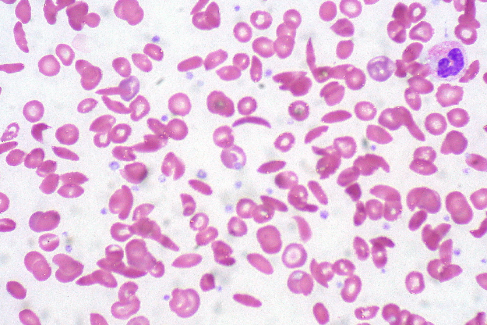
Howell-Jolly bodiesHowell-Jolly BodiesAsplenia in individuals with autosplenectomyAutosplenectomyAsplenia (immature RBCsRBCsErythrocytes, or red blood cells (RBCs), are the most abundant cells in the blood. While erythrocytes in the fetus are initially produced in the yolk sac then the liver, the bone marrow eventually becomes the main site of production.Erythrocytes: Histology with basophilic nuclear remnants)
Reticulocytes
Target cells with HbSC diseaseHbSC diseaseA disease characterized by chronic hemolytic anemia, episodic painful crises, and pathologic involvement of many organs. It is the clinical expression of homozygosity for hemoglobin s.Sickle Cell Disease
Other tests
↑↑ ReticulocyteReticulocyteImmature erythrocytes. In humans, these are erythroid cells that have just undergone extrusion of their cell nucleus. They still contain some organelles that gradually decrease in number as the cells mature. Ribosomes are last to disappear. Certain staining techniques cause components of the ribosomes to precipitate into characteristic ‘reticulum’ (not the same as the endoplasmic reticulum), hence the name reticulocytes.Erythrocytes: Histology count (3%–15%)
↑ BilirubinBilirubinA bile pigment that is a degradation product of heme.Heme Metabolism (indirect)
↑ BUN, ↑ creatinine
Liver function testsLiver function testsLiver function tests, also known as hepatic function panels, are one of the most commonly performed screening blood tests. Such tests are also used to detect, evaluate, and monitor acute and chronic liver diseases.Liver Function Tests: ↑ ASTASTEnzymes of the transferase class that catalyze the conversion of l-aspartate and 2-ketoglutarate to oxaloacetate and l-glutamate.Liver Function Tests/ALTALTAn enzyme that catalyzes the conversion of l-alanine and 2-oxoglutarate to pyruvate and l-glutamate.Liver Function Tests
Treatment of painful episodes includes analgesics and general supportive measures. Transfusions may be needed on occasion if the individual has symptomatic anemiaAnemiaAnemia is a condition in which individuals have low Hb levels, which can arise from various causes. Anemia is accompanied by a reduced number of RBCs and may manifest with fatigue, shortness of breath, pallor, and weakness. Subtypes are classified by the size of RBCs, chronicity, and etiology. Anemia: Overview and Types, including for the complication of acute chest syndrome.
Medications[5,8,9,18,20]
HydroxyureaHydroxyureaAn antineoplastic agent that inhibits DNA synthesis through the inhibition of ribonucleoside diphosphate reductase.Antimetabolite Chemotherapy forprevention and treatment of vasoocclusive events:
Mechanism of action:
Increases HbF levels, RBC water content, deformability of sickle cells
Alters adhesionAdhesionThe process whereby platelets adhere to something other than platelets, e.g., collagen; basement membrane; microfibrils; or other ‘foreign’ surfaces.Coagulation Studies of RBCsRBCsErythrocytes, or red blood cells (RBCs), are the most abundant cells in the blood. While erythrocytes in the fetus are initially produced in the yolk sac then the liver, the bone marrow eventually becomes the main site of production.Erythrocytes: Histology to endotheliumEndotheliumA layer of epithelium that lines the heart, blood vessels (vascular endothelium), lymph vessels (lymphatic endothelium), and the serous cavities of the body.Arteries: Histology
Reduces painPainAn unpleasant sensation induced by noxious stimuli which are detected by nerve endings of nociceptive neurons.Pain: Types and Pathways and other vasoocclusive complications
Decreases hospitalizationHospitalizationThe confinement of a patient in a hospital.Delirium rates
Improves survival
Indicated in all infants ≥ 9 months, children, adolescents
Indications in adults:[11,18]
≥ 3 episodes of moderate to severe vasoocclusive painPainAn unpleasant sensation induced by noxious stimuli which are detected by nerve endings of nociceptive neurons.Pain: Types and Pathways in a 12-month period
CKDCKDChronic kidney disease (CKD) is kidney impairment that lasts for ≥ 3 months, implying that it is irreversible. Hypertension and diabetes are the most common causes; however, there are a multitude of other etiologies. In the early to moderate stages, CKD is usually asymptomatic and is primarily diagnosed by laboratory abnormalities.Chronic Kidney Disease in patientsPatientsIndividuals participating in the health care system for the purpose of receiving therapeutic, diagnostic, or preventive procedures.Clinician–Patient Relationship already treated with erythropoietinErythropoietinGlycoprotein hormone, secreted chiefly by the kidney in the adult and the liver in the fetus, that acts on erythroid stem cells of the bone marrow to stimulate proliferation and differentiation.Erythrocytes: Histology (for anemiaAnemiaAnemia is a condition in which individuals have low Hb levels, which can arise from various causes. Anemia is accompanied by a reduced number of RBCs and may manifest with fatigue, shortness of breath, pallor, and weakness. Subtypes are classified by the size of RBCs, chronicity, and etiology. Anemia: Overview and Types)
Chronic SCD-associated painPainAn unpleasant sensation induced by noxious stimuli which are detected by nerve endings of nociceptive neurons.Pain: Types and Pathways interfering with daily activities or qualityQualityActivities and programs intended to assure or improve the quality of care in either a defined medical setting or a program. The concept includes the assessment or evaluation of the quality of care; identification of problems or shortcomings in the delivery of care; designing activities to overcome these deficiencies; and follow-up monitoring to ensure effectiveness of corrective steps.Quality Measurement and Improvement of life
Severe symptomatic chronic anemiaAnemiaAnemia is a condition in which individuals have low Hb levels, which can arise from various causes. Anemia is accompanied by a reduced number of RBCs and may manifest with fatigue, shortness of breath, pallor, and weakness. Subtypes are classified by the size of RBCs, chronicity, and etiology. Anemia: Overview and Types
Severe or recurrent acute chest syndrome
Dosing:
Initially, 15 mg/kg orally once daily (rounded to the nearest 50-mg or 100-mg strength); monitor blood counts every 2 weeks
Increase by 5 mg/kg/day every 12 weeks until mild myelosuppressionMyelosuppressionOxazolidinones is achieved (absolute neutrophil countAbsolute neutrophil countThe number of neutrophils (as opposed to the percentage of WBCs) circulating per µL of blood .Neutropenia (ANC) 2,000–4,000/mm3) or to a maximum dose of 35 mg/kg/day
New therapies since 2019 for patientsPatientsIndividuals participating in the health care system for the purpose of receiving therapeutic, diagnostic, or preventive procedures.Clinician–Patient Relationship who do not tolerate hydroxyureaHydroxyureaAn antineoplastic agent that inhibits DNA synthesis through the inhibition of ribonucleoside diphosphate reductase.Antimetabolite Chemotherapy or have continued painPainAn unpleasant sensation induced by noxious stimuli which are detected by nerve endings of nociceptive neurons.Pain: Types and Pathways while receiving it:
L-glutamine:
For use in adults or children ≥ 5 years
0.3 g/kg orally twice daily
Crizanlizumab:
P-selectinP-selectinCell adhesion molecule and cd antigen that mediates the adhesion of neutrophils and monocytes to activated platelets and endothelial cells.Tumor Necrosis Factor (TNF) inhibitor
Significantly lowers the frequency of SCD-related painPainAn unpleasant sensation induced by noxious stimuli which are detected by nerve endings of nociceptive neurons.Pain: Types and Pathways crises
May be given with or without hydroxyureaHydroxyureaAn antineoplastic agent that inhibits DNA synthesis through the inhibition of ribonucleoside diphosphate reductase.Antimetabolite Chemotherapy in patientsPatientsIndividuals participating in the health care system for the purpose of receiving therapeutic, diagnostic, or preventive procedures.Clinician–Patient Relationship ≥ 16 years
Dosing: 5 mg/kg IV every 2 weeks for 2 doses, then every 4 weeks
Voxelotor:
A new HbHbThe oxygen-carrying proteins of erythrocytes. They are found in all vertebrates and some invertebrates. The number of globin subunits in the hemoglobin quaternary structure differs between species. Structures range from monomeric to a variety of multimeric arrangements.Gas Exchange S polymerization inhibitor; increases HbHbThe oxygen-carrying proteins of erythrocytes. They are found in all vertebrates and some invertebrates. The number of globin subunits in the hemoglobin quaternary structure differs between species. Structures range from monomeric to a variety of multimeric arrangements.Gas Exchange levels and reduces markers of hemolysis
Studies did not show a significant ↓ in acute painAcute painIntensely discomforting, distressful, or agonizing sensation associated with trauma or disease, with well-defined location, character, and timing.Pain Management events.
May be given with or without hydroxyureaHydroxyureaAn antineoplastic agent that inhibits DNA synthesis through the inhibition of ribonucleoside diphosphate reductase.Antimetabolite Chemotherapy in patientsPatientsIndividuals participating in the health care system for the purpose of receiving therapeutic, diagnostic, or preventive procedures.Clinician–Patient Relationship ≥ 4 years
May cause hepatotoxicityHepatotoxicityAcetaminophen in 1%–2% of patientsPatientsIndividuals participating in the health care system for the purpose of receiving therapeutic, diagnostic, or preventive procedures.Clinician–Patient Relationship (in those with abnormal liverLiverThe liver is the largest gland in the human body. The liver is found in the superior right quadrant of the abdomen and weighs approximately 1.5 kilograms. Its main functions are detoxification, metabolism, nutrient storage (e.g., iron and vitamins), synthesis of coagulation factors, formation of bile, filtration, and storage of blood. Liver: Anatomy tests due to hemolysis, diagnosis can be challenging)
Dosing for adults and children ≥ 12 years: 1.5 g once daily
Dosing for children 4–11 years: 600–900 mg/day based on weight
Therapies with potential to cure, if vasoocclusive complications are not well controlled (but expensive and potentially fatal):
Traditional allogeneic hematopoietic stem cell transplant (HSCT)
Modified HSCT (geneGeneA category of nucleic acid sequences that function as units of heredity and which code for the basic instructions for the development, reproduction, and maintenance of organisms.Basic Terms of Genetics therapy, geneGeneA category of nucleic acid sequences that function as units of heredity and which code for the basic instructions for the development, reproduction, and maintenance of organisms.Basic Terms of Genetics editing)
Upregulating geneGeneA category of nucleic acid sequences that function as units of heredity and which code for the basic instructions for the development, reproduction, and maintenance of organisms.Basic Terms of Genetics expression to raise HbHbThe oxygen-carrying proteins of erythrocytes. They are found in all vertebrates and some invertebrates. The number of globin subunits in the hemoglobin quaternary structure differs between species. Structures range from monomeric to a variety of multimeric arrangements.Gas Exchange F and decrease HbHbThe oxygen-carrying proteins of erythrocytes. They are found in all vertebrates and some invertebrates. The number of globin subunits in the hemoglobin quaternary structure differs between species. Structures range from monomeric to a variety of multimeric arrangements.Gas Exchange S levels
Preventive measures[5,8,9,18,20]
Infection prevention:
Age-appropriate vaccinations:
S. pneumoniaPneumoniaPneumonia or pulmonary inflammation is an acute or chronic inflammation of lung tissue. Causes include infection with bacteria, viruses, or fungi. In more rare cases, pneumonia can also be caused through toxic triggers through inhalation of toxic substances, immunological processes, or in the course of radiotherapy.Pneumonia(pneumococcal conjugate and polysaccharide vaccines)
Seasonal influenzaInfluenzaInfluenza viruses are members of the Orthomyxoviridae family and the causative organisms of influenza, a highly contagious febrile respiratory disease. There are 3 primary influenza viruses (A, B, and C) and various subtypes, which are classified based on their virulent surface antigens, hemagglutinin (HA) and neuraminidase (NA). Influenza typically presents with a fever, myalgia, headache, and symptoms of an upper respiratory infection. Influenza Viruses/Influenza
N. meningitidisN. meningitidisA species of gram-negative, aerobic bacteria. It is a commensal and pathogen only of humans, and can be carried asymptomatically in the nasopharynx. When found in cerebrospinal fluid it is the causative agent of cerebrospinal meningitis. It is also found in venereal discharges and blood. There are at least 13 serogroups based on antigenic differences in the capsular polysaccharides; the ones causing most meningitis infections being a, b, c, y, and w-135. Each serogroup can be further classified by serotype, serosubtype, and immunotype.Neisseria
H. influenzaeH. influenzaeA species of Haemophilus found on the mucous membranes of humans and a variety of animals. The species is further divided into biotypes I through VIII.Haemophilus type b (Hib)
Hepatitis BHepatitis BHepatitis B virus (HBV) is a partially double-stranded DNA virus, which belongs to the Orthohepadnavirus genus and the Hepadnaviridae family. Most individuals with acute HBV infection are asymptomatic or have mild, self-limiting symptoms. Chronic infection can be asymptomatic or create hepatic inflammation, leading to liver cirrhosis and hepatocellular carcinoma (HCC). Hepatitis B Virus
ErythromycinErythromycinA bacteriostatic antibiotic macrolide produced by streptomyces erythreus. Erythromycin a is considered its major active component. In sensitive organisms, it inhibits protein synthesis by binding to 50s ribosomal subunits. This binding process inhibits peptidyl transferase activity and interferes with translocation of amino acids during translation and assembly of proteins.Macrolides and Ketolides if penicillinPenicillinRheumatic FeverallergyAllergyAn abnormal adaptive immune response that may or may not involve antigen-specific IgEType I Hypersensitivity Reaction
Red blood cell transfusion reduces burden of sickled cells and increases oxygen-carrying capacity and perfusion throughout the vascular system.
Delivery:
Simple transfusion:
Infusion of donor blood without removal of patient’s blood
Can cause hyperviscosityHyperviscosityHypercoagulable States, iron overloadIron overloadAn excessive accumulation of iron in the body due to a greater than normal absorption of iron from the gastrointestinal tract or from parenteral injection. This may arise from idiopathic hemochromatosis, excessive iron intake, chronic alcoholism, certain types of refractory anemia, or transfusional hemosiderosis.Hereditary Hemochromatosis
Exchange transfusion (partial or total):
Removal of patient’s blood during or after infusion of donor blood
Can be done manually or by apheresis
Can cause iron overloadIron overloadAn excessive accumulation of iron in the body due to a greater than normal absorption of iron from the gastrointestinal tract or from parenteral injection. This may arise from idiopathic hemochromatosis, excessive iron intake, chronic alcoholism, certain types of refractory anemia, or transfusional hemosiderosis.Hereditary Hemochromatosis
Requires placement of central venous catheterCentral Venous CatheterCentral venous catheters are IV lines placed into the large central veins for monitoring of central venous pressure (CVP), prolonged drug administration, or administration of parenteral nutrition. The most common sites of insertion are the internal jugular and subclavian veins. Central Venous Catheter and an experienced center and staff
Expensive
Indications for transfusion (see table):[9,15,18]
Symptomatic or severe anemiaAnemiaAnemia is a condition in which individuals have low Hb levels, which can arise from various causes. Anemia is accompanied by a reduced number of RBCs and may manifest with fatigue, shortness of breath, pallor, and weakness. Subtypes are classified by the size of RBCs, chronicity, and etiology. Anemia: Overview and Types
Preoperatively
Acute stroke
Multiorgan failure
Acute chest syndrome
Recurrent priapismPriapismA prolonged painful erection that may lasts hours and is not associated with sexual activity. It is seen in patients with sickle cell anemia, advanced malignancy, spinal trauma; and certain drug treatments.Penile Anomalies and Conditions
Table: Transfusion indications in sickle cell diseaseSickle cell diseaseSickle cell disease (SCD) is a group of genetic disorders in which an abnormal Hb molecule (HbS) transforms RBCs into sickle-shaped cells, resulting in chronic anemia, vasoocclusive episodes, pain, and organ damage.Sickle Cell Disease[18]
Acute stroke or TIATIATransient ischemic attack (TIA) is a temporary episode of neurologic dysfunction caused by ischemia without infarction that resolves completely when blood supply is restored. Transient ischemic attack is a neurologic emergency that warrants urgent medical attention. Transient Ischemic Attack (TIA)
Exchange transfusion
Acute hepatic sequestration or cholestasis
Simple or exchange transfusion
Acute chest syndrome
Multiorgan failure
Preoperative
Indications for chronic transfusion
Primary stroke prevention
Simple or exchange transfusion
Secondary stroke prevention
Recurrent vasoocclusive conditions
Other therapeutic recommendations:
Hematopoietic stem cell transplantationHematopoietic stem cell transplantationTransfer of hematopoietic stem cells from bone marrow or blood between individuals within the same species (homologous transplantation) or transfer within the same individual (autologous transplantation). Hematopoietic stem cell transplantation has been used as an alternative to bone marrow transplantation in the treatment of a variety of neoplasms.Organ Transplantation is the only curative option.[20]
Limited donors
Matched siblings offer the best results in children.
Therapies notused for acute vasoocclusive episodes:
O2: unless saturation is below baseline or there are pulmonary symptoms
Meperidine: associated with accumulation of metabolites, leading to seizuresSeizuresA seizure is abnormal electrical activity of the neurons in the cerebral cortex that can manifest in numerous ways depending on the region of the brain affected. Seizures consist of a sudden imbalance that occurs between the excitatory and inhibitory signals in cortical neurons, creating a net excitation. The 2 major classes of seizures are focal and generalized. Seizures
Ketorolac: due to toxicities
Granulocyte colony-stimulating factorGranulocyte colony-stimulating factorA glycoprotein of mw 25 kda containing internal disulfide bonds. It induces the survival, proliferation, and differentiation of neutrophilic granulocyte precursor cells and functionally activates mature blood neutrophils. Among the family of colony-stimulating factors, G-CSF is the most potent inducer of terminal differentiation to granulocytes and macrophages of leukemic myeloid cell lines.White Myeloid Cells: Histology (G-CSF): due to risk of multiorgan failure and death (with rare exceptions)
Complications
Recommendations for acute complications:[1,9,11]
Acute chest syndrome: a new radiodensity on chest radiograph accompanied by feverFeverFever is defined as a measured body temperature of at least 38°C (100.4°F). Fever is caused by circulating endogenous and/or exogenous pyrogens that increase levels of prostaglandin E2 in the hypothalamus. Fever is commonly associated with chills, rigors, sweating, and flushing of the skin. Fever and/or respiratory symptoms [11]
Pathophysiology: usually vasoocclusion in the pulmonary microvasculature due to fat emboli caused by bone marrowBone marrowThe soft tissue filling the cavities of bones. Bone marrow exists in two types, yellow and red. Yellow marrow is found in the large cavities of large bones and consists mostly of fat cells and a few primitive blood cells. Red marrow is a hematopoietic tissue and is the site of production of erythrocytes and granular leukocytes. Bone marrow is made up of a framework of connective tissue containing branching fibers with the frame being filled with marrow cells.Bone Marrow: Composition and Hematopoiesis infarction; in children, infection is a common cause.[17]
A leading cause of death for patientsPatientsIndividuals participating in the health care system for the purpose of receiving therapeutic, diagnostic, or preventive procedures.Clinician–Patient Relationship with sickle cell diseaseSickle cell diseaseSickle cell disease (SCD) is a group of genetic disorders in which an abnormal Hb molecule (HbS) transforms RBCs into sickle-shaped cells, resulting in chronic anemia, vasoocclusive episodes, pain, and organ damage.Sickle Cell Disease; more severe in adults
Needs prompt management to prevent clinical deterioration and death:
RBC transfusion to achieve HbHbThe oxygen-carrying proteins of erythrocytes. They are found in all vertebrates and some invertebrates. The number of globin subunits in the hemoglobin quaternary structure differs between species. Structures range from monomeric to a variety of multimeric arrangements.Gas Exchange > 10 g/dL
Antibiotics (empiric therapyEmpiric TherapyMeningitis in Children for pneumoniaPneumoniaPneumonia or pulmonary inflammation is an acute or chronic inflammation of lung tissue. Causes include infection with bacteria, viruses, or fungi. In more rare cases, pneumonia can also be caused through toxic triggers through inhalation of toxic substances, immunological processes, or in the course of radiotherapy.Pneumonia)
Acute painful episodes:[14, 18]
Individualize painPainAn unpleasant sensation induced by noxious stimuli which are detected by nerve endings of nociceptive neurons.Pain: Types and Pathways protocol. (Discuss options with patient and family.)
HospitalizationHospitalizationThe confinement of a patient in a hospital.Delirium and rapid initiation of parenteral opioidsOpioidsOpiates are drugs that are derived from the sap of the opium poppy. Opiates have been used since antiquity for the relief of acute severe pain. Opioids are synthetic opiates with properties that are substantially similar to those of opiates. Opioid Analgesics in severe cases
MorphineMorphineThe principal alkaloid in opium and the prototype opiate analgesic and narcotic. Morphine has widespread effects in the central nervous system and on smooth muscle.Opioid Analgesics:
Opioid-naive: 0.1–0.15 mg/kg IV within 30 minutes after presentation; reassess painPainAn unpleasant sensation induced by noxious stimuli which are detected by nerve endings of nociceptive neurons.Pain: Types and Pathways in 20 minutes
May repeat doses of 0.02–0.05 mg/kg every 20–30 minutes
HydromorphoneHydromorphoneAn opioid analgesic made from morphine and used mainly as an analgesic. It has a shorter duration of action than morphine.Opioid Analgesics:
Patient weight ≥ 50 kg: 1.5 mg initially; repeat with 20%–50% of original dose every 30 minutes if needed
Patient weight < 50 kg: 0.02–0.05 mg/kg; maximum initial dose, 1.5 mg; repeat as above
Stroke:[12,13,15,16,18]
RegularRegularInsulin annual transcranial DopplerDopplerUltrasonography applying the doppler effect, with frequency-shifted ultrasound reflections produced by moving targets (usually red blood cells) in the bloodstream along the ultrasound axis in direct proportion to the velocity of movement of the targets, to determine both direction and velocity of blood flow.Ultrasound (Sonography) ultrasonography:
Recommended in children ≤ 16 years of age
Helps determine those at high risk (transcranial DopplerDopplerUltrasonography applying the doppler effect, with frequency-shifted ultrasound reflections produced by moving targets (usually red blood cells) in the bloodstream along the ultrasound axis in direct proportion to the velocity of movement of the targets, to determine both direction and velocity of blood flow.Ultrasound (Sonography) velocity ≥ 200 cm/sec)
Exchange transfusion
Chronic red blood cell transfusion maintaining HbHbThe oxygen-carrying proteins of erythrocytes. They are found in all vertebrates and some invertebrates. The number of globin subunits in the hemoglobin quaternary structure differs between species. Structures range from monomeric to a variety of multimeric arrangements.Gas Exchange S at ≤ 30% of total Hgb (primary and secondary prevention)
HydroxyureaHydroxyureaAn antineoplastic agent that inhibits DNA synthesis through the inhibition of ribonucleoside diphosphate reductase.Antimetabolite Chemotherapy
InfectionsInfectionsInvasion of the host organism by microorganisms or their toxins or by parasites that can cause pathological conditions or diseases.Chronic Granulomatous Disease:[11,18]
FeverFeverFever is defined as a measured body temperature of at least 38°C (100.4°F). Fever is caused by circulating endogenous and/or exogenous pyrogens that increase levels of prostaglandin E2 in the hypothalamus. Fever is commonly associated with chills, rigors, sweating, and flushing of the skin. Fever (temperature > 38.3°C (101.5°F)) needs prompt evaluation and antibiotic initiation.
Consider splenectomySplenectomySurgical procedure involving either partial or entire removal of the spleen.Rupture of the Spleen.
Aplastic crisis:[1,8]
Suspect in worsening anemiaAnemiaAnemia is a condition in which individuals have low Hb levels, which can arise from various causes. Anemia is accompanied by a reduced number of RBCs and may manifest with fatigue, shortness of breath, pallor, and weakness. Subtypes are classified by the size of RBCs, chronicity, and etiology. Anemia: Overview and Types, without increase in reticulocytes (transient)
Acute cholecystitisAcute cholecystitisAcute inflammation of the gallbladder wall. It is characterized by the presence of abdominal pain; fever; and leukocytosis. Gallstone obstruction of the cystic duct is present in approximately 90% of the cases.Cholecystitis from gallstonesGallstonesCholelithiasis (gallstones) is the presence of stones in the gallbladder. Most gallstones are cholesterol stones, while the rest are composed of bilirubin (pigment stones) and other mixed components. Patients are commonly asymptomatic but may present with biliary colic (intermittent pain in the right upper quadrant).Cholelithiasis:
Supportive therapy
Surgery
Myocardial infarctionMyocardial infarctionMI is ischemia and death of an area of myocardial tissue due to insufficient blood flow and oxygenation, usually from thrombus formation on a ruptured atherosclerotic plaque in the epicardial arteries. Clinical presentation is most commonly with chest pain, but women and patients with diabetes may have atypical symptoms.Myocardial Infarction:
Management per acute coronary syndrome protocol
Others:[1,11]
Venous thromboembolismThromboembolismObstruction of a blood vessel (embolism) by a blood clot (thrombus) in the blood stream.Systemic Lupus Erythematosus (VTEVTEObstruction of a vein or veins (embolism) by a blood clot (thrombus) in the bloodstream.Hypercoagulable States):
Thromboprophylaxis is recommended for all adults with SCDSCDSickle cell disease (SCD) is a group of genetic disorders in which an abnormal Hb molecule (HbS) transforms RBCs into sickle-shaped cells, resulting in chronic anemia, vasoocclusive episodes, pain, and organ damage.Sickle Cell Disease who are hospitalized (unless contraindicated).
For patientsPatientsIndividuals participating in the health care system for the purpose of receiving therapeutic, diagnostic, or preventive procedures.Clinician–Patient Relationship with legLegThe lower leg, or just “leg” in anatomical terms, is the part of the lower limb between the knee and the ankle joint. The bony structure is composed of the tibia and fibula bones, and the muscles of the leg are grouped into the anterior, lateral, and posterior compartments by extensions of fascia.Leg: Anatomy ulcers, check lower-extremity DopplerDopplerUltrasonography applying the doppler effect, with frequency-shifted ultrasound reflections produced by moving targets (usually red blood cells) in the bloodstream along the ultrasound axis in direct proportion to the velocity of movement of the targets, to determine both direction and velocity of blood flow.Ultrasound (Sonography) for deep vein thrombosisThrombosisFormation and development of a thrombus or blood clot in the blood vessel.Epidemic Typhus.
PriapismPriapismA prolonged painful erection that may lasts hours and is not associated with sexual activity. It is seen in patients with sickle cell anemia, advanced malignancy, spinal trauma; and certain drug treatments.Penile Anomalies and Conditions (a compartment syndromeCompartment SyndromeCompartment syndrome is a surgical emergency usually occurring secondary to trauma. The condition is marked by increased pressure within a compartment that compromises the circulation and function of the tissues within that space.Compartment Syndrome of the penisPenisThe penis is the male organ of copulation and micturition. The organ is composed of a root, body, and glans. The root is attached to the pubic bone by the crura penis. The body consists of the 2 parallel corpora cavernosa and the corpus spongiosum. The glans is ensheathed by the prepuce or foreskin. Penis: Anatomy):
Hydration
PainPainAn unpleasant sensation induced by noxious stimuli which are detected by nerve endings of nociceptive neurons.Pain: Types and Pathways control
Emergent urologic evaluation for episodes > 4 hours (at 6 hours, irreversible tissue damage can begin to occur)
Aspiration of blood from the corpus cavernosum, with saline irrigation and then injection of an alpha-adrenergic agonist (e.g., phenylephrinePhenylephrineAn alpha-1 adrenergic agonist used as a mydriatic, nasal decongestant, and cardiotonic agent.Sympathomimetic Drugs)
Alloimmunization:[15]
Delayed hemolytic transfusion reactionsTransfusion reactionsTransfusion-related complications occur during or after a blood product is given. These complications can be classified as immunologic, non-immunologic and acute, and delayed. Non-immunologic reactions are caused by the transmission of disease in blood products, and immunologic reactions are antigen-antibody-mediated. Transfusion Reactions may present with worsening anemiaAnemiaAnemia is a condition in which individuals have low Hb levels, which can arise from various causes. Anemia is accompanied by a reduced number of RBCs and may manifest with fatigue, shortness of breath, pallor, and weakness. Subtypes are classified by the size of RBCs, chronicity, and etiology. Anemia: Overview and Types, feverFeverFever is defined as a measured body temperature of at least 38°C (100.4°F). Fever is caused by circulating endogenous and/or exogenous pyrogens that increase levels of prostaglandin E2 in the hypothalamus. Fever is commonly associated with chills, rigors, sweating, and flushing of the skin. Fever, fatigueFatigueThe state of weariness following a period of exertion, mental or physical, characterized by a decreased capacity for work and reduced efficiency to respond to stimuli.Fibromyalgia, jaundiceJaundiceJaundice is the abnormal yellowing of the skin and/or sclera caused by the accumulation of bilirubin. Hyperbilirubinemia is caused by either an increase in bilirubin production or a decrease in the hepatic uptake, conjugation, or excretion of bilirubin. Jaundice, dark urine, or painPainAn unpleasant sensation induced by noxious stimuli which are detected by nerve endings of nociceptive neurons.Pain: Types and Pathways within 3–14 days.
Can present up to 3 months later
RBC antigenAntigenSubstances that are recognized by the immune system and induce an immune reaction.Vaccination matching can be used for prevention.
Immunosuppressive therapies may be needed to treat a severe reaction.
Chronic kidney diseaseChronic Kidney DiseaseChronic kidney disease (CKD) is kidney impairment that lasts for ≥ 3 months, implying that it is irreversible. Hypertension and diabetes are the most common causes; however, there are a multitude of other etiologies. In the early to moderate stages, CKD is usually asymptomatic and is primarily diagnosed by laboratory abnormalities.Chronic Kidney Disease:
If protein excretion rate > 300 mg/24 hours → nephrology referral
Consider angiotensin-converting enzyme inhibitor or angiotensin IIAngiotensin IIAn octapeptide that is a potent but labile vasoconstrictor. It is produced from angiotensin I after the removal of two amino acids at the c-terminal by angiotensin converting enzyme. The amino acid in position 5 varies in different species. To block vasoconstriction and hypertension effect of angiotensin II, patients are often treated with ace inhibitors or with angiotensin II type 1 receptor blockers.Renal Sodium and Water RegulationreceptorReceptorReceptors are proteins located either on the surface of or within a cell that can bind to signaling molecules known as ligands (e.g., hormones) and cause some type of response within the cell.Receptors blocker therapy.
Consider erythropoietin-stimulating agents.
HypertensionHypertensionHypertension, or high blood pressure, is a common disease that manifests as elevated systemic arterial pressures. Hypertension is most often asymptomatic and is found incidentally as part of a routine physical examination or during triage for an unrelated medical encounter. Hypertension:
Goal BP: ≤ 130/80 mm Hg
Early treatment if BP is elevated
Pulmonary hypertensionPulmonary HypertensionPulmonary hypertension (PH) or pulmonary arterial hypertension (PAH) is characterized by elevated pulmonary arterial pressure, which can lead to chronic progressive right heart failure. Pulmonary hypertension is grouped into 5 categories based on etiology, which include primary PAH, and PH due to cardiac disease, lung or hypoxic disease, chronic thromboembolic disease, and multifactorial or unclear etiologies. Pulmonary Hypertension:
EchocardiographyEchocardiographyUltrasonic recording of the size, motion, and composition of the heart and surrounding tissues. The standard approach is transthoracic.Tricuspid Valve Atresia (TVA):
In patientsPatientsIndividuals participating in the health care system for the purpose of receiving therapeutic, diagnostic, or preventive procedures.Clinician–Patient Relationship with suspicious symptoms (e.g., chest painPainAn unpleasant sensation induced by noxious stimuli which are detected by nerve endings of nociceptive neurons.Pain: Types and Pathways, dyspneaDyspneaDyspnea is the subjective sensation of breathing discomfort. Dyspnea is a normal manifestation of heavy physical or psychological exertion, but also may be caused by underlying conditions (both pulmonary and extrapulmonary). Dyspnea or hypoxemiaHypoxemiaNeonatal Respiratory Distress Syndrome at rest, syncopeSyncopeSyncope is a short-term loss of consciousness and loss of postural stability followed by spontaneous return of consciousness to the previous neurologic baseline without the need for resuscitation. The condition is caused by transient interruption of cerebral blood flow that may be benign or related to a underlying life-threatening condition. Syncope, new murmur)
In patientsPatientsIndividuals participating in the health care system for the purpose of receiving therapeutic, diagnostic, or preventive procedures.Clinician–Patient Relationship who have known problems associated with pulmonary hypertensionPulmonary HypertensionPulmonary hypertension (PH) or pulmonary arterial hypertension (PAH) is characterized by elevated pulmonary arterial pressure, which can lead to chronic progressive right heart failure. Pulmonary hypertension is grouped into 5 categories based on etiology, which include primary PAH, and PH due to cardiac disease, lung or hypoxic disease, chronic thromboembolic disease, and multifactorial or unclear etiologies. Pulmonary Hypertension (e.g., legLegThe lower leg, or just “leg” in anatomical terms, is the part of the lower limb between the knee and the ankle joint. The bony structure is composed of the tibia and fibula bones, and the muscles of the leg are grouped into the anterior, lateral, and posterior compartments by extensions of fascia.Leg: Anatomy ulcers, priapismPriapismA prolonged painful erection that may lasts hours and is not associated with sexual activity. It is seen in patients with sickle cell anemia, advanced malignancy, spinal trauma; and certain drug treatments.Penile Anomalies and Conditions)
EchocardiographyEchocardiographyUltrasonic recording of the size, motion, and composition of the heart and surrounding tissues. The standard approach is transthoracic.Tricuspid Valve Atresia (TVA) is not recommended in asymptomatic patientsPatientsIndividuals participating in the health care system for the purpose of receiving therapeutic, diagnostic, or preventive procedures.Clinician–Patient Relationship.
Respiratory problems:
High prevalencePrevalenceThe total number of cases of a given disease in a specified population at a designated time. It is differentiated from incidence, which refers to the number of new cases in the population at a given time.Measures of Disease Frequency of asthmaAsthmaAsthma is a chronic inflammatory respiratory condition characterized by bronchial hyperresponsiveness and airflow obstruction. The disease is believed to result from the complex interaction of host and environmental factors that increase disease predisposition, with inflammation causing symptoms and structural changes. Patients typically present with wheezing, cough, and dyspnea. Asthma, especially in children
Perform pulmonary evaluation during visits and add workup (e.g., spirometrySpirometryMeasurement of volume of air inhaled or exhaled by the lung.Pulmonary Function Tests) as indicated.
Neurocognitive dysfunction:
Evaluate for signs of cognitive dysfunction (e.g., memoryMemoryComplex mental function having four distinct phases: (1) memorizing or learning, (2) retention, (3) recall, and (4) recognition. Clinically, it is usually subdivided into immediate, recent, and remote memory.Psychiatric Assessment problems, decline in school performance), as patientsPatientsIndividuals participating in the health care system for the purpose of receiving therapeutic, diagnostic, or preventive procedures.Clinician–Patient Relationship may have silent cerebral infarcts.
Depression also needs to be screened.
Use a validated screeningScreeningPreoperative Care tool or refer to a specialist for in-depth behavioral and cognitive assessment.
Annual examination of joints (painPainAn unpleasant sensation induced by noxious stimuli which are detected by nerve endings of nociceptive neurons.Pain: Types and Pathways and ↓ range of motionRange of motionThe distance and direction to which a bone joint can be extended. Range of motion is a function of the condition of the joints, muscles, and connective tissues involved. Joint flexibility can be improved through appropriate muscle strength exercises.Examination of the Upper Limbs especially in the hip and shoulder)
May require orthopedic surgery consultation for hip or shoulder arthroplastyArthroplastySurgical reconstruction of a joint to relieve pain or restore motion.Osteoarthritis
Other boneBoneBone is a compact type of hardened connective tissue composed of bone cells, membranes, an extracellular mineralized matrix, and central bone marrow. The 2 primary types of bone are compact and spongy. Bones: Structure and Types health issues:
Vitamin D deficiencyVitamin D DeficiencyA nutritional condition produced by a deficiency of vitamin D in the diet, insufficient production of vitamin D in the skin, inadequate absorption of vitamin D from the diet, or abnormal conversion of vitamin D to its bioactive metabolites. It is manifested clinically as rickets in children and osteomalacia in adults.Fat-soluble Vitamins and their Deficiencies is common in SCDSCDSickle cell disease (SCD) is a group of genetic disorders in which an abnormal Hb molecule (HbS) transforms RBCs into sickle-shaped cells, resulting in chronic anemia, vasoocclusive episodes, pain, and organ damage.Sickle Cell Disease.
Annual testing of vitamin DVitamin DA vitamin that includes both cholecalciferols and ergocalciferols, which have the common effect of preventing or curing rickets in animals. It can also be viewed as a hormone since it can be formed in skin by action of ultraviolet rays upon the precursors, 7-dehydrocholesterol and ergosterol, and acts on vitamin D receptors to regulate calcium in opposition to parathyroid hormone.Fat-soluble Vitamins and their Deficiencies levels
BoneBoneBone is a compact type of hardened connective tissue composed of bone cells, membranes, an extracellular mineralized matrix, and central bone marrow. The 2 primary types of bone are compact and spongy. Bones: Structure and Types mineral density test (from age 12 years, repeated 1–3 years)
Chronic painChronic painAching sensation that persists for more than a few months. It may or may not be associated with trauma or disease, and may persist after the initial injury has healed. Its localization, character, and timing are more vague than with acute pain.Pain Management issues:
In addition to vasoocclusion, painPainAn unpleasant sensation induced by noxious stimuli which are detected by nerve endings of nociceptive neurons.Pain: Types and Pathways issues may be complicated by avascular necrosisAvascular NecrosisHip Fractures, compressionCompressionBlunt Chest Trauma fractures, and opioidOpioidCompounds with activity like opiate alkaloids, acting at opioid receptors. Properties include induction of analgesia or narcosis.Constipation use disorder.
Provide adequate opioidOpioidCompounds with activity like opiate alkaloids, acting at opioid receptors. Properties include induction of analgesia or narcosis.Constipation dosing for painPainAn unpleasant sensation induced by noxious stimuli which are detected by nerve endings of nociceptive neurons.Pain: Types and Pathways relief, but with the following precautions:
Avoid unnecessary dose escalation.
Taper dose when appropriate.
Offer nonpharmacologic options (e.g., massage, meditation, cognitive behavioral therapyCognitive behavioral therapyA directive form of psychotherapy based on the interpretation of situations (cognitive structure of experiences) that determine how an individual feels and behaves. It is based on the premise that cognition, the process of acquiring knowledge and forming beliefs, is a primary determinant of mood and behavior. The therapy uses behavioral and verbal techniques to identify and correct negative thinking that is at the root of the aberrant behavior.Psychotherapy).
Mental health referral if depression, anxietyAnxietyFeelings or emotions of dread, apprehension, and impending disaster but not disabling as with anxiety disorders.Generalized Anxiety Disorder, or dependence on painPainAn unpleasant sensation induced by noxious stimuli which are detected by nerve endings of nociceptive neurons.Pain: Types and Pathways medication is present.
LegLegThe lower leg, or just “leg” in anatomical terms, is the part of the lower limb between the knee and the ankle joint. The bony structure is composed of the tibia and fibula bones, and the muscles of the leg are grouped into the anterior, lateral, and posterior compartments by extensions of fascia.Leg: Anatomy ulcers:
Annual ophthalmologic (dilated) examination for those 10 years of age and older
If retinopathyRetinopathyDegenerative changes to the retina due to hypertension.Alport Syndrome is suspected, refer to retinaRetinaThe ten-layered nervous tissue membrane of the eye. It is continuous with the optic nerve and receives images of external objects and transmits visual impulses to the brain. Its outer surface is in contact with the choroid and the inner surface with the vitreous body. The outermost layer is pigmented, whereas the inner nine layers are transparent.Eye: Anatomy specialist.
Iron overloadIron overloadAn excessive accumulation of iron in the body due to a greater than normal absorption of iron from the gastrointestinal tract or from parenteral injection. This may arise from idiopathic hemochromatosis, excessive iron intake, chronic alcoholism, certain types of refractory anemia, or transfusional hemosiderosis.Hereditary Hemochromatosis:
Monitor serum ferritinFerritinIron-containing proteins that are widely distributed in animals, plants, and microorganisms. Their major function is to store iron in a nontoxic bioavailable form. Each ferritin molecule consists of ferric iron in a hollow protein shell (apoferritins) made of 24 subunits of various sequences depending on the species and tissue types.Hereditary Hemochromatosis every 1–3 months; start chelation therapy if >1,000–1,500 µg/L.
LiverLiverThe liver is the largest gland in the human body. The liver is found in the superior right quadrant of the abdomen and weighs approximately 1.5 kilograms. Its main functions are detoxification, metabolism, nutrient storage (e.g., iron and vitamins), synthesis of coagulation factors, formation of bile, filtration, and storage of blood. Liver: Anatomy MRI:
May also be used every 1–2 years to assess liverLiverThe liver is the largest gland in the human body. The liver is found in the superior right quadrant of the abdomen and weighs approximately 1.5 kilograms. Its main functions are detoxification, metabolism, nutrient storage (e.g., iron and vitamins), synthesis of coagulation factors, formation of bile, filtration, and storage of blood. Liver: AnatomyironIronA metallic element with atomic symbol fe, atomic number 26, and atomic weight 55. 85. It is an essential constituent of hemoglobins; cytochromes; and iron-binding proteins. It plays a role in cellular redox reactions and in the transport of oxygen.Trace Elements content/overload
Start chelation therapy when liverLiverThe liver is the largest gland in the human body. The liver is found in the superior right quadrant of the abdomen and weighs approximately 1.5 kilograms. Its main functions are detoxification, metabolism, nutrient storage (e.g., iron and vitamins), synthesis of coagulation factors, formation of bile, filtration, and storage of blood. Liver: AnatomyironIronA metallic element with atomic symbol fe, atomic number 26, and atomic weight 55. 85. It is an essential constituent of hemoglobins; cytochromes; and iron-binding proteins. It plays a role in cellular redox reactions and in the transport of oxygen.Trace Elements is > 3–5 mg/g dry weight.
Chelation therapy:
DeferasiroxDeferasiroxA triazole and benzoate derivative that acts as a selective iron chelator. It is used in the management of chronic iron overload due to blood transfusion or non-transfusion dependent thalassemia.Hereditary Hemochromatosis (preferred)
Discuss testing partners for hemoglobinopathy status testing (if unknown).
Women anticipating pregnancyPregnancyThe status during which female mammals carry their developing young (embryos or fetuses) in utero before birth, beginning from fertilization to birth.Pregnancy: Diagnosis, Physiology, and Care should be tested for red blood cell alloantibodies.
Discuss contraception choices, which include:
Progestin-only contraceptives
Barrier methodsBarrier MethodsMethods of contraception in which physical, chemical, or biological means are used to prevent the sperm from reaching the fertilizable ovum.Nonhormonal Contraception (recommended for patientsPatientsIndividuals participating in the health care system for the purpose of receiving therapeutic, diagnostic, or preventive procedures.Clinician–Patient Relationship on hydroxyureaHydroxyureaAn antineoplastic agent that inhibits DNA synthesis through the inhibition of ribonucleoside diphosphate reductase.Antimetabolite Chemotherapy)
PrognosisPrognosisA prediction of the probable outcome of a disease based on a individual’s condition and the usual course of the disease as seen in similar situations.Non-Hodgkin Lymphomas:[3,7,20]
PatientsPatientsIndividuals participating in the health care system for the purpose of receiving therapeutic, diagnostic, or preventive procedures.Clinician–Patient Relationship with SCDSCDSickle cell disease (SCD) is a group of genetic disorders in which an abnormal Hb molecule (HbS) transforms RBCs into sickle-shaped cells, resulting in chronic anemia, vasoocclusive episodes, pain, and organ damage.Sickle Cell Disease:
Survival for individuals with SCDSCDSickle cell disease (SCD) is a group of genetic disorders in which an abnormal Hb molecule (HbS) transforms RBCs into sickle-shaped cells, resulting in chronic anemia, vasoocclusive episodes, pain, and organ damage.Sickle Cell Disease has improved dramatically in the past 40 years in the US.
Major cause of death has shifted from infectionsInfectionsInvasion of the host organism by microorganisms or their toxins or by parasites that can cause pathological conditions or diseases.Chronic Granulomatous Disease to progressive end-organ damage.
Median life expectancyLife expectancyBased on known statistical data, the number of years which any person of a given age may reasonably expected to live.Population Pyramids for adults with SCDSCDSickle cell disease (SCD) is a group of genetic disorders in which an abnormal Hb molecule (HbS) transforms RBCs into sickle-shaped cells, resulting in chronic anemia, vasoocclusive episodes, pain, and organ damage.Sickle Cell Disease is ≥ 20 years shorter than African Americans without SCDSCDSickle cell disease (SCD) is a group of genetic disorders in which an abnormal Hb molecule (HbS) transforms RBCs into sickle-shaped cells, resulting in chronic anemia, vasoocclusive episodes, pain, and organ damage.Sickle Cell Disease living in the US.
Acute and chronic complications greatly increase morbidityMorbidityThe proportion of patients with a particular disease during a given year per given unit of population.Measures of Health Status.
SCDSCDSickle cell disease (SCD) is a group of genetic disorders in which an abnormal Hb molecule (HbS) transforms RBCs into sickle-shaped cells, resulting in chronic anemia, vasoocclusive episodes, pain, and organ damage.Sickle Cell Disease is also associated with reduced qualityQualityActivities and programs intended to assure or improve the quality of care in either a defined medical setting or a program. The concept includes the assessment or evaluation of the quality of care; identification of problems or shortcomings in the delivery of care; designing activities to overcome these deficiencies; and follow-up monitoring to ensure effectiveness of corrective steps.Quality Measurement and Improvement of life and work/school productivity.
Summary of the management of sickle cell diseaseSickle cell diseaseSickle cell disease (SCD) is a group of genetic disorders in which an abnormal Hb molecule (HbS) transforms RBCs into sickle-shaped cells, resulting in chronic anemia, vasoocclusive episodes, pain, and organ damage.Sickle Cell Disease
Presentation
Management
Acute painAcute painIntensely discomforting, distressful, or agonizing sensation associated with trauma or disease, with well-defined location, character, and timing.Pain Management episodes/ vasoocclusive events
Oral hydration
PainPainAn unpleasant sensation induced by noxious stimuli which are detected by nerve endings of nociceptive neurons.Pain: Types and Pathways control with fast-acting oral (or IV if needed) opiate analgesics
IV fluidsIV fluidsIntravenous fluids are one of the most common interventions administered in medicine to approximate physiologic bodily fluids. Intravenous fluids are divided into 2 categories: crystalloid and colloid solutions. Intravenous fluids have a wide variety of indications, including intravascular volume expansion, electrolyte manipulation, and maintenance fluids. Intravenous Fluids if hypovolemic and for painPainAn unpleasant sensation induced by noxious stimuli which are detected by nerve endings of nociceptive neurons.Pain: Types and Pathways control
Incentive spirometrySpirometryMeasurement of volume of air inhaled or exhaled by the lung.Pulmonary Function Tests to reduce the risk of acute chest syndrome
HydroxyureaHydroxyureaAn antineoplastic agent that inhibits DNA synthesis through the inhibition of ribonucleoside diphosphate reductase.Antimetabolite Chemotherapy
Folic acid (for folate deficiencyFolate deficiencyA nutritional condition produced by a deficiency of folic acid in the diet. Many plant and animal tissues contain folic acid, abundant in green leafy vegetables, yeast, liver, and mushrooms but destroyed by long-term cooking. Alcohol interferes with its intermediate metabolism and absorption. Folic acid deficiency may develop in long-term anticonvulsant therapy or with use of oral contraceptives. This deficiency causes anemia, macrocytic anemia, and megaloblastic anemia. It is indistinguishable from vitamin B12 deficiency in peripheral blood and bone marrow findings, but the neurologic lesions seen in B12 deficiency do not occur.Megaloblastic Anemia)
Exchange transfusion (in case of HbHbThe oxygen-carrying proteins of erythrocytes. They are found in all vertebrates and some invertebrates. The number of globin subunits in the hemoglobin quaternary structure differs between species. Structures range from monomeric to a variety of multimeric arrangements.Gas Exchange < 6 g/dL or acute chest syndrome)
SplenectomySplenectomySurgical procedure involving either partial or entire removal of the spleen.Rupture of the Spleen (delay until the age of 3–5 years)
InfectionsInfectionsInvasion of the host organism by microorganisms or their toxins or by parasites that can cause pathological conditions or diseases.Chronic Granulomatous Disease
PriapismPriapismA prolonged painful erection that may lasts hours and is not associated with sexual activity. It is seen in patients with sickle cell anemia, advanced malignancy, spinal trauma; and certain drug treatments.Penile Anomalies and Conditions
Hydration
Analgesics
If episode > 4 hours, obtain emergent urologic evaluation
Aspiration of blood from the corpus cavernosum, with saline irrigation and then injection of an alpha-adrenergic agonist (e.g., phenylephrinePhenylephrineAn alpha-1 adrenergic agonist used as a mydriatic, nasal decongestant, and cardiotonic agent.Sympathomimetic Drugs)
Stroke: annual transcranial DopplerDopplerUltrasonography applying the doppler effect, with frequency-shifted ultrasound reflections produced by moving targets (usually red blood cells) in the bloodstream along the ultrasound axis in direct proportion to the velocity of movement of the targets, to determine both direction and velocity of blood flow.Ultrasound (Sonography) exam (age 2–16 years)
RetinopathyRetinopathyDegenerative changes to the retina due to hypertension.Alport Syndrome: annual retinal exam (beginning at age 10 years)
EchocardiographyEchocardiographyUltrasonic recording of the size, motion, and composition of the heart and surrounding tissues. The standard approach is transthoracic.Tricuspid Valve Atresia (TVA): for pulmonary hypertensionPulmonary HypertensionPulmonary hypertension (PH) or pulmonary arterial hypertension (PAH) is characterized by elevated pulmonary arterial pressure, which can lead to chronic progressive right heart failure. Pulmonary hypertension is grouped into 5 categories based on etiology, which include primary PAH, and PH due to cardiac disease, lung or hypoxic disease, chronic thromboembolic disease, and multifactorial or unclear etiologies. Pulmonary Hypertension (childhood/early adulthood)
UrinalysisUrinalysisExamination of urine by chemical, physical, or microscopic means. Routine urinalysis usually includes performing chemical screening tests, determining specific gravity, observing any unusual color or odor, screening for bacteriuria, and examining the sediment microscopically.Urinary Tract Infections (UTIs) in Children: annually for proteinuriaProteinuriaThe presence of proteins in the urine, an indicator of kidney diseases.Nephrotic Syndrome in Children/casts
Refractory
Hematopoietic stem cell transplant (HSCT) if:
Refractory to hydroxyureaHydroxyureaAn antineoplastic agent that inhibits DNA synthesis through the inhibition of ribonucleoside diphosphate reductase.Antimetabolite Chemotherapy
End-organ damage
Child (age < 18)
GeneGeneA category of nucleic acid sequences that function as units of heredity and which code for the basic instructions for the development, reproduction, and maintenance of organisms.Basic Terms of Genetics therapy or gene-editing techniques that reduce the amount of HbHbThe oxygen-carrying proteins of erythrocytes. They are found in all vertebrates and some invertebrates. The number of globin subunits in the hemoglobin quaternary structure differs between species. Structures range from monomeric to a variety of multimeric arrangements.Gas Exchange S are currently in clinical trials.
Differential Diagnosis
Hemolytic anemiaHemolytic AnemiaHemolytic anemia (HA) is the term given to a large group of anemias that are caused by the premature destruction/hemolysis of circulating red blood cells (RBCs). Hemolysis can occur within (intravascular hemolysis) or outside the blood vessels (extravascular hemolysis). Hemolytic Anemia: group of conditions defined by the breakdown of RBCsRBCsErythrocytes, or red blood cells (RBCs), are the most abundant cells in the blood. While erythrocytes in the fetus are initially produced in the yolk sac then the liver, the bone marrow eventually becomes the main site of production.Erythrocytes: Histology, reduction in hemoglobin levels, and reticulocytosis. Sickle cell diseaseSickle cell diseaseSickle cell disease (SCD) is a group of genetic disorders in which an abnormal Hb molecule (HbS) transforms RBCs into sickle-shaped cells, resulting in chronic anemia, vasoocclusive episodes, pain, and organ damage.Sickle Cell Disease is a form of hemolytic anemiaHemolytic AnemiaHemolytic anemia (HA) is the term given to a large group of anemias that are caused by the premature destruction/hemolysis of circulating red blood cells (RBCs). Hemolysis can occur within (intravascular hemolysis) or outside the blood vessels (extravascular hemolysis). Hemolytic Anemia with vasoocclusive episodes. Other causes of hemolytic anemiaHemolytic AnemiaHemolytic anemia (HA) is the term given to a large group of anemias that are caused by the premature destruction/hemolysis of circulating red blood cells (RBCs). Hemolysis can occur within (intravascular hemolysis) or outside the blood vessels (extravascular hemolysis). Hemolytic Anemia must be considered in the differential diagnosis.
ThalassemiaThalassemiaThalassemia is a hereditary cause of microcytic hypochromic anemia and results from a deficiency in either the α or β globin chains, resulting in hemoglobinopathy. The presentation of thalassemia depends on the number of defective chains present and can range from being asymptomatic to rendering the more severely affected patients to be transfusion dependent. Thalassemia: inherited deficiency in either alpha- or beta-globin chain resulting in hypochromic anemiaAnemiaAnemia is a condition in which individuals have low Hb levels, which can arise from various causes. Anemia is accompanied by a reduced number of RBCs and may manifest with fatigue, shortness of breath, pallor, and weakness. Subtypes are classified by the size of RBCs, chronicity, and etiology. Anemia: Overview and Types. Presentation is mainly with severe anemiaAnemiaAnemia is a condition in which individuals have low Hb levels, which can arise from various causes. Anemia is accompanied by a reduced number of RBCs and may manifest with fatigue, shortness of breath, pallor, and weakness. Subtypes are classified by the size of RBCs, chronicity, and etiology. Anemia: Overview and Types, and more severely affected individuals are transfusion-dependent. Diagnosis is confirmed by HbHbThe oxygen-carrying proteins of erythrocytes. They are found in all vertebrates and some invertebrates. The number of globin subunits in the hemoglobin quaternary structure differs between species. Structures range from monomeric to a variety of multimeric arrangements.Gas ExchangeelectrophoresisElectrophoresisAn electrochemical process in which macromolecules or colloidal particles with a net electric charge migrate in a solution under the influence of an electric current.Blotting Techniques. Management includes folateFolateFolate and vitamin B12 are 2 of the most clinically important water-soluble vitamins. Deficiencies can present with megaloblastic anemia, GI symptoms, neuropsychiatric symptoms, and adverse pregnancy complications, including neural tube defects. Folate and Vitamin B12 supplementation, transfusions, ironIronA metallic element with atomic symbol fe, atomic number 26, and atomic weight 55. 85. It is an essential constituent of hemoglobins; cytochromes; and iron-binding proteins. It plays a role in cellular redox reactions and in the transport of oxygen.Trace Elements chelation, and splenectomySplenectomySurgical procedure involving either partial or entire removal of the spleen.Rupture of the Spleen.
Autoimmune hemolytic anemiaAutoimmune Hemolytic AnemiaAutoimmune hemolytic anemia (AIHA) is a rare type of hemolytic anemia characterized by antibody production against self RBCs, leading to destruction of these cells in the spleen and other reticuloendothelial tissues. The disease is generally categorized as warm or cold, depending on the thermal reactivity of the autoantibodies. Autoimmune Hemolytic Anemia: rare type of anemiaAnemiaAnemia is a condition in which individuals have low Hb levels, which can arise from various causes. Anemia is accompanied by a reduced number of RBCs and may manifest with fatigue, shortness of breath, pallor, and weakness. Subtypes are classified by the size of RBCs, chronicity, and etiology. Anemia: Overview and Types resulting from immune-mediated tagging and destruction of RBCsRBCsErythrocytes, or red blood cells (RBCs), are the most abundant cells in the blood. While erythrocytes in the fetus are initially produced in the yolk sac then the liver, the bone marrow eventually becomes the main site of production.Erythrocytes: Histology by the spleenSpleenThe spleen is the largest lymphoid organ in the body, located in the LUQ of the abdomen, superior to the left kidney and posterior to the stomach at the level of the 9th-11th ribs just below the diaphragm. The spleen is highly vascular and acts as an important blood filter, cleansing the blood of pathogens and damaged erythrocytes. Spleen: Anatomy. Presentation is with symptoms of hemolysis, and the diagnosis is made by a positive Coombs test. Management involves supportive care, removal of the inciting agent, use of steroidsSteroidsA group of polycyclic compounds closely related biochemically to terpenes. They include cholesterol, numerous hormones, precursors of certain vitamins, bile acids, alcohols (sterols), and certain natural drugs and poisons. Steroids have a common nucleus, a fused, reduced 17-carbon atom ring system, cyclopentanoperhydrophenanthrene. Most steroids also have two methyl groups and an aliphatic side-chain attached to the nucleus.Benign Liver Tumors and immunosuppressive agents, and splenectomySplenectomySurgical procedure involving either partial or entire removal of the spleen.Rupture of the Spleen in refractory cases.
Hereditary spherocytosisHereditary SpherocytosisHereditary spherocytosis (HS) is the most common type of hereditary hemolytic anemia. The condition is caused by a cytoskeletal protein deficiency in the RBC membrane. This results in loss of membrane stability and deformability of the RBC, giving the cell its spherical shape (spherocyte). Hereditary Spherocytosis: most common type of hereditary hemolytic anemiaHemolytic AnemiaHemolytic anemia (HA) is the term given to a large group of anemias that are caused by the premature destruction/hemolysis of circulating red blood cells (RBCs). Hemolysis can occur within (intravascular hemolysis) or outside the blood vessels (extravascular hemolysis). Hemolytic Anemia. Hereditary spherocytosisHereditary SpherocytosisHereditary spherocytosis (HS) is the most common type of hereditary hemolytic anemia. The condition is caused by a cytoskeletal protein deficiency in the RBC membrane. This results in loss of membrane stability and deformability of the RBC, giving the cell its spherical shape (spherocyte). Hereditary Spherocytosis is caused by an abnormality in the RBC membrane protein, resulting in RBCsRBCsErythrocytes, or red blood cells (RBCs), are the most abundant cells in the blood. While erythrocytes in the fetus are initially produced in the yolk sac then the liver, the bone marrow eventually becomes the main site of production.Erythrocytes: Histology with a spherical shape that get trapped within the splenic tissue. Presentation is with hemolysis, anemiaAnemiaAnemia is a condition in which individuals have low Hb levels, which can arise from various causes. Anemia is accompanied by a reduced number of RBCs and may manifest with fatigue, shortness of breath, pallor, and weakness. Subtypes are classified by the size of RBCs, chronicity, and etiology. Anemia: Overview and Types, jaundiceJaundiceJaundice is the abnormal yellowing of the skin and/or sclera caused by the accumulation of bilirubin. Hyperbilirubinemia is caused by either an increase in bilirubin production or a decrease in the hepatic uptake, conjugation, or excretion of bilirubin. Jaundice, and splenomegalySplenomegalySplenomegaly is pathologic enlargement of the spleen that is attributable to numerous causes, including infections, hemoglobinopathies, infiltrative processes, and outflow obstruction of the portal vein. Splenomegaly. Management is with splenectomySplenectomySurgical procedure involving either partial or entire removal of the spleen.Rupture of the Spleen.
Glucose-6-phosphate dehydrogenaseGlucose-6-phosphate dehydrogenasePentose Phosphate Pathway (G6PDG6PDPentose Phosphate Pathway) deficiency: type of intravascular hemolytic anemiaHemolytic AnemiaHemolytic anemia (HA) is the term given to a large group of anemias that are caused by the premature destruction/hemolysis of circulating red blood cells (RBCs). Hemolysis can occur within (intravascular hemolysis) or outside the blood vessels (extravascular hemolysis). Hemolytic Anemia. Individuals experience episodic hemolysisEpisodic HemolysisGlucose-6-phosphate Dehydrogenase (G6PD) Deficiency due to an identified oxidative stressor that causes damage to RBCsRBCsErythrocytes, or red blood cells (RBCs), are the most abundant cells in the blood. While erythrocytes in the fetus are initially produced in the yolk sac then the liver, the bone marrow eventually becomes the main site of production.Erythrocytes: Histology and presents with classic signs of hemolysis and anemiaAnemiaAnemia is a condition in which individuals have low Hb levels, which can arise from various causes. Anemia is accompanied by a reduced number of RBCs and may manifest with fatigue, shortness of breath, pallor, and weakness. Subtypes are classified by the size of RBCs, chronicity, and etiology. Anemia: Overview and Types. Diagnosis is made by lab testing; peripheral blood smearPeripheral Blood SmearAnemia: Overview and Types may show Heinz bodiesHeinz bodiesAbnormal intracellular inclusions, composed of denatured hemoglobin, found on the membrane of red blood cells. They are seen in thalassemias, enzymopathies, hemoglobinopathies, and after splenectomy.Asplenia and bite cells. Management includes avoiding oxidative stressors and transfusion, if needed.
Paroxysmal nocturnal hemoglobinuriaHemoglobinuriaThe presence of free hemoglobin in the urine, indicating hemolysis of erythrocytes within the vascular system. After saturating the hemoglobin-binding proteins (haptoglobins), free hemoglobin begins to appear in the urine.Transfusion Reactions: acquired mutationMutationGenetic mutations are errors in DNA that can cause protein misfolding and dysfunction. There are various types of mutations, including chromosomal, point, frameshift, and expansion mutations. Types of Mutations that leads to intravascular hemolytic anemiaHemolytic AnemiaHemolytic anemia (HA) is the term given to a large group of anemias that are caused by the premature destruction/hemolysis of circulating red blood cells (RBCs). Hemolysis can occur within (intravascular hemolysis) or outside the blood vessels (extravascular hemolysis). Hemolytic Anemia that is Coombs-negative and episodic. Presentation is with the classic triad of hemolytic anemiaHemolytic AnemiaHemolytic anemia (HA) is the term given to a large group of anemias that are caused by the premature destruction/hemolysis of circulating red blood cells (RBCs). Hemolysis can occur within (intravascular hemolysis) or outside the blood vessels (extravascular hemolysis). Hemolytic Anemia, bone marrow failureBone marrow failureInherited or acquired diseases characterized by insufficient and/or dysplastic blood cells.Paroxysmal Nocturnal Hemoglobinuria, and thrombophiliaThrombophiliaA disorder of hemostasis in which there is a tendency for the occurrence of thrombosis.Hypercoagulable States. Individuals may also have progressive renal failureRenal failureConditions in which the kidneys perform below the normal level in the ability to remove wastes, concentrate urine, and maintain electrolyte balance; blood pressure; and calcium metabolism. Renal insufficiency can be classified by the degree of kidney damage (as measured by the level of proteinuria) and reduction in glomerular filtration rate.Crush Syndrome with a very high risk of thrombosisThrombosisFormation and development of a thrombus or blood clot in the blood vessel.Epidemic Typhus in atypical locations. Individuals may be treated with the monoclonal antibody eculizumab or with stem cell transplantation.
Gaucher diseaseGaucher diseaseGaucher Disease (GD) is an autosomal recessive lysosomal storage disorder caused by a deficiency of glucocerebrosidase enzyme activity, resulting in accumulation of glucocerebroside in cells and certain organs. The disease is categorized into 3 types with variable clinical presentation. Gaucher Disease (GDGDGaucher disease (GD) is an autosomal recessive lysosomal storage disorder caused by a deficiency of glucocerebrosidase enzyme activity, resulting in accumulation of glucocerebroside in cells and certain organs. The disease is categorized into 3 types with variable clinical presentation.Gaucher Disease): inherited disorder that leads to the accumulation of undegraded glycolipid substrates in cells and certain organs, with bone marrowBone marrowThe soft tissue filling the cavities of bones. Bone marrow exists in two types, yellow and red. Yellow marrow is found in the large cavities of large bones and consists mostly of fat cells and a few primitive blood cells. Red marrow is a hematopoietic tissue and is the site of production of erythrocytes and granular leukocytes. Bone marrow is made up of a framework of connective tissue containing branching fibers with the frame being filled with marrow cells.Bone Marrow: Composition and Hematopoiesis infarction and splenomegalySplenomegalySplenomegaly is pathologic enlargement of the spleen that is attributable to numerous causes, including infections, hemoglobinopathies, infiltrative processes, and outflow obstruction of the portal vein. Splenomegaly. These symptoms contrast with the splenic infarctionSplenic InfarctionInsufficiency of arterial or venous blood supply to the spleen due to emboli, thrombi, vascular torsion, or pressure that produces a macroscopic area of necrosis. .Imaging of the Spleen seen in patientsPatientsIndividuals participating in the health care system for the purpose of receiving therapeutic, diagnostic, or preventive procedures.Clinician–Patient Relationship with SCDSCDSickle cell disease (SCD) is a group of genetic disorders in which an abnormal Hb molecule (HbS) transforms RBCs into sickle-shaped cells, resulting in chronic anemia, vasoocclusive episodes, pain, and organ damage.Sickle Cell Disease. Symptoms of GDGDGaucher disease (GD) is an autosomal recessive lysosomal storage disorder caused by a deficiency of glucocerebrosidase enzyme activity, resulting in accumulation of glucocerebroside in cells and certain organs. The disease is categorized into 3 types with variable clinical presentation.Gaucher Disease are bruising, lethargyLethargyA general state of sluggishness, listless, or uninterested, with being tired, and having difficulty concentrating and doing simple tasks. It may be related to depression or drug addiction.Hyponatremia, anemiaAnemiaAnemia is a condition in which individuals have low Hb levels, which can arise from various causes. Anemia is accompanied by a reduced number of RBCs and may manifest with fatigue, shortness of breath, pallor, and weakness. Subtypes are classified by the size of RBCs, chronicity, and etiology. Anemia: Overview and Types, and hepatosplenomegalyHepatosplenomegalyCytomegalovirus. Diagnosis is based on clinical suspicion and confirmed by measurement of glucocerebrosidase activity. Management is supportive, with enzyme replacementEnzyme replacementTherapeutic replacement or supplementation of defective or missing enzymes to alleviate the effects of enzyme deficiency (e.g., glucosylceramidase replacement for gaucher disease).Severe Combined Immunodeficiency (SCID) therapy for symptoms and to improve qualityQualityActivities and programs intended to assure or improve the quality of care in either a defined medical setting or a program. The concept includes the assessment or evaluation of the quality of care; identification of problems or shortcomings in the delivery of care; designing activities to overcome these deficiencies; and follow-up monitoring to ensure effectiveness of corrective steps.Quality Measurement and Improvement of life.
Billing and Coding
Diagnosis Codes:
Sickle Cell DiseaseSickle cell diseaseSickle cell disease (SCD) is a group of genetic disorders in which an abnormal Hb molecule (HbS) transforms RBCs into sickle-shaped cells, resulting in chronic anemia, vasoocclusive episodes, pain, and organ damage.Sickle Cell Disease is coded based on the specific type of hemoglobinopathy and the presence of a crisis, such as a vaso-occlusive crisis or acute chest syndrome.
Coding System
Code
Description
ICD-10-CM
D57.00
Hb-SS disease with crisis, unspecified
ICD-10-CM
D57.01
Hb-SS disease with acute chest syndrome
ICD-10-CM
D57.1
Sickle-cell disease without crisis
Medications:
This code is for hydroxyureaHydroxyureaAn antineoplastic agent that inhibits DNA synthesis through the inhibition of ribonucleoside diphosphate reductase.Antimetabolite Chemotherapy, a medication used to reduce the frequency of painful crises and the need for blood transfusionsBlood transfusionsThe introduction of whole blood or blood component directly into the bloodstream.Transfusion Products in patientsPatientsIndividuals participating in the health care system for the purpose of receiving therapeutic, diagnostic, or preventive procedures.Clinician–Patient Relationship with sickle cell diseaseSickle cell diseaseSickle cell disease (SCD) is a group of genetic disorders in which an abnormal Hb molecule (HbS) transforms RBCs into sickle-shaped cells, resulting in chronic anemia, vasoocclusive episodes, pain, and organ damage.Sickle Cell Disease by increasing fetal hemoglobin production.
Coding System
Code
Description
RxNorm
5526
HydroxyureaHydroxyureaAn antineoplastic agent that inhibits DNA synthesis through the inhibition of ribonucleoside diphosphate reductase.Antimetabolite Chemotherapy (ingredient)
Complications:
This code is for acute chest syndrome, a form of acute lung injury and a leading cause of death in patientsPatientsIndividuals participating in the health care system for the purpose of receiving therapeutic, diagnostic, or preventive procedures.Clinician–Patient Relationship with sickle cell diseaseSickle cell diseaseSickle cell disease (SCD) is a group of genetic disorders in which an abnormal Hb molecule (HbS) transforms RBCs into sickle-shaped cells, resulting in chronic anemia, vasoocclusive episodes, pain, and organ damage.Sickle Cell Disease, characterized by feverFeverFever is defined as a measured body temperature of at least 38°C (100.4°F). Fever is caused by circulating endogenous and/or exogenous pyrogens that increase levels of prostaglandin E2 in the hypothalamus. Fever is commonly associated with chills, rigors, sweating, and flushing of the skin. Fever, chest painPainAn unpleasant sensation induced by noxious stimuli which are detected by nerve endings of nociceptive neurons.Pain: Types and Pathways, and a new infiltrate on chest X-rayX-rayPenetrating electromagnetic radiation emitted when the inner orbital electrons of an atom are excited and release radiant energy. X-ray wavelengths range from 1 pm to 10 nm. Hard x-rays are the higher energy, shorter wavelength x-rays. Soft x-rays or grenz rays are less energetic and longer in wavelength. The short wavelength end of the x-ray spectrum overlaps the gamma rays wavelength range. The distinction between gamma rays and x-rays is based on their radiation source.Pulmonary Function Tests.
Lubeck, D., Agodoa, I., Bhakta, N., et al. (2019). Estimated life expectancy and income of patients with sickle cell disease compared with those without sickle cell disease. JAMA Network Open, 2(11), e1915374. doi:10.1001/jamanetworkopen.2019.15374
DeBaun, M. R., et al. (2019) Decreased median survival of adults with sickle cell disease after adjusting for left truncation bias: a pooled analysis. Blood, 133(6), 615–617. https://doi.org/10.1182/blood-2018-10-880575
Damon, L. E., Andreadis, C. B. (2023). Blood disorders. In Papadakis, M. A., et al. (Eds.), Current Medical Diagnosis & Treatment (62nd ed., pp. 512–514).
Patel, B. G., et al. (2023). Hematologic disorders and transfusion therapy. In Ancha, S., et al. (Eds.), The Washington Manual of Medical Therapeutics (37th ed., pp. 765–769).
Thakker, R. V. (2019). Sickle cell disease and other hemoglobinopathies. In Crow, M. K., et al. (Eds.), Goldman-Cecil Medicine (26th ed., vol. 1, pp. 1061–1068).
Onimoe, G., Rotz, S. (2020). Sickle cell disease: a primary care update. Cleveland Clinic Journal of Medicine, 87(1), 19–27. https://doi.org/10.3949/ccjm.87a.18051
Hafiz, T. A., Aldharman, S. S., AlSubaie, R. N., Alzahrani, L. D., Albalawi, I. A. J., Alali, Y. A., Yousuf, M. M., Alharbi, H. M., Alamri, N. S., Jamil, S. F. (2022). Blood transfusion vs. hydroxyurea for stroke prevention in children with sickle cell anemia: a systematic review and meta-analysis. Cureus, 14(11), e31778. https://doi.org/10.7759/cureus.31778
Adams, R. J., McKie, V. C., Hsu, L., Files, B., Vichinsky, E., Pegelow, C., Abboud, M., Gallagher, D., Kutlar, A., Nichols, F. T., Bonds, D. R., Brambilla, D. (1998). Prevention of a first stroke by transfusions in children with sickle cell anemia and abnormal results on transcranial Doppler ultrasonography. New England Journal of Medicine, 339(1), 5–11. https://doi.org/10.1056/NEJM199807023390102
Brandow, A. M., Carroll, C. P., Creary, S., Edwards-Elliott, R., Glassberg, J., Hurley, R. W., Kutlar, A., Seisa, M., Stinson, J., Strouse, J. J., Yusuf, F., Zempsky, W., Lang, E. (2020). American Society of Hematology 2020 guidelines for sickle cell disease: management of acute and chronic pain. Blood Advances, 4(12), 2656–2701. https://doi.org/10.1182/bloodadvances.2020001851
Liem, R. I., Lanzkron, S., Coates, T. D., DeCastro, L., Desai, A. A., Ataga, K. I., Cohen, R. T., Haynes, J., Osunkwo, I., Lebensburger, J. D., Lash, J. P., Wun, T., Verhovsek, M., Ontala, E., Blaylark, R., Alahdab, F., Katabi, A., Mustafa, R. A. (2019). American Society of Hematology 2019 guidelines for sickle cell disease: cardiopulmonary and kidney disease. Blood Advances, 3(23), 3867–3897. https://doi.org/10.1182/bloodadvances.2019000916
Vichinsky, E. P., Neumayr, L. D., Earles, A. N., Williams, R., Lennette, E. T., Dean, D., Nickerson, B., Orringer, E., McKie, V., Bellevue, R., Daeschner, C., Manci, E. A. (2000). Causes and outcomes of the acute chest syndrome in sickle cell disease. New England Journal of Medicine, 342(25), 1855–1865. https://doi.org/10.1056/NEJM200006223422502
Han, H., Hensch, L., Tubman, V. N. (2021). Indications for transfusion in the management of sickle cell disease. Hematology American Society of Hematology. Education Program, 2021(1), 696–703. https://doi.org/10.1182/hematology.2021000307
Yawn, B. P., Buchanan, G. R., Afenyi-Annan, A. N., Ballas, S. K., Hassell, K. L., James, A. H., Jordan, L., Lanzkron, S. M., Lottenberg, R., Savage, W. J., Tanabe, P. J., Ware, R. E., Murad, M. H., Goldsmith, J. C., Ortiz, E., Fulwood, R., Horton, A., John-Sowah, J. (2014). Management of sickle cell disease: summary of the 2014 evidence-based report by expert panel members. JAMA, 312(10), 1033–1048. https://doi.org/10.1001/jama.2014.10517