An ischemic strokeIschemic StrokeAn ischemic stroke (also known as cerebrovascular accident) is an acute neurologic injury that occurs as a result of brain ischemia; this condition may be due to cerebral blood vessel occlusion by thrombosis or embolism, or rarely due to systemic hypoperfusion. Ischemic Stroke (also known as cerebrovascular accidentCerebrovascular accidentAn ischemic stroke (also known as cerebrovascular accident) is an acute neurologic injury that occurs as a result of brain ischemia; this condition may be due to cerebral blood vessel occlusion by thrombosis or embolism, or rarely due to systemic hypoperfusion. Ischemic Stroke) is an acute neurologic injury that occurs as a result of brain ischemiaBrain IschemiaLocalized reduction of blood flow to brain tissue due to arterial obstruction or systemic hypoperfusion. This frequently occurs in conjunction with brain hypoxia. Prolonged ischemia is associated with brain infarction.Ischemic Stroke; this condition may be due to cerebral blood vessel occlusion by thrombosisThrombosisFormation and development of a thrombus or blood clot in the blood vessel.Epidemic Typhus or embolism, or rarely due to systemic hypoperfusionSystemic HypoperfusionIschemic Stroke. The clinical presentation includes neurologic symptoms with varying degrees of motorMotorNeurons which send impulses peripherally to activate muscles or secretory cells.Nervous System: Histology and sensorySensoryNeurons which conduct nerve impulses to the central nervous system.Nervous System: Histology loss corresponding to the area of the brainBrainThe part of central nervous system that is contained within the skull (cranium). Arising from the neural tube, the embryonic brain is comprised of three major parts including prosencephalon (the forebrain); mesencephalon (the midbrain); and rhombencephalon (the hindbrain). The developed brain consists of cerebrum; cerebellum; and other structures in the brain stem.Nervous System: Anatomy, Structure, and Classification that is affected and the extent of tissue damage. Diagnosis is made by physical examination and imaging. Management is ideally with thrombolytic therapy to restore blood flowBlood flowBlood flow refers to the movement of a certain volume of blood through the vasculature over a given unit of time (e.g., mL per minute).Vascular Resistance, Flow, and Mean Arterial Pressure, depending on the time frame and clinical situation. Long-term rehabilitation with physical, occupational, and speech therapies is important after the acute event.
An ischemic strokeIschemic StrokeAn ischemic stroke (also known as cerebrovascular accident) is an acute neurologic injury that occurs as a result of brain ischemia; this condition may be due to cerebral blood vessel occlusion by thrombosis or embolism, or rarely due to systemic hypoperfusion. Ischemic Stroke is an acute neurologic injury that occurs as a result of brain ischemiaBrain IschemiaLocalized reduction of blood flow to brain tissue due to arterial obstruction or systemic hypoperfusion. This frequently occurs in conjunction with brain hypoxia. Prolonged ischemia is associated with brain infarction.Ischemic Stroke; this condition may be due to cerebral blood vessel occlusion by thrombosisThrombosisFormation and development of a thrombus or blood clot in the blood vessel.Epidemic Typhus or embolism or, rarely, due to systemic hypoperfusionSystemic HypoperfusionIschemic Stroke.
Epidemiology[4,12]
IncidenceIncidenceThe number of new cases of a given disease during a given period in a specified population. It also is used for the rate at which new events occur in a defined population. It is differentiated from prevalence, which refers to all cases in the population at a given time.Measures of Disease Frequency: 800,000 cases per year in the US
2nd leading cause of death worldwide and 5th leading cause of death in the US
2nd most common cause of disabilityDisabilityDetermination of the degree of a physical, mental, or emotional handicap. The diagnosis is applied to legal qualification for benefits and income under disability insurance and to eligibility for social security and workman’s compensation benefits.ABCDE Assessmentin adults
PrevalencePrevalenceThe total number of cases of a given disease in a specified population at a designated time. It is differentiated from incidence, which refers to the number of new cases in the population at a given time.Measures of Disease Frequency increases with age.
Approximately 80%–85% of all strokes in the US are ischemic (15%–20% are hemorrhagic).
Of the ischemic strokes, approximately 80% are thrombotic (20% are embolic).
The most common embolic cause is atrial fibrillationAtrial fibrillationAtrial fibrillation (AF or Afib) is a supraventricular tachyarrhythmia and the most common kind of arrhythmia. It is caused by rapid, uncontrolled atrial contractions and uncoordinated ventricular responses. Atrial Fibrillation with thromboembolismThromboembolismObstruction of a blood vessel (embolism) by a blood clot (thrombus) in the blood stream.Systemic Lupus Erythematosus.
Etiology/classification[6]
Thrombotic strokeThrombotic strokeA type of ischemic stroke resulting from obstruction due to a blood clot formed within in a cerebral artery often associated with atherosclerosis. A stroke due to a blood clot in a cerebral vein is a venous infarction.Ischemic Stroke:
Large-vessel occlusion: middle cerebral arteryMiddle cerebral arteryThe largest of the cerebral arteries. It trifurcates into temporal, frontal, and parietal branches supplying blood to most of the parenchyma of these lobes in the cerebral cortex. These are the areas involved in motor, sensory, and speech activities.Cerebrovascular System: Anatomy (MCA), posterior cerebral arteryPosterior cerebral arteryArtery formed by the bifurcation of the basilar artery. Branches of the posterior cerebral artery supply portions of the occipital lobe; parietal lobe; inferior temporal gyrus, brainstem, and choroid plexus.Cerebrovascular System: Anatomy, anterior cerebral arteryAnterior cerebral arteryArtery formed by the bifurcation of the internal carotid artery. Branches of the anterior cerebral artery supply the caudate nucleus; internal capsule; putamen; septal nuclei; gyrus cinguli; and surfaces of the frontal lobe and parietal lobe.Cerebrovascular System: Anatomy
Causes:
AtherosclerosisAtherosclerosisAtherosclerosis is a common form of arterial disease in which lipid deposition forms a plaque in the blood vessel walls. Atherosclerosis is an incurable disease, for which there are clearly defined risk factors that often can be reduced through a change in lifestyle and behavior of the patient. Atherosclerosis (most common cause) → large-vessel thrombosisThrombosisFormation and development of a thrombus or blood clot in the blood vessel.Epidemic Typhus due to a clot on an established plaquePlaquePrimary Skin Lesions
Arteritis/vasculitisVasculitisInflammation of any one of the blood vessels, including the arteries; veins; and rest of the vasculature system in the body.Systemic Lupus Erythematosus
Moyamoya disease (spontaneous occlusion of the arteriesArteriesArteries are tubular collections of cells that transport oxygenated blood and nutrients from the heart to the tissues of the body. The blood passes through the arteries in order of decreasing luminal diameter, starting in the largest artery (the aorta) and ending in the small arterioles. Arteries are classified into 3 types: large elastic arteries, medium muscular arteries, and small arteries and arterioles. Arteries: Histology around the circle of WillisCircle of WillisA polygonal anastomosis at the base of the brain formed by the internal carotid, proximal parts of the anterior, middle, and posterior cerebral arteries, the anterior communicating artery and the posterior communicating arteries.Subarachnoid Hemorrhage (rare))
Most common locations:
Bifurcation of the common carotid arteryCommon carotid arteryThe two principal arteries supplying the structures of the head and neck. They ascend in the neck, one on each side, and at the level of the upper border of the thyroid cartilage, each divides into two branches, the external and internal carotid arteries.Carotid Arterial System: Anatomy
MCA
Intracranial vertebral arteriesArteriesArteries are tubular collections of cells that transport oxygenated blood and nutrients from the heart to the tissues of the body. The blood passes through the arteries in order of decreasing luminal diameter, starting in the largest artery (the aorta) and ending in the small arterioles. Arteries are classified into 3 types: large elastic arteries, medium muscular arteries, and small arteries and arterioles. Arteries: Histology proximal to the middle basilar artery
Origin of the vertebral arteriesArteriesArteries are tubular collections of cells that transport oxygenated blood and nutrients from the heart to the tissues of the body. The blood passes through the arteries in order of decreasing luminal diameter, starting in the largest artery (the aorta) and ending in the small arterioles. Arteries are classified into 3 types: large elastic arteries, medium muscular arteries, and small arteries and arterioles. Arteries: Histology
Less common locations:
Origin of the common carotid arteryCommon carotid arteryThe two principal arteries supplying the structures of the head and neck. They ascend in the neck, one on each side, and at the level of the upper border of the thyroid cartilage, each divides into two branches, the external and internal carotid arteries.Carotid Arterial System: Anatomy
Posterior cerebral arteryPosterior cerebral arteryArtery formed by the bifurcation of the basilar artery. Branches of the posterior cerebral artery supply portions of the occipital lobe; parietal lobe; inferior temporal gyrus, brainstem, and choroid plexus.Cerebrovascular System: Anatomy
Origin of the major branches of the vertebral basilar arteriesArteriesArteries are tubular collections of cells that transport oxygenated blood and nutrients from the heart to the tissues of the body. The blood passes through the arteries in order of decreasing luminal diameter, starting in the largest artery (the aorta) and ending in the small arterioles. Arteries are classified into 3 types: large elastic arteries, medium muscular arteries, and small arteries and arterioles. Arteries: Histology
Origin of the branches of the anterior, middle, and posterior cerebral arteriesArteriesArteries are tubular collections of cells that transport oxygenated blood and nutrients from the heart to the tissues of the body. The blood passes through the arteries in order of decreasing luminal diameter, starting in the largest artery (the aorta) and ending in the small arterioles. Arteries are classified into 3 types: large elastic arteries, medium muscular arteries, and small arteries and arterioles. Arteries: Histology
Small vessel occlusion = lacunar infarcts:
Causes:
Lipohyalinosis due to hypertensionHypertensionHypertension, or high blood pressure, is a common disease that manifests as elevated systemic arterial pressures. Hypertension is most often asymptomatic and is found incidentally as part of a routine physical examination or during triage for an unrelated medical encounter. Hypertension
AtherosclerosisAtherosclerosisAtherosclerosis is a common form of arterial disease in which lipid deposition forms a plaque in the blood vessel walls. Atherosclerosis is an incurable disease, for which there are clearly defined risk factors that often can be reduced through a change in lifestyle and behavior of the patient. Atherosclerosis at the origin of the small vessel or large parent artery
Least likely to have embolic occlusion
Location:
Small penetrating arteriesArteriesArteries are tubular collections of cells that transport oxygenated blood and nutrients from the heart to the tissues of the body. The blood passes through the arteries in order of decreasing luminal diameter, starting in the largest artery (the aorta) and ending in the small arterioles. Arteries are classified into 3 types: large elastic arteries, medium muscular arteries, and small arteries and arterioles. Arteries: Histology that arise from the distal vertebral arteryVertebral arteryThe first branch of the subclavian artery with distribution to muscles of the neck; vertebrae; spinal cord; cerebellum; and interior of the cerebrum.Lateral Medullary Syndrome (Wallenberg Syndrome), basilar artery, stem of the MCA, or the circle of WillisCircle of WillisA polygonal anastomosis at the base of the brain formed by the internal carotid, proximal parts of the anterior, middle, and posterior cerebral arteries, the anterior communicating artery and the posterior communicating arteries.Subarachnoid Hemorrhage
Most often in the deep penetrating vessels that reach the white matterWhite MatterThe region of central nervous system that appears lighter in color than the other type, gray matter. It mainly consists of myelinated nerve fibers and contains few neuronal cell bodies or dendrites.Brown-Séquard Syndrome and deep gray matterGray matterRegion of central nervous system that appears darker in color than the other type, white matter. It is composed of neuronal cell bodies; neuropil; glial cells and capillaries but few myelinated nerve fibers.Cerebral Cortex: Anatomy in the thalamusThalamusThe thalamus is a large, ovoid structure in the dorsal part of the diencephalon that is located between the cerebral cortex and midbrain. It consists of several interconnected nuclei of grey matter separated by the laminae of white matter. The thalamus is the main conductor of information that passes between the cerebral cortex and the periphery, spinal cord, or brain stem.Thalamus: Anatomy, basal gangliaBasal GangliaBasal ganglia are a group of subcortical nuclear agglomerations involved in movement, and are located deep to the cerebral hemispheres. Basal ganglia include the striatum (caudate nucleus and putamen), globus pallidus, substantia nigra, and subthalamic nucleus. Basal Ganglia: Anatomy, or ponsPonsThe front part of the hindbrain (rhombencephalon) that lies between the medulla and the midbrain (mesencephalon) ventral to the cerebellum. It is composed of two parts, the dorsal and the ventral. The pons serves as a relay station for neural pathways between the cerebellum to the cerebrum.Brain Stem: Anatomy
Embolic strokeEmbolic strokeAn ischemic stroke due to a blood clot, emboli or other types of blockage which forms somewhere other than the brain and subsequently travels near and restricts blood flow to the brain. Most often the origin of the clot is from the heart and is referred to as cardioembolic stroke.Ischemic Stroke:
Cardiac sources:
Left atrial thrombiLeft Atrial ThrombiTransient Ischemic Attack (TIA)/atrial fibrillationAtrial fibrillationAtrial fibrillation (AF or Afib) is a supraventricular tachyarrhythmia and the most common kind of arrhythmia. It is caused by rapid, uncontrolled atrial contractions and uncoordinated ventricular responses. Atrial Fibrillation (most frequent isolated cause)
Left ventricular thrombus
Atrial fibrillationAtrial fibrillationAtrial fibrillation (AF or Afib) is a supraventricular tachyarrhythmia and the most common kind of arrhythmia. It is caused by rapid, uncontrolled atrial contractions and uncoordinated ventricular responses. Atrial Fibrillation
Recent MIMIMI is ischemia and death of an area of myocardial tissue due to insufficient blood flow and oxygenation, usually from thrombus formation on a ruptured atherosclerotic plaque in the epicardial arteries. Clinical presentation is most commonly with chest pain, but women and patients with diabetes may have atypical symptoms.Myocardial Infarction (< 1 month)
Rheumatic mitral or aortic valveAortic valveThe valve between the left ventricle and the ascending aorta which prevents backflow into the left ventricle.Heart: Anatomy disease
Bioprosthetic and mechanical heart valve emboli
Carotid atherosclerosisAtherosclerosisAtherosclerosis is a common form of arterial disease in which lipid deposition forms a plaque in the blood vessel walls. Atherosclerosis is an incurable disease, for which there are clearly defined risk factors that often can be reduced through a change in lifestyle and behavior of the patient. Atherosclerosis
Bacterial endocarditisBacterial endocarditisInflammation of the endocardium caused by bacteria that entered the bloodstream. The strains of bacteria vary with predisposing factors, such as congenital heart defects; heart valve diseases; heart valve prosthesis implantation; or intravenous drug use.Endocarditis (septic emboli)
Patent foramen ovaleForamen ovaleAn opening in the wall between the right and the left upper chambers (heart atria) of a fetal heart. Oval foramen normally closes soon after birth; when it fails to close the condition is called patent oval foramen.Patent Foramen Ovale
Arterial dissectionArterial dissectionArterial dissection is a violation of the structural integrity of the arterial wall that results in blood accumulating between the layers.Dissection of the Carotid and Vertebral Arteries (rupture of an arterial wall followed by thrombus formation and embolizationEmbolizationA method of hemostasis utilizing various agents such as gelfoam, silastic, metal, glass, or plastic pellets, autologous clot, fat, and muscle as emboli. It has been used in the treatment of spinal cord and intracranial arteriovenous malformations, renal arteriovenous fistulas, gastrointestinal bleeding, epistaxis, hypersplenism, certain highly vascular tumors, traumatic rupture of blood vessels, and control of operative hemorrhage.Gastrointestinal Bleeding)
AtherosclerosisAtherosclerosisAtherosclerosis is a common form of arterial disease in which lipid deposition forms a plaque in the blood vessel walls. Atherosclerosis is an incurable disease, for which there are clearly defined risk factors that often can be reduced through a change in lifestyle and behavior of the patient. Atherosclerosis may also be embolic (in addition to thrombotic).
Usually global/bilateral and does not affect isolated regions
Watershed regions between major cerebral arteriesArteriesArteries are tubular collections of cells that transport oxygenated blood and nutrients from the heart to the tissues of the body. The blood passes through the arteries in order of decreasing luminal diameter, starting in the largest artery (the aorta) and ending in the small arterioles. Arteries are classified into 3 types: large elastic arteries, medium muscular arteries, and small arteries and arterioles. Arteries: Histology are the most vulnerable.
Causes:
Heart failureHeart FailureA heterogeneous condition in which the heart is unable to pump out sufficient blood to meet the metabolic need of the body. Heart failure can be caused by structural defects, functional abnormalities (ventricular dysfunction), or a sudden overload beyond its capacity. Chronic heart failure is more common than acute heart failure which results from sudden insult to cardiac function, such as myocardial infarction.Total Anomalous Pulmonary Venous Return (TAPVR) with severely reduced cardiac outputCardiac outputThe volume of blood passing through the heart per unit of time. It is usually expressed as liters (volume) per minute so as not to be confused with stroke volume (volume per beat).Cardiac Mechanics
Cardiac arrestCardiac arrestCardiac arrest is the sudden, complete cessation of cardiac output with hemodynamic collapse. Patients present as pulseless, unresponsive, and apneic. Rhythms associated with cardiac arrest are ventricular fibrillation/tachycardia, asystole, or pulseless electrical activity. Cardiac Arrest or serious arrhythmia
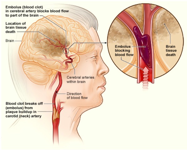
How an ischemic stroke can occur in the brain: If a blood clot breaks away from plaque buildup in the carotid (neck) artery, it can travel to and lodge in an artery in the brain. The clot can block blood flow to a part of the brain, causing brain tissue death.
Image: “The illustration shows how an ischemic stroke can occur in the brain” by National Heart Lung and Blood Institute (NIH). License: Public Domain
Risk factors[10]
HypertensionHypertensionHypertension, or high blood pressure, is a common disease that manifests as elevated systemic arterial pressures. Hypertension is most often asymptomatic and is found incidentally as part of a routine physical examination or during triage for an unrelated medical encounter. Hypertension (most important)
DiabetesDiabetesDiabetes mellitus (DM) is a metabolic disease characterized by hyperglycemia and dysfunction of the regulation of glucose metabolism by insulin. Type 1 DM is diagnosed mostly in children and young adults as the result of autoimmune destruction of β cells in the pancreas and the resulting lack of insulin. Type 2 DM has a significant association with obesity and is characterized by insulin resistance.Diabetes Mellitus mellitus
SmokingSmokingWillful or deliberate act of inhaling and exhaling smoke from burning substances or agents held by hand.Interstitial Lung Diseases
Hyperlipidemia
ObesityObesityObesity is a condition associated with excess body weight, specifically with the deposition of excessive adipose tissue. Obesity is considered a global epidemic. Major influences come from the western diet and sedentary lifestyles, but the exact mechanisms likely include a mixture of genetic and environmental factors. Obesity: The fastest-growing risk factor for stroke between 1990 and 2019 was high BMIBMIAn indicator of body density as determined by the relationship of body weight to body height. Bmi=weight (kg)/height squared (m2). Bmi correlates with body fat (adipose tissue). Their relationship varies with age and gender. For adults, bmi falls into these categories: below 18. 5 (underweight); 18. 5-24. 9 (normal); 25. 0-29. 9 (overweight); 30. 0 and above (obese).Obesity.
Previous history of stroke or transient ischemic attackTransient ischemic attackTransient ischemic attack (TIA) is a temporary episode of neurologic dysfunction caused by ischemia without infarction that resolves completely when blood supply is restored. Transient ischemic attack is a neurologic emergency that warrants urgent medical attention. Transient Ischemic Attack (TIA) (TIATIATransient ischemic attack (TIA) is a temporary episode of neurologic dysfunction caused by ischemia without infarction that resolves completely when blood supply is restored. Transient ischemic attack is a neurologic emergency that warrants urgent medical attention. Transient Ischemic Attack (TIA))
Heart disease:
Atrial fibrillationAtrial fibrillationAtrial fibrillation (AF or Afib) is a supraventricular tachyarrhythmia and the most common kind of arrhythmia. It is caused by rapid, uncontrolled atrial contractions and uncoordinated ventricular responses. Atrial Fibrillation
Valvular heart disease
CardiomyopathyCardiomyopathyCardiomyopathy refers to a group of myocardial diseases associated with structural changes of the heart muscles (myocardium) and impaired systolic and/or diastolic function in the absence of other heart disorders (coronary artery disease, hypertension, valvular disease, and congenital heart disease). Cardiomyopathy: Overview and Types
Older age
Migraine with auraMigraine with AuraA subtype of migraine disorder, characterized by recurrent attacks of reversible neurological symptoms (aura) that precede or accompany the headache. Aura may include a combination of sensory disturbances, such as blurred vision; hallucinations; vertigo; numbness; and difficulty in concentrating and speaking. Aura is usually followed by features of the common migraine, such as photophobia; phonophobia; and nausea.Migraine Headache
HypercoagulableHypercoagulableHypercoagulable states (also referred to as thrombophilias) are a group of hematologic diseases defined by an increased risk of clot formation (i.e., thrombosis) due to either an increase in procoagulants, a decrease in anticoagulants, or a decrease in fibrinolysis. Hypercoagulable States states:
Antiphospholipid syndromeAntiphospholipid syndromeAntiphospholipid syndrome (APLS) is an acquired autoimmune disorder characterized by the persistent presence of antiphospholipid antibodies, which create a hypercoagulable state. These antibodies are most commonly discovered during a workup for a thrombotic event or recurrent pregnancy loss, which are the 2 most common clinical manifestations.Antiphospholipid Syndrome
PregnancyPregnancyThe status during which female mammals carry their developing young (embryos or fetuses) in utero before birth, beginning from fertilization to birth.Pregnancy: Diagnosis, Physiology, and Care
Oral contraceptiveOral contraceptiveCompounds, usually hormonal, taken orally in order to block ovulation and prevent the occurrence of pregnancy. The hormones are generally estrogen or progesterone or both.Benign Liver Tumors use (especially in women > 35 years who smoke)
Genetic conditions: cerebral autosomal dominantAutosomal dominantAutosomal inheritance, both dominant and recessive, refers to the transmission of genes from the 22 autosomal chromosomes. Autosomal dominant diseases are expressed when only 1 copy of the dominant allele is inherited. Autosomal Recessive and Autosomal Dominant Inheritance arteriopathy with subcortical infarcts and leukoencephalopathy (CADASIL)
Blood disorders:
Sickle cell diseaseSickle cell diseaseSickle cell disease (SCD) is a group of genetic disorders in which an abnormal Hb molecule (HbS) transforms RBCs into sickle-shaped cells, resulting in chronic anemia, vasoocclusive episodes, pain, and organ damage.Sickle Cell Disease
Protein C or protein S deficiencyProtein S deficiencyAn autosomal dominant disorder showing decreased levels of plasma protein S antigen or activity, associated with venous thrombosis and pulmonary embolism. Protein s is a vitamin k-dependent plasma protein that inhibits blood clotting by serving as a cofactor for activated protein C (also a vitamin K-dependent protein), and the clinical manifestations of its deficiency are virtually identical to those of protein C deficiency. Treatment with heparin for acute thrombotic processes is usually followed by maintenance administration of coumarin drugs for the prevention of recurrent thrombosis.Hypercoagulable States (usually congenital; can also be acquired)
A complete reduction in cerebral blood flowBlood flowBlood flow refers to the movement of a certain volume of blood through the vasculature over a given unit of time (e.g., mL per minute).Vascular Resistance, Flow, and Mean Arterial Pressure results in the death of brainBrainThe part of central nervous system that is contained within the skull (cranium). Arising from the neural tube, the embryonic brain is comprised of three major parts including prosencephalon (the forebrain); mesencephalon (the midbrain); and rhombencephalon (the hindbrain). The developed brain consists of cerebrum; cerebellum; and other structures in the brain stem.Nervous System: Anatomy, Structure, and Classification tissue within 4–10 minutes:[4,11]
Occlusion of intracranial artery
Reduction in blood flowBlood flowBlood flow refers to the movement of a certain volume of blood through the vasculature over a given unit of time (e.g., mL per minute).Vascular Resistance, Flow, and Mean Arterial Pressure to the region supplied by the vessel
Cerebral tissue starts to undergo ischemiaIschemiaA hypoperfusion of the blood through an organ or tissue caused by a pathologic constriction or obstruction of its blood vessels, or an absence of blood circulation.Ischemic Cell Damage as neuronsNeuronsThe basic cellular units of nervous tissue. Each neuron consists of a body, an axon, and dendrites. Their purpose is to receive, conduct, and transmit impulses in the nervous system.Nervous System: Histology are deprived of glucoseGlucoseA primary source of energy for living organisms. It is naturally occurring and is found in fruits and other parts of plants in its free state. It is used therapeutically in fluid and nutrient replacement.Lactose Intolerance and oxygen → failure of mitochondriaMitochondriaSemiautonomous, self-reproducing organelles that occur in the cytoplasm of all cells of most, but not all, eukaryotes. Each mitochondrion is surrounded by a double limiting membrane. The inner membrane is highly invaginated, and its projections are called cristae. Mitochondria are the sites of the reactions of oxidative phosphorylation, which result in the formation of ATP. They contain distinctive ribosomes, transfer RNAs; amino Acyl tRNA synthetases; and elongation and termination factors. Mitochondria depend upon genes within the nucleus of the cells in which they reside for many essential messenger RNAs. Mitochondria are believed to have arisen from aerobic bacteria that established a symbiotic relationship with primitive protoeukaryotes.The Cell: Organelles to produce ATP
Membrane ion pumps stop functioning due to lack of ATP, and neuronsNeuronsThe basic cellular units of nervous tissue. Each neuron consists of a body, an axon, and dendrites. Their purpose is to receive, conduct, and transmit impulses in the nervous system.Nervous System: Histology depolarize → intracellular calciumCalciumA basic element found in nearly all tissues. It is a member of the alkaline earth family of metals with the atomic symbol ca, atomic number 20, and atomic weight 40. Calcium is the most abundant mineral in the body and combines with phosphorus to form calcium phosphate in the bones and teeth. It is essential for the normal functioning of nerves and muscles and plays a role in blood coagulation (as factor IV) and in many enzymatic processes.Electrolytes increases and glutamateGlutamateDerivatives of glutamic acid. Included under this heading are a broad variety of acid forms, salts, esters, and amides that contain the 2-aminopentanedioic acid structure.Synthesis of Nonessential Amino Acids is released from the presynaptic terminals
Excess extracellular glutamateGlutamateDerivatives of glutamic acid. Included under this heading are a broad variety of acid forms, salts, esters, and amides that contain the 2-aminopentanedioic acid structure.Synthesis of Nonessential Amino Acids induces greater uptake of calciumCalciumA basic element found in nearly all tissues. It is a member of the alkaline earth family of metals with the atomic symbol ca, atomic number 20, and atomic weight 40. Calcium is the most abundant mineral in the body and combines with phosphorus to form calcium phosphate in the bones and teeth. It is essential for the normal functioning of nerves and muscles and plays a role in blood coagulation (as factor IV) and in many enzymatic processes.Electrolytes by neuronsNeuronsThe basic cellular units of nervous tissue. Each neuron consists of a body, an axon, and dendrites. Their purpose is to receive, conduct, and transmit impulses in the nervous system.Nervous System: Histology → neurotoxicity
Free radicalsFree radicalsHighly reactive molecules with an unsatisfied electron valence pair. Free radicals are produced in both normal and pathological processes. They are proven or suspected agents of tissue damage in a wide variety of circumstances including radiation, damage from environment chemicals, and aging. Natural and pharmacological prevention of free radical damage is being actively investigated.Ischemic Cell Damage are produced and accumulate within neuronsNeuronsThe basic cellular units of nervous tissue. Each neuron consists of a body, an axon, and dendrites. Their purpose is to receive, conduct, and transmit impulses in the nervous system.Nervous System: Histology → catalytic destruction of membranes
Infarction or necrosisNecrosisThe death of cells in an organ or tissue due to disease, injury or failure of the blood supply.Ischemic Cell Damage of brainBrainThe part of central nervous system that is contained within the skull (cranium). Arising from the neural tube, the embryonic brain is comprised of three major parts including prosencephalon (the forebrain); mesencephalon (the midbrain); and rhombencephalon (the hindbrain). The developed brain consists of cerebrum; cerebellum; and other structures in the brain stem.Nervous System: Anatomy, Structure, and Classification tissue will occur if blood flowBlood flowBlood flow refers to the movement of a certain volume of blood through the vasculature over a given unit of time (e.g., mL per minute).Vascular Resistance, Flow, and Mean Arterial Pressure is not restored within a few minutes.
An area of ischemic penumbraIschemic PenumbraIschemic Stroke is produced around the area of infarction, where injury and corresponding neurologic dysfunction are still reversible.
Restoration of blood flowBlood flowBlood flow refers to the movement of a certain volume of blood through the vasculature over a given unit of time (e.g., mL per minute).Vascular Resistance, Flow, and Mean Arterial Pressure can induce the formation of more free radicalsFree radicalsHighly reactive molecules with an unsatisfied electron valence pair. Free radicals are produced in both normal and pathological processes. They are proven or suspected agents of tissue damage in a wide variety of circumstances including radiation, damage from environment chemicals, and aging. Natural and pharmacological prevention of free radical damage is being actively investigated.Ischemic Cell Damage and reperfusion injuryReperfusion injuryAdverse functional, metabolic, or structural changes in ischemic tissues resulting from the restoration of blood flow to the tissue (reperfusion), including swelling; hemorrhage; necrosis; and damage from free radicals. The most common instance is myocardial reperfusion injury.Ischemic Cell Damage.
Amount of time passed since symptom onset (the last time seen as “normal”):
If the individual woke up with neurologic symptoms, the last time point considered “normal” is the night before.
Treatment decisions for reperfusion therapy are based on the time the affected individual was last seen without stroke symptoms.
PatientsPatientsIndividuals participating in the health care system for the purpose of receiving therapeutic, diagnostic, or preventive procedures.Clinician–Patient Relationship with “wake-up strokes” (20% of all ischemic strokes) or unknown stroke-onset time may be eligible for reperfusion (based on advanced imaging), even if the last known “normal” time is 4.5–24 hours.[13,20]
Precise account of the event
Cardiovascular disease and other risk factors
Past episodes of TIAs
Medications
Physical examination[1,3,4]
Neurologic exam findings represent ischemiaIschemiaA hypoperfusion of the blood through an organ or tissue caused by a pathologic constriction or obstruction of its blood vessels, or an absence of blood circulation.Ischemic Cell Damage in different vascular territories:
Anterior cerebral arteryAnterior cerebral arteryArtery formed by the bifurcation of the internal carotid artery. Branches of the anterior cerebral artery supply the caudate nucleus; internal capsule; putamen; septal nuclei; gyrus cinguli; and surfaces of the frontal lobe and parietal lobe.Cerebrovascular System: Anatomy:
Lower extremity weakness and sensorySensoryNeurons which conduct nerve impulses to the central nervous system.Nervous System: Histology loss
Limb apraxiaApraxiaA group of cognitive disorders characterized by the inability to perform previously learned skills that cannot be attributed to deficits of motor or sensory function. The two major subtypes of this condition are ideomotor and ideational apraxia, which refers to loss of the ability to mentally formulate the processes involved with performing an action. For example, dressing apraxia may result from an inability to mentally formulate the act of placing clothes on the body. Apraxias are generally associated with lesions of the dominant parietal lobe and supramarginal gyrus.Cranial Nerve Palsies
Incontinence
MCA:
Contralateral hemiparesisHemiparesisThe term hemiparesis refers to mild to moderate weakness involving one side of the body.Epidural Hemorrhage and sensorySensoryNeurons which conduct nerve impulses to the central nervous system.Nervous System: Histology deficits
The face and upper limb are more profoundly affected than the legLegThe lower leg, or just “leg” in anatomical terms, is the part of the lower limb between the knee and the ankle joint. The bony structure is composed of the tibia and fibula bones, and the muscles of the leg are grouped into the anterior, lateral, and posterior compartments by extensions of fascia.Leg: Anatomy.
AphasiaAphasiaA cognitive disorder marked by an impaired ability to comprehend or express language in its written or spoken form. This condition is caused by diseases which affect the language areas of the dominant hemisphere. Clinical features are used to classify the various subtypes of this condition. General categories include receptive, expressive, and mixed forms of aphasia.Ischemic Stroke: inability to understand and utilize language (dominant hemisphere)
Ataxic hemiparesisHemiparesisThe term hemiparesis refers to mild to moderate weakness involving one side of the body.Epidural Hemorrhage and dysarthriaDysarthriaDisorders of speech articulation caused by imperfect coordination of pharynx, larynx, tongue, or face muscles. This may result from cranial nerve diseases; neuromuscular diseases; cerebellar diseases; basal ganglia diseases; brain stem diseases; or diseases of the corticobulbar tracts. The cortical language centers are intact in this condition.Wilson Disease (also seen with lacunar strokeLacunar strokeStroke caused by lacunar infarction or other small vessel diseases of the brain. It features hemiparesis, hemisensory, or hemisensory motor loss.Ischemic Stroke)
Hemianopsia
Posterior cerebral arteryPosterior cerebral arteryArtery formed by the bifurcation of the basilar artery. Branches of the posterior cerebral artery supply portions of the occipital lobe; parietal lobe; inferior temporal gyrus, brainstem, and choroid plexus.Cerebrovascular System: Anatomy:
Posterior inferior cerebellar arteryPosterior inferior cerebellar arteryCerebrovascular System: Anatomy (PICAPicaPica is an eating disorder characterized by a desire or recurrent compulsion to eat substances that are nonnutritive and not food. These compulsions and ingested substances are inappropriate for age or culture. Pica) = Wallenberg’s syndrome
DysphoniaDysphoniaDifficulty and/or pain in phonation or speaking.Epiglottitis (difficulty in phonating speech) and dysphagiaDysphagiaDysphagia is the subjective sensation of difficulty swallowing. Symptoms can range from a complete inability to swallow, to the sensation of solids or liquids becoming “stuck.” Dysphagia is classified as either oropharyngeal or esophageal, with esophageal dysphagia having 2 sub-types: functional and mechanical. Dysphagia (from cranial nervesCranial nervesThere are 12 pairs of cranial nerves (CNs), which run from the brain to various parts of the head, neck, and trunk. The CNs can be sensory or motor or both. The CNs are named and numbered in Roman numerals according to their location, from the front to the back of the brain.The 12 Cranial Nerves: Overview and Functions IX and X being affected)
Ipsilateral ataxiaAtaxiaImpairment of the ability to perform smoothly coordinated voluntary movements. This condition may affect the limbs, trunk, eyes, pharynx, larynx, and other structures. Ataxia may result from impaired sensory or motor function. Sensory ataxia may result from posterior column injury or peripheral nerve diseases. Motor ataxia may be associated with cerebellar diseases; cerebral cortex diseases; thalamic diseases; basal ganglia diseases; injury to the red nucleus; and other conditions.Ataxia-telangiectasia with a tendency to fall to the affected side (inferior cerebellar hemisphere, spinocerebellar fibers, and cerebellar peduncle)
Horner syndromeHorner syndromeHorner syndrome is a condition resulting from an interruption of the sympathetic innervation of the eyes. The syndrome is usually idiopathic but can be directly caused by head and neck trauma, cerebrovascular disease, or a tumor of the CNS. Horner Syndrome: miosisMiosisPupil: Physiology and Abnormalities, ptosisPtosisCranial Nerve Palsies, and anhidrosis (sympathetic fibers)
Ipsilateral painPainAn unpleasant sensation induced by noxious stimuli which are detected by nerve endings of nociceptive neurons.Pain: Types and Pathways and numbness on the face (loss of facial sensation)
Contralateral numbness on the body (as opposed to on the face)
DysarthriaDysarthriaDisorders of speech articulation caused by imperfect coordination of pharynx, larynx, tongue, or face muscles. This may result from cranial nerve diseases; neuromuscular diseases; cerebellar diseases; basal ganglia diseases; brain stem diseases; or diseases of the corticobulbar tracts. The cortical language centers are intact in this condition.Wilson Disease (difficulty in articulating speech)
VertigoVertigoVertigo is defined as the perceived sensation of rotational motion while remaining still. A very common complaint in primary care and the ER, vertigo is more frequently experienced by women and its prevalence increases with age. Vertigo is classified into peripheral or central based on its etiology. Vertigo with nystagmusNystagmusInvoluntary movements of the eye that are divided into two types, jerk and pendular. Jerk nystagmus has a slow phase in one direction followed by a corrective fast phase in the opposite direction, and is usually caused by central or peripheral vestibular dysfunction. Pendular nystagmus features oscillations that are of equal velocity in both directions and this condition is often associated with visual loss early in life.Albinism
NauseaNauseaAn unpleasant sensation in the stomach usually accompanied by the urge to vomit. Common causes are early pregnancy, sea and motion sickness, emotional stress, intense pain, food poisoning, and various enteroviruses.Antiemetics and vomitingVomitingThe forcible expulsion of the contents of the stomach through the mouth.Hypokalemia associated with vertigoVertigoVertigo is defined as the perceived sensation of rotational motion while remaining still. A very common complaint in primary care and the ER, vertigo is more frequently experienced by women and its prevalence increases with age. Vertigo is classified into peripheral or central based on its etiology. Vertigo; sometimes intractable hiccups
Small-vessel infarcts (lacunar infarcts): pure motorMotorNeurons which send impulses peripherally to activate muscles or secretory cells.Nervous System: Histology or sensorySensoryNeurons which conduct nerve impulses to the central nervous system.Nervous System: Histology strokes
Pure motorMotorNeurons which send impulses peripherally to activate muscles or secretory cells.Nervous System: Histology hemiplegia: contralateral ponsPonsThe front part of the hindbrain (rhombencephalon) that lies between the medulla and the midbrain (mesencephalon) ventral to the cerebellum. It is composed of two parts, the dorsal and the ventral. The pons serves as a relay station for neural pathways between the cerebellum to the cerebrum.Brain Stem: Anatomy or internal capsuleCapsuleAn envelope of loose gel surrounding a bacterial cell which is associated with the virulence of pathogenic bacteria. Some capsules have a well-defined border, whereas others form a slime layer that trails off into the medium. Most capsules consist of relatively simple polysaccharides but there are some bacteria whose capsules are made of polypeptides.Bacteroides
Pure sensorySensoryNeurons which conduct nerve impulses to the central nervous system.Nervous System: Histology stroke: lacunar infarctInfarctArea of necrotic cells in an organ, arising mainly from hypoxia and ischemiaIschemic Cell Damage in the contralateral thalamusThalamusThe thalamus is a large, ovoid structure in the dorsal part of the diencephalon that is located between the cerebral cortex and midbrain. It consists of several interconnected nuclei of grey matter separated by the laminae of white matter. The thalamus is the main conductor of information that passes between the cerebral cortex and the periphery, spinal cord, or brain stem.Thalamus: Anatomy
Cardiovascular examinationCardiovascular examinationExamination of the cardiovascular system (CVS) is a critical component of a thorough physical examination. As with all components of a complete physical examination, the CVS examination consists of inspection, palpation, and auscultation. The evaluation of the CVS focuses on the heart, but also includes an assessment of the arterial system throughout the body. Cardiovascular Examination:
Cardiac arrhythmia (e.g., atrial fibrillationAtrial fibrillationAtrial fibrillation (AF or Afib) is a supraventricular tachyarrhythmia and the most common kind of arrhythmia. It is caused by rapid, uncontrolled atrial contractions and uncoordinated ventricular responses. Atrial Fibrillation)
Cardiac murmurs
Carotid bruits
Ophthalmologic examination: fundoscopic signs of hypertensive or diabetic retinopathyDiabetic retinopathyDisease of the retina as a complication of diabetes mellitus. It is characterized by the progressive microvascular complications, such as aneurysm, intraretinal edema, and intraocular pathologic neovascularization.Chronic Diabetic Complications
Diagnosis is made with a suggestive history and clinical examination findings and confirmed by neuroimagingNeuroimagingNon-invasive methods of visualizing the central nervous system, especially the brain, by various imaging modalities.Febrile Infant.
Formal stroke severity ratings are used in all patientsPatientsIndividuals participating in the health care system for the purpose of receiving therapeutic, diagnostic, or preventive procedures.Clinician–Patient Relationship in whom ischemic strokeIschemic StrokeAn ischemic stroke (also known as cerebrovascular accident) is an acute neurologic injury that occurs as a result of brain ischemia; this condition may be due to cerebral blood vessel occlusion by thrombosis or embolism, or rarely due to systemic hypoperfusion. Ischemic Stroke is suspected to assess the degree of neurologic impairment and determine their appropriateness for intervention.
Note the time interval when the test is performed:
Baseline
2 hours post-treatment
24 hours post-onset of symptoms
7–10 days
Administer in the order listed and record the performance in each category:
Level of consciousness
Best gaze-testing of horizontal eye movements
Visual fields
Facial palsyPalsyparalysis of an area of the body, thus incapable of voluntary movementCranial Nerve Palsies
MotorMotorNeurons which send impulses peripherally to activate muscles or secretory cells.Nervous System: Histology evaluation in arms and legs
Limb ataxiaAtaxiaImpairment of the ability to perform smoothly coordinated voluntary movements. This condition may affect the limbs, trunk, eyes, pharynx, larynx, and other structures. Ataxia may result from impaired sensory or motor function. Sensory ataxia may result from posterior column injury or peripheral nerve diseases. Motor ataxia may be associated with cerebellar diseases; cerebral cortex diseases; thalamic diseases; basal ganglia diseases; injury to the red nucleus; and other conditions.Ataxia-telangiectasia
Evaluation of dysarthriaDysarthriaDisorders of speech articulation caused by imperfect coordination of pharynx, larynx, tongue, or face muscles. This may result from cranial nerve diseases; neuromuscular diseases; cerebellar diseases; basal ganglia diseases; brain stem diseases; or diseases of the corticobulbar tracts. The cortical language centers are intact in this condition.Wilson Disease
Unresponsive/comaComaComa is defined as a deep state of unarousable unresponsiveness, characterized by a score of 3 points on the GCS. A comatose state can be caused by a multitude of conditions, making the precise epidemiology and prognosis of coma difficult to determine. Coma
MotorMotorNeurons which send impulses peripherally to activate muscles or secretory cells.Nervous System: Histology function of the arms (left and right tested)
No drift
0
Drift occurs within 10 seconds
1
Falls within 10 seconds
2
No effort against gravity
3
No movement
4
MotorMotorNeurons which send impulses peripherally to activate muscles or secretory cells.Nervous System: Histology function of the legs (left and right tested)
No drift
0
Drift occurs within 10 seconds
1
Falls within 10 seconds
2
No effort against gravity
3
No movement
4
Limb ataxiaAtaxiaImpairment of the ability to perform smoothly coordinated voluntary movements. This condition may affect the limbs, trunk, eyes, pharynx, larynx, and other structures. Ataxia may result from impaired sensory or motor function. Sensory ataxia may result from posterior column injury or peripheral nerve diseases. Motor ataxia may be associated with cerebellar diseases; cerebral cortex diseases; thalamic diseases; basal ganglia diseases; injury to the red nucleus; and other conditions.Ataxia-telangiectasia
Mild aphasiaAphasiaA cognitive disorder marked by an impaired ability to comprehend or express language in its written or spoken form. This condition is caused by diseases which affect the language areas of the dominant hemisphere. Clinical features are used to classify the various subtypes of this condition. General categories include receptive, expressive, and mixed forms of aphasia.Ischemic Stroke
1
Severe aphasiaAphasiaA cognitive disorder marked by an impaired ability to comprehend or express language in its written or spoken form. This condition is caused by diseases which affect the language areas of the dominant hemisphere. Clinical features are used to classify the various subtypes of this condition. General categories include receptive, expressive, and mixed forms of aphasia.Ischemic Stroke
2
Global aphasiaAphasiaA cognitive disorder marked by an impaired ability to comprehend or express language in its written or spoken form. This condition is caused by diseases which affect the language areas of the dominant hemisphere. Clinical features are used to classify the various subtypes of this condition. General categories include receptive, expressive, and mixed forms of aphasia.Ischemic Stroke
3
Articulation
Normal
0
Mild dysarthriaDysarthriaDisorders of speech articulation caused by imperfect coordination of pharynx, larynx, tongue, or face muscles. This may result from cranial nerve diseases; neuromuscular diseases; cerebellar diseases; basal ganglia diseases; brain stem diseases; or diseases of the corticobulbar tracts. The cortical language centers are intact in this condition.Wilson Disease
1
Severe dysarthriaDysarthriaDisorders of speech articulation caused by imperfect coordination of pharynx, larynx, tongue, or face muscles. This may result from cranial nerve diseases; neuromuscular diseases; cerebellar diseases; basal ganglia diseases; brain stem diseases; or diseases of the corticobulbar tracts. The cortical language centers are intact in this condition.Wilson Disease
2
InattentionInattentionAttention Deficit Hyperactivity Disorder or extinctionExtinctionThe procedure of presenting the conditioned stimulus without reinforcement to an organism previously conditioned. It refers also to the diminution of a conditioned response resulting from this procedure.Psychotherapy
Loss of consciousness or syncopeSyncopeSyncope is a short-term loss of consciousness and loss of postural stability followed by spontaneous return of consciousness to the previous neurologic baseline without the need for resuscitation. The condition is caused by transient interruption of cerebral blood flow that may be benign or related to a underlying life-threatening condition. Syncope
CBC including plateletsPlateletsPlatelets are small cell fragments involved in hemostasis. Thrombopoiesis takes place primarily in the bone marrow through a series of cell differentiation and is influenced by several cytokines. Platelets are formed after fragmentation of the megakaryocyte cytoplasm. Platelets: Histology → screen for infection, obtain baseline in case there is bleeding after reperfusion therapy
UrinalysisUrinalysisExamination of urine by chemical, physical, or microscopic means. Routine urinalysis usually includes performing chemical screening tests, determining specific gravity, observing any unusual color or odor, screening for bacteriuria, and examining the sediment microscopically.Urinary Tract Infections (UTIs) in Children → exclude urinary tractUrinary tractThe urinary tract is located in the abdomen and pelvis and consists of the kidneys, ureters, urinary bladder, and urethra. The structures permit the excretion of urine from the body. Urine flows from the kidneys through the ureters to the urinary bladder and out through the urethra.Urinary Tract: Anatomy infection
Chemistries: electrolytesElectrolytesElectrolytes are mineral salts that dissolve in water and dissociate into charged particles called ions, which can be either be positively (cations) or negatively (anions) charged. Electrolytes are distributed in the extracellular and intracellular compartments in different concentrations. Electrolytes are essential for various basic life-sustaining functions.Electrolytes, glucoseGlucoseA primary source of energy for living organisms. It is naturally occurring and is found in fruits and other parts of plants in its free state. It is used therapeutically in fluid and nutrient replacement.Lactose Intolerance, BUN, and creatinine → exclude metabolic causes (e.g., hyponatremiaHyponatremiaHyponatremia is defined as a decreased serum sodium (sNa+) concentration less than 135 mmol/L. Serum sodium is the greatest contributor to plasma osmolality, which is very tightly controlled via antidiuretic hormone (ADH) release from the hypothalamus and by the thirst mechanism.Hyponatremia, hypoglycemiaHypoglycemiaHypoglycemia is an emergency condition defined as a serum glucose level ≤ 70 mg/dL (≤ 3.9 mmol/L) in diabetic patients. In nondiabetic patients, there is no specific or defined limit for normal serum glucose levels, and hypoglycemia is defined mainly by its clinical features. Hypoglycemia, uremiaUremiaA clinical syndrome associated with the retention of renal waste products or uremic toxins in the blood. It is usually the result of renal insufficiency. Most uremic toxins are end products of protein or nitrogen catabolism, such as urea or creatinine. Severe uremia can lead to multiple organ dysfunctions with a constellation of symptoms.Acute Kidney Injury)
Serum lipid profileLipid profileLipid Disorders → provides a baseline, but may not be accurate in acute emergencies
PT and PTT → obtain baseline in preparation for reperfusion therapy
Cardiac enzymesEnzymesEnzymes are complex protein biocatalysts that accelerate chemical reactions without being consumed by them. Due to the body’s constant metabolic needs, the absence of enzymes would make life unsustainable, as reactions would occur too slowly without these molecules. Basics of Enzymes → baseline recommended in all patientsPatientsIndividuals participating in the health care system for the purpose of receiving therapeutic, diagnostic, or preventive procedures.Clinician–Patient Relationship
Chest X-rayX-rayPenetrating electromagnetic radiation emitted when the inner orbital electrons of an atom are excited and release radiant energy. X-ray wavelengths range from 1 pm to 10 nm. Hard x-rays are the higher energy, shorter wavelength x-rays. Soft x-rays or grenz rays are less energetic and longer in wavelength. The short wavelength end of the x-ray spectrum overlaps the gamma rays wavelength range. The distinction between gamma rays and x-rays is based on their radiation source.Pulmonary Function Tests → usefulness in ischemic strokeIschemic StrokeAn ischemic stroke (also known as cerebrovascular accident) is an acute neurologic injury that occurs as a result of brain ischemia; this condition may be due to cerebral blood vessel occlusion by thrombosis or embolism, or rarely due to systemic hypoperfusion. Ischemic Stroke without evidence of concurrent acute pulmonary or cardiac disease is unclear
ECGECGAn electrocardiogram (ECG) is a graphic representation of the electrical activity of the heart plotted against time. Adhesive electrodes are affixed to the skin surface allowing measurement of cardiac impulses from many angles. The ECG provides 3-dimensional information about the conduction system of the heart, the myocardium, and other cardiac structures. Electrocardiogram (ECG) → baseline is recommended in all patientsPatientsIndividuals participating in the health care system for the purpose of receiving therapeutic, diagnostic, or preventive procedures.Clinician–Patient Relationship
NeuroimagingNeuroimagingNon-invasive methods of visualizing the central nervous system, especially the brain, by various imaging modalities.Febrile Infant[2,4,7,13,20,31]
All patientsPatientsIndividuals participating in the health care system for the purpose of receiving therapeutic, diagnostic, or preventive procedures.Clinician–Patient Relationship with a suspected stroke should undergo emergency brainBrainThe part of central nervous system that is contained within the skull (cranium). Arising from the neural tube, the embryonic brain is comprised of three major parts including prosencephalon (the forebrain); mesencephalon (the midbrain); and rhombencephalon (the hindbrain). The developed brain consists of cerebrum; cerebellum; and other structures in the brain stem.Nervous System: Anatomy, Structure, and Classification imaging.
Goals:
To exclude hemorrhagic strokeHemorrhagic strokeStroke due to rupture of a weakened blood vessel in the brain (e.g., cerebral hemispheres; cerebellum; subarachnoid space).Subarachnoid Hemorrhage or stroke mimics (e.g., tumorTumorInflammation)
To identify the vascular lesion causing ischemiaIschemiaA hypoperfusion of the blood through an organ or tissue caused by a pathologic constriction or obstruction of its blood vessels, or an absence of blood circulation.Ischemic Cell Damage
To assess the degree of reversible infarction and estimate the extent of the ischemic penumbraIschemic PenumbraIschemic Stroke (to find potentially salvageable brainBrainThe part of central nervous system that is contained within the skull (cranium). Arising from the neural tube, the embryonic brain is comprised of three major parts including prosencephalon (the forebrain); mesencephalon (the midbrain); and rhombencephalon (the hindbrain). The developed brain consists of cerebrum; cerebellum; and other structures in the brain stem.Nervous System: Anatomy, Structure, and Classification tissue)
Help guide interventions and identify patientsPatientsIndividuals participating in the health care system for the purpose of receiving therapeutic, diagnostic, or preventive procedures.Clinician–Patient Relationship for reperfusion therapies (thrombolysis or mechanical thrombectomyThrombectomySurgical removal of an obstructing clot or foreign material from a blood vessel at the point of its formation. Removal of a clot arising from a distant site is called embolectomy.Vascular Surgery)
Noncontrast head CT scan:
Only for differentiation between ischemic and hemorrhagic strokes
Acute ischemic zones are not visualized (can be mistaken for a normal scan).
Preferred emergency image modality
MRI brainBrainThe part of central nervous system that is contained within the skull (cranium). Arising from the neural tube, the embryonic brain is comprised of three major parts including prosencephalon (the forebrain); mesencephalon (the midbrain); and rhombencephalon (the hindbrain). The developed brain consists of cerebrum; cerebellum; and other structures in the brain stem.Nervous System: Anatomy, Structure, and Classification:
Allows visualization of acute ischemiaIschemiaA hypoperfusion of the blood through an organ or tissue caused by a pathologic constriction or obstruction of its blood vessels, or an absence of blood circulation.Ischemic Cell Damage and ischemic penumbraIschemic PenumbraIschemic Stroke = greater sensitivity
Greater resolution but longer time for imaging
Less availability
Advanced technologies can distinguish potentially salvageable brainBrainThe part of central nervous system that is contained within the skull (cranium). Arising from the neural tube, the embryonic brain is comprised of three major parts including prosencephalon (the forebrain); mesencephalon (the midbrain); and rhombencephalon (the hindbrain). The developed brain consists of cerebrum; cerebellum; and other structures in the brain stem.Nervous System: Anatomy, Structure, and Classification tissue from irreversible infarction:[14,22]
If the last known “normal” time is unknown:
A multimodal CT with angiographyAngiographyRadiography of blood vessels after injection of a contrast medium.Cardiac Surgery (CTACTAA non-invasive method that uses a ct scanner for capturing images of blood vessels and tissues. A contrast material is injected, which helps produce detailed images that aid in diagnosing vascular diseases.Pulmonary Function Tests) and perfusion (CTPCTPPurine and Pyrimidine Metabolism) is indicated.
These patientsPatientsIndividuals participating in the health care system for the purpose of receiving therapeutic, diagnostic, or preventive procedures.Clinician–Patient Relationship may be eligible for endovascular therapy (mechanical thrombectomyThrombectomySurgical removal of an obstructing clot or foreign material from a blood vessel at the point of its formation. Removal of a clot arising from a distant site is called embolectomy.Vascular Surgery) in the extended 6- to 24-hour window.
In patientsPatientsIndividuals participating in the health care system for the purpose of receiving therapeutic, diagnostic, or preventive procedures.Clinician–Patient Relationship with “wake-up strokes” (20% of all ischemic strokes):[13]
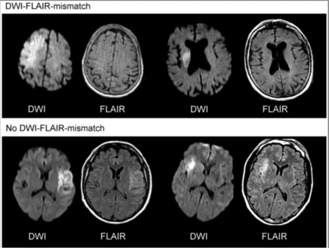
The presence of a DWIDWIImaging of the Head and Brain–FLAIRFLAIRMagnetic Resonance Imaging (MRI) mismatch indicates stroke onset within 4.5 hours and potential benefit with IV tissue plasminogen activatorTissue plasminogen activatorA proteolytic enzyme in the serine protease family found in many tissues which converts plasminogen to fibrinolysin. It has fibrin-binding activity and is immunologically different from urokinase-type plasminogen activator. The primary sequence, composed of 527 amino acids, is identical in both the naturally occurring and synthetic proteases.Hemostasis (tPAtPAIschemic Stroke).
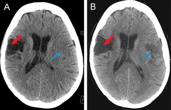
Computed tomogram of an individual with middle cerebral artery stroke illustrating hypodense areas within the temporal and frontal lobes
Image: “CT scan of a patient with middle cerebral artery stroke” by 0475ramosk. License: CC BY-SA 4.0
Imaging approach to identify patients with “wake-up stroke” who are likely to benefit from thrombolysis:
The upper row gives 2 examples of a clearly visible acute ischemic lesion on diffusion-weighted imaging (DWI), while no marked parenchymal hyperintensity is detected on fluid-attenuated inversion recovery (FLAIR) images, indicating DWI–FLAIR mismatch. In the lower row, a clear hyperintensity can be seen on FLAIR images in the area of the acute DWI lesion (no DWI–FLAIR mismatch).
Image: “DWI-FLAIR-mismatch” by Rimmele DL, Thomalia G. License: CC BY 3.0
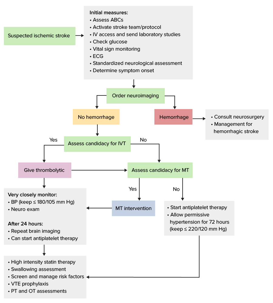
The goal of stroke management is to ensure prompt intervention and optimal outcomes. If possible, restoration of adequate blood flowBlood flowBlood flow refers to the movement of a certain volume of blood through the vasculature over a given unit of time (e.g., mL per minute).Vascular Resistance, Flow, and Mean Arterial Pressure to the injured regions and saving the ischemic penumbraIschemic PenumbraIschemic Stroke from permanent injury should be attempted.
NeurosurgeryNeurosurgeryNeurosurgery is a specialized field focused on the surgical management of pathologies of the brain, spine, spinal cord, and peripheral nerves. General neurosurgery includes cases of trauma and emergencies. There are a number of specialized neurosurgical practices, including oncologic neurosurgery, spinal neurosurgery, and pediatric neurosurgery. Neurosurgery, if evidence of brainBrainThe part of central nervous system that is contained within the skull (cranium). Arising from the neural tube, the embryonic brain is comprised of three major parts including prosencephalon (the forebrain); mesencephalon (the midbrain); and rhombencephalon (the hindbrain). The developed brain consists of cerebrum; cerebellum; and other structures in the brain stem.Nervous System: Anatomy, Structure, and ClassificationedemaEdemaEdema is a condition in which excess serous fluid accumulates in the body cavity or interstitial space of connective tissues. Edema is a symptom observed in several medical conditions. It can be categorized into 2 types, namely, peripheral (in the extremities) and internal (in an organ or body cavity). Edema or herniationHerniationOmphalocele
All patientsPatientsIndividuals participating in the health care system for the purpose of receiving therapeutic, diagnostic, or preventive procedures.Clinician–Patient Relationship with suspected stroke should have an urgent evaluation, including:
NIHSS
BrainBrainThe part of central nervous system that is contained within the skull (cranium). Arising from the neural tube, the embryonic brain is comprised of three major parts including prosencephalon (the forebrain); mesencephalon (the midbrain); and rhombencephalon (the hindbrain). The developed brain consists of cerebrum; cerebellum; and other structures in the brain stem.Nervous System: Anatomy, Structure, and Classification imaging
Finger-stick blood glucoseGlucoseA primary source of energy for living organisms. It is naturally occurring and is found in fruits and other parts of plants in its free state. It is used therapeutically in fluid and nutrient replacement.Lactose Intolerance
Continuous monitoring:
Temperature
Heart rateHeart rateThe number of times the heart ventricles contract per unit of time, usually per minute.Cardiac Physiology
Treat hypoglycemiaHypoglycemiaHypoglycemia is an emergency condition defined as a serum glucose level ≤ 70 mg/dL (≤ 3.9 mmol/L) in diabetic patients. In nondiabetic patients, there is no specific or defined limit for normal serum glucose levels, and hypoglycemia is defined mainly by its clinical features. Hypoglycemia or hyperglycemiaHyperglycemiaAbnormally high blood glucose level.Diabetes Mellitus accordingly (may masquerade as stroke).
IV fluidsIV fluidsIntravenous fluids are one of the most common interventions administered in medicine to approximate physiologic bodily fluids. Intravenous fluids are divided into 2 categories: crystalloid and colloid solutions. Intravenous fluids have a wide variety of indications, including intravascular volume expansion, electrolyte manipulation, and maintenance fluids. Intravenous Fluids
Assess for reperfusion therapy: ideally administered within 60 minutes of arrival to the ED
Those receiving reperfusion therapy should be admitted to a stroke unit or ICUICUHospital units providing continuous surveillance and care to acutely ill patients.West Nile Virus for close monitoring.
Many patientsPatientsIndividuals participating in the health care system for the purpose of receiving therapeutic, diagnostic, or preventive procedures.Clinician–Patient Relationship who present with acute ischemic strokeIschemic StrokeAn ischemic stroke (also known as cerebrovascular accident) is an acute neurologic injury that occurs as a result of brain ischemia; this condition may be due to cerebral blood vessel occlusion by thrombosis or embolism, or rarely due to systemic hypoperfusion. Ischemic Stroke will benefit from restoring blood flowBlood flowBlood flow refers to the movement of a certain volume of blood through the vasculature over a given unit of time (e.g., mL per minute).Vascular Resistance, Flow, and Mean Arterial Pressure to brainBrainThe part of central nervous system that is contained within the skull (cranium). Arising from the neural tube, the embryonic brain is comprised of three major parts including prosencephalon (the forebrain); mesencephalon (the midbrain); and rhombencephalon (the hindbrain). The developed brain consists of cerebrum; cerebellum; and other structures in the brain stem.Nervous System: Anatomy, Structure, and Classification tissue that is ischemic but “salvageable” (not yet infarcted). Those who are treated with either IV thrombolysis or mechanical thrombectomyThrombectomySurgical removal of an obstructing clot or foreign material from a blood vessel at the point of its formation. Removal of a clot arising from a distant site is called embolectomy.Vascular Surgery have improved functional outcomes several months after the acute event.
Diagnosis of ischemic strokeIschemic StrokeAn ischemic stroke (also known as cerebrovascular accident) is an acute neurologic injury that occurs as a result of brain ischemia; this condition may be due to cerebral blood vessel occlusion by thrombosis or embolism, or rarely due to systemic hypoperfusion. Ischemic Stroke causing neurologic deficitsNeurologic DeficitsHigh-Risk Headaches (and not improving)
Absolute contraindicationsContraindicationsA condition or factor associated with a recipient that makes the use of a drug, procedure, or physical agent improper or inadvisable. Contraindications may be absolute (life threatening) or relative (higher risk of complications in which benefits may outweigh risks).Noninvasive Ventilation to thrombolytic therapy:
Intracranial hemorrhageIntracranial hemorrhageSubarachnoid hemorrhage (SAH) is a type of cerebrovascular accident (stroke) resulting from intracranial hemorrhage into the subarachnoid space between the arachnoid and the pia mater layers of the meninges surrounding the brain. Most sahs originate from a saccular aneurysm in the circle of willis but may also occur as a result of trauma, uncontrolled hypertension, vasculitis, anticoagulant use, or stimulant use.Subarachnoid Hemorrhage on CT
Clinical presentation suggestive of subarachnoid hemorrhageSubarachnoid HemorrhageSubarachnoid hemorrhage (SAH) is a type of cerebrovascular accident (stroke) resulting from intracranial hemorrhage into the subarachnoid space between the arachnoid and the pia mater layers of the meninges surrounding the brain. Most SAHs originate from a saccular aneurysm in the circle of Willis but may also occur as a result of trauma, uncontrolled hypertension, vasculitis, anticoagulant use, or stimulant use. Subarachnoid Hemorrhage
NeurosurgeryNeurosurgeryNeurosurgery is a specialized field focused on the surgical management of pathologies of the brain, spine, spinal cord, and peripheral nerves. General neurosurgery includes cases of trauma and emergencies. There are a number of specialized neurosurgical practices, including oncologic neurosurgery, spinal neurosurgery, and pediatric neurosurgery. Neurosurgery, head traumaHead traumaHead trauma occurs when external forces are directed to the skull and brain structures, resulting in damage to the skull, brain, and intracranial structures. Head injuries can be classified as open (penetrating) or closed (blunt), and primary (from the initial trauma) or secondary (indirect brain injury), and range from mild to severe and life-threatening. Head Trauma, or stroke in the previous 3 months
Uncontrolled hypertensionUncontrolled hypertensionAlthough hypertension is defined as a blood pressure of > 130/80 mm Hg, individuals can present with comorbidities of severe asymptomatic or “uncontrolled” hypertension (≥ 180 mm Hg systolic and/or ≥ 120 mm Hg diastolic) that carries with it a significant risk of morbidity and mortality. Uncontrolled Hypertension (systolic BP > 185 mm Hg or diastolic BP > 110 mm Hg)
History of intracranial hemorrhageIntracranial hemorrhageSubarachnoid hemorrhage (SAH) is a type of cerebrovascular accident (stroke) resulting from intracranial hemorrhage into the subarachnoid space between the arachnoid and the pia mater layers of the meninges surrounding the brain. Most sahs originate from a saccular aneurysm in the circle of willis but may also occur as a result of trauma, uncontrolled hypertension, vasculitis, anticoagulant use, or stimulant use.Subarachnoid Hemorrhage
Known intracranial arteriovenous malformationArteriovenous malformationAbnormal formation of blood vessels that shunt arterial blood directly into veins without passing through the capillaries. They usually are crooked, dilated, and with thick vessel walls. A common type is the congenital arteriovenous fistula. The lack of blood flow and oxygen in the capillaries can lead to tissue damage in the affected areas.Erysipelas, neoplasm, or intracranial aneurysmIntracranial aneurysmAbnormal outpouching in the wall of intracranial blood vessels. Most common are the saccular (berry) aneurysms located at branch points in circle of willis at the base of the brain. Vessel rupture results in subarachnoid hemorrhage or intracranial hemorrhages. Giant aneurysms (>2. 5 cm in diameter) may compress adjacent structures, including the oculomotor nerve.Brain Aneurysms
Active internal bleeding
Suspected or confirmed endocarditisEndocarditisEndocarditis is an inflammatory disease involving the inner lining (endometrium) of the heart, most commonly affecting the cardiac valves. Both infectious and noninfectious etiologies lead to vegetations on the valve leaflets. Patients may present with nonspecific symptoms such as fever and fatigue. Endocarditis
Elevated PTT with heparin administered in the past 48 hours or with the use of oral anticoagulantsAnticoagulantsAnticoagulants are drugs that retard or interrupt the coagulation cascade. The primary classes of available anticoagulants include heparins, vitamin K-dependent antagonists (e.g., warfarin), direct thrombin inhibitors, and factor Xa inhibitors. Anticoagulants
GlucoseGlucoseA primary source of energy for living organisms. It is naturally occurring and is found in fruits and other parts of plants in its free state. It is used therapeutically in fluid and nutrient replacement.Lactose Intolerance < 50 mg/dL
Relative contraindicationsContraindicationsA condition or factor associated with a recipient that makes the use of a drug, procedure, or physical agent improper or inadvisable. Contraindications may be absolute (life threatening) or relative (higher risk of complications in which benefits may outweigh risks).Noninvasive Ventilation:
Recent GI or urinary tractUrinary tractThe urinary tract is located in the abdomen and pelvis and consists of the kidneys, ureters, urinary bladder, and urethra. The structures permit the excretion of urine from the body. Urine flows from the kidneys through the ureters to the urinary bladder and out through the urethra.Urinary Tract: Anatomy bleeding (past 21 days)
Minor or rapidly improving stroke symptoms
Major surgery or serious nonhead trauma in the past 14 days
Seizure at stroke onset
Recent arterial puncture at a noncompressible site
Post-MI pericarditisPericarditisPericarditis is an inflammation of the pericardium, often with fluid accumulation. It can be caused by infection (often viral), myocardial infarction, drugs, malignancies, metabolic disorders, autoimmune disorders, or trauma. Acute, subacute, and chronic forms exist. Pericarditis
PregnancyPregnancyThe status during which female mammals carry their developing young (embryos or fetuses) in utero before birth, beginning from fertilization to birth.Pregnancy: Diagnosis, Physiology, and Care
Other additional cautions:
Age > 80 years
History of prior stroke and diabetesDiabetesDiabetes mellitus (DM) is a metabolic disease characterized by hyperglycemia and dysfunction of the regulation of glucose metabolism by insulin. Type 1 DM is diagnosed mostly in children and young adults as the result of autoimmune destruction of β cells in the pancreas and the resulting lack of insulin. Type 2 DM has a significant association with obesity and is characterized by insulin resistance.Diabetes Mellitus
Use of any active anticoagulantsAnticoagulantsAnticoagulants are drugs that retard or interrupt the coagulation cascade. The primary classes of available anticoagulants include heparins, vitamin K-dependent antagonists (e.g., warfarin), direct thrombin inhibitors, and factor Xa inhibitors. Anticoagulants (even with INR < 1.7)
NIHSS score > 25 (severe stroke)
CT with multilobar infarction (hypodensity > ⅓ of a cerebral hemisphere)
Not recommended for mild, nondisabling symptoms.
IVT management:[2,7]
Before IVT administration, lower BP if > 185/110 mm Hg:
LabetalolLabetalolA salicylamide derivative that is a non-cardioselective blocker of beta-adrenergic receptors and alpha-1 adrenergic receptors.Subarachnoid Hemorrhage:
10–20 mg IV over 1–2 minutes
May repeat once
NicardipineNicardipineA potent calcium channel blockader with marked vasodilator action. It has antihypertensive properties and is effective in the treatment of angina and coronary spasms without showing cardiodepressant effects. It has also been used in the treatment of asthma and enhances the action of specific antineoplastic agents.Class 4 Antiarrhythmic Drugs (Calcium Channel Blockers):
5 mg/hr IV
Titrate up by 2.5 mg/hr every 5–15 minutes until desired BP is reached
Do not give tPAtPAIschemic Stroke if BP is not maintained at ≤ 185/110 mm Hg
If potential candidate for mechanical thrombectomyThrombectomySurgical removal of an obstructing clot or foreign material from a blood vessel at the point of its formation. Removal of a clot arising from a distant site is called embolectomy.Vascular Surgery → tenecteplaseTenecteplaseA tissue plasminogen activator enzyme that acts as a fibrinolytic agent; it is used for the dissolution of blood clots, such as those that occur in acute myocardial infarction.Thrombolytics is also an option:
0.25 mg/kg IV bolus
Maximum: 25 mg
Monitor BP closely over the next 24 hours and maintain at ≤ 180/105 mm Hg:
Monitor BP every 15 minutes for 2 hours after the start of tPAtPAIschemic Stroke therapy.
Then monitor BP every 30 minutes for 6 hours.
Then monitor BP every hour for 16 hours.
Monitor for complications (e.g., bleeding, angioedemaAngioedemaAngioedema is a localized, self-limited (but potentially life-threatening), nonpitting, asymmetrical edema occurring in the deep layers of the skin and mucosal tissue. The common underlying pathophysiology involves inflammatory mediators triggering significant vasodilation and increased capillary permeability. Angioedema)
Watch for signs or symptoms:
Severe headacheHeadacheThe symptom of pain in the cranial region. It may be an isolated benign occurrence or manifestation of a wide variety of headache disorders.Brain Abscess
Acute hypertensionHypertensionHypertension, or high blood pressure, is a common disease that manifests as elevated systemic arterial pressures. Hypertension is most often asymptomatic and is found incidentally as part of a routine physical examination or during triage for an unrelated medical encounter. Hypertension
NauseaNauseaAn unpleasant sensation in the stomach usually accompanied by the urge to vomit. Common causes are early pregnancy, sea and motion sickness, emotional stress, intense pain, food poisoning, and various enteroviruses.Antiemetics and/or vomitingVomitingThe forcible expulsion of the contents of the stomach through the mouth.Hypokalemia
Worsening neurologic examination
Send for an emergent head CT if any worrisome changes.
Do not give aspirinAspirinThe prototypical analgesic used in the treatment of mild to moderate pain. It has anti-inflammatory and antipyretic properties and acts as an inhibitor of cyclooxygenase which results in the inhibition of the biosynthesis of prostaglandins. Aspirin also inhibits platelet aggregation and is used in the prevention of arterial and venous thrombosis.Nonsteroidal Antiinflammatory Drugs (NSAIDs) or other antithrombotic agents for the 1st 24 hours after tPAtPAIschemic Stroke.
Avoid/delay indwelling catheterIndwelling catheterCatheters designed to be left within an organ or passage for an extended period of time.Urinary Tract Infections (UTIs) placement and blood draws, when possible.
Endovascular therapy (mechanical thrombectomyThrombectomySurgical removal of an obstructing clot or foreign material from a blood vessel at the point of its formation. Removal of a clot arising from a distant site is called embolectomy.Vascular Surgery):[2,23–28,31]
Background:
About 20% of patientsPatientsIndividuals participating in the health care system for the purpose of receiving therapeutic, diagnostic, or preventive procedures.Clinician–Patient Relationship with acute ischemic strokes have large-vessel occlusion.
Indicated in occlusion of larger vessels (e.g., MCA) regardless of previous IVT administration
Early intervention with mechanical thrombectomyThrombectomySurgical removal of an obstructing clot or foreign material from a blood vessel at the point of its formation. Removal of a clot arising from a distant site is called embolectomy.Vascular Surgery (MT) is safe and can improve functional outcomes.
MT is superior to IVT alone for ischemic strokeIschemic StrokeAn ischemic stroke (also known as cerebrovascular accident) is an acute neurologic injury that occurs as a result of brain ischemia; this condition may be due to cerebral blood vessel occlusion by thrombosis or embolism, or rarely due to systemic hypoperfusion. Ischemic Stroke,even in patientsPatientsIndividuals participating in the health care system for the purpose of receiving therapeutic, diagnostic, or preventive procedures.Clinician–Patient Relationship with clinical pre-stroke disabilityDisabilityDetermination of the degree of a physical, mental, or emotional handicap. The diagnosis is applied to legal qualification for benefits and income under disability insurance and to eligibility for social security and workman’s compensation benefits.ABCDE Assessment.
Timing:
Ideally within 6–16 hours after last known normal
Recent evidence supports expansion of the time window for emergency reperfusion in patientsPatientsIndividuals participating in the health care system for the purpose of receiving therapeutic, diagnostic, or preventive procedures.Clinician–Patient Relationship with “wake-up stroke” or delayed presentation (up to 24 hours from the last symptom-free time).[8,19,26,32]
Maintain BP at ≤ 180/105 mm Hg during the 1st 24 hours.
Antithrombotic therapy[2,31]
Antiplatelet therapy:
Monotherapy:
Indicated if no cardioembolic source at presentation
Options:
AspirinAspirinThe prototypical analgesic used in the treatment of mild to moderate pain. It has anti-inflammatory and antipyretic properties and acts as an inhibitor of cyclooxygenase which results in the inhibition of the biosynthesis of prostaglandins. Aspirin also inhibits platelet aggregation and is used in the prevention of arterial and venous thrombosis.Nonsteroidal Antiinflammatory Drugs (NSAIDs): 162–325 mg daily (can also be given rectally for patientsPatientsIndividuals participating in the health care system for the purpose of receiving therapeutic, diagnostic, or preventive procedures.Clinician–Patient Relationship with dysphagiaDysphagiaDysphagia is the subjective sensation of difficulty swallowing. Symptoms can range from a complete inability to swallow, to the sensation of solids or liquids becoming “stuck.” Dysphagia is classified as either oropharyngeal or esophageal, with esophageal dysphagia having 2 sub-types: functional and mechanical. Dysphagia)
ClopidogrelClopidogrelA ticlopidine analog and platelet purinergic p2y receptor antagonist that inhibits adenosine diphosphate-mediated platelet aggregation. It is used to prevent thromboembolism in patients with arterial occlusive diseases; myocardial infarction; stroke; or atrial fibrillation.Antiplatelet Drugs: 75 mg daily
Dual antiplatelet therapy:
Indications:
Minor ischemic strokeIschemic StrokeAn ischemic stroke (also known as cerebrovascular accident) is an acute neurologic injury that occurs as a result of brain ischemia; this condition may be due to cerebral blood vessel occlusion by thrombosis or embolism, or rarely due to systemic hypoperfusion. Ischemic Stroke (NIHSS < 3) with no cardioembolic source who did not receive tPAtPAIschemic Stroke
Large intracranial artery atherosclerosisAtherosclerosisAtherosclerosis is a common form of arterial disease in which lipid deposition forms a plaque in the blood vessel walls. Atherosclerosis is an incurable disease, for which there are clearly defined risk factors that often can be reduced through a change in lifestyle and behavior of the patient. Atherosclerosis
Options:
AspirinAspirinThe prototypical analgesic used in the treatment of mild to moderate pain. It has anti-inflammatory and antipyretic properties and acts as an inhibitor of cyclooxygenase which results in the inhibition of the biosynthesis of prostaglandins. Aspirin also inhibits platelet aggregation and is used in the prevention of arterial and venous thrombosis.Nonsteroidal Antiinflammatory Drugs (NSAIDs) plus clopidogrelClopidogrelA ticlopidine analog and platelet purinergic p2y receptor antagonist that inhibits adenosine diphosphate-mediated platelet aggregation. It is used to prevent thromboembolism in patients with arterial occlusive diseases; myocardial infarction; stroke; or atrial fibrillation.Antiplatelet Drugs
AspirinAspirinThe prototypical analgesic used in the treatment of mild to moderate pain. It has anti-inflammatory and antipyretic properties and acts as an inhibitor of cyclooxygenase which results in the inhibition of the biosynthesis of prostaglandins. Aspirin also inhibits platelet aggregation and is used in the prevention of arterial and venous thrombosis.Nonsteroidal Antiinflammatory Drugs (NSAIDs) plus ticagrelorTicagrelorAn adenosine triphosphate analogue and reversible p2y12 purinoceptor antagonist that inhibits adp-mediated platelet aggregation. It is used for the prevention of thromboembolism by patients with acute coronary syndrome or a history of myocardial infarction.Antiplatelet Drugs
Duration:
Generally given for 21 days for minor stroke
May be extended to 90 days in cases of large intracranial artery atherosclerosisAtherosclerosisAtherosclerosis is a common form of arterial disease in which lipid deposition forms a plaque in the blood vessel walls. Atherosclerosis is an incurable disease, for which there are clearly defined risk factors that often can be reduced through a change in lifestyle and behavior of the patient. Atherosclerosis
At that point, switch to indefinite aspirinAspirinThe prototypical analgesic used in the treatment of mild to moderate pain. It has anti-inflammatory and antipyretic properties and acts as an inhibitor of cyclooxygenase which results in the inhibition of the biosynthesis of prostaglandins. Aspirin also inhibits platelet aggregation and is used in the prevention of arterial and venous thrombosis.Nonsteroidal Antiinflammatory Drugs (NSAIDs) or clopidogrelClopidogrelA ticlopidine analog and platelet purinergic p2y receptor antagonist that inhibits adenosine diphosphate-mediated platelet aggregation. It is used to prevent thromboembolism in patients with arterial occlusive diseases; myocardial infarction; stroke; or atrial fibrillation.Antiplatelet Drugs monotherapy.
Can start within 4–14 days after stroke onset (depends on severity; exact timing is controversial and still under investigation)
Consider delaying if high risk for hemorrhagic transformationTransformationChange brought about to an organism’s genetic composition by unidirectional transfer (transfection; transduction, genetic; conjugation, genetic, etc.) and incorporation of foreign DNA into prokaryotic or eukaryotic cells by recombination of part or all of that DNA into the cell’s genome.Bacteriology
Patient already on anticoagulationAnticoagulationPulmonary Hypertension Drugs at the time of ischemic strokeIschemic StrokeAn ischemic stroke (also known as cerebrovascular accident) is an acute neurologic injury that occurs as a result of brain ischemia; this condition may be due to cerebral blood vessel occlusion by thrombosis or embolism, or rarely due to systemic hypoperfusion. Ischemic Stroke:
Temporarily stop:
While assessing eligibility for reperfusion therapy
If risk for hemorrhagic transformationTransformationChange brought about to an organism’s genetic composition by unidirectional transfer (transfection; transduction, genetic; conjugation, genetic, etc.) and incorporation of foreign DNA into prokaryotic or eukaryotic cells by recombination of part or all of that DNA into the cell’s genome.Bacteriology (large stroke area) → can start aspirinAspirinThe prototypical analgesic used in the treatment of mild to moderate pain. It has anti-inflammatory and antipyretic properties and acts as an inhibitor of cyclooxygenase which results in the inhibition of the biosynthesis of prostaglandins. Aspirin also inhibits platelet aggregation and is used in the prevention of arterial and venous thrombosis.Nonsteroidal Antiinflammatory Drugs (NSAIDs) in the meantime
Can restart when patient stable (typically 24–48 hours after onset of stroke)
Management when ineligible for reperfusion therapy
Antithrombotic therapy (if not eligible for reperfusion therapy):[2]
Start aspirinAspirinThe prototypical analgesic used in the treatment of mild to moderate pain. It has anti-inflammatory and antipyretic properties and acts as an inhibitor of cyclooxygenase which results in the inhibition of the biosynthesis of prostaglandins. Aspirin also inhibits platelet aggregation and is used in the prevention of arterial and venous thrombosis.Nonsteroidal Antiinflammatory Drugs (NSAIDs) or other antiplatelet agentsAntiplatelet agentsAntiplatelet agents are medications that inhibit platelet aggregation, a critical step in the formation of the initial platelet plug. Abnormal, or inappropriate, platelet aggregation is a key step in the pathophysiology of arterial ischemic events. The primary categories of antiplatelet agents include aspirin, ADP inhibitors, phosphodiesterase/adenosine uptake inhibitors, and glycoprotein IIb/IIIa inhibitors. Antiplatelet Drugs within 24–48 hours after symptom onset (after imaging confirms no hemorrhage):
No need to wait for imaging results if patient is not a candidate for reperfusion therapy.
Continuous BP monitoring:[2,7]
Avoid BP reduction in the 1st 72 hours unless systolic BP > 220 mm Hg or diastolic BP > 120 mm Hg
Thereafter, lower to 140/90 mm Hg
Subsequent management and rehabilitation[2,30,31]
Initiate lipid lowering with high-intensity statin therapy
Thigh-length intermittent pneumatic compressionCompressionBlunt Chest Trauma: Start within 72 hours of acute ischemic strokeIschemic StrokeAn ischemic stroke (also known as cerebrovascular accident) is an acute neurologic injury that occurs as a result of brain ischemia; this condition may be due to cerebral blood vessel occlusion by thrombosis or embolism, or rarely due to systemic hypoperfusion. Ischemic Stroke onset in patientsPatientsIndividuals participating in the health care system for the purpose of receiving therapeutic, diagnostic, or preventive procedures.Clinician–Patient Relationship with restricted mobility.
EnoxaparinEnoxaparinLow-molecular-weight fragment of heparin, having a 4-enopyranosuronate sodium structure at the non-reducing end of the chain. It is prepared by depolymerization of the benzylic ester of porcine mucosal heparin. Therapeutically, it is used as an antithrombotic agent.Anticoagulants 40 mg SC once daily, or
DalteparinDalteparinA low-molecular-weight fragment of heparin, prepared by nitrous acid depolymerization of porcine mucosal heparin. The mean molecular weight is 4000-6000 daltons. It is used therapeutically as an antithrombotic agent.Anticoagulants 5,000 units SC once daily, or
Low-dose unfractionated heparinUnfractionated heparinA highly acidic mucopolysaccharide formed of equal parts of sulfated d-glucosamine and d-glucuronic acid with sulfaminic bridges. The molecular weight ranges from six to twenty thousand. Heparin occurs in and is obtained from liver, lung, mast cells, etc. , of vertebrates. Its function is unknown, but it is used to prevent blood clotting in vivo and vitro, in the form of many different salts.Anticoagulants: 5,000 units 2–3 times daily
Caution in patientsPatientsIndividuals participating in the health care system for the purpose of receiving therapeutic, diagnostic, or preventive procedures.Clinician–Patient Relationship with significant bleeding risk
Screen for comorbiditiesComorbiditiesThe presence of co-existing or additional diseases with reference to an initial diagnosis or with reference to the index condition that is the subject of study. Comorbidity may affect the ability of affected individuals to function and also their survival; it may be used as a prognostic indicator for length of hospital stay, cost factors, and outcome or survival.St. Louis Encephalitis Virus or causes:
Cardiac monitoring (for at least 24 hours) → assess for potential atrial fibrillationAtrial fibrillationAtrial fibrillation (AF or Afib) is a supraventricular tachyarrhythmia and the most common kind of arrhythmia. It is caused by rapid, uncontrolled atrial contractions and uncoordinated ventricular responses. Atrial Fibrillation
EchocardiographyEchocardiographyUltrasonic recording of the size, motion, and composition of the heart and surrounding tissues. The standard approach is transthoracic.Tricuspid Valve Atresia (TVA) → detect intracardiacIntracardiacTotal Anomalous Pulmonary Venous Return (TAPVR) thrombus or patent foramen ovaleForamen ovaleAn opening in the wall between the right and the left upper chambers (heart atria) of a fetal heart. Oval foramen normally closes soon after birth; when it fails to close the condition is called patent oval foramen.Patent Foramen Ovale
Carotid imaging (US, CTACTAA non-invasive method that uses a ct scanner for capturing images of blood vessels and tissues. A contrast material is injected, which helps produce detailed images that aid in diagnosing vascular diseases.Pulmonary Function Tests) → evaluate for carotid stenosisStenosisHypoplastic Left Heart Syndrome (HLHS)
GlucoseGlucoseA primary source of energy for living organisms. It is naturally occurring and is found in fruits and other parts of plants in its free state. It is used therapeutically in fluid and nutrient replacement.Lactose Intolerance and hemoglobin A1c → screen for diabetesDiabetesDiabetes mellitus (DM) is a metabolic disease characterized by hyperglycemia and dysfunction of the regulation of glucose metabolism by insulin. Type 1 DM is diagnosed mostly in children and young adults as the result of autoimmune destruction of β cells in the pancreas and the resulting lack of insulin. Type 2 DM has a significant association with obesity and is characterized by insulin resistance.Diabetes Mellitus
Lipid panel → assess for hyperlipidemia
PT to improve neurologic function and reduce the risk of contracturesContracturesProlonged shortening of the muscle or other soft tissue around a joint, preventing movement of the joint.Wound Healing
Occupational therapyOccupational TherapySkilled treatment that helps individuals achieve independence in all facets of their lives. It assists in the development of skills needed for independent living.Fetal Alcohol Spectrum Disorder:
Improves the qualityQualityActivities and programs intended to assure or improve the quality of care in either a defined medical setting or a program. The concept includes the assessment or evaluation of the quality of care; identification of problems or shortcomings in the delivery of care; designing activities to overcome these deficiencies; and follow-up monitoring to ensure effectiveness of corrective steps.Quality Measurement and Improvement of life
May need splinting of hands to prevent contracturesContracturesProlonged shortening of the muscle or other soft tissue around a joint, preventing movement of the joint.Wound Healing
SwallowingSwallowingThe act of taking solids and liquids into the gastrointestinal tract through the mouth and throat.Gastrointestinal Motility assessment to prevent aspiration
If persistent dysphagiaDysphagiaDysphagia is the subjective sensation of difficulty swallowing. Symptoms can range from a complete inability to swallow, to the sensation of solids or liquids becoming “stuck.” Dysphagia is classified as either oropharyngeal or esophageal, with esophageal dysphagia having 2 sub-types: functional and mechanical. Dysphagia → evaluate for percutaneous gastrostomy tube placementTube placementSurgical procedure involving the creation of an opening (stoma) into the chest cavity for drainage; used in the treatment of pleural effusion; pneumothorax; hemothorax; and empyema.Thoracic Surgery
Nutrition consultation:
Can assist with optimal tube feeding options (if applicable)
Supplementation may be necessary for those at risk of malnourishment.
Speech therapySpeech TherapyTreatment for individuals with speech defects and disorders that involves counseling and use of various exercises and AIDS to help the development of new speech habits.Myotonic Dystrophies
Pay careful attentionAttentionFocusing on certain aspects of current experience to the exclusion of others. It is the act of heeding or taking notice or concentrating.Psychiatric Assessment to prevent complications due to immobility:
PneumoniaPneumoniaPneumonia or pulmonary inflammation is an acute or chronic inflammation of lung tissue. Causes include infection with bacteria, viruses, or fungi. In more rare cases, pneumonia can also be caused through toxic triggers through inhalation of toxic substances, immunological processes, or in the course of radiotherapy.Pneumonia
Pressure ulcers
Limb contracturesContracturesProlonged shortening of the muscle or other soft tissue around a joint, preventing movement of the joint.Wound Healing
Careful assessment of the ability to drive safely
Family and patient education:
Continuation of preventive measures at home
The FAST mnemonic is helpful for lay individuals to identify recurrence:
SmokingSmokingWillful or deliberate act of inhaling and exhaling smoke from burning substances or agents held by hand.Interstitial Lung Diseases cessation counseling if applicable
Risk-factor management with improved control of:
DiabetesDiabetesDiabetes mellitus (DM) is a metabolic disease characterized by hyperglycemia and dysfunction of the regulation of glucose metabolism by insulin. Type 1 DM is diagnosed mostly in children and young adults as the result of autoimmune destruction of β cells in the pancreas and the resulting lack of insulin. Type 2 DM has a significant association with obesity and is characterized by insulin resistance.Diabetes Mellitus
HypertensionHypertensionHypertension, or high blood pressure, is a common disease that manifests as elevated systemic arterial pressures. Hypertension is most often asymptomatic and is found incidentally as part of a routine physical examination or during triage for an unrelated medical encounter. Hypertension
Hyperlipidemia
Medications:
Dual antiplatelet therapy unless contraindicated
AspirinAspirinThe prototypical analgesic used in the treatment of mild to moderate pain. It has anti-inflammatory and antipyretic properties and acts as an inhibitor of cyclooxygenase which results in the inhibition of the biosynthesis of prostaglandins. Aspirin also inhibits platelet aggregation and is used in the prevention of arterial and venous thrombosis.Nonsteroidal Antiinflammatory Drugs (NSAIDs)
ClopidogrelClopidogrelA ticlopidine analog and platelet purinergic p2y receptor antagonist that inhibits adenosine diphosphate-mediated platelet aggregation. It is used to prevent thromboembolism in patients with arterial occlusive diseases; myocardial infarction; stroke; or atrial fibrillation.Antiplatelet Drugs or extended-release dipyridamoleDipyridamoleA phosphodiesterase inhibitor that blocks uptake and metabolism of adenosine by erythrocytes and vascular endothelial cells. Dipyridamole also potentiates the antiaggregating action of prostacyclin.Phosphodiesterase Inhibitors
StatinsStatinsStatins are competitive inhibitors of HMG-CoA reductase in the liver. HMG-CoA reductase is the rate-limiting step in cholesterol synthesis. Inhibition results in lowered intrahepatocytic cholesterol formation, resulting in up-regulation of LDL receptors and, ultimately, lowering levels of serum LDL and triglycerides.Statins (e.g., atorvastatinAtorvastatinA pyrrole and heptanoic acid derivative, hydroxymethylglutaryl-CoA reductase inhibitor (statin), and anticholesteremic agent that is used to reduce serum levels of ldl-cholesterol; apolipoprotein b; and triglycerides. It is used to increase serum levels of hdl-cholesterol in the treatment of hyperlipidemias, and for the prevention of cardiovascular diseases in patients with multiple risk factors.Statins) in clinically important atherosclerotic disease
Treatment of underlying causes (e.g., atrial fibrillationAtrial fibrillationAtrial fibrillation (AF or Afib) is a supraventricular tachyarrhythmia and the most common kind of arrhythmia. It is caused by rapid, uncontrolled atrial contractions and uncoordinated ventricular responses. Atrial Fibrillation) and anticoagulationAnticoagulationPulmonary Hypertension Drugs as indicated
Emerging therapies[15–18]
Further human study and replication of recent studies are needed before these are available.
Neuroprotective treatments are in clinical trials.
Remote ischemic conditioning uses repeated occlusion/release cycles to induce ischemiaIschemiaA hypoperfusion of the blood through an organ or tissue caused by a pathologic constriction or obstruction of its blood vessels, or an absence of blood circulation.Ischemic Cell Damage in 1 area (e.g., bilateral upper limb arteriesArteriesArteries are tubular collections of cells that transport oxygenated blood and nutrients from the heart to the tissues of the body. The blood passes through the arteries in order of decreasing luminal diameter, starting in the largest artery (the aorta) and ending in the small arterioles. Arteries are classified into 3 types: large elastic arteries, medium muscular arteries, and small arteries and arterioles. Arteries: Histology), hopefully leading to ischemic toleranceTolerancePharmacokinetics and Pharmacodynamics in other organs (i.e., brainBrainThe part of central nervous system that is contained within the skull (cranium). Arising from the neural tube, the embryonic brain is comprised of three major parts including prosencephalon (the forebrain); mesencephalon (the midbrain); and rhombencephalon (the hindbrain). The developed brain consists of cerebrum; cerebellum; and other structures in the brain stem.Nervous System: Anatomy, Structure, and Classification).
Nerinetide (a drug that acts by targeting a postsynaptic scaffolding protein) and remote ischemic perconditioning are being studied in combination with reperfusion therapy in clinical trials.
Nanomedicines and adaptations of diverse techniques are being evaluated to maximize drug efficiency in targeting ischemic brainBrainThe part of central nervous system that is contained within the skull (cranium). Arising from the neural tube, the embryonic brain is comprised of three major parts including prosencephalon (the forebrain); mesencephalon (the midbrain); and rhombencephalon (the hindbrain). The developed brain consists of cerebrum; cerebellum; and other structures in the brain stem.Nervous System: Anatomy, Structure, and Classification tissues.
Adjunct intra-arterial alteplaseAlteplaseThrombolytics after successful reperfusion following MT
Acute intracranial stenting in addition to MT[29]
Complications and prognosisPrognosisA prediction of the probable outcome of a disease based on a individual’s condition and the usual course of the disease as seen in similar situations.Non-Hodgkin Lymphomas[5]
The amount of time to reperfusion is not the only important factor in patientsPatientsIndividuals participating in the health care system for the purpose of receiving therapeutic, diagnostic, or preventive procedures.Clinician–Patient Relationship with stroke; the blood circulationCirculationThe movement of the blood as it is pumped through the cardiovascular system.ABCDE Assessment pattern/collateral status is also an important determinant of outcome.
Complications:
Cerebral edemaCerebral edemaIncreased intracellular or extracellular fluid in brain tissue. Cytotoxic brain edema (swelling due to increased intracellular fluid) is indicative of a disturbance in cell metabolism, and is commonly associated with hypoxic or ischemic injuries. An increase in extracellular fluid may be caused by increased brain capillary permeability (vasogenic edema), an osmotic gradient, local blockages in interstitial fluid pathways, or by obstruction of CSF flow (e.g., obstructive hydrocephalus).Increased Intracranial Pressure (ICP):
Accumulation of water in the cerebral parenchyma if ischemic injury goes without management for the 1st few days
May lead to obstructing hydrocephalusHydrocephalusExcessive accumulation of cerebrospinal fluid within the cranium which may be associated with dilation of cerebral ventricles, intracranial.Subarachnoid Hemorrhage or brainBrainThe part of central nervous system that is contained within the skull (cranium). Arising from the neural tube, the embryonic brain is comprised of three major parts including prosencephalon (the forebrain); mesencephalon (the midbrain); and rhombencephalon (the hindbrain). The developed brain consists of cerebrum; cerebellum; and other structures in the brain stem.Nervous System: Anatomy, Structure, and ClassificationherniationHerniationOmphalocele
Myocardial infarctionMyocardial infarctionMI is ischemia and death of an area of myocardial tissue due to insufficient blood flow and oxygenation, usually from thrombus formation on a ruptured atherosclerotic plaque in the epicardial arteries. Clinical presentation is most commonly with chest pain, but women and patients with diabetes may have atypical symptoms.Myocardial Infarction
Heart failureHeart FailureA heterogeneous condition in which the heart is unable to pump out sufficient blood to meet the metabolic need of the body. Heart failure can be caused by structural defects, functional abnormalities (ventricular dysfunction), or a sudden overload beyond its capacity. Chronic heart failure is more common than acute heart failure which results from sudden insult to cardiac function, such as myocardial infarction.Total Anomalous Pulmonary Venous Return (TAPVR)
DysphagiaDysphagiaDysphagia is the subjective sensation of difficulty swallowing. Symptoms can range from a complete inability to swallow, to the sensation of solids or liquids becoming “stuck.” Dysphagia is classified as either oropharyngeal or esophageal, with esophageal dysphagia having 2 sub-types: functional and mechanical. Dysphagia
Aspiration pneumoniaAspiration pneumoniaA type of lung inflammation resulting from the aspiration of food, liquid, or gastric contents into the upper respiratory tract.Pneumonia
Medical problems associated with immobility:
Pressure sores
Orthopedic contracturesContracturesProlonged shortening of the muscle or other soft tissue around a joint, preventing movement of the joint.Wound Healing
PrognosisPrognosisA prediction of the probable outcome of a disease based on a individual’s condition and the usual course of the disease as seen in similar situations.Non-Hodgkin Lymphomas depends on these predictors of outcomes:[19,25]
Stroke severity/extent of infarction
Age
Time elapsed from symptom onset to diagnosis and management
Length and intensity of therapy
Collateral blood circulationCirculationThe movement of the blood as it is pumped through the cardiovascular system.ABCDE Assessment pattern
TIATIATransient ischemic attack (TIA) is a temporary episode of neurologic dysfunction caused by ischemia without infarction that resolves completely when blood supply is restored. Transient ischemic attack is a neurologic emergency that warrants urgent medical attention. Transient Ischemic Attack (TIA): a temporary episode of neurologic dysfunction caused by ischemiaIschemiaA hypoperfusion of the blood through an organ or tissue caused by a pathologic constriction or obstruction of its blood vessels, or an absence of blood circulation.Ischemic Cell Damage without evidence of infarction that resolves completely when blood supply is restored. No time limitLimitA value (e.g., pressure or time) that should not be exceeded and which is specified by the operator to protect the lungInvasive Mechanical Ventilation is defined as was used previously. Causes may be small clots or thromboemboli imposed on blood vessels compromised by atherosclerosisAtherosclerosisAtherosclerosis is a common form of arterial disease in which lipid deposition forms a plaque in the blood vessel walls. Atherosclerosis is an incurable disease, for which there are clearly defined risk factors that often can be reduced through a change in lifestyle and behavior of the patient. Atherosclerosis, inflammationInflammationInflammation is a complex set of responses to infection and injury involving leukocytes as the principal cellular mediators in the body’s defense against pathogenic organisms. Inflammation is also seen as a response to tissue injury in the process of wound healing. The 5 cardinal signs of inflammation are pain, heat, redness, swelling, and loss of function. Inflammation, or amyloid, or due to inadequate cerebral blood flowInadequate Cerebral Blood FlowSyncope due to vasoconstrictionVasoconstrictionThe physiological narrowing of blood vessels by contraction of the vascular smooth muscle.Vascular Resistance, Flow, and Mean Arterial Pressure or severe hypotensionHypotensionHypotension is defined as low blood pressure, specifically < 90/60 mm Hg, and is most commonly a physiologic response. Hypotension may be mild, serious, or life threatening, depending on the cause. Hypotension. Clinical presentation is with transient neurologic deficitsNeurologic DeficitsHigh-Risk Headaches that resolve spontaneously. Management includes risk-factor reduction to reduce the risk of a future stroke.
Hemorrhagic strokeHemorrhagic strokeStroke due to rupture of a weakened blood vessel in the brain (e.g., cerebral hemispheres; cerebellum; subarachnoid space).Subarachnoid Hemorrhage: a stroke with resulting neurologic dysfunction caused by rupture and bleeding of an intracranial vessel. Hemorrhagic strokeHemorrhagic strokeStroke due to rupture of a weakened blood vessel in the brain (e.g., cerebral hemispheres; cerebellum; subarachnoid space).Subarachnoid Hemorrhage is subdivided into intracerebral hemorrhageIntracerebral HemorrhageIntracerebral hemorrhage (ICH) refers to a spontaneous or traumatic bleed into the brain parenchyma and is the 2nd-most common cause of cerebrovascular accidents (CVAs), commonly known as stroke, after ischemic CVAs. Intracerebral Hemorrhage and subarachnoid (intracranial) hemorrhage. Clinically, hemorrhages are indistinguishable from ischemic strokeIschemic StrokeAn ischemic stroke (also known as cerebrovascular accident) is an acute neurologic injury that occurs as a result of brain ischemia; this condition may be due to cerebral blood vessel occlusion by thrombosis or embolism, or rarely due to systemic hypoperfusion. Ischemic Stroke and require imaging for definitive diagnosis. Management may include mechanical ventilationVentilationThe total volume of gas inspired or expired per unit of time, usually measured in liters per minute.Ventilation: Mechanics of Breathing, blood pressure control, reversal of anticoagulationAnticoagulationPulmonary Hypertension Drugs, and risk-factor reduction for subsequent strokes.
MigraineMigraineMigraine headache is a primary headache disorder and is among the most prevalent disorders in the world. Migraine is characterized by episodic, moderate to severe headaches that may be associated with increased sensitivity to light and sound, as well as nausea and/or vomiting. Migraine Headache: a common primary headacheHeadacheThe symptom of pain in the cranial region. It may be an isolated benign occurrence or manifestation of a wide variety of headache disorders.Brain Abscess disorder characterized by episodic, moderate-to-severe headaches that may be associated with increased sensitivity to light and sound, and nauseaNauseaAn unpleasant sensation in the stomach usually accompanied by the urge to vomit. Common causes are early pregnancy, sea and motion sickness, emotional stress, intense pain, food poisoning, and various enteroviruses.Antiemetics and/or vomitingVomitingThe forcible expulsion of the contents of the stomach through the mouth.Hypokalemia. Migraines may present as a “stroke mimic” with vasoconstrictionVasoconstrictionThe physiological narrowing of blood vessels by contraction of the vascular smooth muscle.Vascular Resistance, Flow, and Mean Arterial Pressure, causing ischemiaIschemiaA hypoperfusion of the blood through an organ or tissue caused by a pathologic constriction or obstruction of its blood vessels, or an absence of blood circulation.Ischemic Cell Damage and neurologic deficitsNeurologic DeficitsHigh-Risk Headaches similar to those seen in an ischemic strokeIschemic StrokeAn ischemic stroke (also known as cerebrovascular accident) is an acute neurologic injury that occurs as a result of brain ischemia; this condition may be due to cerebral blood vessel occlusion by thrombosis or embolism, or rarely due to systemic hypoperfusion. Ischemic Stroke, including hemiplegia. Diagnosis is by clinical history, physical exam, and imaging. Management includes medications to treat migraines and by avoiding triggers.
Intracranial tumorTumorInflammation: can be a primary neoplasm derived from intracranial tissues (astrocytomaAstrocytomaAstrocytomas are neuroepithelial tumors that arise from astrocytes, which are star-shaped glial cells (supporting tissues of the CNS). Astrocytomas are a type of glioma. There are 4 grades of astrocytomas. Astrocytoma, oligodendrogliomaOligodendrogliomaOligodendrogliomas are malignant CNS tumors arising from neural glial cell precursors. Oligodendrogliomas often arise in the frontal lobes of the brain and have a generally favorable prognosis when compared to other gliomas. Oligodendrogliomas are the 3rd most common CNS tumor. The most frequent presenting symptom is a seizure.Oligodendroglioma, meningiomaMeningiomaMeningiomas are slow-growing tumors that arise from the meninges of the brain and spinal cord. The vast majority are benign. These tumors commonly occur in individuals with a history of high doses of skull radiation, head trauma, and neurofibromatosis 2. Meningioma) or a metastatic process from other malignancies such as lung or breast cancerBreast cancerBreast cancer is a disease characterized by malignant transformation of the epithelial cells of the breast. Breast cancer is the most common form of cancer and 2nd most common cause of cancer-related death among women. Breast Cancer. As the massMassThree-dimensional lesion that occupies a space within the breastImaging of the Breast grows, neurologic presentations similar to those seen in ischemic strokeIschemic StrokeAn ischemic stroke (also known as cerebrovascular accident) is an acute neurologic injury that occurs as a result of brain ischemia; this condition may be due to cerebral blood vessel occlusion by thrombosis or embolism, or rarely due to systemic hypoperfusion. Ischemic Stroke may develop. Diagnosis is by imaging, and management includes addressing the underlying cause.
BrainBrainThe part of central nervous system that is contained within the skull (cranium). Arising from the neural tube, the embryonic brain is comprised of three major parts including prosencephalon (the forebrain); mesencephalon (the midbrain); and rhombencephalon (the hindbrain). The developed brain consists of cerebrum; cerebellum; and other structures in the brain stem.Nervous System: Anatomy, Structure, and ClassificationabscessAbscessAccumulation of purulent material in tissues, organs, or circumscribed spaces, usually associated with signs of infection.Chronic Granulomatous Disease: a life-threatening condition that involves the collection of pus in the brainBrainThe part of central nervous system that is contained within the skull (cranium). Arising from the neural tube, the embryonic brain is comprised of three major parts including prosencephalon (the forebrain); mesencephalon (the midbrain); and rhombencephalon (the hindbrain). The developed brain consists of cerebrum; cerebellum; and other structures in the brain stem.Nervous System: Anatomy, Structure, and Classification parenchyma due to an infection or as a complication of trauma or surgery. The most common presentations include headacheHeadacheThe symptom of pain in the cranial region. It may be an isolated benign occurrence or manifestation of a wide variety of headache disorders.Brain Abscess, fever with chillsFever With ChillsBrain Abscess, seizuresSeizuresA seizure is abnormal electrical activity of the neurons in the cerebral cortex that can manifest in numerous ways depending on the region of the brain affected. Seizures consist of a sudden imbalance that occurs between the excitatory and inhibitory signals in cortical neurons, creating a net excitation. The 2 major classes of seizures are focal and generalized. Seizures, and neurologic deficitsNeurologic DeficitsHigh-Risk Headaches. Diagnosis is by MRI imaging and/or CT-guided aspiration of the contents for culture. Management requires surgical drainage and antibiotics.
Billing and Coding
Diagnosis Codes:
Ischemic strokeIschemic StrokeAn ischemic stroke (also known as cerebrovascular accident) is an acute neurologic injury that occurs as a result of brain ischemia; this condition may be due to cerebral blood vessel occlusion by thrombosis or embolism, or rarely due to systemic hypoperfusion. Ischemic Stroke is coded with high specificity based on the affected cerebral artery, which has significant clinical and prognostic implications.
Coding System
Code
Description
ICD-10-CM
I63.9
Cerebral infarction, unspecified
ICD-10-CM
I63.311
Cerebral infarction due to thrombosisThrombosisFormation and development of a thrombus or blood clot in the blood vessel.Epidemic Typhus of right middle cerebral arteryMiddle cerebral arteryThe largest of the cerebral arteries. It trifurcates into temporal, frontal, and parietal branches supplying blood to most of the parenchyma of these lobes in the cerebral cortex. These are the areas involved in motor, sensory, and speech activities.Cerebrovascular System: Anatomy
ICD-10-CM
I63.412
Cerebral infarction due to embolism of left middle cerebral arteryMiddle cerebral arteryThe largest of the cerebral arteries. It trifurcates into temporal, frontal, and parietal branches supplying blood to most of the parenchyma of these lobes in the cerebral cortex. These are the areas involved in motor, sensory, and speech activities.Cerebrovascular System: Anatomy
Evaluation & Workup:
These codes are for the emergent imaging required to diagnose an acute stroke. A non-contrast head CT is done immediately to rule out hemorrhage, and a CT angiographyAngiographyRadiography of blood vessels after injection of a contrast medium.Cardiac Surgery is performed to identify large vessel occlusions amenable to thrombectomyThrombectomySurgical removal of an obstructing clot or foreign material from a blood vessel at the point of its formation. Removal of a clot arising from a distant site is called embolectomy.Vascular Surgery.
Coding System
Code
Description
CPT
70450
Computed tomography, head or brainBrainThe part of central nervous system that is contained within the skull (cranium). Arising from the neural tube, the embryonic brain is comprised of three major parts including prosencephalon (the forebrain); mesencephalon (the midbrain); and rhombencephalon (the hindbrain). The developed brain consists of cerebrum; cerebellum; and other structures in the brain stem.Nervous System: Anatomy, Structure, and Classification; without contrast material
CPT
70496
Computed tomography angiographyAngiographyRadiography of blood vessels after injection of a contrast medium.Cardiac Surgery, head, with contrast material(s)
Medications:
This code is for alteplase (tPA), the thrombolytic (“clot-busting”) medication that can be given within a narrow time window (typically 3-4.5 hours) of symptom onset to dissolve the clot and restore blood flowBlood flowBlood flow refers to the movement of a certain volume of blood through the vasculature over a given unit of time (e.g., mL per minute).Vascular Resistance, Flow, and Mean Arterial Pressure.
This code is for a mechanical thrombectomyThrombectomySurgical removal of an obstructing clot or foreign material from a blood vessel at the point of its formation. Removal of a clot arising from a distant site is called embolectomy.Vascular Surgery, an endovascular procedure where a catheter is used to retrieve a large clot from a cerebral artery, which is a highly effective treatment for large vessel occlusion strokes.
Coding System
Code
Description
CPT
61645
Percutaneous arterial transluminal mechanical thrombectomyThrombectomySurgical removal of an obstructing clot or foreign material from a blood vessel at the point of its formation. Removal of a clot arising from a distant site is called embolectomy.Vascular Surgery and/or infusion for intracranial arterial disease
Powers, W. J., et al. (2019). Guidelines for the early management of patients with acute ischemic stroke: 2019 update to the 2018 guidelines for the early management of acute ischemic stroke: a guideline for healthcare professionals from the American Heart Association/American Stroke Association. Stroke, 50, e344–e418. https://doi.org/10.1161/STR.0000000000000211
Douglas, V. C., Aminoff, M. J. (2023). Nervous system disorders. In Papadakis, M. A., et al. (Eds.), Current Medical Diagnosis & Treatment (62nd ed., pp. 988–997).
Ma, H., Campbell, B. C. V., Parsons, M. W., et al. (2019). Thrombolysis guided by perfusion imaging up to 9 hours after onset of stroke. New England Journal of Medicine, 380(19), 1795–1803. https://doi.org/10.1056/NEJMoa1813046
GBD 2019 Stroke Collaborators. (2021). Global, regional, and national burden of stroke and its risk factors, 1990–2019: a systematic analysis for the Global Burden of Disease Study 2019. Lancet: Neurology, 20(10), 795–820. https://doi.org/10.1016/S1474-4422(21)00252-0
Maida, C. D., Norrito, R. L., Daidone, M., Tuttolomondo, A., Pinto, A. (2020). Neuroinflammatory mechanisms in ischemic stroke: focus on cardioembolic stroke, background, and therapeutic approaches. International Journal of Molecular Sciences, 21(18), 6454. https://doi.org/10.3390/ijms21186454
Tsao, C. W., Aday, A. W., et al. (2022). Heart disease and stroke statistics—2022 update: a report from the American Heart Association. Circulation, 145(8), e153–e639. https://doi.org/10.1161/CIR.0000000000001052
Thomalla, G., Gerloff, C. (2019). Acute imaging for evidence-based treatment of ischemic stroke. Current Opinion in Neurology, 32(4), 521–529. https://doi.org/10.1097/WCO.0000000000000716
Chamorro, Á., Lo, E. H., Renú, A., van Leyen, K., Lyden, P. D. (2021). The future of neuroprotection in stroke. Journal of Neurology, Neurosurgery, and Psychiatry, 92(2), 129–135. https://doi.org/10.1136/jnnp-2020-324283
Pico, F., Lapergue, B., et al. (2020). Effect of in-hospital remote ischemic perconditioning on brain infarction growth and clinical outcomes in patients with acute ischemic stroke: the rescue brain randomized clinical trial. JAMA Neurology, 77(6), 725–734. https://doi.org/10.1001/jamaneurol.2020.0326
Chen, H.-S., Cui, Y., et al. (2022). Effect of remote ischemic conditioning vs usual care on neurologic function in patients with acute moderate ischemic stroke: the RICAMIS randomized clinical trial. JAMA, 328(7), 627–636. https://doi.org/10.1001/jama.2022.13123
Chen, W., Jiang, L., et al. (2021). Nanomedicines, an emerging therapeutic regimen for treatment of ischemic cerebral stroke: A review. Journal of Controlled Release, 340, 342–360. https://doi.org/10.1016/j.jconrel.2021.10.020
Odland, A., Særvoll, P., Advani, R., Kurz, M. W., Kurz, K. D. (2015). Are the current MRI criteria using the DWI-FLAIR mismatch concept for selection of patients with wake-up stroke to thrombolysis excluding too many patients? Scandinavian Journal of Trauma, Resuscitation and Emergency Medicine, 23, 22. https://doi.org/10.1186/s13049-015-0101-7
Rimmele, D. L., Thomalla, G. (2014). Wake-up stroke: clinical characteristics, imaging findings, and treatment option–an update. Frontiers in Neurology, 5, 35. https://doi.org/10.3389/fneur.2014.00035
Simonsen, C. Z., Leslie-Mazwi, T. M., Thomalla, G. (2021). Which imaging approach should be used for stroke of unknown time of onset? Stroke, 52(1), 373–380. https://doi.org/10.1161/STROKEAHA.120.032020
Saver, J. L., Goyal, M., et al. (2015). Stent-retriever thrombectomy after intravenous t-PA vs. T-PA alone in stroke. New England Journal of Medicine, 372(24), 2285–2295. https://doi.org/10.1056/NEJMoa1415061
Campbell, B. C. V., Donnan, G. A., et al. (2015). Endovascular stent thrombectomy: the new standard of care for large vessel ischaemic stroke. Lancet: Neurology, 14(8), 846–854. https://doi.org/10.1016/S1474-4422(15)00140-4
van den Berg, L. A., Dijkgraaf, M. G. W., et al. (2017). Two-year outcome after endovascular treatment for acute ischemic stroke. New England Journal of Medicine, 376(14), 1341–1349. https://doi.org/10.1056/NEJMoa1612136
Jovin, T. G., Nogueira, R. G., et al. (2022). Thrombectomy for anterior circulation stroke beyond 6 h from time last known well (Aurora): a systematic review and individual patient data meta-analysis. Lancet, 399(10321), 249–258. https://doi.org/10.1016/S0140-6736(21)01341-6
Millán, M., Ramos-Pachón, A., et al. (2022). Predictors of functional outcome after thrombectomy in patients with prestroke disability in clinical practice. Stroke, 53(3), 845–854. https://doi.org/10.1161/STROKEAHA.121.034960
Ganesh, A., Fraser, J. F., Gordon Perue, G. L., Amin-Hanjani, S., Leslie-Mazwi, T. M., Greenberg, S. M., Couillard, P., Asdaghi, N., Goyal, M., on behalf of the American Heart Association Stroke Council. (2022). Endovascular treatment and thrombolysis for acute ischemic stroke in patients with premorbid disability or dementia: a scientific statement from the American Heart Association/American Stroke Association. Stroke, 53(5). https://doi.org/10.1161/STR.0000000000000406
Hassan, A. E., Ringheanu, V. M., et al. (2022). Acute intracranial stenting with mechanical thrombectomy is safe and efficacious in patients diagnosed with underlying intracranial atherosclerotic disease. Interventional Neuroradiology, 28(4), 419–425. https://doi.org/10.1177/15910199211039403