Kawasaki disease (KD), also known as mucocutaneous lymph node syndrome or infantile polyarteritis, is a medium-sized necrotizing febrile vasculitis that predominantly affects children < 5 years of age. The etiology is currently unknown, but it is postulated to involve a combination of environmental and genetic factors. Multiple systems are involved, the disease displays a predilection for the coronary arteries, which may lead to serious complications. Management involves intravenous immunoglobulin and high-dose aspirin. Follow-up requires serial echocardiograms to monitor for coronary artery aneurysm.
One of the most common vasculitidesVasculitidesVasculitides are a group of conditions characterized by vasculitis, ischemia, and damage to the organs supplied by the affected vessels. The affected arteries are of different sizes and locations and vary by the type of vasculitis. Vasculitides of childhood
Boys are more commonly affected than girls.
80%–90% of cases are in children younger than 5 years of age.
Geographic variation:
Highest incidenceIncidenceThe number of new cases of a given disease during a given period in a specified population. It also is used for the rate at which new events occur in a defined population. It is differentiated from prevalence, which refers to all cases in the population at a given time.Measures of Disease Frequency in children who live in East AsiaASIASpinal Cord Injuries or are of Asian ancestry living in other parts of the world
Overall annual incidenceIncidenceThe number of new cases of a given disease during a given period in a specified population. It also is used for the rate at which new events occur in a defined population. It is differentiated from prevalence, which refers to all cases in the population at a given time.Measures of Disease Frequency of 20 per 100,000 children younger than 5 years in the United States
Etiology[1,3–5]
The etiology of Kawasaki diseaseKawasaki diseaseAn acute, febrile, mucocutaneous condition accompanied by swelling of cervical lymph nodes in infants and young children. The principal symptoms are fever, congestion of the ocular conjunctivae, reddening of the lips and oral cavity, protuberance of tongue papillae, and edema or erythema of the extremities.Kawasaki Disease (KDKDAn acute, febrile, mucocutaneous condition accompanied by swelling of cervical lymph nodes in infants and young children. The principal symptoms are fever, congestion of the ocular conjunctivae, reddening of the lips and oral cavity, protuberance of tongue papillae, and edema or erythema of the extremities.Kawasaki Disease) is unknown. There are several theories:
Inflammatory cells are found infiltrating medium-sized arteriesMedium-Sized ArteriesKawasaki Disease: neutrophilsNeutrophilsGranular leukocytes having a nucleus with three to five lobes connected by slender threads of chromatin, and cytoplasm containing fine inconspicuous granules and stainable by neutral dyes.Innate Immunity: Phagocytes and Antigen Presentation, CD8+ T cellsT cellsLymphocytes responsible for cell-mediated immunity. Two types have been identified – cytotoxic (t-lymphocytes, cytotoxic) and helper T-lymphocytes (t-lymphocytes, helper-inducer). They are formed when lymphocytes circulate through the thymus gland and differentiate to thymocytes. When exposed to an antigen, they divide rapidly and produce large numbers of new T cells sensitized to that antigen.T cells: Types and Functions, eosinophilsEosinophilsGranular leukocytes with a nucleus that usually has two lobes connected by a slender thread of chromatin, and cytoplasm containing coarse, round granules that are uniform in size and stainable by eosin.Innate Immunity: Phagocytes and Antigen Presentation, IgAIgARepresents 15-20% of the human serum immunoglobulins, mostly as the 4-chain polymer in humans or dimer in other mammals. Secretory iga is the main immunoglobulin in secretions.Immunoglobulins: Types and FunctionsplasmaPlasmaThe residual portion of blood that is left after removal of blood cells by centrifugation without prior blood coagulation.Transfusion Products cells, macrophagesMacrophagesThe relatively long-lived phagocytic cell of mammalian tissues that are derived from blood monocytes. Main types are peritoneal macrophages; alveolar macrophages; histiocytes; kupffer cells of the liver; and osteoclasts. They may further differentiate within chronic inflammatory lesions to epithelioid cells or may fuse to form foreign body giant cells or langhans giant cells.Innate Immunity: Phagocytes and Antigen Presentation.
Inflammatory geneGeneA category of nucleic acid sequences that function as units of heredity and which code for the basic instructions for the development, reproduction, and maintenance of organisms.Basic Terms of Genetics expression via adrenomedullin, grancalcin, and granulin is high during the acute phaseAcute phaseShort Bowel Syndrome of illness.
The stimulus for this geneGeneA category of nucleic acid sequences that function as units of heredity and which code for the basic instructions for the development, reproduction, and maintenance of organisms.Basic Terms of Genetics expression and inflammatory infiltration is unknown.
Similarities with other pediatric infectious conditions:
Febrile exanthemExanthemDiseases in which skin eruptions or rashes are a prominent manifestation. Classically, six such diseases were described with similar rashes; they were numbered in the order in which they were reported. Only the fourth (Duke’s disease), fifth (erythema infectiosum), and sixth (exanthema subitum) numeric designations survive as occasional synonyms in current terminology.Varicella-Zoster Virus/Chickenpox with lymphadenitisLymphadenitisInflammation of the lymph nodes.Peritonsillar Abscess and mucositisMucositisStomatitis is a general term referring to inflammation of the mucous membranes of the mouth, which may include sores. Stomatitis can be caused by infections, autoimmune disorders, allergic reactions, or exposure to irritants. The typical presentation may be either solitary or a group of painful oral lesions.Stomatitis
IncidenceIncidenceThe number of new cases of a given disease during a given period in a specified population. It also is used for the rate at which new events occur in a defined population. It is differentiated from prevalence, which refers to all cases in the population at a given time.Measures of Disease Frequency during winterWinterPityriasis Rosea and summer
Occurs in epidemicsEpidemicsSudden outbreaks of a disease in a country or region not previously recognized in that area, or a rapid increase in the number of new cases of a previous existing endemic disease. Epidemics can also refer to outbreaks of disease in animal or plant populations.Influenza Viruses/Influenza
Boys > girls
IncidenceIncidenceThe number of new cases of a given disease during a given period in a specified population. It also is used for the rate at which new events occur in a defined population. It is differentiated from prevalence, which refers to all cases in the population at a given time.Measures of Disease Frequency in younger children
No single causative organism has been confirmed; possible infectious triggers:
Viral: adenovirusAdenovirusAdenovirus (member of the family Adenoviridae) is a nonenveloped, double-stranded DNA virus. Adenovirus is transmitted in a variety of ways, and it can have various presentations based on the site of entry. Presentation can include febrile pharyngitis, conjunctivitis, acute respiratory disease, atypical pneumonia, and gastroenteritis. Adenovirus, cytomegalovirusCytomegalovirusCMV is a ubiquitous double-stranded DNA virus belonging to the Herpesviridae family. CMV infections can be transmitted in bodily fluids, such as blood, saliva, urine, semen, and breast milk. The initial infection is usually asymptomatic in the immunocompetent host, or it can present with symptoms of mononucleosis. Cytomegalovirus, rotavirusRotavirusA genus of Reoviridae, causing acute gastroenteritis in birds and mammals, including humans. Transmission is horizontal and by environmental contamination. Seven species (rotaviruses A through G) are recognized.Rotavirus
Bacterial: MycoplasmaMycoplasmaMycoplasma is a species of pleomorphic bacteria that lack a cell wall, which makes them difficult to target with conventional antibiotics and causes them to not gram stain well. Mycoplasma bacteria commonly target the respiratory and urogenital epithelium. Mycoplasma pneumoniae (M. pneumoniae), the causative agent of atypical or “walking” pneumonia.Mycoplasma
Pleomorphism of plasma-activating factor acetylhydrolase in resistanceResistancePhysiologically, the opposition to flow of air caused by the forces of friction. As a part of pulmonary function testing, it is the ratio of driving pressure to the rate of air flow.Ventilation: Mechanics of Breathing to immunoglobulinsImmunoglobulinsImmunoglobulins (Igs), also known as antibodies, are glycoprotein molecules produced by plasma cells that act in immune responses by recognizing and binding particular antigens. The various Ig classes are IgG (the most abundant), IgM, IgE, IgD, and IgA, which differ in their biologic features, structure, target specificity, and distribution.Immunoglobulins: Types and Functions
GenesGenesA category of nucleic acid sequences that function as units of heredity and which code for the basic instructions for the development, reproduction, and maintenance of organisms.DNA Types and Structure implied: single nucleotide polymorphisms (SNPs) of ITPKC (inositol 1,4,5-trisphosphate 3-kinase C) geneGeneA category of nucleic acid sequences that function as units of heredity and which code for the basic instructions for the development, reproduction, and maintenance of organisms.Basic Terms of Genetics (a negative regulator of T cell activationT cell activationAdaptive Cell-mediated Immunity)
Environmental factors theoryEnvironmental Factors TheoryKawasaki Disease: hypothesized to be a triggerTriggerThe type of signal that initiates the inspiratory phase by the ventilatorInvasive Mechanical Ventilation for KDKDAn acute, febrile, mucocutaneous condition accompanied by swelling of cervical lymph nodes in infants and young children. The principal symptoms are fever, congestion of the ocular conjunctivae, reddening of the lips and oral cavity, protuberance of tongue papillae, and edema or erythema of the extremities.Kawasaki Disease but supporting evidence is lacking
MercuryMercuryA silver metallic element that exists as a liquid at room temperature. It has the atomic symbol Hg (from hydrargyrum, liquid silver), atomic number 80, and atomic weight 200. 59. Mercury is used in many industrial applications and its salts have been employed therapeutically as purgatives, antisyphilitics, disinfectants, and astringents. It can be absorbed through the skin and mucous membranes which leads to mercury poisoning. Because of its toxicity, the clinical use of mercury and mercurials is diminishing.Renal Tubular Acidosis
Dust mitesMitesAny arthropod of the subclass acari except the ticks. They are minute animals related to the spiders, usually having transparent or semitransparent bodies. They may be parasitic on humans and domestic animals, producing various irritations of the skin (mite infestations). Many mite species are important to human and veterinary medicine as both parasite and vector. Mites also infest plants.Scabies
Kawasaki diseaseKawasaki diseaseAn acute, febrile, mucocutaneous condition accompanied by swelling of cervical lymph nodes in infants and young children. The principal symptoms are fever, congestion of the ocular conjunctivae, reddening of the lips and oral cavity, protuberance of tongue papillae, and edema or erythema of the extremities.Kawasaki Disease is a systemic, inflammatory illness that affects medium-sized arteriesMedium-Sized ArteriesKawasaki Disease, especially the coronary arteriesArteriesArteries are tubular collections of cells that transport oxygenated blood and nutrients from the heart to the tissues of the body. The blood passes through the arteries in order of decreasing luminal diameter, starting in the largest artery (the aorta) and ending in the small arterioles. Arteries are classified into 3 types: large elastic arteries, medium muscular arteries, and small arteries and arterioles. Arteries: Histology.[3,4]
Multiple organs and tissues are involved, but long-term sequelae occur only in arteriesArteriesArteries are tubular collections of cells that transport oxygenated blood and nutrients from the heart to the tissues of the body. The blood passes through the arteries in order of decreasing luminal diameter, starting in the largest artery (the aorta) and ending in the small arterioles. Arteries are classified into 3 types: large elastic arteries, medium muscular arteries, and small arteries and arterioles. Arteries: Histology.
Immune-mediated inflammationInflammationInflammation is a complex set of responses to infection and injury involving leukocytes as the principal cellular mediators in the body’s defense against pathogenic organisms. Inflammation is also seen as a response to tissue injury in the process of wound healing. The 5 cardinal signs of inflammation are pain, heat, redness, swelling, and loss of function. Inflammation: blood vessel damage results from inflammatory cell infiltration into vascular tissues.
Early-stage: vascular media and endotheliumEndotheliumA layer of epithelium that lines the heart, blood vessels (vascular endothelium), lymph vessels (lymphatic endothelium), and the serous cavities of the body.Arteries: Histology become edematous.
Late-stage: an influx of neutrophilsNeutrophilsGranular leukocytes having a nucleus with three to five lobes connected by slender threads of chromatin, and cytoplasm containing fine inconspicuous granules and stainable by neutral dyes.Innate Immunity: Phagocytes and Antigen Presentation followed by proliferation of IgAIgARepresents 15-20% of the human serum immunoglobulins, mostly as the 4-chain polymer in humans or dimer in other mammals. Secretory iga is the main immunoglobulin in secretions.Immunoglobulins: Types and FunctionsplasmaPlasmaThe residual portion of blood that is left after removal of blood cells by centrifugation without prior blood coagulation.Transfusion Products cells and CD8+ T cellsT cellsLymphocytes responsible for cell-mediated immunity. Two types have been identified – cytotoxic (t-lymphocytes, cytotoxic) and helper T-lymphocytes (t-lymphocytes, helper-inducer). They are formed when lymphocytes circulate through the thymus gland and differentiate to thymocytes. When exposed to an antigen, they divide rapidly and produce large numbers of new T cells sensitized to that antigen.T cells: Types and Functions
EosinophilsEosinophilsGranular leukocytes with a nucleus that usually has two lobes connected by a slender thread of chromatin, and cytoplasm containing coarse, round granules that are uniform in size and stainable by eosin.Innate Immunity: Phagocytes and Antigen Presentation and macrophagesMacrophagesThe relatively long-lived phagocytic cell of mammalian tissues that are derived from blood monocytes. Main types are peritoneal macrophages; alveolar macrophages; histiocytes; kupffer cells of the liver; and osteoclasts. They may further differentiate within chronic inflammatory lesions to epithelioid cells or may fuse to form foreign body giant cells or langhans giant cells.Innate Immunity: Phagocytes and Antigen Presentation can also be prominent.
Multiple cytokinesCytokinesNon-antibody proteins secreted by inflammatory leukocytes and some non-leukocytic cells, that act as intercellular mediators. They differ from classical hormones in that they are produced by a number of tissue or cell types rather than by specialized glands. They generally act locally in a paracrine or autocrine rather than endocrine manner.Adaptive Immune Response and matrix metalloproteinasesMatrix metalloproteinasesA family of zinc-dependent metalloendopeptidases that is involved in the degradation of extracellular matrix components.Hypertrophic and Keloid Scars are secreted by inflammatory cells that result in vascular damage.
FibrousFibrousFibrocystic Changeconnective tissueConnective tissueConnective tissues originate from embryonic mesenchyme and are present throughout the body except inside the brain and spinal cord. The main function of connective tissues is to provide structural support to organs. Connective tissues consist of cells and an extracellular matrix.Connective Tissue: Histology within the vascular wall can develop and cause a thickening of the intima, narrowing of the vessel lumen, and formation of a thrombus.
Destruction of elastin and collagenCollagenA polypeptide substance comprising about one third of the total protein in mammalian organisms. It is the main constituent of skin; connective tissue; and the organic substance of bones (bone and bones) and teeth (tooth).Connective Tissue: Histology fibers can cause a loss of structural integrity of the arterial wall leading to dilation and aneurysmAneurysmAn aneurysm is a bulging, weakened area of a blood vessel that causes an abnormal widening of its diameter > 1.5 times the size of the native vessel. Aneurysms occur more often in arteries than in veins and are at risk of dissection and rupture, which can be life-threatening. Thoracic Aortic Aneurysms formation.
Clinical Presentation
Commonly presenting symptoms[6,14]
FeverFeverFever is defined as a measured body temperature of at least 38°C (100.4°F). Fever is caused by circulating endogenous and/or exogenous pyrogens that increase levels of prostaglandin E2 in the hypothalamus. Fever is commonly associated with chills, rigors, sweating, and flushing of the skin. Fever (most consistent manifestation of KDKDAn acute, febrile, mucocutaneous condition accompanied by swelling of cervical lymph nodes in infants and young children. The principal symptoms are fever, congestion of the ocular conjunctivae, reddening of the lips and oral cavity, protuberance of tongue papillae, and edema or erythema of the extremities.Kawasaki Disease):
ConjunctivitisConjunctivitisConjunctivitis is a common inflammation of the bulbar and/or palpebral conjunctiva. It can be classified into infectious (mostly viral) and noninfectious conjunctivitis, which includes allergic causes. Patients commonly present with red eyes, increased tearing, burning, foreign body sensation, and photophobia. Conjunctivitis:
Bilateral
Nonexudative
Bulbar
Limbic sparing
Begins within days of feverFeverFever is defined as a measured body temperature of at least 38°C (100.4°F). Fever is caused by circulating endogenous and/or exogenous pyrogens that increase levels of prostaglandin E2 in the hypothalamus. Fever is commonly associated with chills, rigors, sweating, and flushing of the skin. Fever
Sometimes seen with photophobiaPhotophobiaAbnormal sensitivity to light. This may occur as a manifestation of eye diseases; migraine; subarachnoid hemorrhage; meningitis; and other disorders. Photophobia may also occur in association with depression and other mental disorders.Migraine Headache
MucositisMucositisStomatitis is a general term referring to inflammation of the mucous membranes of the mouth, which may include sores. Stomatitis can be caused by infections, autoimmune disorders, allergic reactions, or exposure to irritants. The typical presentation may be either solitary or a group of painful oral lesions.Stomatitis:
ErythemaErythemaRedness of the skin produced by congestion of the capillaries. This condition may result from a variety of disease processes.Chalazion of the lipsLipsThe lips are the soft and movable most external parts of the oral cavity. The blood supply of the lips originates from the external carotid artery, and the innervation is through cranial nerves.Lips and Tongue: Anatomy and oral mucosaOral mucosaLining of the oral cavity, including mucosa on the gums; the palate; the lip; the cheek; floor of the mouth; and other structures. The mucosa is generally a nonkeratinized stratified squamous epithelium covering muscle, bone, or glands but can show varying degree of keratinization at specific locations.Stomatitis
SwellingSwellingInflammation and/or erythemaErythemaRedness of the skin produced by congestion of the capillaries. This condition may result from a variety of disease processes.Chalazion on palms and soles
Cervical lymphadenopathyLymphadenopathyLymphadenopathy is lymph node enlargement (> 1 cm) and is benign and self-limited in most patients. Etiologies include malignancy, infection, and autoimmune disorders, as well as iatrogenic causes such as the use of certain medications. Generalized lymphadenopathy often indicates underlying systemic disease. Lymphadenopathy:
Least consistent manifestation of KDKDAn acute, febrile, mucocutaneous condition accompanied by swelling of cervical lymph nodes in infants and young children. The principal symptoms are fever, congestion of the ocular conjunctivae, reddening of the lips and oral cavity, protuberance of tongue papillae, and edema or erythema of the extremities.Kawasaki Disease
Tends to involve anterior cervical chain nodes
Nonspecific prodromeProdromeSymptoms that appear 24–48 hours prior to migraine onset.Migraine Headache of respiratory or gastrointestinal (GI) symptoms
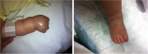
Edema and a polymorphous rash on the hands and feet in a 3-month-old patient with KD
Image: “Patient’s limbs” by Unit of Broncho-Pneumology and Cystic Fibrosis, Department of Medical and Pediatric Science, University of Catania, Via Santa Sofia 78, Catania 95123, Italy. License: CC BY 2.0
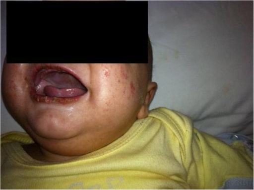
Lip fissuring alongside a polymorphous rash in KD
Image: “Patient’s face” by Unit of Broncho-Pneumology and Cystic Fibrosis, Department of Medical and Pediatric Science, University of Catania, Via Santa Sofia 78, Catania 95123, Italy. License: CC BY 2.0
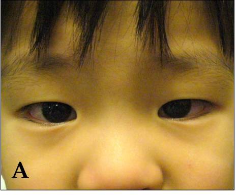
Bilateral, non-exudative conjunctivitis observed in a patient with KD
Image: “Kawasaki symptoms A” by Dong Soo Kim. License: CC BY 2.0
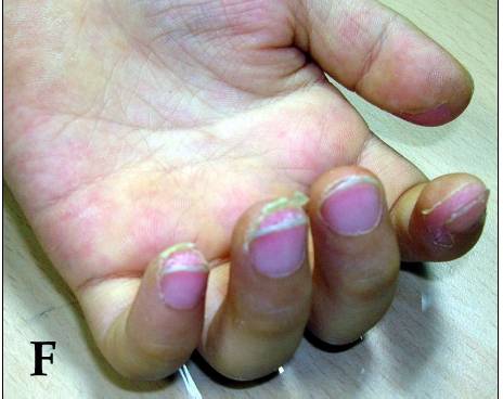
Desquamation of the fingertips observed in KD at 10–14 days
Image: “Desquamation of the fingers” by Dong Soo Kim. License: CC BY 2.0
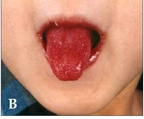
Strawberry tongue and bright-red, swollen lips with vertical cracking and bleeding in a patient with KD
Image: “Kawasaki disease” by Dong Soo Kim. License: CC BY 2.0
Less common manifestations
Table: Other manifestations of Kawasaki diseaseKawasaki diseaseAn acute, febrile, mucocutaneous condition accompanied by swelling of cervical lymph nodes in infants and young children. The principal symptoms are fever, congestion of the ocular conjunctivae, reddening of the lips and oral cavity, protuberance of tongue papillae, and edema or erythema of the extremities.Kawasaki Disease[6]
System
Manifestations
Gastrointestinal
DiarrheaDiarrheaDiarrhea is defined as ≥ 3 watery or loose stools in a 24-hour period. There are a multitude of etiologies, which can be classified based on the underlying mechanism of disease. The duration of symptoms (acute or chronic) and characteristics of the stools (e.g., watery, bloody, steatorrheic, mucoid) can help guide further diagnostic evaluation. Diarrhea, abdominal painAbdominal PainAcute Abdomen, vomitingVomitingThe forcible expulsion of the contents of the stomach through the mouth.Hypokalemia, liverLiverThe liver is the largest gland in the human body. The liver is found in the superior right quadrant of the abdomen and weighs approximately 1.5 kilograms. Its main functions are detoxification, metabolism, nutrient storage (e.g., iron and vitamins), synthesis of coagulation factors, formation of bile, filtration, and storage of blood. Liver: Anatomy dysfunction, pancreatitisPancreatitisInflammation of the pancreas. Pancreatitis is classified as acute unless there are computed tomographic or endoscopic retrograde cholangiopancreatographic findings of chronic pancreatitis. The two most common forms of acute pancreatitis are alcoholic pancreatitis and gallstone pancreatitis.Acute Pancreatitis, hydropsHydropsCholecystitisgallbladderGallbladderThe gallbladder is a pear-shaped sac, located directly beneath the liver, that sits on top of the superior part of the duodenum. The primary functions of the gallbladder include concentrating and storing up to 50 mL of bile. Gallbladder and Biliary Tract: Anatomy, ascitesAscitesAscites is the pathologic accumulation of fluid within the peritoneal cavity that occurs due to an osmotic and/or hydrostatic pressure imbalance secondary to portal hypertension (cirrhosis, heart failure) or non-portal hypertension (hypoalbuminemia, malignancy, infection).Ascites, splenic infarctionSplenic InfarctionInsufficiency of arterial or venous blood supply to the spleen due to emboli, thrombi, vascular torsion, or pressure that produces a macroscopic area of necrosis. .Imaging of the Spleen
MyocarditisMyocarditisMyocarditis is an inflammatory disease of the myocardium, which may occur alone or in association with a systemic process. There are numerous etiologies of myocarditis, but all lead to inflammation and myocyte injury, most often leading to signs and symptoms of heart failure. Myocarditis, pericarditisPericarditisPericarditis is an inflammation of the pericardium, often with fluid accumulation. It can be caused by infection (often viral), myocardial infarction, drugs, malignancies, metabolic disorders, autoimmune disorders, or trauma. Acute, subacute, and chronic forms exist. Pericarditis, tachycardiaTachycardiaAbnormally rapid heartbeat, usually with a heart rate above 100 beats per minute for adults. Tachycardia accompanied by disturbance in the cardiac depolarization (cardiac arrhythmia) is called tachyarrhythmia.Sepsis in Children, valvular heart disease
Genitourinary
UrethritisUrethritisInflammation involving the urethra. Similar to cystitis, clinical symptoms range from vague discomfort to painful urination (dysuria), urethral discharge, or both.Urinary Tract Infections (UTIs), prostatitisProstatitisProstatitis is inflammation or an irritative condition of the prostate that presents as different syndromes: acute bacterial, chronic bacterial, chronic prostatitis/chronic pelvic pain, and asymptomatic. Bacterial prostatitis is easier to identify clinically and the management (antibiotics) is better established. Prostatitis, cystitisCystitisInflammation of the urinary bladder, either from bacterial or non-bacterial causes. Cystitis is usually associated with painful urination (dysuria), increased frequency, urgency, and suprapubic pain.Urinary Tract Infections (UTIs), interstitial nephritis, nephrotic syndromeNephrotic syndromeNephrotic syndrome is characterized by severe proteinuria, hypoalbuminemia, and peripheral edema. In contrast, the nephritic syndromes present with hematuria, variable loss of renal function, and hypertension, although there is sometimes overlap of > 1 glomerular disease in the same individual. Nephrotic Syndrome
LethargyLethargyA general state of sluggishness, listless, or uninterested, with being tired, and having difficulty concentrating and doing simple tasks. It may be related to depression or drug addiction.Hyponatremia, increased irritability, aseptic meningitisMeningitisMeningitis is inflammation of the meninges, the protective membranes of the brain, and spinal cord. The causes of meningitis are varied, with the most common being bacterial or viral infection. The classic presentation of meningitis is a triad of fever, altered mental status, and nuchal rigidity. Meningitis, sensorineural deafness
Respiratory
Shortness of breathShortness of breathDyspnea is the subjective sensation of breathing discomfort. Dyspnea is a normal manifestation of heavy physical or psychological exertion, but also may be caused by underlying conditions (both pulmonary and extrapulmonary).Dyspnea, influenza-like illness, pleural effusionPleural EffusionPleural effusion refers to the accumulation of fluid between the layers of the parietal and visceral pleura. Common causes of this condition include infection, malignancy, autoimmune disorders, or volume overload. Clinical manifestations include chest pain, cough, and dyspnea. Pleural Effusion, cough, rhinorrheaRhinorrheaExcess nasal drainage.Respiratory Syncytial Virus
SkinSkinThe skin, also referred to as the integumentary system, is the largest organ of the body. The skin is primarily composed of the epidermis (outer layer) and dermis (deep layer). The epidermis is primarily composed of keratinocytes that undergo rapid turnover, while the dermis contains dense layers of connective tissue.Skin: Structure and Functions
ErythemaErythemaRedness of the skin produced by congestion of the capillaries. This condition may result from a variety of disease processes.Chalazion and indurationIndurationDermatologic Examination at BCGBCGAn active immunizing agent and a viable avirulent attenuated strain of Mycobacterium bovis, which confers immunity to mycobacterial infections. It is used also in immunotherapy of neoplasms due to its stimulation of antibodies and non-specific immunity.Cancer Immunotherapy (bacille Calmette-Guérin) vaccinationVaccinationVaccination is the administration of a substance to induce the immune system to develop protection against a disease. Unlike passive immunization, which involves the administration of pre-performed antibodies, active immunization constitutes the administration of a vaccine to stimulate the body to produce its own antibodies.Vaccination site, Beau’s linesBeau’s linesKawasaki Disease, finger gangreneFinger GangreneKawasaki Disease
General
Irritability, decreased oral intake, lethargyLethargyA general state of sluggishness, listless, or uninterested, with being tired, and having difficulty concentrating and doing simple tasks. It may be related to depression or drug addiction.Hyponatremia
Diagnosis
Diagnostic criteria[6,7,14]
The diagnosis of KDKDAn acute, febrile, mucocutaneous condition accompanied by swelling of cervical lymph nodes in infants and young children. The principal symptoms are fever, congestion of the ocular conjunctivae, reddening of the lips and oral cavity, protuberance of tongue papillae, and edema or erythema of the extremities.Kawasaki Disease is primarily clinical, utilizing lab criteria to reinforce diagnostic suspicion in cases of incomplete disease. UK and US diagnostic criteria roughly coincide, with the caveat that KDKDAn acute, febrile, mucocutaneous condition accompanied by swelling of cervical lymph nodes in infants and young children. The principal symptoms are fever, congestion of the ocular conjunctivae, reddening of the lips and oral cavity, protuberance of tongue papillae, and edema or erythema of the extremities.Kawasaki Disease is much less frequent in the UK.
FeverFeverFever is defined as a measured body temperature of at least 38°C (100.4°F). Fever is caused by circulating endogenous and/or exogenous pyrogens that increase levels of prostaglandin E2 in the hypothalamus. Fever is commonly associated with chills, rigors, sweating, and flushing of the skin. Fever lasting ≥ 5 days AND at least 4 of the following:
Bilateral, non-exudative conjunctival injection
Oral mucosal changes:
Cracked lipsLipsThe lips are the soft and movable most external parts of the oral cavity. The blood supply of the lips originates from the external carotid artery, and the innervation is through cranial nerves.Lips and Tongue: Anatomy
Injected pharynxPharynxThe pharynx is a component of the digestive system that lies posterior to the nasal cavity, oral cavity, and larynx. The pharynx can be divided into the oropharynx, nasopharynx, and laryngopharynx. Pharyngeal muscles play an integral role in vital processes such as breathing, swallowing, and speaking. Pharynx: Anatomy
Peripheral extremity changes:
ErythemaErythemaRedness of the skin produced by congestion of the capillaries. This condition may result from a variety of disease processes.Chalazion of palms/soles
EdemaEdemaEdema is a condition in which excess serous fluid accumulates in the body cavity or interstitial space of connective tissues. Edema is a symptom observed in several medical conditions. It can be categorized into 2 types, namely, peripheral (in the extremities) and internal (in an organ or body cavity). Edema of hands/feet
Cervical lymphadenopathyLymphadenopathyLymphadenopathy is lymph node enlargement (> 1 cm) and is benign and self-limited in most patients. Etiologies include malignancy, infection, and autoimmune disorders, as well as iatrogenic causes such as the use of certain medications. Generalized lymphadenopathy often indicates underlying systemic disease. Lymphadenopathy: at least 1 node > 1.5 cm in diameter
If < 4 criteria → consider atypical (incomplete) Kawasaki diseaseKawasaki diseaseAn acute, febrile, mucocutaneous condition accompanied by swelling of cervical lymph nodes in infants and young children. The principal symptoms are fever, congestion of the ocular conjunctivae, reddening of the lips and oral cavity, protuberance of tongue papillae, and edema or erythema of the extremities.Kawasaki Disease, supplement with laboratories outlined below
Diagnostic algorithm for children with fever/diagnosis of Kawasaki disease
Units: CRP in mg/dL; erythrocyte sedimentation rate (ESR) in mm/hr
Positive labs (≥ 3 supplemental laboratory findings) include:
Anemia (for age)
↑ Platelet count ≥ 450,000 after day 7 of fever
↑ WBC ≥ 15,000/mm3
↓ Albumin ≤ 3 g/dL
↑ ALT
Pyuria (≥ 10 WBC/high-power field (HPF) on urinalysis)
Image by Lecturio.
Laboratory and imaging studies[14,16]
Laboratory:
CBC with differential:
↑ WBC (≥ 15,000/mm3)
↑ Platelet countsPlatelet countsThe number of platelets per unit volume in a sample of venous blood.Coagulation Studies (≥ 450,000 after day 7 of feverFeverFever is defined as a measured body temperature of at least 38°C (100.4°F). Fever is caused by circulating endogenous and/or exogenous pyrogens that increase levels of prostaglandin E2 in the hypothalamus. Fever is commonly associated with chills, rigors, sweating, and flushing of the skin. Fever); may be normal or low in early stages
AnemiaAnemiaAnemia is a condition in which individuals have low Hb levels, which can arise from various causes. Anemia is accompanied by a reduced number of RBCs and may manifest with fatigue, shortness of breath, pallor, and weakness. Subtypes are classified by the size of RBCs, chronicity, and etiology. Anemia: Overview and Types
Liver function testsLiver function testsLiver function tests, also known as hepatic function panels, are one of the most commonly performed screening blood tests. Such tests are also used to detect, evaluate, and monitor acute and chronic liver diseases.Liver Function Tests (LFTs):
↑ AspartateAspartateOne of the non-essential amino acids commonly occurring in the l-form. It is found in animals and plants, especially in sugar cane and sugar beets. It may be a neurotransmitter.Synthesis of Nonessential Amino Acids aminotransferase (ASTASTEnzymes of the transferase class that catalyze the conversion of l-aspartate and 2-ketoglutarate to oxaloacetate and l-glutamate.Liver Function Tests) and alanineAlanineA non-essential amino acid that occurs in high levels in its free state in plasma. It is produced from pyruvate by transamination. It is involved in sugar and acid metabolism, increases immunity, and provides energy for muscle tissue, brain, and the central nervous system.Synthesis of Nonessential Amino Acids aminotransferase (ALTALTAn enzyme that catalyzes the conversion of l-alanine and 2-oxoglutarate to pyruvate and l-glutamate.Liver Function Tests)
↓ AlbuminAlbuminSerum albumin from humans. It is an essential carrier of both endogenous substances, such as fatty acids and bilirubin, and of xenobiotics in the blood.Liver Function Tests (≤ 3 g/dL)
UrinalysisUrinalysisExamination of urine by chemical, physical, or microscopic means. Routine urinalysis usually includes performing chemical screening tests, determining specific gravity, observing any unusual color or odor, screening for bacteriuria, and examining the sediment microscopically.Urinary Tract Infections (UTIs) in Children: sterileSterileBasic ProcedurespyuriaPyuriaThe presence of white blood cells (leukocytes) in the urine. It is often associated with bacterial infections of the urinary tract. Pyuria without bacteriuria can be caused by tuberculosis, stones, or cancer.Urinary Tract Infections (UTIs) (≥ 10 WBCs/HPFs on urinalysisUrinalysisExamination of urine by chemical, physical, or microscopic means. Routine urinalysis usually includes performing chemical screening tests, determining specific gravity, observing any unusual color or odor, screening for bacteriuria, and examining the sediment microscopically.Urinary Tract Infections (UTIs) in Children)
EchocardiographyEchocardiographyUltrasonic recording of the size, motion, and composition of the heart and surrounding tissues. The standard approach is transthoracic.Tricuspid Valve Atresia (TVA):
↑ Size (internal diameter) of the left anterior descending (LAD) artery or right coronary arteryRight coronary arteryHeart: Anatomy (RCA) (small aneurysmAneurysmAn aneurysm is a bulging, weakened area of a blood vessel that causes an abnormal widening of its diameter > 1.5 times the size of the native vessel. Aneurysms occur more often in arteries than in veins and are at risk of dissection and rupture, which can be life-threatening. Thoracic Aortic Aneurysms: z-scoreZ-scoreStandard deviation difference between patient’s bone mass density and that of age-matched population.Osteoporosis ≥ 2.5 to <5)
Coronary arteryCoronary ArteryTruncus ArteriosusaneurysmAneurysmAn aneurysm is a bulging, weakened area of a blood vessel that causes an abnormal widening of its diameter > 1.5 times the size of the native vessel. Aneurysms occur more often in arteries than in veins and are at risk of dissection and rupture, which can be life-threatening. Thoracic Aortic Aneurysms (other than LAD or RCA) observed
Supportive findings, particularly if ≥3 are present.:
Coronary arteryCoronary ArteryTruncus Arteriosus abnormalities (such as perivascular brightness, lack of tapering of the coronary arteriesArteriesArteries are tubular collections of cells that transport oxygenated blood and nutrients from the heart to the tissues of the body. The blood passes through the arteries in order of decreasing luminal diameter, starting in the largest artery (the aorta) and ending in the small arterioles. Arteries are classified into 3 types: large elastic arteries, medium muscular arteries, and small arteries and arterioles. Arteries: Histology)
Pericardial effusionPericardial effusionFluid accumulation within the pericardium. Serous effusions are associated with pericardial diseases. Hemopericardium is associated with trauma. Lipid-containing effusion (chylopericardium) results from leakage of thoracic duct. Severe cases can lead to cardiac tamponade.Pericardial Effusion and Cardiac Tamponade
LAD and RCA dilation (z-scoreZ-scoreStandard deviation difference between patient’s bone mass density and that of age-matched population.Osteoporosis 2‒2.5)
Should be performed in all patientsPatientsIndividuals participating in the health care system for the purpose of receiving therapeutic, diagnostic, or preventive procedures.Clinician–Patient Relationship with KDKDAn acute, febrile, mucocutaneous condition accompanied by swelling of cervical lymph nodes in infants and young children. The principal symptoms are fever, congestion of the ocular conjunctivae, reddening of the lips and oral cavity, protuberance of tongue papillae, and edema or erythema of the extremities.Kawasaki Disease
Establish a reference point for longitudinal follow-up
Determine treatment efficacy
AngiographyAngiographyRadiography of blood vessels after injection of a contrast medium.Cardiac Surgery:
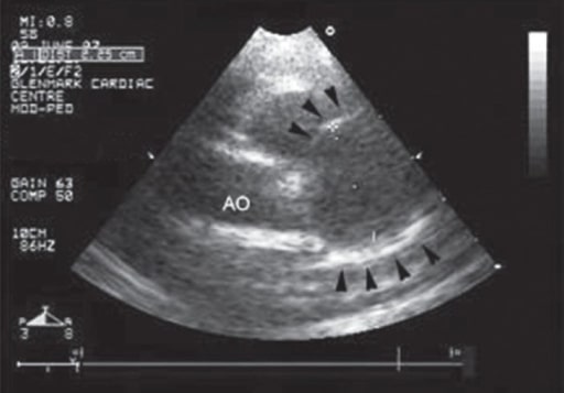
Echocardiography view at the level of aortic valve demonstrating an increase in the size of a coronary artery aneurysm (arrowheads) secondary to KD
Image: “Follow-up echocardiography” by Department of Cardiology, Dr. Balabhai Nanavati Hospital, Mumbai, India. License: CC BY 2.0
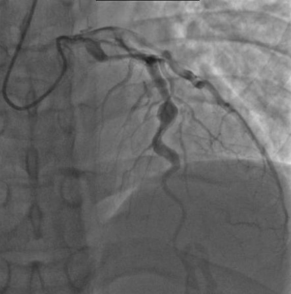
Angiography of a patient with KD showing ectatic left anterior descending coronary artery, with the largest aneurysm measuring 6.5 mm in diameter.
Image: “Angiography showing ectatic LAD,” by mprice18. License: CC BY 3.0
Tests to rule out disease that presents similarly to Kawasaki diseaseKawasaki diseaseAn acute, febrile, mucocutaneous condition accompanied by swelling of cervical lymph nodes in infants and young children. The principal symptoms are fever, congestion of the ocular conjunctivae, reddening of the lips and oral cavity, protuberance of tongue papillae, and edema or erythema of the extremities.Kawasaki Disease[6,10]
Rapid viral testing (PCRPCRPolymerase chain reaction (PCR) is a technique that amplifies DNA fragments exponentially for analysis. The process is highly specific, allowing for the targeting of specific genomic sequences, even with minuscule sample amounts. The PCR cycles multiple times through 3 phases: denaturation of the template DNA, annealing of a specific primer to the individual DNA strands, and synthesis/elongation of new DNA molecules.Polymerase Chain Reaction (PCR)): useful for ruling out infectious causes such as adenovirusAdenovirusAdenovirus (member of the family Adenoviridae) is a nonenveloped, double-stranded DNA virus. Adenovirus is transmitted in a variety of ways, and it can have various presentations based on the site of entry. Presentation can include febrile pharyngitis, conjunctivitis, acute respiratory disease, atypical pneumonia, and gastroenteritis. Adenovirus
Bacterial cultures: rule out SIRS from bacterial infectionsInfectionsInvasion of the host organism by microorganisms or their toxins or by parasites that can cause pathological conditions or diseases.Chronic Granulomatous Disease
Severe acute respiratory syndrome coronavirusSevere acute respiratory syndrome coronavirusA viral disorder characterized by high fever, dry cough, shortness of breath (dyspnea) or breathing difficulties, and atypical pneumonia. A virus in the genus Coronavirus is the suspected agent.Coronavirus 2 (SARS-CoV-2) testing
Mnemonic
CRASH and Burn:
Conjunctivitis
Rash
Adenopathy
Strawberry tongueTongueThe tongue, on the other hand, is a complex muscular structure that permits tasting and facilitates the process of mastication and communication. The blood supply of the tongue originates from the external carotid artery, and the innervation is through cranial nerves.Lips and Tongue: Anatomy
Hands and feet
Burn:feverFeverFever is defined as a measured body temperature of at least 38°C (100.4°F). Fever is caused by circulating endogenous and/or exogenous pyrogens that increase levels of prostaglandin E2 in the hypothalamus. Fever is commonly associated with chills, rigors, sweating, and flushing of the skin. Fever
Management
Kawasaki diseaseKawasaki diseaseAn acute, febrile, mucocutaneous condition accompanied by swelling of cervical lymph nodes in infants and young children. The principal symptoms are fever, congestion of the ocular conjunctivae, reddening of the lips and oral cavity, protuberance of tongue papillae, and edema or erythema of the extremities.Kawasaki Disease is self-limited!
Treatment is aimed at preventing complications and reducing symptoms.
2 g/kg administered as a single infusion (slow) over 8–12 hours
Started within 10 days after feverFeverFever is defined as a measured body temperature of at least 38°C (100.4°F). Fever is caused by circulating endogenous and/or exogenous pyrogens that increase levels of prostaglandin E2 in the hypothalamus. Fever is commonly associated with chills, rigors, sweating, and flushing of the skin. Fever onset; reduces the risk of coronary arteryCoronary ArteryTruncus Arteriosus aneurysms
Observe for 24 hours following completion of IVIGIVIGDermatomyositis infusion to confirm feverFeverFever is defined as a measured body temperature of at least 38°C (100.4°F). Fever is caused by circulating endogenous and/or exogenous pyrogens that increase levels of prostaglandin E2 in the hypothalamus. Fever is commonly associated with chills, rigors, sweating, and flushing of the skin. Fever resolution.
AspirinAspirinThe prototypical analgesic used in the treatment of mild to moderate pain. It has anti-inflammatory and antipyretic properties and acts as an inhibitor of cyclooxygenase which results in the inhibition of the biosynthesis of prostaglandins. Aspirin also inhibits platelet aggregation and is used in the prevention of arterial and venous thrombosis.Nonsteroidal Antiinflammatory Drugs (NSAIDs) (ASAASAAnterior Cord Syndrome):
30–50 mg/kg daily divided into 4 doses (maximum: 4 g/day)
High dose (80–100 mg/kg daily) is also used, but has not been shown to be superior to medium doses (30–50 mg/kg daily).
Antiinflammatory and antiplatelet effects
Prevents thrombus in coronary arteriesArteriesArteries are tubular collections of cells that transport oxygenated blood and nutrients from the heart to the tissues of the body. The blood passes through the arteries in order of decreasing luminal diameter, starting in the largest artery (the aorta) and ending in the small arterioles. Arteries are classified into 3 types: large elastic arteries, medium muscular arteries, and small arteries and arterioles. Arteries: Histology
Change to low dose (3–5 mg/kg daily; maximum: 81–325 mg daily) when the patient has been afebrile for about 48–72 hours.
With long-term ASAASAAnterior Cord Syndrome use, children should receive influenzaInfluenzaInfluenza viruses are members of the Orthomyxoviridae family and the causative organisms of influenza, a highly contagious febrile respiratory disease. There are 3 primary influenza viruses (A, B, and C) and various subtypes, which are classified based on their virulent surface antigens, hemagglutinin (HA) and neuraminidase (NA). Influenza typically presents with a fever, myalgia, headache, and symptoms of an upper respiratory infection. Influenza Viruses/Influenza and varicella vaccinationVaccinationVaccination is the administration of a substance to induce the immune system to develop protection against a disease. Unlike passive immunization, which involves the administration of pre-performed antibodies, active immunization constitutes the administration of a vaccine to stimulate the body to produce its own antibodies.Vaccination (as Reye syndromeReye syndromeA form of encephalopathy with fatty infiltration of the liver, characterized by brain edema and vomiting that may rapidly progress to seizures; coma; and death. It is caused by a generalized loss of mitochondrial function leading to disturbances in fatty acid and carnitine metabolism.Varicella-Zoster Virus/Chickenpox has been noted in the setting of ASAASAAnterior Cord Syndrome intake and varicella or influenzaInfluenzaInfluenza viruses are members of the Orthomyxoviridae family and the causative organisms of influenza, a highly contagious febrile respiratory disease. There are 3 primary influenza viruses (A, B, and C) and various subtypes, which are classified based on their virulent surface antigens, hemagglutinin (HA) and neuraminidase (NA). Influenza typically presents with a fever, myalgia, headache, and symptoms of an upper respiratory infection. Influenza Viruses/Influenza infection)
Avoid ibuprofenIbuprofenA nonsteroidal anti-inflammatory agent with analgesic properties used in the treatment of rheumatism and arthritis.Nonsteroidal Antiinflammatory Drugs (NSAIDs) or other NSAIDNSAIDNonsteroidal antiinflammatory drugs (NSAIDs) are a class of medications consisting of aspirin, reversible NSAIDs, and selective NSAIDs. NSAIDs are used as antiplatelet, analgesic, antipyretic, and antiinflammatory agents. Nonsteroidal Antiinflammatory Drugs (NSAIDs), which interferes with aspirinAspirinThe prototypical analgesic used in the treatment of mild to moderate pain. It has anti-inflammatory and antipyretic properties and acts as an inhibitor of cyclooxygenase which results in the inhibition of the biosynthesis of prostaglandins. Aspirin also inhibits platelet aggregation and is used in the prevention of arterial and venous thrombosis.Nonsteroidal Antiinflammatory Drugs (NSAIDs)’s antiplatelet effect.
PrednisolonePrednisoloneA glucocorticoid with the general properties of the corticosteroids. It is the drug of choice for all conditions in which routine systemic corticosteroid therapy is indicated, except adrenal deficiency states.Immunosuppressants: 2 mg/kg IV per day divided every 8 h for 5 days (maximum 60 mg/day), transition to oral and taper (over 2-3 weeks) when CRP becomes normal
MethylprednisoloneMethylprednisoloneA prednisolone derivative with similar anti-inflammatory action.Immunosuppressants: 2 mg/kg IV per day divided every 12 hours for 5 days (maximum 60 mg/day); transition to oral and taper (over 2-3 weeks) when CRP becomes normal
InfliximabInfliximabA chimeric monoclonal antibody to tnf-alpha that is used in the treatment of rheumatoid arthritis; ankylosing spondylitis; psoriatic arthritis and Crohn’s disease.Disease-Modifying Antirheumatic Drugs (DMARDs): 10 mg/kg IV given over 2 hours
EtanerceptEtanerceptA recombinant version of soluble human tnf receptor fused to an IgG Fc fragment that binds specifically to tumor necrosis factor and inhibits its binding with endogenous tnf receptors. It prevents the inflammatory effect of tnf and is used to treat rheumatoid arthritis; psoriatic arthritis and ankylosing spondylitis.Immunosuppressants: 0.8 mg/kg subcutaneous injection weekly ×3 doses
Interleukin-1Interleukin-1A soluble factor produced by monocytes; macrophages, and other cells which activates T-lymphocytes and potentiates their response to mitogens or antigens. Interleukin-1 is a general term refers to either of the two distinct proteins, interleukin-1alpha and interleukin-1beta. The biological effects of il-1 include the ability to replace macrophage requirements for t-cell activation.Interleukins inhibition (anakinraAnakinraImmunosuppressants): 10 mg/kg per day (IV divided every 12 hours; may be given subcutaneously but IV is preferred)
CyclosporineCyclosporineA cyclic undecapeptide from an extract of soil fungi. It is a powerful immunosupressant with a specific action on T-lymphocytes. It is used for the prophylaxis of graft rejection in organ and tissue transplantation.Immunosuppressants: 5 mg/kg per day orally divided every 12 hours (check 2 hour level after 3rd dose with a goal of 300–600 ng/mL); taper when CRP normalizes or with clinical improvement
CyclophosphamideCyclophosphamidePrecursor of an alkylating nitrogen mustard antineoplastic and immunosuppressive agent that must be activated in the liver to form the active aldophosphamide. It has been used in the treatment of lymphoma and leukemia. Its side effect, alopecia, has been used for defleecing sheep. Cyclophosphamide may also cause sterility, birth defects, mutations, and cancer.Immunosuppressants (for extreme refractory cases): 10 mg/kg IV per day in 1 or 2 doses
Large coronary arteryCoronary ArteryTruncus Arteriosus aneurysms: high risk for luminal thrombosisThrombosisFormation and development of a thrombus or blood clot in the blood vessel.Epidemic Typhus so antiplatelet therapy (aspirinAspirinThe prototypical analgesic used in the treatment of mild to moderate pain. It has anti-inflammatory and antipyretic properties and acts as an inhibitor of cyclooxygenase which results in the inhibition of the biosynthesis of prostaglandins. Aspirin also inhibits platelet aggregation and is used in the prevention of arterial and venous thrombosis.Nonsteroidal Antiinflammatory Drugs (NSAIDs) or dual therapy) and anticoagulationAnticoagulationPulmonary Hypertension Drugs are given.
Either warfarinWarfarinAn anticoagulant that acts by inhibiting the synthesis of vitamin K-dependent coagulation factors. Warfarin is indicated for the prophylaxis and/or treatment of venous thrombosis and its extension, pulmonary embolism, and atrial fibrillation with embolization. It is also used as an adjunct in the prophylaxis of systemic embolism after myocardial infarction. Warfarin is also used as a rodenticide.Anticoagulants or low molecular weight heparin (LMWH); direct oral anticoagulantsAnticoagulantsAnticoagulants are drugs that retard or interrupt the coagulation cascade. The primary classes of available anticoagulants include heparins, vitamin K-dependent antagonists (e.g., warfarin), direct thrombin inhibitors, and factor Xa inhibitors. Anticoagulants (DOACsDOACsAnticoagulants) are still under study.
Consultation with KDKDAn acute, febrile, mucocutaneous condition accompanied by swelling of cervical lymph nodes in infants and young children. The principal symptoms are fever, congestion of the ocular conjunctivae, reddening of the lips and oral cavity, protuberance of tongue papillae, and edema or erythema of the extremities.Kawasaki Disease expert is recommended especially with the associated increased risk of bleeding.
Laboratory monitoring:[6]
CRP: normalizes in 1–2 weeks (essentially, as inflammationInflammationInflammation is a complex set of responses to infection and injury involving leukocytes as the principal cellular mediators in the body’s defense against pathogenic organisms. Inflammation is also seen as a response to tissue injury in the process of wound healing. The 5 cardinal signs of inflammation are pain, heat, redness, swelling, and loss of function. Inflammation resolves)
Follow-up with serial echocardiograms at 2 and 6 weeks
Table: Other medications that can be used
Medication
Indication
ClopidogrelClopidogrelA ticlopidine analog and platelet purinergic p2y receptor antagonist that inhibits adenosine diphosphate-mediated platelet aggregation. It is used to prevent thromboembolism in patients with arterial occlusive diseases; myocardial infarction; stroke; or atrial fibrillation.Antiplatelet Drugs, dipyridamoleDipyridamoleA phosphodiesterase inhibitor that blocks uptake and metabolism of adenosine by erythrocytes and vascular endothelial cells. Dipyridamole also potentiates the antiaggregating action of prostacyclin.Phosphodiesterase Inhibitors, and other antiplatelet drugs
For small-to-medium-sized aneurysms of the coronary arteriesArteriesArteries are tubular collections of cells that transport oxygenated blood and nutrients from the heart to the tissues of the body. The blood passes through the arteries in order of decreasing luminal diameter, starting in the largest artery (the aorta) and ending in the small arterioles. Arteries are classified into 3 types: large elastic arteries, medium muscular arteries, and small arteries and arterioles. Arteries: Histology and a high risk for thrombus formation
Low-molecular-weight heparin, warfarinWarfarinAn anticoagulant that acts by inhibiting the synthesis of vitamin K-dependent coagulation factors. Warfarin is indicated for the prophylaxis and/or treatment of venous thrombosis and its extension, pulmonary embolism, and atrial fibrillation with embolization. It is also used as an adjunct in the prophylaxis of systemic embolism after myocardial infarction. Warfarin is also used as a rodenticide.Anticoagulants, and other anticoagulantsAnticoagulantsAnticoagulants are drugs that retard or interrupt the coagulation cascade. The primary classes of available anticoagulants include heparins, vitamin K-dependent antagonists (e.g., warfarin), direct thrombin inhibitors, and factor Xa inhibitors. Anticoagulants
For large aneurysms of the coronary arteriesArteriesArteries are tubular collections of cells that transport oxygenated blood and nutrients from the heart to the tissues of the body. The blood passes through the arteries in order of decreasing luminal diameter, starting in the largest artery (the aorta) and ending in the small arterioles. Arteries are classified into 3 types: large elastic arteries, medium muscular arteries, and small arteries and arterioles. Arteries: Histology with a high risk of thrombus formation
InfliximabInfliximabA chimeric monoclonal antibody to tnf-alpha that is used in the treatment of rheumatoid arthritis; ankylosing spondylitis; psoriatic arthritis and Crohn’s disease.Disease-Modifying Antirheumatic Drugs (DMARDs), cyclophosphamideCyclophosphamidePrecursor of an alkylating nitrogen mustard antineoplastic and immunosuppressive agent that must be activated in the liver to form the active aldophosphamide. It has been used in the treatment of lymphoma and leukemia. Its side effect, alopecia, has been used for defleecing sheep. Cyclophosphamide may also cause sterility, birth defects, mutations, and cancer.Immunosuppressants, and cyclosporineCyclosporineA cyclic undecapeptide from an extract of soil fungi. It is a powerful immunosupressant with a specific action on T-lymphocytes. It is used for the prophylaxis of graft rejection in organ and tissue transplantation.Immunosuppressants
Refractory cases with coronary aneurysmAneurysmAn aneurysm is a bulging, weakened area of a blood vessel that causes an abnormal widening of its diameter > 1.5 times the size of the native vessel. Aneurysms occur more often in arteries than in veins and are at risk of dissection and rupture, which can be life-threatening. Thoracic Aortic Aneurysms
PatientsPatientsIndividuals participating in the health care system for the purpose of receiving therapeutic, diagnostic, or preventive procedures.Clinician–Patient Relationship who do not respond to standard treatments and therapies or are at risk for IVIGIVIGDermatomyositisresistanceResistancePhysiologically, the opposition to flow of air caused by the forces of friction. As a part of pulmonary function testing, it is the ratio of driving pressure to the rate of air flow.Ventilation: Mechanics of Breathing
Complications
ShockShockShock is a life-threatening condition associated with impaired circulation that results in tissue hypoxia. The different types of shock are based on the underlying cause: distributive (↑ cardiac output (CO), ↓ systemic vascular resistance (SVR)), cardiogenic (↓ CO, ↑ SVR), hypovolemic (↓ CO, ↑ SVR), obstructive (↓ CO), and mixed. Types of Shock:[6,15]
KD shock syndromeKD shock syndromeKawasaki Disease (KDSSKDSSKawasaki Disease) is sustained systolic hypotensionHypotensionHypotension is defined as low blood pressure, specifically < 90/60 mm Hg, and is most commonly a physiologic response. Hypotension may be mild, serious, or life threatening, depending on the cause. Hypotension or clinical signs of poor perfusion.
Accompanying characteristics that are highly suggestive of KDSSKDSSKawasaki Disease:
Coronary artery dilationCoronary Artery DilationKawasaki Disease, aneurysmAneurysmAn aneurysm is a bulging, weakened area of a blood vessel that causes an abnormal widening of its diameter > 1.5 times the size of the native vessel. Aneurysms occur more often in arteries than in veins and are at risk of dissection and rupture, which can be life-threatening. Thoracic Aortic Aneurysms, and/or stenosisStenosisHypoplastic Left Heart Syndrome (HLHS) (Giant aneurysms ~>8 mm have a high long-term risk for thrombosisThrombosisFormation and development of a thrombus or blood clot in the blood vessel.Epidemic Typhus, stenosisStenosisHypoplastic Left Heart Syndrome (HLHS), myocardial infarctionMyocardial infarctionMI is ischemia and death of an area of myocardial tissue due to insufficient blood flow and oxygenation, usually from thrombus formation on a ruptured atherosclerotic plaque in the epicardial arteries. Clinical presentation is most commonly with chest pain, but women and patients with diabetes may have atypical symptoms.Myocardial Infarction, and sudden death)
Myocardial ischemiaMyocardial ischemiaA disorder of cardiac function caused by insufficient blood flow to the muscle tissue of the heart. The decreased blood flow may be due to narrowing of the coronary arteries (coronary artery disease), to obstruction by a thrombus (coronary thrombosis), or less commonly, to diffuse narrowing of arterioles and other small vessels within the heart.Coronary Heart Disease or infarction
Macrophage activation syndromeMacrophage activation syndromeA serious complication of childhood systemic inflammatory disorders that is thought to be caused by excessive activation and proliferation of T-lymphocytes and macrophages. It is seen predominantly in children with systemic onset juvenile idiopathic arthritis.Juvenile Idiopathic Arthritis:
Activation and proliferation of macrophagesMacrophagesThe relatively long-lived phagocytic cell of mammalian tissues that are derived from blood monocytes. Main types are peritoneal macrophages; alveolar macrophages; histiocytes; kupffer cells of the liver; and osteoclasts. They may further differentiate within chronic inflammatory lesions to epithelioid cells or may fuse to form foreign body giant cells or langhans giant cells.Innate Immunity: Phagocytes and Antigen Presentation and T cellsT cellsLymphocytes responsible for cell-mediated immunity. Two types have been identified – cytotoxic (t-lymphocytes, cytotoxic) and helper T-lymphocytes (t-lymphocytes, helper-inducer). They are formed when lymphocytes circulate through the thymus gland and differentiate to thymocytes. When exposed to an antigen, they divide rapidly and produce large numbers of new T cells sensitized to that antigen.T cells: Types and Functions
Can lead to life-threatening complications, such as:
ThrombosisThrombosisFormation and development of a thrombus or blood clot in the blood vessel.Epidemic Typhus
IschemiaIschemiaA hypoperfusion of the blood through an organ or tissue caused by a pathologic constriction or obstruction of its blood vessels, or an absence of blood circulation.Ischemic Cell Damage, gangreneGangreneDeath and putrefaction of tissue usually due to a loss of blood supply.Small Bowel Obstruction:
Caused by peripheral arterial obstruction
Can involve the viscera or limbs
Urinary abnormalities:
SterileSterileBasic ProcedurespyuriaPyuriaThe presence of white blood cells (leukocytes) in the urine. It is often associated with bacterial infections of the urinary tract. Pyuria without bacteriuria can be caused by tuberculosis, stones, or cancer.Urinary Tract Infections (UTIs) (common)
Acute interstitial nephritisAcute interstitial nephritisInflammation of the interstitial tissue of the kidney. This term is generally used for primary inflammation of kidney tubules and/or surrounding interstitium. For primary inflammation of glomerular interstitium, see glomerulonephritis. Infiltration of the inflammatory cells into the interstitial compartment results in edema, increased spaces between the tubules, and tubular renal dysfunction.Acute Kidney Injury
Acute kidney injuryAcute Kidney InjuryAcute kidney injury refers to sudden and often reversible loss of renal function, which develops over days or weeks. Azotemia refers to elevated levels of nitrogen-containing substances in the blood that accompany AKI, which include BUN and creatinine. Acute Kidney Injury
GI abnormalities:
HydropsHydropsCholecystitis of the gallbladderGallbladderThe gallbladder is a pear-shaped sac, located directly beneath the liver, that sits on top of the superior part of the duodenum. The primary functions of the gallbladder include concentrating and storing up to 50 mL of bile. Gallbladder and Biliary Tract: Anatomy is a common finding during KDKDAn acute, febrile, mucocutaneous condition accompanied by swelling of cervical lymph nodes in infants and young children. The principal symptoms are fever, congestion of the ocular conjunctivae, reddening of the lips and oral cavity, protuberance of tongue papillae, and edema or erythema of the extremities.Kawasaki Disease’s acute phaseAcute phaseShort Bowel Syndrome
Irritability is a common feature of KDKDAn acute, febrile, mucocutaneous condition accompanied by swelling of cervical lymph nodes in infants and young children. The principal symptoms are fever, congestion of the ocular conjunctivae, reddening of the lips and oral cavity, protuberance of tongue papillae, and edema or erythema of the extremities.Kawasaki Disease’s acute phaseAcute phaseShort Bowel Syndrome
Sensorineural hearing lossSensorineural hearing lossHearing loss resulting from damage to the cochlea and the sensorineural elements which lie internally beyond the oval and round windows. These elements include the auditory nerve and its connections in the brainstem.Hearing Loss: can occur in KDKDAn acute, febrile, mucocutaneous condition accompanied by swelling of cervical lymph nodes in infants and young children. The principal symptoms are fever, congestion of the ocular conjunctivae, reddening of the lips and oral cavity, protuberance of tongue papillae, and edema or erythema of the extremities.Kawasaki Disease’s acute phaseAcute phaseShort Bowel Syndrome, but rarely persists
Multisystem inflammatory syndrome in children (MIS-C)[7]
Rare but serious inflammatory syndrome[9]
Seen after pediatric cases of COVID-19COVID-19Coronavirus disease 2019 (COVID-19) is an infectious disease caused by the severe acute respiratory syndrome coronavirus 2 (SARS-CoV-2) that mainly affects the respiratory system but can also cause damage to other body systems (cardiovascular, gastrointestinal, renal, and central nervous systems)., about 2 to 6 weeks after infection
Seen in < 1% of confirmed positive cases
Median age: 7 to 12 years
Symptoms that overlap with KDKDAn acute, febrile, mucocutaneous condition accompanied by swelling of cervical lymph nodes in infants and young children. The principal symptoms are fever, congestion of the ocular conjunctivae, reddening of the lips and oral cavity, protuberance of tongue papillae, and edema or erythema of the extremities.Kawasaki Disease include:[10]
FeverFeverFever is defined as a measured body temperature of at least 38°C (100.4°F). Fever is caused by circulating endogenous and/or exogenous pyrogens that increase levels of prostaglandin E2 in the hypothalamus. Fever is commonly associated with chills, rigors, sweating, and flushing of the skin. Fever
ConjunctivitisConjunctivitisConjunctivitis is a common inflammation of the bulbar and/or palpebral conjunctiva. It can be classified into infectious (mostly viral) and noninfectious conjunctivitis, which includes allergic causes. Patients commonly present with red eyes, increased tearing, burning, foreign body sensation, and photophobia. Conjunctivitis
Swollen lipsLipsThe lips are the soft and movable most external parts of the oral cavity. The blood supply of the lips originates from the external carotid artery, and the innervation is through cranial nerves.Lips and Tongue: Anatomy/strawberry tongueStrawberry tongueKawasaki Disease
Swollen hands/feet
LymphadenopathyLymphadenopathyLymphadenopathy is lymph node enlargement (> 1 cm) and is benign and self-limited in most patients. Etiologies include malignancy, infection, and autoimmune disorders, as well as iatrogenic causes such as the use of certain medications. Generalized lymphadenopathy often indicates underlying systemic disease. Lymphadenopathy
Complication:
Multiorgan failure
How to distinguish from KDKDAn acute, febrile, mucocutaneous condition accompanied by swelling of cervical lymph nodes in infants and young children. The principal symptoms are fever, congestion of the ocular conjunctivae, reddening of the lips and oral cavity, protuberance of tongue papillae, and edema or erythema of the extremities.Kawasaki Disease:
History:
Previous documented COVID-19COVID-19Coronavirus disease 2019 (COVID-19) is an infectious disease caused by the severe acute respiratory syndrome coronavirus 2 (SARS-CoV-2) that mainly affects the respiratory system but can also cause damage to other body systems (cardiovascular, gastrointestinal, renal, and central nervous systems). infection
Lab testing:
SARS-CoV-2 testing
Inflammatory markers are more elevated in MIS-C.[11]
Absolute lymphocyte and platelet countsPlatelet countsThe number of platelets per unit volume in a sample of venous blood.Coagulation Studies are lower in MIS-C.[12]
Management:[13]
If KDKDAn acute, febrile, mucocutaneous condition accompanied by swelling of cervical lymph nodes in infants and young children. The principal symptoms are fever, congestion of the ocular conjunctivae, reddening of the lips and oral cavity, protuberance of tongue papillae, and edema or erythema of the extremities.Kawasaki Disease criteria or incomplete KDKDAn acute, febrile, mucocutaneous condition accompanied by swelling of cervical lymph nodes in infants and young children. The principal symptoms are fever, congestion of the ocular conjunctivae, reddening of the lips and oral cavity, protuberance of tongue papillae, and edema or erythema of the extremities.Kawasaki Disease criteria are metMETPreoperative Care, IVIGIVIGDermatomyositis should be administered.
Supportive care
Address shockShockShock is a life-threatening condition associated with impaired circulation that results in tissue hypoxia. The different types of shock are based on the underlying cause: distributive (↑ cardiac output (CO), ↓ systemic vascular resistance (SVR)), cardiogenic (↓ CO, ↑ SVR), hypovolemic (↓ CO, ↑ SVR), obstructive (↓ CO), and mixed. Types of Shock.
Scarlet feverScarlet feverInfection with group a Streptococci that is characterized by tonsillitis and pharyngitis. An erythematous rash is commonly present.Scarlet Fever: a disease that occurs as a result of a group A streptococcusGroup A StreptococcusA species of gram-positive, coccoid bacteria isolated from skin lesions, blood, inflammatory exudates, and the upper respiratory tract of humans. It is a group a hemolytic Streptococcus that can cause scarlet fever and rheumatic fever.Postinfectious Glomerulonephritis infection, also known as StreptococcusStreptococcusStreptococcus is one of the two medically important genera of gram-positive cocci, the other being Staphylococcus. Streptococci are identified as different species on blood agar on the basis of their hemolytic pattern and sensitivity to optochin and bacitracin. There are many pathogenic species of streptococci, including S. pyogenes, S. agalactiae, S. pneumoniae, and the viridans streptococci.Streptococcus pyogenes. Signs and symptoms include sore throatSore throatPharyngitis is an inflammation of the back of the throat (pharynx). Pharyngitis is usually caused by an upper respiratory tract infection, which is viral in most cases. It typically results in a sore throat and fever. Other symptoms may include a runny nose, cough, headache, and hoarseness.Pharyngitis, feverFeverFever is defined as a measured body temperature of at least 38°C (100.4°F). Fever is caused by circulating endogenous and/or exogenous pyrogens that increase levels of prostaglandin E2 in the hypothalamus. Fever is commonly associated with chills, rigors, sweating, and flushing of the skin. Fever, headaches, swollen lymph nodesLymph NodesThey are oval or bean shaped bodies (1 – 30 mm in diameter) located along the lymphatic system.Lymphatic Drainage System: Anatomy, a characteristic rashRashRocky Mountain Spotted Fever (red and sandpaper-like), and red/bumpy tongueTongueThe tongue, on the other hand, is a complex muscular structure that permits tasting and facilitates the process of mastication and communication. The blood supply of the tongue originates from the external carotid artery, and the innervation is through cranial nerves.Lips and Tongue: Anatomy. The exudative pharyngitisPharyngitisPharyngitis is an inflammation of the back of the throat (pharynx). Pharyngitis is usually caused by an upper respiratory tract infection, which is viral in most cases. It typically results in a sore throat and fever. Other symptoms may include a runny nose, cough, headache, and hoarseness. Pharyngitis in KDKDAn acute, febrile, mucocutaneous condition accompanied by swelling of cervical lymph nodes in infants and young children. The principal symptoms are fever, congestion of the ocular conjunctivae, reddening of the lips and oral cavity, protuberance of tongue papillae, and edema or erythema of the extremities.Kawasaki Disease can be confused with streptococcal pharyngitisStreptococcal PharyngitisRheumatic Fever.
MeaslesMeaslesMeasles (also known as rubeola) is caused by a single-stranded, linear, negative-sense RNA virus of the family Paramyxoviridae. It is highly contagious and spreads by respiratory droplets or direct-contact transmission from an infected person. Typically a disease of childhood, measles classically starts with cough, coryza, and conjunctivitis, followed by a maculopapular rash. Measles Virus: infection by the paramyxovirusParamyxovirusMumps Virus/Mumps that presents with feverFeverFever is defined as a measured body temperature of at least 38°C (100.4°F). Fever is caused by circulating endogenous and/or exogenous pyrogens that increase levels of prostaglandin E2 in the hypothalamus. Fever is commonly associated with chills, rigors, sweating, and flushing of the skin. Fever, conjunctivitisConjunctivitisConjunctivitis is a common inflammation of the bulbar and/or palpebral conjunctiva. It can be classified into infectious (mostly viral) and noninfectious conjunctivitis, which includes allergic causes. Patients commonly present with red eyes, increased tearing, burning, foreign body sensation, and photophobia. Conjunctivitis, desquamationDesquamationStaphylococcal Scalded Skin Syndrome (SSSS), and a polymorphous rashRashRocky Mountain Spotted Fever that is highly contagious. Discrete intraoral lesions of KDKDAn acute, febrile, mucocutaneous condition accompanied by swelling of cervical lymph nodes in infants and young children. The principal symptoms are fever, congestion of the ocular conjunctivae, reddening of the lips and oral cavity, protuberance of tongue papillae, and edema or erythema of the extremities.Kawasaki Disease can be confused with Koplik spots of measlesMeaslesMeasles (also known as rubeola) is caused by a single-stranded, linear, negative-sense RNA virus of the family Paramyxoviridae. It is highly contagious and spreads by respiratory droplets or direct-contact transmission from an infected person. Typically a disease of childhood, measles classically starts with cough, coryza, and conjunctivitis, followed by a maculopapular rash. Measles Virus. Diagnosis is made by viral polymerase chain reactionPolymerase chain reactionPolymerase chain reaction (PCR) is a technique that amplifies DNA fragments exponentially for analysis. The process is highly specific, allowing for the targeting of specific genomic sequences, even with minuscule sample amounts. The PCR cycles multiple times through 3 phases: denaturation of the template DNA, annealing of a specific primer to the individual DNA strands, and synthesis/elongation of new DNA molecules.Polymerase Chain Reaction (PCR) (PCRPCRPolymerase chain reaction (PCR) is a technique that amplifies DNA fragments exponentially for analysis. The process is highly specific, allowing for the targeting of specific genomic sequences, even with minuscule sample amounts. The PCR cycles multiple times through 3 phases: denaturation of the template DNA, annealing of a specific primer to the individual DNA strands, and synthesis/elongation of new DNA molecules.Polymerase Chain Reaction (PCR)), and management involves isolation and supportive treatment.
Lyme diseaseLyme diseaseLyme disease is a tick-borne infection caused by the gram-negative spirochete Borrelia burgdorferi. Lyme disease is transmitted by the black-legged Ixodes tick (known as a deer tick), which is only found in specific geographic regions. Patient presentation can vary depending on the stage of the disease and may include a characteristic erythema migrans rash. Lyme Disease: an infectious disease caused by Borrelia burgdorferiBorrelia burgdorferiA specific species of bacteria, part of the borrelia burgdorferi group, whose common name is lyme disease spirochete.Borrelia, which spreads by ticksTicksBlood-sucking acarid parasites of the order ixodida comprising two families: the softbacked ticks (argasidae) and hardbacked ticks (ixodidae). Ticks are larger than their relatives, the mites. They penetrate the skin of their host by means of highly specialized, hooked mouth parts and feed on its blood. Ticks attack all groups of terrestrial vertebrates. In humans they are responsible for many tick-borne diseases, including the transmission of rocky mountain spotted fever; tularemia; babesiosis; african swine fever; and relapsing fever.Coxiella/Q Fever. The most common sign is erythemaErythemaRedness of the skin produced by congestion of the capillaries. This condition may result from a variety of disease processes.Chalazion migrans that appear at the site of a tick bite about a week after it occurred. Other symptoms are joint painPainAn unpleasant sensation induced by noxious stimuli which are detected by nerve endings of nociceptive neurons.Pain: Types and Pathways, severe headacheHeadacheThe symptom of pain in the cranial region. It may be an isolated benign occurrence or manifestation of a wide variety of headache disorders.Brain Abscess, neck stiffnessNeck StiffnessMeningitis or heart palpitationsPalpitationsEbstein’s Anomaly. Diagnosis relies on clinical findings and tick exposure, and is supported by serologic testing. Antibiotics, such as doxycycline, are used for treatment.
Rocky Mountain spotted feverFeverFever is defined as a measured body temperature of at least 38°C (100.4°F). Fever is caused by circulating endogenous and/or exogenous pyrogens that increase levels of prostaglandin E2 in the hypothalamus. Fever is commonly associated with chills, rigors, sweating, and flushing of the skin. Fever: A bacterial infection that spreads by a bite from an infected tick. Symptoms include vomitingVomitingThe forcible expulsion of the contents of the stomach through the mouth.Hypokalemia, a sudden high feverFeverFever is defined as a measured body temperature of at least 38°C (100.4°F). Fever is caused by circulating endogenous and/or exogenous pyrogens that increase levels of prostaglandin E2 in the hypothalamus. Fever is commonly associated with chills, rigors, sweating, and flushing of the skin. Fever, headacheHeadacheThe symptom of pain in the cranial region. It may be an isolated benign occurrence or manifestation of a wide variety of headache disorders.Brain Abscess, abdominal painAbdominal PainAcute Abdomen, rashRashRocky Mountain Spotted Fever, and muscle aches. Diagnosis is based on the clinical features, biopsyBiopsyRemoval and pathologic examination of specimens from the living body.Ewing Sarcoma of the rashRashRocky Mountain Spotted Fever, and serologic testing. Treatment involves antibiotics, such as doxycycline.
RotavirusRotavirusA genus of Reoviridae, causing acute gastroenteritis in birds and mammals, including humans. Transmission is horizontal and by environmental contamination. Seven species (rotaviruses A through G) are recognized.Rotavirus: RotavirusRotavirusA genus of Reoviridae, causing acute gastroenteritis in birds and mammals, including humans. Transmission is horizontal and by environmental contamination. Seven species (rotaviruses A through G) are recognized.Rotavirus is a common cause of severe gastroenteritisGastroenteritisGastroenteritis is inflammation of the stomach and intestines, commonly caused by infections from bacteria, viruses, or parasites. Transmission may be foodborne, fecal-oral, or through animal contact. Common clinical features include abdominal pain, diarrhea, vomiting, fever, and dehydration.Gastroenteritis. The nonspecific prodromeProdromeSymptoms that appear 24–48 hours prior to migraine onset.Migraine Headache of gastrointestinal (GI) symptoms such as diarrheaDiarrheaDiarrhea is defined as ≥ 3 watery or loose stools in a 24-hour period. There are a multitude of etiologies, which can be classified based on the underlying mechanism of disease. The duration of symptoms (acute or chronic) and characteristics of the stools (e.g., watery, bloody, steatorrheic, mucoid) can help guide further diagnostic evaluation. Diarrhea, abdominal painAbdominal PainAcute Abdomen, and vomitingVomitingThe forcible expulsion of the contents of the stomach through the mouth.Hypokalemia that can be seen in KDKDAn acute, febrile, mucocutaneous condition accompanied by swelling of cervical lymph nodes in infants and young children. The principal symptoms are fever, congestion of the ocular conjunctivae, reddening of the lips and oral cavity, protuberance of tongue papillae, and edema or erythema of the extremities.Kawasaki Disease can be confused with an infection due to rotavirusRotavirusA genus of Reoviridae, causing acute gastroenteritis in birds and mammals, including humans. Transmission is horizontal and by environmental contamination. Seven species (rotaviruses A through G) are recognized.Rotavirus. Diagnostic testing for rotavirusRotavirusA genus of Reoviridae, causing acute gastroenteritis in birds and mammals, including humans. Transmission is horizontal and by environmental contamination. Seven species (rotaviruses A through G) are recognized.Rotavirus is generally not required. Oral rehydrationRehydrationDengue Virus therapy is the mainstay of treatment.
PneumoniaPneumoniaPneumonia or pulmonary inflammation is an acute or chronic inflammation of lung tissue. Causes include infection with bacteria, viruses, or fungi. In more rare cases, pneumonia can also be caused through toxic triggers through inhalation of toxic substances, immunological processes, or in the course of radiotherapy.Pneumonia: an acute or chronic inflammationChronic InflammationInflammation of lung tissue caused by infection with bacteriaBacteriaBacteria are prokaryotic single-celled microorganisms that are metabolically active and divide by binary fission. Some of these organisms play a significant role in the pathogenesis of diseases. Bacteriology, virusesVirusesMinute infectious agents whose genomes are composed of DNA or RNA, but not both. They are characterized by a lack of independent metabolism and the inability to replicate outside living host cells.Virology, or fungiFungiA kingdom of eukaryotic, heterotrophic organisms that live parasitically as saprobes, including mushrooms; yeasts; smuts, molds, etc. They reproduce either sexually or asexually, and have life cycles that range from simple to complex. Filamentous fungi, commonly known as molds, refer to those that grow as multicellular colonies.Mycology that is considered a routine childhood illness. The nonspecific prodromeProdromeSymptoms that appear 24–48 hours prior to migraine onset.Migraine Headache of respiratory and GI symptoms of KDKDAn acute, febrile, mucocutaneous condition accompanied by swelling of cervical lymph nodes in infants and young children. The principal symptoms are fever, congestion of the ocular conjunctivae, reddening of the lips and oral cavity, protuberance of tongue papillae, and edema or erythema of the extremities.Kawasaki Disease can be mistaken for pneumoniaPneumoniaPneumonia or pulmonary inflammation is an acute or chronic inflammation of lung tissue. Causes include infection with bacteria, viruses, or fungi. In more rare cases, pneumonia can also be caused through toxic triggers through inhalation of toxic substances, immunological processes, or in the course of radiotherapy.Pneumonia. Diagnosis of pneumoniaPneumoniaPneumonia or pulmonary inflammation is an acute or chronic inflammation of lung tissue. Causes include infection with bacteria, viruses, or fungi. In more rare cases, pneumonia can also be caused through toxic triggers through inhalation of toxic substances, immunological processes, or in the course of radiotherapy.Pneumonia is based on the clinical history and examination, and supported by imaging (such as a chest X-rayX-rayPenetrating electromagnetic radiation emitted when the inner orbital electrons of an atom are excited and release radiant energy. X-ray wavelengths range from 1 pm to 10 nm. Hard x-rays are the higher energy, shorter wavelength x-rays. Soft x-rays or grenz rays are less energetic and longer in wavelength. The short wavelength end of the x-ray spectrum overlaps the gamma rays wavelength range. The distinction between gamma rays and x-rays is based on their radiation source.Pulmonary Function Tests). Management involves supportive care and antimicrobial agents based on the etiology.
AdenovirusAdenovirusAdenovirus (member of the family Adenoviridae) is a nonenveloped, double-stranded DNA virus. Adenovirus is transmitted in a variety of ways, and it can have various presentations based on the site of entry. Presentation can include febrile pharyngitis, conjunctivitis, acute respiratory disease, atypical pneumonia, and gastroenteritis. Adenovirus: causes mild upper respiratory infectionsInfectionsInvasion of the host organism by microorganisms or their toxins or by parasites that can cause pathological conditions or diseases.Chronic Granulomatous Disease in young children. The prodromeProdromeSymptoms that appear 24–48 hours prior to migraine onset.Migraine Headache of nonspecific respiratory symptoms and accompanying nonexudative conjunctivitisConjunctivitisConjunctivitis is a common inflammation of the bulbar and/or palpebral conjunctiva. It can be classified into infectious (mostly viral) and noninfectious conjunctivitis, which includes allergic causes. Patients commonly present with red eyes, increased tearing, burning, foreign body sensation, and photophobia. Conjunctivitis in KDKDAn acute, febrile, mucocutaneous condition accompanied by swelling of cervical lymph nodes in infants and young children. The principal symptoms are fever, congestion of the ocular conjunctivae, reddening of the lips and oral cavity, protuberance of tongue papillae, and edema or erythema of the extremities.Kawasaki Disease can be mistaken for a common upper respiratory infectionUpper respiratory infectionRhinitis caused by adenovirusAdenovirusAdenovirus (member of the family Adenoviridae) is a nonenveloped, double-stranded DNA virus. Adenovirus is transmitted in a variety of ways, and it can have various presentations based on the site of entry. Presentation can include febrile pharyngitis, conjunctivitis, acute respiratory disease, atypical pneumonia, and gastroenteritis. Adenovirus. Diagnosis of adenovirusAdenovirusAdenovirus (member of the family Adenoviridae) is a nonenveloped, double-stranded DNA virus. Adenovirus is transmitted in a variety of ways, and it can have various presentations based on the site of entry. Presentation can include febrile pharyngitis, conjunctivitis, acute respiratory disease, atypical pneumonia, and gastroenteritis. Adenovirus is confirmed with PCRPCRPolymerase chain reaction (PCR) is a technique that amplifies DNA fragments exponentially for analysis. The process is highly specific, allowing for the targeting of specific genomic sequences, even with minuscule sample amounts. The PCR cycles multiple times through 3 phases: denaturation of the template DNA, annealing of a specific primer to the individual DNA strands, and synthesis/elongation of new DNA molecules.Polymerase Chain Reaction (PCR) and antigenAntigenSubstances that are recognized by the immune system and induce an immune reaction.Vaccination testing. Most infectionsInfectionsInvasion of the host organism by microorganisms or their toxins or by parasites that can cause pathological conditions or diseases.Chronic Granulomatous Disease are self-limited, so management is generally supportive.
MeningitisMeningitisMeningitis is inflammation of the meninges, the protective membranes of the brain, and spinal cord. The causes of meningitis are varied, with the most common being bacterial or viral infection. The classic presentation of meningitis is a triad of fever, altered mental status, and nuchal rigidity. Meningitis: inflammationInflammationInflammation is a complex set of responses to infection and injury involving leukocytes as the principal cellular mediators in the body’s defense against pathogenic organisms. Inflammation is also seen as a response to tissue injury in the process of wound healing. The 5 cardinal signs of inflammation are pain, heat, redness, swelling, and loss of function. Inflammation of the meningesMeningesThe brain and the spinal cord are enveloped by 3 overlapping layers of connective tissue called the meninges. The layers are, from the most external layer to the most internal layer, the dura mater, arachnoid mater, and pia mater. Between these layers are 3 potential spaces called the epidural, subdural, and subarachnoid spaces. Meninges: Anatomy most commonly caused by bacteriaBacteriaBacteria are prokaryotic single-celled microorganisms that are metabolically active and divide by binary fission. Some of these organisms play a significant role in the pathogenesis of diseases. Bacteriology and virusesVirusesMinute infectious agents whose genomes are composed of DNA or RNA, but not both. They are characterized by a lack of independent metabolism and the inability to replicate outside living host cells.Virology. Irritability is a common manifestation of KDKDAn acute, febrile, mucocutaneous condition accompanied by swelling of cervical lymph nodes in infants and young children. The principal symptoms are fever, congestion of the ocular conjunctivae, reddening of the lips and oral cavity, protuberance of tongue papillae, and edema or erythema of the extremities.Kawasaki Disease and meningitisMeningitisMeningitis is inflammation of the meninges, the protective membranes of the brain, and spinal cord. The causes of meningitis are varied, with the most common being bacterial or viral infection. The classic presentation of meningitis is a triad of fever, altered mental status, and nuchal rigidity. Meningitis. Other presenting signs and symptoms include feverFeverFever is defined as a measured body temperature of at least 38°C (100.4°F). Fever is caused by circulating endogenous and/or exogenous pyrogens that increase levels of prostaglandin E2 in the hypothalamus. Fever is commonly associated with chills, rigors, sweating, and flushing of the skin. Fever, altered mental statusAltered Mental StatusSepsis in Children, and nuchal rigidityNuchal RigidityMeningitis. Diagnosis is confirmed with CSF analysisCSF analysisMeningitis. Management includes immediate broad-spectrumBroad-SpectrumFluoroquinolones antimicrobial therapy, which can be tailored once the offending pathogen is identified.
Stevens-Johnson syndromeStevens-Johnson syndromeStevens-Johnson syndrome (SJS) is a cutaneous, immune-mediated hypersensitivity reaction that is commonly triggered by medications, including antiepileptics and antibiotics. The condition runs on a spectrum with toxic epidermal necrolysis (TEN) based on the amount of body surface area (BSA) involved. Stevens-Johnson Syndrome(SJSSJSStevens-Johnson syndrome (SJS) is a cutaneous, immune-mediated hypersensitivity reaction that is commonly triggered by medications, including antiepileptics and antibiotics. The condition runs on a spectrum with toxic epidermal necrolysis (TEN) based on the amount of body surface area (BSA) involved. Stevens-Johnson Syndrome): immune-complex mediated hypersensitivity reaction that can be triggered by infectious etiologiesInfectious EtiologiesHigh-Risk Headaches or the use of anticonvulsants, antibiotics, or other drugs. The syndrome is characterized by epidermal necrolysis, separation of the epidermisEpidermisThe external, nonvascular layer of the skin. It is made up, from within outward, of five layers of epithelium: (1) basal layer (stratum basale epidermidis); (2) spinous layer (stratum spinosum epidermidis); (3) granular layer (stratum granulosum epidermidis); (4) clear layer (stratum lucidum epidermidis); and (5) horny layer (stratum corneum epidermidis).Skin: Structure and Functions from the dermisDermisA layer of vascularized connective tissue underneath the epidermis. The surface of the dermis contains innervated papillae. Embedded in or beneath the dermis are sweat glands; hair follicles; and sebaceous glands.Skin: Structure and Functions, and the formation of skinSkinThe skin, also referred to as the integumentary system, is the largest organ of the body. The skin is primarily composed of the epidermis (outer layer) and dermis (deep layer). The epidermis is primarily composed of keratinocytes that undergo rapid turnover, while the dermis contains dense layers of connective tissue.Skin: Structure and Functions blisters and bullaeBullaeErythema Multiforme on the face, lipsLipsThe lips are the soft and movable most external parts of the oral cavity. The blood supply of the lips originates from the external carotid artery, and the innervation is through cranial nerves.Lips and Tongue: Anatomy, throatThroatThe pharynx is a component of the digestive system that lies posterior to the nasal cavity, oral cavity, and larynx. The pharynx can be divided into the oropharynx, nasopharynx, and laryngopharynx. Pharyngeal muscles play an integral role in vital processes such as breathing, swallowing, and speaking.Pharynx: Anatomy, and extremities. The bullous or vesicular rashRashRocky Mountain Spotted Fever in KDKDAn acute, febrile, mucocutaneous condition accompanied by swelling of cervical lymph nodes in infants and young children. The principal symptoms are fever, congestion of the ocular conjunctivae, reddening of the lips and oral cavity, protuberance of tongue papillae, and edema or erythema of the extremities.Kawasaki Disease can be mistaken for SJSSJSStevens-Johnson syndrome (SJS) is a cutaneous, immune-mediated hypersensitivity reaction that is commonly triggered by medications, including antiepileptics and antibiotics. The condition runs on a spectrum with toxic epidermal necrolysis (TEN) based on the amount of body surface area (BSA) involved. Stevens-Johnson Syndrome. The diagnosis of SJSSJSStevens-Johnson syndrome (SJS) is a cutaneous, immune-mediated hypersensitivity reaction that is commonly triggered by medications, including antiepileptics and antibiotics. The condition runs on a spectrum with toxic epidermal necrolysis (TEN) based on the amount of body surface area (BSA) involved. Stevens-Johnson Syndrome is usually clinical, and management is largely supportive. Withdrawal of any causative agent is required.
Epstein-Barr virusEpstein-Barr VirusEpstein-Barr virus (EBV) is a linear, double-stranded DNA virus belonging to the Herpesviridae family. This highly prevalent virus is mostly transmitted through contact with oropharyngeal secretions from an infected individual. The virus can infect epithelial cells and B lymphocytes, where it can undergo lytic replication or latency. Epstein-Barr Virus: causes infectious mononucleosisMononucleosisInfectious mononucleosis (IM), also known as “the kissing disease,” is a highly contagious viral infection caused by the Epstein-Barr virus. Its common name is derived from its main method of transmission: the spread of infected saliva via kissing. Clinical manifestations of IM include fever, tonsillar pharyngitis, and lymphadenopathy. Mononucleosis and is associated with Burkitt’s lymphomaLymphomaA general term for various neoplastic diseases of the lymphoid tissue.Imaging of the Mediastinum, hemophagocytic lymphohistiocytosisHemophagocytic lymphohistiocytosisA group of related disorders characterized by lymphocytosis; histiocytosis; and hemophagocytosis. The two major forms are familial and reactive.Epstein-Barr Virus, and Hodgkin’s lymphomaLymphomaA general term for various neoplastic diseases of the lymphoid tissue.Imaging of the Mediastinum. The generalized lymphadenopathyGeneralized LymphadenopathyLymphadenopathy observed in KDKDAn acute, febrile, mucocutaneous condition accompanied by swelling of cervical lymph nodes in infants and young children. The principal symptoms are fever, congestion of the ocular conjunctivae, reddening of the lips and oral cavity, protuberance of tongue papillae, and edema or erythema of the extremities.Kawasaki Disease can be mistaken for the generalized lymphadenopathyGeneralized LymphadenopathyLymphadenopathy of infectious mononucleosisMononucleosisInfectious mononucleosis (IM), also known as “the kissing disease,” is a highly contagious viral infection caused by the Epstein-Barr virus. Its common name is derived from its main method of transmission: the spread of infected saliva via kissing. Clinical manifestations of IM include fever, tonsillar pharyngitis, and lymphadenopathy. Mononucleosis. The diagnosis is clinical and confirmed through heterophile antibody testing or serologySerologyThe study of serum, especially of antigen-antibody reactions in vitro.Yellow Fever Virus. Management is supportive.
Billing and Coding
Diagnosis Codes:
These codes are used to formally document a diagnosis of Kawasaki diseaseKawasaki diseaseAn acute, febrile, mucocutaneous condition accompanied by swelling of cervical lymph nodes in infants and young children. The principal symptoms are fever, congestion of the ocular conjunctivae, reddening of the lips and oral cavity, protuberance of tongue papillae, and edema or erythema of the extremities.Kawasaki Disease, an acute febrile illness of childhood characterized by vasculitisVasculitisInflammation of any one of the blood vessels, including the arteries; veins; and rest of the vasculature system in the body.Systemic Lupus Erythematosus, which is critical for guiding treatment and long-term cardiac monitoring.
Kawasaki diseaseKawasaki diseaseAn acute, febrile, mucocutaneous condition accompanied by swelling of cervical lymph nodes in infants and young children. The principal symptoms are fever, congestion of the ocular conjunctivae, reddening of the lips and oral cavity, protuberance of tongue papillae, and edema or erythema of the extremities.Kawasaki Disease
SNOMED CT
82327001
Kawasaki’s disease (disorder)
Evaluation & Workup:
These CPT codes are used to order an echocardiogramEchocardiogramTransposition of the Great Arteries, the most important diagnostic test to detect and monitor for the development of coronary arteryCoronary ArteryTruncus Arteriosus aneurysms, the most serious complication of Kawasaki diseaseKawasaki diseaseAn acute, febrile, mucocutaneous condition accompanied by swelling of cervical lymph nodes in infants and young children. The principal symptoms are fever, congestion of the ocular conjunctivae, reddening of the lips and oral cavity, protuberance of tongue papillae, and edema or erythema of the extremities.Kawasaki Disease.
Domain
Code
Description
CPT
93306
EchocardiographyEchocardiographyUltrasonic recording of the size, motion, and composition of the heart and surrounding tissues. The standard approach is transthoracic.Tricuspid Valve Atresia (TVA), transthoracic, real-time with image documentationDocumentationSystematic organization, storage, retrieval, and dissemination of specialized information, especially of a scientific or technical nature. It often involves authenticating or validating information.Advance Directives
CPT
85652
Sedimentation rate, erythrocyte, automated
CPT
86140
C-reactive protein
LOINC
30522-7
C reactive protein MassMassThree-dimensional lesion that occupies a space within the breastImaging of the Breast/volume in Serum or PlasmaPlasmaThe residual portion of blood that is left after removal of blood cells by centrifugation without prior blood coagulation.Transfusion Products
Medications:
These codes are used to bill for and track the essential treatments for Kawasaki diseaseKawasaki diseaseAn acute, febrile, mucocutaneous condition accompanied by swelling of cervical lymph nodes in infants and young children. The principal symptoms are fever, congestion of the ocular conjunctivae, reddening of the lips and oral cavity, protuberance of tongue papillae, and edema or erythema of the extremities.Kawasaki Disease: high-dose intravenous immunoglobulin (IVIGIVIGDermatomyositis) to reduce inflammationInflammationInflammation is a complex set of responses to infection and injury involving leukocytes as the principal cellular mediators in the body’s defense against pathogenic organisms. Inflammation is also seen as a response to tissue injury in the process of wound healing. The 5 cardinal signs of inflammation are pain, heat, redness, swelling, and loss of function. Inflammation and aspirinAspirinThe prototypical analgesic used in the treatment of mild to moderate pain. It has anti-inflammatory and antipyretic properties and acts as an inhibitor of cyclooxygenase which results in the inhibition of the biosynthesis of prostaglandins. Aspirin also inhibits platelet aggregation and is used in the prevention of arterial and venous thrombosis.Nonsteroidal Antiinflammatory Drugs (NSAIDs) to prevent blood clots.
AspirinAspirinThe prototypical analgesic used in the treatment of mild to moderate pain. It has anti-inflammatory and antipyretic properties and acts as an inhibitor of cyclooxygenase which results in the inhibition of the biosynthesis of prostaglandins. Aspirin also inhibits platelet aggregation and is used in the prevention of arterial and venous thrombosis.Nonsteroidal Antiinflammatory Drugs (NSAIDs) (ingredient)
ATC
M01BA03
GlucocorticoidsGlucocorticoidsGlucocorticoids are a class within the corticosteroid family. Glucocorticoids are chemically and functionally similar to endogenous cortisol. There are a wide array of indications, which primarily benefit from the antiinflammatory and immunosuppressive effects of this class of drugs.Glucocorticoids (for refractory cases)
Complications & Supportive Procedures:
These ICD-10 codesICD-10 CodesKawasaki Disease (Clinical) are used to document the primary and most dangerous complication of Kawasaki diseaseKawasaki diseaseAn acute, febrile, mucocutaneous condition accompanied by swelling of cervical lymph nodes in infants and young children. The principal symptoms are fever, congestion of the ocular conjunctivae, reddening of the lips and oral cavity, protuberance of tongue papillae, and edema or erythema of the extremities.Kawasaki Disease: the formation of coronary arteryCoronary ArteryTruncus Arteriosus aneurysms, which can lead to myocardial infarctionMyocardial infarctionMI is ischemia and death of an area of myocardial tissue due to insufficient blood flow and oxygenation, usually from thrombus formation on a ruptured atherosclerotic plaque in the epicardial arteries. Clinical presentation is most commonly with chest pain, but women and patients with diabetes may have atypical symptoms.Myocardial Infarction or sudden death if not monitored.
Domain
Code
Description
ICD-10-CM
I25.41
Coronary arteryCoronary ArteryTruncus ArteriosusaneurysmAneurysmAn aneurysm is a bulging, weakened area of a blood vessel that causes an abnormal widening of its diameter > 1.5 times the size of the native vessel. Aneurysms occur more often in arteries than in veins and are at risk of dissection and rupture, which can be life-threatening. Thoracic Aortic Aneurysms
ICD-10-CM
I21.9
Acute myocardial infarctionMyocardial infarctionMI is ischemia and death of an area of myocardial tissue due to insufficient blood flow and oxygenation, usually from thrombus formation on a ruptured atherosclerotic plaque in the epicardial arteries. Clinical presentation is most commonly with chest pain, but women and patients with diabetes may have atypical symptoms.Myocardial Infarction, unspecified
Holman, R. C., Curns, A. T., Belay, E. D., Steiner, C. A., Schonberger, L. B. (2003). Kawasaki syndrome hospitalizations in the United States, 1997 and 2000. Pediatrics, 112(3 Pt 1), 495–501. https://doi.org/10.1542/peds.112.3.495
Takahashi, K., Oharaseki, T., Yokouchi, Y. (2011). Pathogenesis of Kawasaki disease. Clinical and Experimental Immunology, 164(Suppl 1), 20–22. https://doi.org/10.1111/j.1365-2249.2011.04361.x
Amano, S., Hazama, F., Kubagawa, H., Tasaka, K., Haebara, H., Hamashima, Y. (1980). General pathology of Kawasaki disease: on the morphological alterations corresponding to the clinical manifestations. Acta Pathologica Japonica, 30(5), 681–694.
Fujita, Y., Nakamura, Y., Sakata, K., Hara, N., Kobayashi, M., Nagai, M., Yanagawa, H., Kawasaki, T. (1989). Kawasaki disease in families. Pediatrics, 84(4), 666–669.
McCrindle, Brian W., et al. (2017). Diagnosis, treatment, and long-term management of Kawasaki disease: a scientific statement for health professionals from the American Heart Association. Circulation, 135(17). https://doi.org/10.1161/CIR.0000000000000484.
Riphagen, S., Gomez, X., Gonzalez-Martinez, C., Wilkinson, N., Theocharis, P. (2020). Hyperinflammatory shock in children during COVID-19 pandemic. Lancet, 395(10237), 1607–1608. https://doi.org/10.1016/S0140-6736(20)31094-1
Dufort, E. M., Koumans, E. H., Chow, E. J., Rosenthal, E. M., Muse, A., Rowlands, J., Barranco, M. A., Maxted, A. M., Rosenberg, E. S., Easton, D., Udo, T., Kumar, J., Pulver, W., Smith, L., Hutton, B., Blog, D., Zucker, H., New York State and Centers for Disease Control and Prevention Multisystem Inflammatory Syndrome in Children Investigation Team. (2020). Multisystem inflammatory syndrome in children in New York State. New England Journal of Medicine, 383(4), 347–358. https://doi.org/10.1056/NEJMoa2021756
Feldstein, L. R., Rose, E. B., Horwitz, S. M., Collins, J. P., Newhams, M. M., Son, M., Newburger, J. W., Kleinman, L. C., Heidemann, S. M., Martin, A. A., Singh, A. R., Li, S., Tarquinio, K. M., Jaggi, P., Oster, M. E., Zackai, S. P., Gillen, J., Ratner, A. J., Walsh, R. F., Fitzgerald, J. C., … CDC COVID-19 Response Team. (2020). Multisystem inflammatory syndrome in U.S. children and adolescents. New England Journal of Medicine, 383(4), 334–346. https://doi.org/10.1056/NEJMoa2021680
Whittaker, E., Bamford, A., Kenny, J., Kaforou, M., Jones, C. E., Shah, P., Ramnarayan, P., Fraisse, A., Miller, O., Davies, P., Kucera, F., Brierley, J., McDougall, M., Carter, M., Tremoulet, A., Shimizu, C., Herberg, J., Burns, J. C., Lyall, H., Levin, M., … PIMS-TS Study Group and EUCLIDS and PERFORM Consortia. (2020). Clinical characteristics of 58 children with a pediatric inflammatory multisystem syndrome temporally associated with SARS-CoV-2. JAMA, 324(3), 259–269. https://doi.org/10.1001/jama.2020.10369
Henderson, L. A., Canna, S. W., Friedman, K. G., Gorelik, M., Lapidus, S. K., Bassiri, H., Behrens, E. M., Ferris, A., Kernan, K. F., Schulert, G. S., Seo, P., F Son, M. B., Tremoulet, A. H., Yeung, R., Mudano, A. S., Turner, A. S., Karp, D. R., Mehta, J. J. (2020). American College of Rheumatology clinical guidance for multisystem inflammatory syndrome in children associated with SARS-CoV-2 and hyperinflammation in pediatric COVID-19: version 1. Arthritis & Rheumatology, 72(11), 1791–1805. https://doi.org/10.1002/art.41454
American Academy of Pediatrics. (2018). Kawasaki disease. In Kimberlin, D. W., Brady, M. T., Jackson, M. A., Long, S. S. (Eds). Red Book: 2018 Report of the Committee on Infectious Diseases (31st ed., p. 490). Itasca, IL.
Jone, P. N., Tremoulet, A., Choueiter, N., Dominguez, S. R., Harahsheh, A. S., Mitani, Y., Zimmerman, M., Lin, M. T., Friedman, K. G., & American Heart Association Rheumatic Fever, Endocarditis, and Kawasaki Disease Committee of the Council on Lifelong Congenital Heart Disease and Heart Health in the Young; Council on Cardiovascular and Stroke Nursing; Council on Cardiovascular Radiology and Intervention; and Council on Clinical Cardiology (2024). Update on Diagnosis and Management of Kawasaki Disease: A Scientific Statement From the American Heart Association. Circulation, 150(23), e481–e500. https://doi.org/10.1161/CIR.0000000000001295
Create your free account or log in to continue reading!