Definition and Epidemiology
Types of pneumonia
- Community-acquired pneumonia (CAP): pneumonia acquired outside of the hospital setting
- Hospital-acquired pneumonia (HAP): pneumonia acquired 48 hours after admission into the hospital for another reason in a patient who does not meet the criteria for ventilator-associated pneumonia (VAP)
- Ventilator-associated pneumonia: pneumonia acquired 48 hours after endotracheal intubation or within 48 hours after extubation
- Nosocomial pneumonia: encompasses the definition of both HAP and VAP
- Healthcare-associated pneumonia (HCAP): an inaccurate term based on low-quality evidence
- Aspiration pneumonia: pneumonia in the setting of increased risk of aspiration such as poor gag reflex or a critically ill status
- Atypical pneumonia: pneumonia caused by atypical organisms (e.g., Mycoplasma pneumoniae, Chlamydia pneumoniae, and respiratory viruses) and clinically characterized by milder symptoms with no lobar infiltrates on X-ray
Epidemiology
- Most common cause of death due to infection in the United States
- Higher mortality rates in developing countries
- Leading cause of death in children under 5 years of age worldwide
- More common in winter and in colder climates
- Higher incidence and mortality rate in advanced age
Related videos
Risk Factors and Pathophysiology
Risk factors for multidrug-resistant (MDR) pathogens
- Too much emphasis on the management of pneumonia based on the above definitions has led to the inappropriate use of broad-spectrum antibiotics and worse outcomes.
- More recently, the emphasis has been on the identification of risk factors that increase the likelihood of infection with drug-resistant pathogens including methicillin-resistant Staphylococcus aureus (MRSA).
- Identification of ≥ 2 of the risk factors outlined in Table 1 guides the choice of antibiotic therapy.
MDR gram-negative bacteria and MRSA | Nosocomial (HAP and VAP) MRSA | Community-acquired MRSA |
---|---|---|
|
|
|
Pathophysiology
- Main route: small-volume aspiration of pathogens such as bacteria → access to and proliferation in alveolar space → immune response through alveolar macrophages → localized capillary leak and alveolar infiltration → symptoms and signs of pneumonia such as rales on auscultation and consolidation on X-ray
- Respiratory defense mechanisms that must be overcome: nasal hair and turbinates, the gag and cough reflex, the tracheobronchial tree and its mucociliary lining, and alveolar macrophages
- Other routes:
- Hematogenous (e.g., right heart endocarditis)
- Contiguous spread (pleural or mediastinal infection)
- Alternative pathogenesis: A defect in the normal defense mechanism of the airways facilitates overgrowth of the normal airway microbiota, causing pneumonia.
- Typical pathologic phases for bacterial lobar pneumonia:
- Edema: alveolar exudate containing the pathogenic organism which transits quickly to the next phase
- Red hepatization: predominant presence of erythrocytes with neutrophils and occasional bacteria in the exudate
- Gray hepatization (successful control of infection): predominant presence of neutrophils and fibrin with few erythrocytes and no bacteria
- Resolution: predominant presence of macrophages with the clearing of neutrophils and fibrin
Community-Acquired Pneumonia
Etiology
Common pathogens that cause CAP to vary based on the severity of CAP (i.e., requiring treatment as an outpatient or as an inpatient outside or inside the ICU) (see Table 2):
Outpatient | Non-intensive care unit (ICU) Inpatient | ICU |
---|---|---|
|
|
|
Newly identified pathogens | ||
|
2Atypical pathogen (note: atypical pathogens are resistant to β-lactams, and must be treated with macrolides, fluoroquinolones, or a tetracycline)
3Respiratory viruses include influenza viruses, adenoviruses, human metapneumovirus, and respiratory syncytial virus.
An anaerobic etiology is suggested only when a history of aspiration is present days to weeks before the diagnosis of pneumonia.
- Major risk factor for aspiration pneumonia: unprotected airway (e.g., alcohol/drug overdose or seizure) + significant gingivitis
- Common complications of aspiration pneumonia: abscess formation and empyema
Epidemiology
- 80% of CAP cases are treated as outpatients.
- Most common cause of death from infection in patients > 65 years
- Almost 1 out of 5 CAP inpatients are rehospitalized within 1 month.
- CAP mortality rate is highest at age extremes.
Risk factors
General
- Age > 65 years and < 2 years
- Immunosuppression
- Chronic conditions (especially cardiopulmonary diseases, asthma)
- Reduced gag reflex
- Smoking
- Institutionalization (e.g., hospital, nursing home)
- Living in crowded conditions
- Alcoholism
- Asthma
- Immunosuppression
- Institutionalization (e.g., nursing homes)
- Age ≥ 70 years (e.g., reduced gag/cough reflex)
Specific
- Pneumococcal pneumonia: dementia, seizure disorders, heart failure, cerebrovascular disease, alcoholism, smoking, COPD, HIV
- Legionella infection: immunosuppression, diabetes, malignancy, HIV, smoking, male sex, and a recent hotel stay or ship cruise
- P. aeruginosa: structural lung diseases such as severe COPD, bronchiectasis, or cystic fibrosis
- H. influenza: smoking, COPD
- S. aureus: influenza infection
- Gram-negative bacilli (e.g., Klebsiella pneumonia): increased risk of aspiration such as alcohol abuse
Clinical manifestations
- Cough:
- Productive (mucoid, purulent, or blood-tinged sputum)
- Nonproductive (mostly with atypical pneumonias)
- Gross hemoptysis suggests CA-MRSA
- Dyspnea (mild to severe)
- Pleuritic chest pain
- Physical examination:
- Increased respiratory rate and use of accessory muscles
- Increased tactile fremitus and dull percussion: consolidation
- Decreased tactile fremitus and flat percussion: pleural effusion
- Auscultation: crackles, bronchial sounds in the periphery, pleural friction rub
- Nonspecific symptoms: fever, palpitations, chills, night sweats, fatigue, nausea, vomiting, headache, myalgia, arthralgia
- Pneumonia in the elderly may present with confusion.
- Severe cases may present with signs of septic shock and multiorgan failure.
- Cardiovascular complications including myocardial infarction and arrhythmias.
Clinical diagnosis
- Most outpatients: symptoms and signs + chest X-ray (images 1–3)
- Some patients may require lung CT to evaluate for suspected tumors, foreign bodies, cavitary lesions, etc.
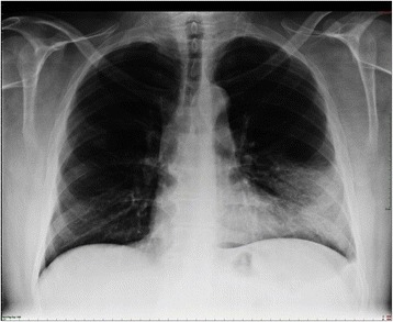
Lobar pneumonia. Dense infiltration in the left lower lobe has caused a silhouette of the left cardiac border (dashed line). Air bronchogram is a typical feature of consolidation.
Image: “Chest x-ray of the patient with dense infiltration in the left lower lobe” by Ortega et al. License: CC BY 4.0, edited by Lecturio.
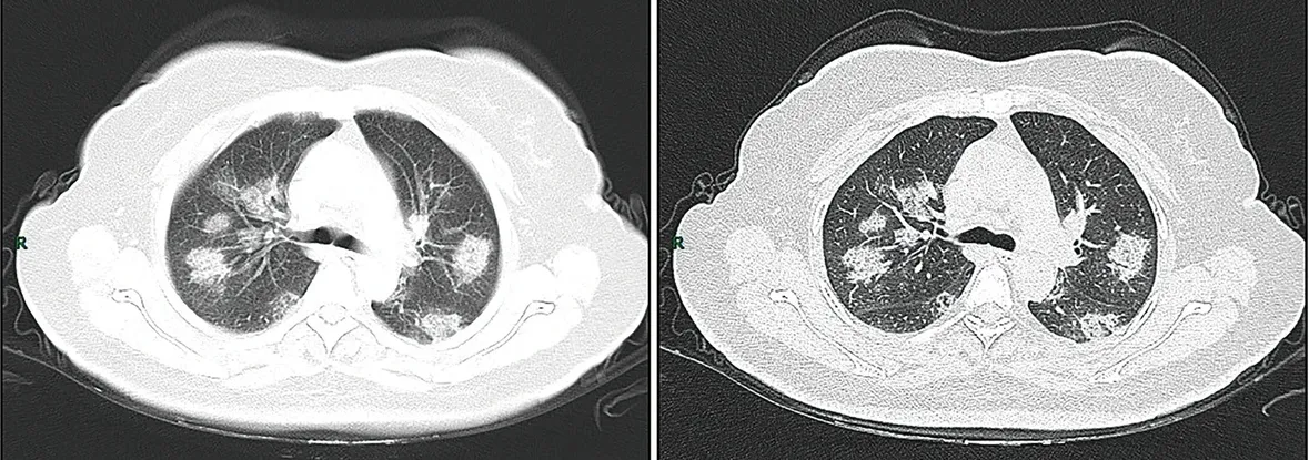
CT imaging of rapid progression stage. A 50-year-old female with anorexia, fatigue, muscle soreness, nasal congestion, and runny nose for 1 week, sore and itching throat for 2 days. Laboratory test: increased erythrocyte sedimentation rate (25 mm/h), normal white blood cells (4.08 × 10⁹/L), decreased lymphocytes (0.96 × 10⁹/ L), increased C-reaction protein (60.8 mg/L). Imaging examination: a (thin layer CT) and b (high-resolution CT) showed multiple patchy and light consolidation in both lungs and grid-like thickness of interlobular septa.
Image: “COVID19 CT chest” by Jin, Y., Cai, L., Cheng, Z. et al. License: CC BY 4.0
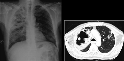
Necrotizing cavitating pneumonia. Chest X-ray and CT depicting necrotizing cavitating pneumonia due to Staphylococcus aureus in a 29-year-old man with acquired immunodeficiency syndrome.
Image: “Necrotizing cavitating pneumonia” by Annals of Thoracic Medicine. License: CC BY 2.0
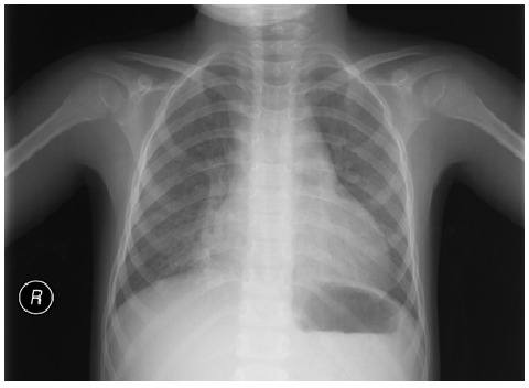
Atypical pneumonia: mild diffuse interstitial infiltration. No lobar consolidation, effusion, or pneumothorax is observed.
Image: “Chest X-ray demonstrating interstitial infiltration” by Department of pediatrics, Ege University Faculty of Medicine, Izmir, 35100 Bornova, Turkey. License: CC BY 3.0
Etiologic diagnosis
- Sputum and blood cultures are recommended only in severe pneumonia or likely infection with MRSA or Pseudomonas aeruginosa.
- Polymerase chain reaction (PCR) of nasopharyngeal swabs:
- During influenza season, testing for influenza is recommended.
- Testing may be indicated for a specific virus during outbreaks based on local or regional health-care policies and availability of tests. For example, testing priorities for SARS-CoV-2 during the pandemic include:
- Hospitalized patients
- Symptomatic healthcare workers/first responders
- Symptomatic patients who are in long-term care facilities, are ≥ 65 years old, or have comorbidities
- Asymptomatic critical infrastructure workers, healthcare workers, or first responders
- Symptomatic individuals who do not meet the above categories
- Mildly symptomatic individuals in communities with high hospitalization rates for COVID-19
- Can also detect bacteria including Legionella species, M. pneumoniae, C. pneumoniae, and mycobacterium
- Urinary antigen test may detect Legionalla pneumophila or pneumococcus
Noninfectious differential diagnoses
- Pulmonary edema: Bilateral infiltration with central predominance and abnormal ECG is suggestive.
- Pulmonary embolism: rarely presents with productive cough or infiltrations visible on chest X-ray
- Lung carcinoma: A history of smoking, constitutional symptoms (e.g., significant weight loss), or chronic cough may be suggestive.
- Radiation
- Hypersensitivity pneumonitis: diagnostic criteria including a compatible exposure history
- Connective tissue disease involving the lung: Most often, a prior diagnosis or symptoms of underlying disease is already present.
Infectious differential diagnoses
- Acute bronchitis
- Exacerbation of COPD
- Tuberculosis
- Lung abscess
Risk assessment
In addition to clinical judgment, a validated prediction tool is recommended to determine the need for hospitalization:
- Preferred tool: Pneumonia Severity Index (PSI) (based on a combination of patient demographics, comorbidities, physical examination findings, and laboratory and imaging studies including arterial pH, blood urea nitrogen, serum sodium and glucose, hematocrit, partial pressure of oxygen, and pleural effusion)
- Risk class 1 or 2 is sent home on oral antibiotics.
- Risk class 3 may be sent home on oral antibiotics or admitted for short-term monitoring and antibiotic therapy based on home environment and follow-up.
- Risk class 4 or 5 is hospitalized.
- Alternative tool: CURB-65 (1 point for each of the following: confusion, blood urea nitrogen ≥ 20 mg/dL, respirations ≥ 30/min, systolic blood pressure < 90 mm Hg or diastolic blood pressure < 60 mm Hg, age ≥ 65 years):
- 0–1 (mortality rate < 1%–3%): Patient should be sent home on oral antibiotics.
- 2 (mortality rate 7%): Patient may be sent home on oral antibiotics or admitted for short-term monitoring and antibiotic therapy based on home environment and follow-up.
- 3–5 (mortality rate 14%–28%): Hospitalize the patient.
Severe CAP or CAP requiring ICU admission is defined by CAP plus at least 1 of the following:
- Septic shock requiring vasopressors
- Respiratory failure requiring mechanical ventilation
- 3 or more of the following: respirations ≥ 30/min, PaO2/FiO2 ≤ 250, multilobar pneumonia, confusion, blood urea nitrogen ≥ 20 mg/dL, WBC < 4,000 cells/µL, platelet < 100,000/µL, hypothermia, hypotension requiring aggressive fluid management)
Management
- Serum procalcitonin levels should not influence the decision to treat pneumonia with antibiotics.
- Initial antibiotic treatment for outpatients with CAP:
- No comorbidities or risk factors for MRSA or P. aeruginosa:
- Amoxicillin OR
- Doxycycline OR
- Macrolide (azithromycin, clarithromycin)
- With comorbidities (e.g., congestive heart disease, chronic lung disease, malignancy, cerebrovascular disease, renal disease, liver disease):
- Amoxicillin/clavulanate (or cephalosporin) AND macrolide (or doxycycline) OR
- Respiratory fluoroquinolone (levofloxacin, moxifloxacin, gemifloxacin)
- No comorbidities or risk factors for MRSA or P. aeruginosa:
- Initial antibiotic treatment for inpatients with CAP:
- Nonsevere:
- Β-lactam and macrolide OR
- Respiratory fluoroquinolone
- If ≥ 2 risk factors for MRSA or P. aeruginosa are present, add coverage (see below) only if culture/PCR is positive.
- If lung abscess or empyema is suspected, add anaerobic coverage. (Note: suspected aspiration pneumonia is not an indication for anaerobic coverage.)
- Severe:
- Β-lactam and macrolide (preferred) OR
- Β-lactam and respiratory fluoroquinolone
- If ≥ 2 risk factors for MRSA or P. aeruginosa are present, add coverage
- If lung abscess or empyema is suspected, add anaerobic coverage. (Note: suspected aspiration pneumonia is not an indication for anaerobic coverage.)
- Nonsevere:
- Empiric coverage for MRSA includes vancomycin or linezolid.
- Empiric coverage for P. aeruginosa includes piperacillin-tazobactam, cefepime, ceftazidime, aztreonam, meropenem, or imipenem.
- Outpatients and inpatients with CAP who test positive for influenza should receive anti-influenza treatment (e.g., oseltamivir) in addition to standard therapy for CAP.
- Adjunctive measures in the treatment of CAP:
- Hydration, oxygen therapy for hypoxemia, vasopressors for shock, and mechanical ventilation for respiratory failure
- Corticosteroids are only used in refractory septic shock.
- Duration of antibiotic therapy in outpatients and inpatients with CAP is at least 5 days and based on improvement of vital signs, mentation, ability to eat, and the patient’s overall clinical condition.
- Follow-up chest imaging is not routinely recommended in patients with resolution of CAP within 1 week.
- If no improvement by day 3 or progressively worsening condition despite receiving antibiotics, evaluate:
- Noninfectious differential diagnosis
- Infections other than pneumonia
- Nosocomial superinfection
- Focus such as lung abscess or empyema blocking antibiotic access to pathogen
- Resistance to or wrong dose of antibiotic(s)
- Presence of unsuspected pathogens such as CA-MRSA, tuberculosis, or fungus
Complications
- General:
- Respiratory failure
- Shock and multiorgan failure
- Coagulopathy
- Specific:
- Metastatic infection (rare) such as brain abscess or left side endocarditis
- Lung abscess: suggests aspiration pneumonia (mixed anaerobic-aerobic infection), CA-MRSA, or P. aeruginosa
- Complicated pleural effusion (pus, pH < 7, glucose < 2.2 mmol/L, lactate dehydrogenase > 1,000 U/L, or positive culture for bacteria): complete drainage usually with a chest tube ± delayed video-assisted thoracoscopy
- Relapse or recurrence in same lung segment: evaluate for underlying neoplasm
Prevention
- Vaccination:
- Pneumococcal vaccine (e.g., PCV13) is recommended in children, the elderly, and immunocompromised patients
- Inactive or recombinant form of influenza vaccine is recommended.
- During outbreaks, patients without prior immunization and at risk for complications should receive the vaccine + antiviral chemoprophylaxis (oseltamivir or zanamivir) for 2 weeks until the vaccine induces sufficient protection
- Smokers should be encouraged to quit smoking.
Related videos
Ventilator-Associated Pneumonia and Hospital-Acquired Pneumonia
Etiology
- Hospital/ICU dependent
- Common non-MDR pathogens: S. pneumonia, H. influenza, methicillin-sensitive S. aureus (MSSA), antibiotic-sensitive Enterobacteriaceae
- More common in HAP than in VAP
- More common in VAP developing in the first week of admission
- Common MDR pathogens: P. aeruginosa, MRSA, antibiotic-resistant Enterobacteriaceae, L. pneumophila, Aspergillus spp., etc.
- More common in VAP than in HAP
- More common in VAP developing after 1st week of admission
- Anaerobes are more common in HAP than in VAP
Epidemiology
Approximately 10% of ICU patients have pneumonia, mostly VAP.
Pathogenesis of VAP
- Oropharyngeal colonization with pathogenic organism
- Aspiration of the pathogen
- Compromise of the normal host defense mechanism
Risk factors
- Risk factors for CAP
- Endotracheal tube: increases risk of microaspiration
- Endotracheal tube: allows pathogenic bacteria to form a layer of resistant biofilm
- Suctioning: damages the endotracheal mucosa and dislodges bacteria in biofilm
- Poor hand hygiene: increases risk of cross-infection from other patients
- Antibiotic exposure: increases risk of MDR pathogens
- Severely ill state with sepsis, trauma, and/or hyperglycemia: causes immunoparalysis
Clinical manifestations and diagnosis
- Similarities with CAP: tachypnea and increased minute ventilation, tachycardia, fever, increased sputum, leukocytosis, worsening oxygenation, and signs of consolidation
- VAP is more difficult to diagnose:
- Prior infiltrates are common in ventilated patients
- Anterior-posterior view on chest X-ray is more difficult to interpret
- Signs and symptoms such as fever and leukocytosis could be due to a variety of other causes such as sepsis, other infections, and medication
- Cultures from tracheal aspirates or more distal bronchial aspirates are used for etiologic diagnosis
- Absence of bacteria in gram stains of endotracheal aspirates suggests an alternative diagnosis for symptoms of fever and pulmonary infiltrates
- Culture-based diagnosis is more difficult in HAP because cultures are more difficult to obtain
Differential diagnosis
- Pulmonary edema
- Pulmonary contusion
- Alveolar hemorrhage
- Hypersensitivity pneumonitis
- Acute respiratory distress syndrome
- Pulmonary embolism
Treatment
- Empiric broad-spectrum antibiotics in patients with risk factors for MDR pathogens (most patients with VAP and HAP, especially VAP)
- Specific antibiotic therapy: once the etiologic diagnosis is known; usually a single agent
- Appropriate clinical response is expected within 48–72 hours.
Complications
- Shock, multiorgan failure, coagulopathy, complicated pleural effusion, lung abscess, metastatic infection, etc.
Prognosis
- Due to frequent presence of comorbidities, VAP mortality rate is 50%–70%.
- VAP with MDR pathogens has higher mortality.
- HAP has a better prognosis due to better host immunity and lower prevalence of MDR pathogens.
Prevention
- Consistent infection-control measures and hand washing
- Avoiding unnecessary intubation
- Appropriate use of noninvasive ventilation
- Minimizing duration of intubation
- Judicious use of antibiotics
- Head elevation (30°–45°)
- Endotracheal tubes with special cuffs that reduce microaspiration
- Reducing patient transportation outside the ICU
References
- Metlay JP, Waterer GW, Long AC, et al. Diagnosis and Treatment of Adults with Community-acquired Pneumonia. An Official Clinical Practice Guideline of the American Thoracic Society and Infectious Diseases Society of America. Am J Respir Crit Care Med. 2019 Oct 1;200(7):e45-e67. doi: 10.1164/rccm.201908-1581ST. Retrieved from https://pubmed.ncbi.nlm.nih.gov/31573350/#
- Kasper DL, Fausi AS, Hauser SL, Longo DL, Lameson JL, Loscalzo J. Harrison’s Principles of Internal Medicine. New York, NY: McGraw-Hill Education; 2018.