Intestinal ischemia occurs when perfusion fails to meet the demands of the intestines, resulting in ischemic tissue injury that can be life-threatening if bowel necrosis and/or perforation occurs. Symptoms can range from mild indigestion or diarrhea to severe abdominal pain. Imaging techniques including CT and angiography are used to detect stenosis or occlusion. The chronic form of intestinal ischemia benefits from medical therapies and revascularization procedures (stents, bypass surgery) while acute forms require urgent interventions to restore blood flow and remove any dead bowel tissue. Delay in the diagnosis and management of acute intestinal ischemia results in high mortality and severe complications, including intestinal perforation and sepsis.
Intestinal ischemiaIschemiaA hypoperfusion of the blood through an organ or tissue caused by a pathologic constriction or obstruction of its blood vessels, or an absence of blood circulation.Ischemic Cell Damage is a decrease in blood flowBlood flowBlood flow refers to the movement of a certain volume of blood through the vasculature over a given unit of time (e.g., mL per minute).Vascular Resistance, Flow, and Mean Arterial Pressure to the intestines resulting in hypoperfusion that may lead to bowel infarction. Mucosal sloughing occurs after approximately 3 hours of ischemiaIschemiaA hypoperfusion of the blood through an organ or tissue caused by a pathologic constriction or obstruction of its blood vessels, or an absence of blood circulation.Ischemic Cell Damage, and necrosisNecrosisThe death of cells in an organ or tissue due to disease, injury or failure of the blood supply.Ischemic Cell Damage occurs after approximately 6–12 hours of ischemiaIschemiaA hypoperfusion of the blood through an organ or tissue caused by a pathologic constriction or obstruction of its blood vessels, or an absence of blood circulation.Ischemic Cell Damage.
Types of intestinal ischemiaIschemiaA hypoperfusion of the blood through an organ or tissue caused by a pathologic constriction or obstruction of its blood vessels, or an absence of blood circulation.Ischemic Cell Damage[1]
Ischemic colitisIschemic colitisInflammation of the colon due to colonic ischemia resulting from alterations in systemic circulation or local vasculature.Large Bowel Obstruction (ischemiaIschemiaA hypoperfusion of the blood through an organ or tissue caused by a pathologic constriction or obstruction of its blood vessels, or an absence of blood circulation.Ischemic Cell Damage of colonColonThe large intestines constitute the last portion of the digestive system. The large intestine consists of the cecum, appendix, colon (with ascending, transverse, descending, and sigmoid segments), rectum, and anal canal. The primary function of the colon is to remove water and compact the stool prior to expulsion from the body via the rectum and anal canal. Colon, Cecum, and Appendix: Anatomy): hypoperfusion of the large bowel
Acute mesenteric ischemiaAcute Mesenteric IschemiaMesenteric Ischemia: acute loss of blood flowBlood flowBlood flow refers to the movement of a certain volume of blood through the vasculature over a given unit of time (e.g., mL per minute).Vascular Resistance, Flow, and Mean Arterial Pressure to the small intestineSmall intestineThe small intestine is the longest part of the GI tract, extending from the pyloric orifice of the stomach to the ileocecal junction. The small intestine is the major organ responsible for chemical digestion and absorption of nutrients. It is divided into 3 segments: the duodenum, the jejunum, and the ileum. Small Intestine: Anatomy
Chronic mesenteric ischemiaChronic Mesenteric IschemiaMesenteric Ischemia: constant or episodic hypoperfusion of the small intestineSmall intestineThe small intestine is the longest part of the GI tract, extending from the pyloric orifice of the stomach to the ileocecal junction. The small intestine is the major organ responsible for chemical digestion and absorption of nutrients. It is divided into 3 segments: the duodenum, the jejunum, and the ileum. Small Intestine: Anatomy
Epidemiology[1,3,4]
For all types, intestinal ischemiaIschemiaA hypoperfusion of the blood through an organ or tissue caused by a pathologic constriction or obstruction of its blood vessels, or an absence of blood circulation.Ischemic Cell Damage primarily affects adults > 60 years of age.
Ischemic colitisIschemic colitisInflammation of the colon due to colonic ischemia resulting from alterations in systemic circulation or local vasculature.Large Bowel Obstruction: most common type of intestinal ischemiaIschemiaA hypoperfusion of the blood through an organ or tissue caused by a pathologic constriction or obstruction of its blood vessels, or an absence of blood circulation.Ischemic Cell Damage (approximately 60%–70%)
In addition to older adults, may also occur in younger people with:
Atrial fibrillationAtrial fibrillationAtrial fibrillation (AF or Afib) is a supraventricular tachyarrhythmia and the most common kind of arrhythmia. It is caused by rapid, uncontrolled atrial contractions and uncoordinated ventricular responses. Atrial Fibrillation (AfibAfibAtrial fibrillation (AF or Afib) is a supraventricular tachyarrhythmia and the most common kind of arrhythmia. It is caused by rapid, uncontrolled atrial contractions and uncoordinated ventricular responses. Atrial Fibrillation)
HypercoagulableHypercoagulableHypercoagulable states (also referred to as thrombophilias) are a group of hematologic diseases defined by an increased risk of clot formation (i.e., thrombosis) due to either an increase in procoagulants, a decrease in anticoagulants, or a decrease in fibrinolysis. Hypercoagulable States states
Occlusive mesenteric infarction has a ↑↑ mortalityMortalityAll deaths reported in a given population.Measures of Health Status rate (approximately 60%)
Chronic mesenteric ischemiaChronic Mesenteric IschemiaMesenteric Ischemia: ↓ incidenceIncidenceThe number of new cases of a given disease during a given period in a specified population. It also is used for the rate at which new events occur in a defined population. It is differentiated from prevalence, which refers to all cases in the population at a given time.Measures of Disease Frequency
Acute Mesenteric Ischemia and Ischemic Colitis
Etiology
Whether in the colonColonThe large intestines constitute the last portion of the digestive system. The large intestine consists of the cecum, appendix, colon (with ascending, transverse, descending, and sigmoid segments), rectum, and anal canal. The primary function of the colon is to remove water and compact the stool prior to expulsion from the body via the rectum and anal canal. Colon, Cecum, and Appendix: Anatomy or small intestines, acute bowel ischemiaBowel ischemiaMesenteric ischemia is a rare, life-threatening condition caused by inadequate blood flow through the mesenteric vessels, which results in ischemia and necrosis of the intestinal wall. Mesenteric ischemia can be either acute or chronic.Mesenteric Ischemia is the result of reduced blood flowBlood flowBlood flow refers to the movement of a certain volume of blood through the vasculature over a given unit of time (e.g., mL per minute).Vascular Resistance, Flow, and Mean Arterial Pressure. Causes include:[3,4,7,8]
Vessel obstruction due to:
Acute arterial embolism (approximately 50%):
Atrial fibrillationAtrial fibrillationAtrial fibrillation (AF or Afib) is a supraventricular tachyarrhythmia and the most common kind of arrhythmia. It is caused by rapid, uncontrolled atrial contractions and uncoordinated ventricular responses. Atrial Fibrillation
Valvular heart disease (i.e., infective endocarditisInfective endocarditisInfective endocarditis (IE) is caused by infection or inflammation of the inner lining of the heart (endocardium), most commonly affecting the heart valves.Endocarditis)
CholesterolCholesterolThe principal sterol of all higher animals, distributed in body tissues, especially the brain and spinal cord, and in animal fats and oils.Cholesterol Metabolism embolism from ruptured arterial plaques
Air embolismAir embolismBlocking of a blood vessel by air bubbles that enter the circulatory system, usually after trauma; surgical procedures, or changes in atmospheric pressure.Nonthrombotic Embolism (can occur in neurosurgeries)
AtherosclerosisAtherosclerosisAtherosclerosis is a common form of arterial disease in which lipid deposition forms a plaque in the blood vessel walls. Atherosclerosis is an incurable disease, for which there are clearly defined risk factors that often can be reduced through a change in lifestyle and behavior of the patient. Atherosclerosis
Venous thrombosisVenous thrombosisThe formation or presence of a blood clot (thrombus) within a vein.Budd-Chiari Syndrome in mesenteric vessels (approximately 5%, rarely involves the colonColonThe large intestines constitute the last portion of the digestive system. The large intestine consists of the cecum, appendix, colon (with ascending, transverse, descending, and sigmoid segments), rectum, and anal canal. The primary function of the colon is to remove water and compact the stool prior to expulsion from the body via the rectum and anal canal. Colon, Cecum, and Appendix: Anatomy); risk factors include:
HypercoagulableHypercoagulableHypercoagulable states (also referred to as thrombophilias) are a group of hematologic diseases defined by an increased risk of clot formation (i.e., thrombosis) due to either an increase in procoagulants, a decrease in anticoagulants, or a decrease in fibrinolysis. Hypercoagulable States states/thrombophiliasThrombophiliasHypercoagulable states (also referred to as thrombophilias) are a group of hematologic diseases defined by an increased risk of clot formation (i.e., thrombosis) due to either an increase in procoagulants, a decrease in anticoagulants, or a decrease in fibrinolysis.Hypercoagulable States
InfectionsInfectionsInvasion of the host organism by microorganisms or their toxins or by parasites that can cause pathological conditions or diseases.Chronic Granulomatous Disease/inflammationInflammationInflammation is a complex set of responses to infection and injury involving leukocytes as the principal cellular mediators in the body’s defense against pathogenic organisms. Inflammation is also seen as a response to tissue injury in the process of wound healing. The 5 cardinal signs of inflammation are pain, heat, redness, swelling, and loss of function. Inflammation
Estrogen therapyEstrogen therapyThe use of hormonal agents with estrogen-like activity in postmenopausal or other estrogen-deficient women to alleviate effects of hormone deficiency, such as vasomotor symptoms, dyspareunia, and progressive development of osteoporosis. This may also include the use of progestational agents in combination therapy.Menopause (e.g., oral contraceptiveOral contraceptiveCompounds, usually hormonal, taken orally in order to block ovulation and prevent the occurrence of pregnancy. The hormones are generally estrogen or progesterone or both.Benign Liver Tumors pills)
Nonocclusive mesenteric ischemiaIschemiaA hypoperfusion of the blood through an organ or tissue caused by a pathologic constriction or obstruction of its blood vessels, or an absence of blood circulation.Ischemic Cell Damage (20%–30%) may be due to:
HypotensionHypotensionHypotension is defined as low blood pressure, specifically < 90/60 mm Hg, and is most commonly a physiologic response. Hypotension may be mild, serious, or life threatening, depending on the cause. Hypotension/shockShockShock is a life-threatening condition associated with impaired circulation that results in tissue hypoxia. The different types of shock are based on the underlying cause: distributive (↑ cardiac output (CO), ↓ systemic vascular resistance (SVR)), cardiogenic (↓ CO, ↑ SVR), hypovolemic (↓ CO, ↑ SVR), obstructive (↓ CO), and mixed. Types of Shock
SepsisSepsisSystemic inflammatory response syndrome with a proven or suspected infectious etiology. When sepsis is associated with organ dysfunction distant from the site of infection, it is called severe sepsis. When sepsis is accompanied by hypotension despite adequate fluid infusion, it is called septic shock.Sepsis and Septic Shock
Congestive heart failureHeart FailureA heterogeneous condition in which the heart is unable to pump out sufficient blood to meet the metabolic need of the body. Heart failure can be caused by structural defects, functional abnormalities (ventricular dysfunction), or a sudden overload beyond its capacity. Chronic heart failure is more common than acute heart failure which results from sudden insult to cardiac function, such as myocardial infarction.Total Anomalous Pulmonary Venous Return (TAPVR)
VasculitisVasculitisInflammation of any one of the blood vessels, including the arteries; veins; and rest of the vasculature system in the body.Systemic Lupus Erythematosus
CocaineCocaineAn alkaloid ester extracted from the leaves of plants including coca. It is a local anesthetic and vasoconstrictor and is clinically used for that purpose, particularly in the eye, ear, nose, and throat. It also has powerful central nervous system effects similar to the amphetamines and is a drug of abuse. Cocaine, like amphetamines, acts by multiple mechanisms on brain catecholaminergic neurons; the mechanism of its reinforcing effects is thought to involve inhibition of dopamine uptake.Local Anesthetics use disorder
Pathophysiology
Mechanisms:[1–4,11]
Hypoperfusion → intestinal hypoxiaHypoxiaSub-optimal oxygen levels in the ambient air of living organisms.Ischemic Cell Damage → bowel wall damage/inflammationInflammationInflammation is a complex set of responses to infection and injury involving leukocytes as the principal cellular mediators in the body’s defense against pathogenic organisms. Inflammation is also seen as a response to tissue injury in the process of wound healing. The 5 cardinal signs of inflammation are pain, heat, redness, swelling, and loss of function. Inflammation
May progress to infarction and necrosisNecrosisThe death of cells in an organ or tissue due to disease, injury or failure of the blood supply.Ischemic Cell Damage within 6–12 hours
Venous obstruction may lead to volvulusVolvulusA volvulus is the twisting or axial rotation of a portion of the bowel around its mesentery. The most common site of volvulus in adults is the colon; most frequently the sigmoid volvulus. Patients typically present with symptoms of bowel obstruction such as abdominal pain, distension, vomiting, and constipation/obstipation. Volvulus (twisting of bowel) or incarcerationIncarcerationInguinal Canal: Anatomy and Hernias, leading to hypoperfusion.
Commonly involved sites in AMI:[1–4,11]
Superior mesenteric arterySuperior mesenteric arteryA large vessel supplying the whole length of the small intestine except the superior part of the duodenum. It also supplies the cecum and the ascending part of the colon and about half the transverse part of the colon. It arises from the anterior surface of the aorta below the celiac artery at the level of the first lumbar vertebra.Small Intestine: Anatomy (SMA)
90% of cases
Supplies: the distal duodenumDuodenumThe shortest and widest portion of the small intestine adjacent to the pylorus of the stomach. It is named for having the length equal to about the width of 12 fingers.Small Intestine: Anatomy, jejunumJejunumThe middle portion of the small intestine, between duodenum and ileum. It represents about 2/5 of the remaining portion of the small intestine below duodenum.Small Intestine: Anatomy, ileumIleumThe distal and narrowest portion of the small intestine, between the jejunum and the ileocecal valve of the large intestine.Small Intestine: Anatomy, and colonColonThe large intestines constitute the last portion of the digestive system. The large intestine consists of the cecum, appendix, colon (with ascending, transverse, descending, and sigmoid segments), rectum, and anal canal. The primary function of the colon is to remove water and compact the stool prior to expulsion from the body via the rectum and anal canal. Colon, Cecum, and Appendix: Anatomy to the splenic flexureSplenic flexureSmall Intestine: Anatomy
Superior mesenteric vein: drains blood from the small intestineSmall intestineThe small intestine is the longest part of the GI tract, extending from the pyloric orifice of the stomach to the ileocecal junction. The small intestine is the major organ responsible for chemical digestion and absorption of nutrients. It is divided into 3 segments: the duodenum, the jejunum, and the ileum. Small Intestine: Anatomy
Inferior mesenteric arteryInferior mesenteric arteryThe artery supplying nearly all the left half of the transverse colon, the whole of the descending colon, the sigmoid colon, and the greater part of the rectum. It is smaller than the superior mesenteric artery and arises from the aorta above its bifurcation into the common iliac arteries.Small Intestine: Anatomy (IMA) (uncommon)
Celiac artery (uncommon)
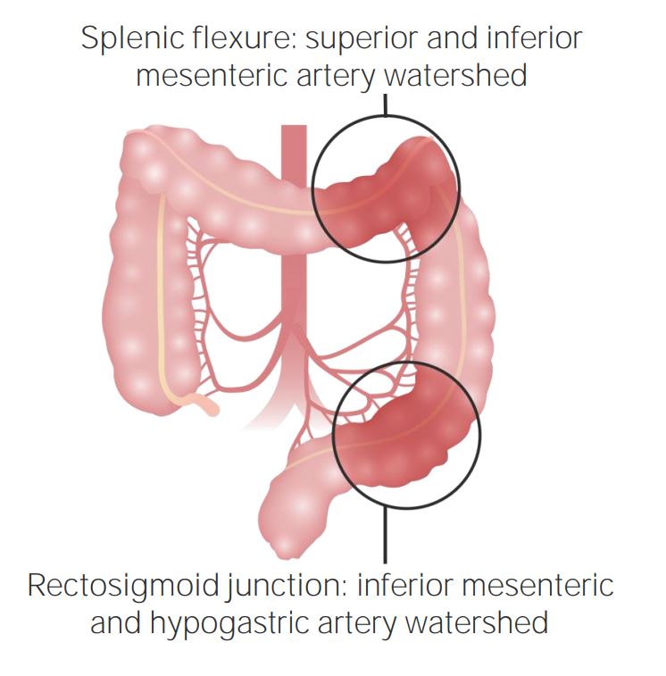
Involved sites in ischemic colitisIschemic colitisInflammation of the colon due to colonic ischemia resulting from alterations in systemic circulation or local vasculature.Large Bowel Obstruction: tends to occur in watershed areasWatershed areasThe regions have dual blood supply, but are located at the most distal reaches of the arteries.Ischemic Cell Damage of the colonColonThe large intestines constitute the last portion of the digestive system. The large intestine consists of the cecum, appendix, colon (with ascending, transverse, descending, and sigmoid segments), rectum, and anal canal. The primary function of the colon is to remove water and compact the stool prior to expulsion from the body via the rectum and anal canal. Colon, Cecum, and Appendix: Anatomy with limited collateral circulationCirculationThe movement of the blood as it is pumped through the cardiovascular system.ABCDE Assessment
Classic finding: sudden onset of abdominal painAbdominal PainAcute Abdomen out of proportion to physical findings (i.e., intense painPainAn unpleasant sensation induced by noxious stimuli which are detected by nerve endings of nociceptive neurons.Pain: Types and Pathways without peritoneal signs early in presentation).
Ischemic colitisIschemic colitisInflammation of the colon due to colonic ischemia resulting from alterations in systemic circulation or local vasculature.Large Bowel Obstruction:[2,3,8,11]
NauseaNauseaAn unpleasant sensation in the stomach usually accompanied by the urge to vomit. Common causes are early pregnancy, sea and motion sickness, emotional stress, intense pain, food poisoning, and various enteroviruses.Antiemetics or vomitingVomitingThe forcible expulsion of the contents of the stomach through the mouth.Hypokalemia
DiarrheaDiarrheaDiarrhea is defined as ≥ 3 watery or loose stools in a 24-hour period. There are a multitude of etiologies, which can be classified based on the underlying mechanism of disease. The duration of symptoms (acute or chronic) and characteristics of the stools (e.g., watery, bloody, steatorrheic, mucoid) can help guide further diagnostic evaluation. Diarrhea, which may be bloody in later stages, indicating infarction
If necrosisNecrosisThe death of cells in an organ or tissue due to disease, injury or failure of the blood supply.Ischemic Cell Damage and/or perforationPerforationA pathological hole in an organ, blood vessel or other soft part of the body, occurring in the absence of external force.Esophagitis present:
Septic shockSeptic shockSepsis associated with hypotension or hypoperfusion despite adequate fluid resuscitation. Perfusion abnormalities may include, but are not limited to lactic acidosis; oliguria; or acute alteration in mental status.Sepsis and Septic Shock: feverFeverFever is defined as a measured body temperature of at least 38°C (100.4°F). Fever is caused by circulating endogenous and/or exogenous pyrogens that increase levels of prostaglandin E2 in the hypothalamus. Fever is commonly associated with chills, rigors, sweating, and flushing of the skin. Fever, hypotensionHypotensionHypotension is defined as low blood pressure, specifically < 90/60 mm Hg, and is most commonly a physiologic response. Hypotension may be mild, serious, or life threatening, depending on the cause. Hypotension, signs of end-organ damage
Venous thrombosisVenous thrombosisThe formation or presence of a blood clot (thrombus) within a vein.Budd-Chiari Syndrome:more gradual onset of symptoms
Diagnosis of AMI
Laboratory studies:[8,9]
No laboratory studies are sensitive or specific enough to identify ischemic bowel.
↑ WBC in > 90%
↑ Lactic acid in 88% (> 2 mmol/L associated with irreversible ischemiaIschemiaA hypoperfusion of the blood through an organ or tissue caused by a pathologic constriction or obstruction of its blood vessels, or an absence of blood circulation.Ischemic Cell Damage)
↑ AmylaseAmylaseA group of amylolytic enzymes that cleave starch, glycogen, and related alpha-1, 4-glucans.Digestion and Absorption in 50%
Imaging:[8,9]
Computed tomographic angiographyAngiographyRadiography of blood vessels after injection of a contrast medium.Cardiac Surgery (CTACTAA non-invasive method that uses a ct scanner for capturing images of blood vessels and tissues. A contrast material is injected, which helps produce detailed images that aid in diagnosing vascular diseases.Pulmonary Function Tests) is 1st-line method and should be performed as soon as possible.
Renal failureRenal failureConditions in which the kidneys perform below the normal level in the ability to remove wastes, concentrate urine, and maintain electrolyte balance; blood pressure; and calcium metabolism. Renal insufficiency can be classified by the degree of kidney damage (as measured by the level of proteinuria) and reduction in glomerular filtration rate.Crush Syndrome is not a contraindication, as the risks of AMI to the kidneysKidneysThe kidneys are a pair of bean-shaped organs located retroperitoneally against the posterior wall of the abdomen on either side of the spine. As part of the urinary tract, the kidneys are responsible for blood filtration and excretion of water-soluble waste in the urine.Kidneys: Anatomy are higher than the negative effects of intravenous contrast.
Sensitivity, 93%; specificity, 100%
Includes:
Precontrast scans detect vascular calcifications, intravascular thrombus, and intramural hemorrhage.
Multiplanar reconstructions (MPRs) assess the origin of mesenteric arteriesArteriesArteries are tubular collections of cells that transport oxygenated blood and nutrients from the heart to the tissues of the body. The blood passes through the arteries in order of decreasing luminal diameter, starting in the largest artery (the aorta) and ending in the small arterioles. Arteries are classified into 3 types: large elastic arteries, medium muscular arteries, and small arteries and arterioles. Arteries: Histology.
CTACTAA non-invasive method that uses a ct scanner for capturing images of blood vessels and tissues. A contrast material is injected, which helps produce detailed images that aid in diagnosing vascular diseases.Pulmonary Function Tests findings of AMI include:
Lack of bowel-wall enhancement
Pneumatosis intestinalisPneumatosis intestinalisA condition characterized by the presence of multiple gas-filled cysts in the intestinal wall, the submucosa and/or subserosa of the intestine. The majority of the cysts are found in the jejunum and the ileum.Necrotizing Enterocolitis: air in the bowel wall
Portal veinPortal veinA short thick vein formed by union of the superior mesenteric vein and the splenic vein.Liver: Anatomy gas
Distended intestinal loops
Bowel wall thickening
Air–fluid levels
MRMRCalculated as the ratio of the total number of people who die due to all causes over a specific time period to the total number of people in the selected population.Measures of Health StatusangiographyAngiographyRadiography of blood vessels after injection of a contrast medium.Cardiac Surgery:
Findings similar to those seen on CT angiographyAngiographyRadiography of blood vessels after injection of a contrast medium.Cardiac Surgery
Higher cost and more time-consuming than CTACTAA non-invasive method that uses a ct scanner for capturing images of blood vessels and tissues. A contrast material is injected, which helps produce detailed images that aid in diagnosing vascular diseases.Pulmonary Function Tests
Conventional mesenteric angiographyAngiographyRadiography of blood vessels after injection of a contrast medium.Cardiac Surgery may be performed if the diagnosis is unclear.
Pros: can treat and diagnose concomitantly; the traditional gold standard
Done initially to rule out pneumoperitoneumPneumoperitoneumA condition with trapped gas or air in the peritoneal cavity, usually secondary to perforation of the internal organs such as the lung and the gastrointestinal tract, or to recent surgery. Pneumoperitoneum may be purposely introduced to aid radiological examination.Perforated Viscus or free air in the peritoneumPeritoneumThe peritoneum is a serous membrane lining the abdominopelvic cavity. This lining is formed by connective tissue and originates from the mesoderm. The membrane lines both the abdominal walls (as parietal peritoneum) and all of the visceral organs (as visceral peritoneum).Peritoneum: Anatomy (which mandates immediate surgery)
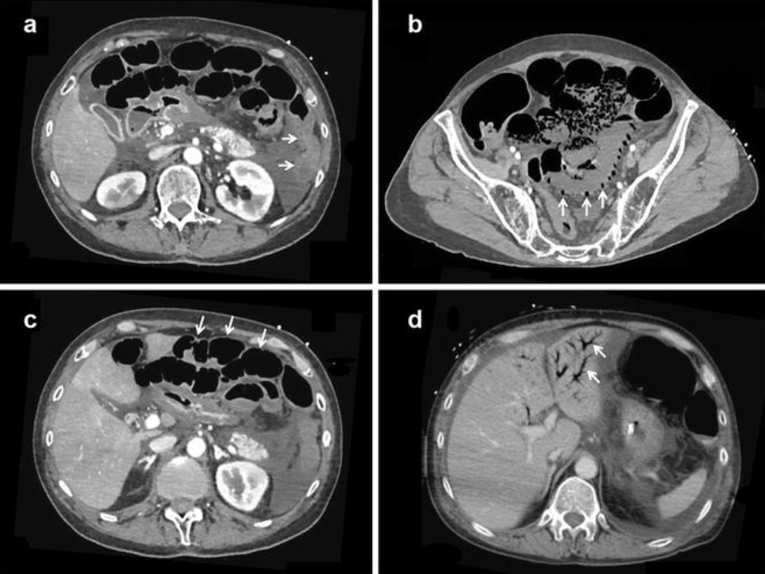
Representative CT scan findings of nonocclusive mesenteric ischemia: a: Absence of contrast-induced bowel wall enhancement (arrows) b: Pneumatosis intestinalis and absence of contrast-induced bowel wall enhancement (arrows) c: Bowel dilatation and absence of contrast-induced bowel wall enhancement (arrows) d: Portal venous gas (arrows)
Image: “Diagnosis of non-occlusive acute mesenteric ischemia in the intensive care unit” by Annals of Intensive Care. License: CC BY 4.0
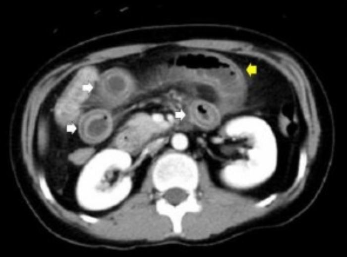
CT scans of the abdomen: Image shows distention of the bowel (yellow arrow), diffuse bowel wall thickening, abnormal bowel wall enhancement (double halo, or target, sign; white arrows).
Image: “The use of tacrolimus for recurrent lupus enteritis: a case report” by Shirai T, Hirabayashi Y, Watanabe R, Tajima Y, Fujii H, Takasawa N, Ishii T, Harigae H. License: CC BY 2.0, cropped by Lecturio.
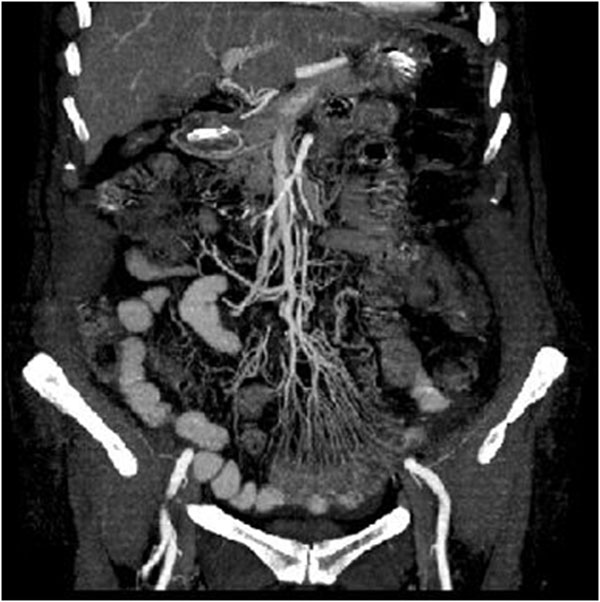
Acute arterial mesenteric ischemia on a contrast-enhanced, multidetector row CT 2-dimensional reconstruction of the coronal plane in early phase:
Image: “Intestinal Ischemia: US-CT findings correlations” by Reginelli A, Genovese E, Cappabianca S, Iacobellis F, Berritto D, Fonio P, Coppolino F, Grassi R. License: CC BY 2.0
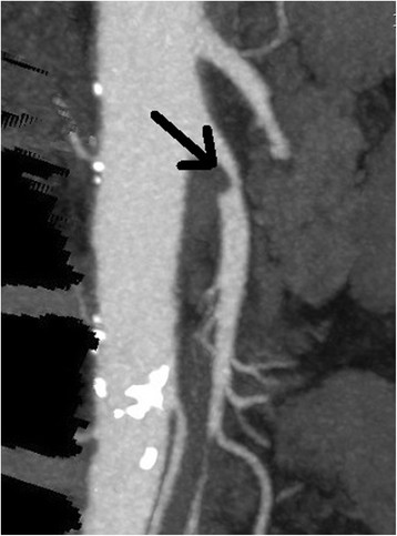
Acute mesenteric ischemia: CT angiogram showing an occlusive embolism of the midportion of the superior mesenteric artery (circle)
Image: “Percutaneous mechanical thrombectomy of superior mesenteric artery embolism” by Dimitrij Kuhelj, Pavel Kavcic, and Peter Popovic. License: CC BY 3.0, edited by Lecturio.
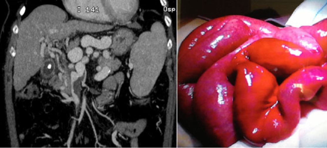
Acute venous mesenteric ischemia on contrast-enhanced multidetector row CT 2-dimensional reconstruction.
The coronal image on the left demonstrates venous thrombosis in the superior mesenteric vein, which was confirmed at surgery (right).
Image: “Intestinal Ischemia: US-CT findings correlations” by Reginelli A, Genovese E, Cappabianca S, Iacobellis F, Berritto D, Fonio P, Coppolino F, Grassi R. License: CC BY 2.0
Diagnosis of ischemic colitisIschemic colitisInflammation of the colon due to colonic ischemia resulting from alterations in systemic circulation or local vasculature.Large Bowel Obstruction
Laboratory studies:[10,11]
Nonspecific
LeukocytosisLeukocytosisA transient increase in the number of leukocytes in a body fluid.West Nile Virus commonly found
↑ Lactate is a sign of severe advanced ischemiaIschemiaA hypoperfusion of the blood through an organ or tissue caused by a pathologic constriction or obstruction of its blood vessels, or an absence of blood circulation.Ischemic Cell Damage.
D-lactate is a product of colonic bacteriaBacteriaBacteria are prokaryotic single-celled microorganisms that are metabolically active and divide by binary fission. Some of these organisms play a significant role in the pathogenesis of diseases. Bacteriology and may be a more specific finding.
Rule out Clostridium difficileClostridium difficileA common inhabitant of the colon flora in human infants and sometimes in adults. The type species clostridioides difficile is formerly known as Clostridium difficile. It is a causative agent for clostridioides infections and is associated with pseudomembranous enterocolitis in patients receiving antibiotic therapy.ClostridiacolitisColitisInflammation of the colon section of the large intestine, usually with symptoms such as diarrhea (often with blood and mucus), abdominal pain, and fever.Pseudomembranous Colitis with C. difficile stool antigenAntigenSubstances that are recognized by the immune system and induce an immune reaction.Vaccination
Imaging:[10,11]
Computed tomography (CT):
Study of choice
Findings include:
Bowel wall thickening
“Thumbprinting” of mucosa
Pericolonic stranding
AscitesAscitesAscites is the pathologic accumulation of fluid within the peritoneal cavity that occurs due to an osmotic and/or hydrostatic pressure imbalance secondary to portal hypertension (cirrhosis, heart failure) or non-portal hypertension (hypoalbuminemia, malignancy, infection).Ascites
Pneumatosis and portal venous gasPortal Venous GasImaging of the Intestines indicate more severe disease, possibly necrotic colonColonThe large intestines constitute the last portion of the digestive system. The large intestine consists of the cecum, appendix, colon (with ascending, transverse, descending, and sigmoid segments), rectum, and anal canal. The primary function of the colon is to remove water and compact the stool prior to expulsion from the body via the rectum and anal canal. Colon, Cecum, and Appendix: Anatomy
Mucosal edemaEdemaEdema is a condition in which excess serous fluid accumulates in the body cavity or interstitial space of connective tissues. Edema is a symptom observed in several medical conditions. It can be categorized into 2 types, namely, peripheral (in the extremities) and internal (in an organ or body cavity). Edema, ulcerationUlcerationCorneal Abrasions, Erosion, and Ulcers, petechial hemorrhage
Colonic single stripe sign: single linear ulcer running along the antimesenteric colonic wall suggestive of ischemic colitisIschemic colitisInflammation of the colon due to colonic ischemia resulting from alterations in systemic circulation or local vasculature.Large Bowel Obstruction
Mucosal sloughing usually seen after 48 hours (“pseudomembranesPseudomembranesRaised yellow or off-white plaques up to 2 cm in diameter that form as a result of mucosal ulcerationPseudomembranous Colitis”)
Gray-green or black mucosa is a sign of transmural ischemiaIschemiaA hypoperfusion of the blood through an organ or tissue caused by a pathologic constriction or obstruction of its blood vessels, or an absence of blood circulation.Ischemic Cell Damage/full-thickness necrosisNecrosisThe death of cells in an organ or tissue due to disease, injury or failure of the blood supply.Ischemic Cell Damage.
AngiographyAngiographyRadiography of blood vessels after injection of a contrast medium.Cardiac Surgery:
Useful if an embolus is suspected, most commonly associated with right colonColonThe large intestines constitute the last portion of the digestive system. The large intestine consists of the cecum, appendix, colon (with ascending, transverse, descending, and sigmoid segments), rectum, and anal canal. The primary function of the colon is to remove water and compact the stool prior to expulsion from the body via the rectum and anal canal. Colon, Cecum, and Appendix: Anatomy and concomitant small bowelSmall bowelThe small intestine is the longest part of the GI tract, extending from the pyloric orifice of the stomach to the ileocecal junction. The small intestine is the major organ responsible for chemical digestion and absorption of nutrients. It is divided into 3 segments: the duodenum, the jejunum, and the ileum.Small Intestine: Anatomy involvement
Otherwise, rarely helpful, as ischemic colitisIschemic colitisInflammation of the colon due to colonic ischemia resulting from alterations in systemic circulation or local vasculature.Large Bowel Obstruction is usually caused by transient small vessel hypoperfusion
Other radiologic tests (limited use):
DopplerDopplerUltrasonography applying the doppler effect, with frequency-shifted ultrasound reflections produced by moving targets (usually red blood cells) in the bloodstream along the ultrasound axis in direct proportion to the velocity of movement of the targets, to determine both direction and velocity of blood flow.Ultrasound (Sonography) ultrasonography
Nuclear medicineNuclear medicineA specialty field of radiology concerned with diagnostic, therapeutic, and investigative use of radioactive compounds.Nuclear Imaging scanning
PainPainAn unpleasant sensation induced by noxious stimuli which are detected by nerve endings of nociceptive neurons.Pain: Types and Pathways control as needed
Minimize pressors and vasoconstricting agents when nonocclusive ischemiaIschemiaA hypoperfusion of the blood through an organ or tissue caused by a pathologic constriction or obstruction of its blood vessels, or an absence of blood circulation.Ischemic Cell Damage is suspected.
Early loss of mucosal barrier may lead to sepsisSepsisSystemic inflammatory response syndrome with a proven or suspected infectious etiology. When sepsis is associated with organ dysfunction distant from the site of infection, it is called severe sepsis. When sepsis is accompanied by hypotension despite adequate fluid infusion, it is called septic shock.Sepsis and Septic Shock.
Antibiotics are indicated empirically if AMI is suspected; options include:[13]
CeftriaxoneCeftriaxoneA broad-spectrum cephalosporin antibiotic and cefotaxime derivative with a very long half-life and high penetrability to meninges, eyes and inner ears.Cephalosporins 1–2 g IV daily + metronidazoleMetronidazoleA nitroimidazole used to treat amebiasis; vaginitis; trichomonas infections; giardiasis; anaerobic bacteria; and treponemal infections.Pyogenic Liver Abscess 500 mg IV every 8 hours
CiprofloxacinCiprofloxacinA broad-spectrum antimicrobial carboxyfluoroquinoline.Fluoroquinolones 400 mg IV daily + metronidazoleMetronidazoleA nitroimidazole used to treat amebiasis; vaginitis; trichomonas infections; giardiasis; anaerobic bacteria; and treponemal infections.Pyogenic Liver Abscess 500 mg IV every 8 hours
AnticoagulationAnticoagulationPulmonary Hypertension Drugs for venous or arterial occlusion and nonocclusive mesenteric ischemiaIschemiaA hypoperfusion of the blood through an organ or tissue caused by a pathologic constriction or obstruction of its blood vessels, or an absence of blood circulation.Ischemic Cell Damage:[8,9]
IV unfractionated heparinUnfractionated heparinA highly acidic mucopolysaccharide formed of equal parts of sulfated d-glucosamine and d-glucuronic acid with sulfaminic bridges. The molecular weight ranges from six to twenty thousand. Heparin occurs in and is obtained from liver, lung, mast cells, etc. , of vertebrates. Its function is unknown, but it is used to prevent blood clotting in vivo and vitro, in the form of many different salts.Anticoagulants should be started unless contraindicated.
May be a definitive treatment for mesenteric venous ischemiaIschemiaA hypoperfusion of the blood through an organ or tissue caused by a pathologic constriction or obstruction of its blood vessels, or an absence of blood circulation.Ischemic Cell Damage
After resuscitationResuscitationThe restoration to life or consciousness of one apparently dead. .Neonatal Respiratory Distress Syndrome, procedure is performed immediately for any patient with peritonitisPeritonitisInflammation of the peritoneum lining the abdominal cavity as the result of infectious, autoimmune, or chemical processes. Primary peritonitis is due to infection of the peritoneal cavity via hematogenous or lymphatic spread and without intra-abdominal source. Secondary peritonitis arises from the abdominal cavity itself through rupture or abscess of intra-abdominal organs.Penetrating Abdominal Injury.
Typically not required for venous ischemiaIschemiaA hypoperfusion of the blood through an organ or tissue caused by a pathologic constriction or obstruction of its blood vessels, or an absence of blood circulation.Ischemic Cell Damage or nonocclusive ischemiaIschemiaA hypoperfusion of the blood through an organ or tissue caused by a pathologic constriction or obstruction of its blood vessels, or an absence of blood circulation.Ischemic Cell Damage (mesenteric vein thrombosisThrombosisFormation and development of a thrombus or blood clot in the blood vessel.Epidemic Typhus)
Embolectomy and angioplastyAngioplastyReconstruction or repair of a blood vessel, which includes the widening of a pathological narrowing of an artery or vein by the removal of atheromatous plaque material and/or the endothelial lining as well, or by dilatation (balloon angioplasty) to compress an atheroma. Except for endarterectomy, usually these procedures are performed via catheterization as minimally invasive endovascular procedures.Cardiac Surgery for arterial emboli
ThrombosisThrombosisFormation and development of a thrombus or blood clot in the blood vessel.Epidemic Typhus at the origin of SMA will frequently require bypass surgery.
Intraoperative angiographyAngiographyRadiography of blood vessels after injection of a contrast medium.Cardiac Surgery should be performed in uncertain cases.
May be used in very early cases of AMI in which bowel necrosisNecrosisThe death of cells in an organ or tissue due to disease, injury or failure of the blood supply.Ischemic Cell Damage is not suspected
Any suspicion of bowel infarction should lead to laparotomyLaparotomyIncision into the side of the abdomen between the ribs and pelvis.Laparotomy and Laparoscopy.
Management of ischemic colitisIschemic colitisInflammation of the colon due to colonic ischemia resulting from alterations in systemic circulation or local vasculature.Large Bowel Obstruction
PatientsPatientsIndividuals participating in the health care system for the purpose of receiving therapeutic, diagnostic, or preventive procedures.Clinician–Patient Relationship without peritonitisPeritonitisInflammation of the peritoneum lining the abdominal cavity as the result of infectious, autoimmune, or chemical processes. Primary peritonitis is due to infection of the peritoneal cavity via hematogenous or lymphatic spread and without intra-abdominal source. Secondary peritonitis arises from the abdominal cavity itself through rupture or abscess of intra-abdominal organs.Penetrating Abdominal Injury and/or signs of full-thickness colonic necrosisNecrosisThe death of cells in an organ or tissue due to disease, injury or failure of the blood supply.Ischemic Cell Damage should be treated nonoperatively.[10,11]
Nonoperative management:
Bowel rest; nasogastric tubeNasogastric tubeMalnutrition in children in resource-limited countries can be placed if there is associated ileusIleusA condition caused by the lack of intestinal peristalsis or intestinal motility without any mechanical obstruction. This interference of the flow of intestinal contents often leads to intestinal obstruction. Ileus may be classified into postoperative, inflammatory, metabolic, neurogenic, and drug-induced.Small Bowel Obstruction.
IV fluidsIV fluidsIntravenous fluids are one of the most common interventions administered in medicine to approximate physiologic bodily fluids. Intravenous fluids are divided into 2 categories: crystalloid and colloid solutions. Intravenous fluids have a wide variety of indications, including intravascular volume expansion, electrolyte manipulation, and maintenance fluids. Intravenous Fluids
Remove all medications contributing to ischemiaIschemiaA hypoperfusion of the blood through an organ or tissue caused by a pathologic constriction or obstruction of its blood vessels, or an absence of blood circulation.Ischemic Cell Damage.
Antibiotics with coverage of enteric flora
EndoscopyEndoscopyProcedures of applying endoscopes for disease diagnosis and treatment. Endoscopy involves passing an optical instrument through a small incision in the skin i.e., percutaneous; or through a natural orifice and along natural body pathways such as the digestive tract; and/or through an incision in the wall of a tubular structure or organ, i.e. Transluminal, to examine or perform surgery on the interior parts of the body.Gastroesophageal Reflux Disease (GERD) (repeat) for reevaluation if no improvement over 48 hours
Surgery:
Indicated for cases of overt peritonitisPeritonitisInflammation of the peritoneum lining the abdominal cavity as the result of infectious, autoimmune, or chemical processes. Primary peritonitis is due to infection of the peritoneal cavity via hematogenous or lymphatic spread and without intra-abdominal source. Secondary peritonitis arises from the abdominal cavity itself through rupture or abscess of intra-abdominal organs.Penetrating Abdominal Injury and evidence of full-thickness colonic necrosisNecrosisThe death of cells in an organ or tissue due to disease, injury or failure of the blood supply.Ischemic Cell Damage or lack of improvement with nonoperative management
Extent of colonic resection depends on the extent of disease.
Decision to perform a primary anastomosis versus colostomy is made based on the clinical picture.
Leaving bowel in discontinuity and performing a 2nd-look laparotomyLaparotomyIncision into the side of the abdomen between the ribs and pelvis.Laparotomy and Laparoscopy is an option if the extent of ischemiaIschemiaA hypoperfusion of the blood through an organ or tissue caused by a pathologic constriction or obstruction of its blood vessels, or an absence of blood circulation.Ischemic Cell Damage is uncertain.
Complications
Reperfusion injuryReperfusion injuryAdverse functional, metabolic, or structural changes in ischemic tissues resulting from the restoration of blood flow to the tissue (reperfusion), including swelling; hemorrhage; necrosis; and damage from free radicals. The most common instance is myocardial reperfusion injury.Ischemic Cell Damage:[2,3]
Can occur following restoration of blood flowBlood flowBlood flow refers to the movement of a certain volume of blood through the vasculature over a given unit of time (e.g., mL per minute).Vascular Resistance, Flow, and Mean Arterial Pressure after a period of ischemiaIschemiaA hypoperfusion of the blood through an organ or tissue caused by a pathologic constriction or obstruction of its blood vessels, or an absence of blood circulation.Ischemic Cell Damage
Complex mechanism involving the release of toxic by-products of ischemic injury and neutrophil activationNeutrophil activationThe process in which the neutrophil is stimulated by diverse substances, resulting in degranulation and/or generation of reactive oxygen products, and culminating in the destruction of invading pathogens. The stimulatory substances, including opsonized particles, immune complexes, and chemotactic factors, bind to specific cell-surface receptors on the neutrophil.Ehrlichiosis and Anaplasmosis
May lead to multisystem organ failure
PerforationPerforationA pathological hole in an organ, blood vessel or other soft part of the body, occurring in the absence of external force.Esophagitis:[2,3]
Necrotic areas of bowel may perforate, spilling contents into the abdominal cavity.
AtherosclerosisAtherosclerosisAtherosclerosis is a common form of arterial disease in which lipid deposition forms a plaque in the blood vessel walls. Atherosclerosis is an incurable disease, for which there are clearly defined risk factors that often can be reduced through a change in lifestyle and behavior of the patient. Atherosclerosis
HypertensionHypertensionHypertension, or high blood pressure, is a common disease that manifests as elevated systemic arterial pressures. Hypertension is most often asymptomatic and is found incidentally as part of a routine physical examination or during triage for an unrelated medical encounter. Hypertension
SmokingSmokingWillful or deliberate act of inhaling and exhaling smoke from burning substances or agents held by hand.Interstitial Lung Diseases
↑ LDL
DiabetesDiabetesDiabetes mellitus (DM) is a metabolic disease characterized by hyperglycemia and dysfunction of the regulation of glucose metabolism by insulin. Type 1 DM is diagnosed mostly in children and young adults as the result of autoimmune destruction of β cells in the pancreas and the resulting lack of insulin. Type 2 DM has a significant association with obesity and is characterized by insulin resistance.Diabetes Mellitus mellitus
Pathophysiology [4,12]
Progressive atherosclerosisAtherosclerosisAtherosclerosis is a common form of arterial disease in which lipid deposition forms a plaque in the blood vessel walls. Atherosclerosis is an incurable disease, for which there are clearly defined risk factors that often can be reduced through a change in lifestyle and behavior of the patient. Atherosclerosis of ≥ 2 main arteriesArteriesArteries are tubular collections of cells that transport oxygenated blood and nutrients from the heart to the tissues of the body. The blood passes through the arteries in order of decreasing luminal diameter, starting in the largest artery (the aorta) and ending in the small arterioles. Arteries are classified into 3 types: large elastic arteries, medium muscular arteries, and small arteries and arterioles. Arteries: Histology → mismatch between the blood flowBlood flowBlood flow refers to the movement of a certain volume of blood through the vasculature over a given unit of time (e.g., mL per minute).Vascular Resistance, Flow, and Mean Arterial Pressure and intestinal metabolic demand (especially after a meal)
Leads to postprandial painPainAn unpleasant sensation induced by noxious stimuli which are detected by nerve endings of nociceptive neurons.Pain: Types and Pathways
Main arteriesArteriesArteries are tubular collections of cells that transport oxygenated blood and nutrients from the heart to the tissues of the body. The blood passes through the arteries in order of decreasing luminal diameter, starting in the largest artery (the aorta) and ending in the small arterioles. Arteries are classified into 3 types: large elastic arteries, medium muscular arteries, and small arteries and arterioles. Arteries: Histology include SMA, IMA, and celiac artery
When only 1 main artery is affected, collateral connections between the arteriesArteriesArteries are tubular collections of cells that transport oxygenated blood and nutrients from the heart to the tissues of the body. The blood passes through the arteries in order of decreasing luminal diameter, starting in the largest artery (the aorta) and ending in the small arterioles. Arteries are classified into 3 types: large elastic arteries, medium muscular arteries, and small arteries and arterioles. Arteries: Histology can form and compensate for the ↓ flowFlowBlood flows through the heart, arteries, capillaries, and veins in a closed, continuous circuit. Flow is the movement of volume per unit of time. Flow is affected by the pressure gradient and the resistance fluid encounters between 2 points. Vascular resistance is the opposition to flow, which is caused primarily by blood friction against vessel walls.Vascular Resistance, Flow, and Mean Arterial Pressure.
Sudden thrombus formation in addition to stenosisStenosisHypoplastic Left Heart Syndrome (HLHS) can lead to acute-on-chronic mesenteric ischemiaIschemiaA hypoperfusion of the blood through an organ or tissue caused by a pathologic constriction or obstruction of its blood vessels, or an absence of blood circulation.Ischemic Cell Damage.
Clinical presentation
Ischemic symptoms occur for ≥ 3 months owing to insufficient blood supply to the gastrointestinal (GI) tract.[4,12]
Other symptoms can include nauseaNauseaAn unpleasant sensation in the stomach usually accompanied by the urge to vomit. Common causes are early pregnancy, sea and motion sickness, emotional stress, intense pain, food poisoning, and various enteroviruses.Antiemetics, early satietyEarly SatietyBariatric Surgery, and diarrheaDiarrheaDiarrhea is defined as ≥ 3 watery or loose stools in a 24-hour period. There are a multitude of etiologies, which can be classified based on the underlying mechanism of disease. The duration of symptoms (acute or chronic) and characteristics of the stools (e.g., watery, bloody, steatorrheic, mucoid) can help guide further diagnostic evaluation. Diarrhea.
May be asymptomatic, owing to the collateral blood supply
Diagnosis
Making the diagnosis:[4,12,14]
Clinical suspicion based on history and physical exam
CT angiographyAngiographyRadiography of blood vessels after injection of a contrast medium.Cardiac Surgery (gold standard) or duplex ultrasonographyDuplex ultrasonographyUltrasonography applying the doppler effect combined with real-time imaging. The real-time image is created by rapid movement of the ultrasound beam. A powerful advantage of this technique is the ability to estimate the velocity of flow from the doppler shift frequency.Hypercoagulable States is used to identify atherosclerotic vascular disease and rule out other abdominal disorders.
Significant stenosisStenosisHypoplastic Left Heart Syndrome (HLHS) (> 70%) within the celiac axis and superior mesenteric arterySuperior mesenteric arteryA large vessel supplying the whole length of the small intestine except the superior part of the duodenum. It also supplies the cecum and the ascending part of the colon and about half the transverse part of the colon. It arises from the anterior surface of the aorta below the celiac artery at the level of the first lumbar vertebra.Small Intestine: Anatomy (SMA), OR
CT angiographyAngiographyRadiography of blood vessels after injection of a contrast medium.Cardiac Surgery (CTACTAA non-invasive method that uses a ct scanner for capturing images of blood vessels and tissues. A contrast material is injected, which helps produce detailed images that aid in diagnosing vascular diseases.Pulmonary Function Tests):
An alternative when CTACTAA non-invasive method that uses a ct scanner for capturing images of blood vessels and tissues. A contrast material is injected, which helps produce detailed images that aid in diagnosing vascular diseases.Pulmonary Function Tests is contraindicated (e.g., contrast allergyAllergyAn abnormal adaptive immune response that may or may not involve antigen-specific IgEType I Hypersensitivity Reaction)
Not readily available
Cannot be performed in patientsPatientsIndividuals participating in the health care system for the purpose of receiving therapeutic, diagnostic, or preventive procedures.Clinician–Patient Relationship with noncompatible pacemakers or mesenteric stents
Duplex ultrasonographyDuplex ultrasonographyUltrasonography applying the doppler effect combined with real-time imaging. The real-time image is created by rapid movement of the ultrasound beam. A powerful advantage of this technique is the ability to estimate the velocity of flow from the doppler shift frequency.Hypercoagulable States:
Digital subtraction angiographyAngiographyRadiography of blood vessels after injection of a contrast medium.Cardiac Surgery:
Used when CTACTAA non-invasive method that uses a ct scanner for capturing images of blood vessels and tissues. A contrast material is injected, which helps produce detailed images that aid in diagnosing vascular diseases.Pulmonary Function Tests results are equivocal
Used in treatment of occlusive mesenteric disease
EndoscopyEndoscopyProcedures of applying endoscopes for disease diagnosis and treatment. Endoscopy involves passing an optical instrument through a small incision in the skin i.e., percutaneous; or through a natural orifice and along natural body pathways such as the digestive tract; and/or through an incision in the wall of a tubular structure or organ, i.e. Transluminal, to examine or perform surgery on the interior parts of the body.Gastroesophageal Reflux Disease (GERD) (esophagogastroduodenoscopy and colonoscopyColonoscopyEndoscopic examination, therapy or surgery of the luminal surface of the colon.Colorectal Cancer Screening):
Quit smokingSmokingWillful or deliberate act of inhaling and exhaling smoke from burning substances or agents held by hand.Interstitial Lung Diseases
Healthy lifestyle habits
Manage chronic conditions (e.g., hypertensionHypertensionHypertension, or high blood pressure, is a common disease that manifests as elevated systemic arterial pressures. Hypertension is most often asymptomatic and is found incidentally as part of a routine physical examination or during triage for an unrelated medical encounter. Hypertension, hyperlipidemia, diabetesDiabetesDiabetes mellitus (DM) is a metabolic disease characterized by hyperglycemia and dysfunction of the regulation of glucose metabolism by insulin. Type 1 DM is diagnosed mostly in children and young adults as the result of autoimmune destruction of β cells in the pancreas and the resulting lack of insulin. Type 2 DM has a significant association with obesity and is characterized by insulin resistance.Diabetes Mellitus)
Endovascular techniques (e.g., angioplastyAngioplastyReconstruction or repair of a blood vessel, which includes the widening of a pathological narrowing of an artery or vein by the removal of atheromatous plaque material and/or the endothelial lining as well, or by dilatation (balloon angioplasty) to compress an atheroma. Except for endarterectomy, usually these procedures are performed via catheterization as minimally invasive endovascular procedures.Cardiac Surgery or stenting): 1st line for symptomatic patientsPatientsIndividuals participating in the health care system for the purpose of receiving therapeutic, diagnostic, or preventive procedures.Clinician–Patient Relationship
Open surgery (e.g., vascular bypass or endarterectomyEndarterectomySurgical excision, performed under general anesthesia, of the atheromatous tunica intima of an artery. When reconstruction of an artery is performed as an endovascular procedure through a catheter, it is called atherectomy.Intestinal Ischemia)
Since intestinal ischemiaIschemiaA hypoperfusion of the blood through an organ or tissue caused by a pathologic constriction or obstruction of its blood vessels, or an absence of blood circulation.Ischemic Cell Damage typically presents with abdominal painAbdominal PainAcute Abdomen, the differential diagnosis includes:
AppendicitisAppendicitisAppendicitis is the acute inflammation of the vermiform appendix and the most common abdominal surgical emergency globally. The condition has a lifetime risk of 8%. Characteristic features include periumbilical abdominal pain that migrates to the right lower quadrant, fever, anorexia, nausea, and vomiting.Appendicitis:acute inflammationAcute InflammationInflammation of the appendixAppendixA worm-like blind tube extension from the cecum.Colon, Cecum, and Appendix: Anatomy. Symptoms are periumbilical painPainAn unpleasant sensation induced by noxious stimuli which are detected by nerve endings of nociceptive neurons.Pain: Types and Pathways that migrates to the RLQ, feverFeverFever is defined as a measured body temperature of at least 38°C (100.4°F). Fever is caused by circulating endogenous and/or exogenous pyrogens that increase levels of prostaglandin E2 in the hypothalamus. Fever is commonly associated with chills, rigors, sweating, and flushing of the skin. Fever, anorexiaAnorexiaThe lack or loss of appetite accompanied by an aversion to food and the inability to eat. It is the defining characteristic of the disorder anorexia nervosa.Anorexia Nervosa, nauseaNauseaAn unpleasant sensation in the stomach usually accompanied by the urge to vomit. Common causes are early pregnancy, sea and motion sickness, emotional stress, intense pain, food poisoning, and various enteroviruses.Antiemetics, and vomitingVomitingThe forcible expulsion of the contents of the stomach through the mouth.Hypokalemia, but appendicitisAppendicitisAppendicitis is the acute inflammation of the vermiform appendix and the most common abdominal surgical emergency globally. The condition has a lifetime risk of 8%. Characteristic features include periumbilical abdominal pain that migrates to the right lower quadrant, fever, anorexia, nausea, and vomiting.Appendicitis can often cause constipationConstipationConstipation is common and may be due to a variety of causes. Constipation is generally defined as bowel movement frequency < 3 times per week. Patients who are constipated often strain to pass hard stools. The condition is classified as primary (also known as idiopathic or functional constipation) or secondary, and as acute or chronic. Constipation, as well. The diagnosis is clinical, but CT imaging is used in cases of uncertainty. The standard management is appendectomyAppendectomyAppendectomy is an invasive surgical procedure performed with the goal of resecting and extracting the vermiform appendix through either an open or a laparoscopic approach. The most common indication is acute appendicitis.Appendectomy, though there can be a role for antibiotics in some cases.
Bowel obstructionBowel obstructionAny impairment, arrest, or reversal of the normal flow of intestinal contents toward the anal canal.Ascaris/Ascariasis: interruption of the flowFlowBlood flows through the heart, arteries, capillaries, and veins in a closed, continuous circuit. Flow is the movement of volume per unit of time. Flow is affected by the pressure gradient and the resistance fluid encounters between 2 points. Vascular resistance is the opposition to flow, which is caused primarily by blood friction against vessel walls.Vascular Resistance, Flow, and Mean Arterial Pressure of the intraluminal contents through the small intestineSmall intestineThe small intestine is the longest part of the GI tract, extending from the pyloric orifice of the stomach to the ileocecal junction. The small intestine is the major organ responsible for chemical digestion and absorption of nutrients. It is divided into 3 segments: the duodenum, the jejunum, and the ileum. Small Intestine: Anatomy. Typically, bowel obstructionBowel obstructionAny impairment, arrest, or reversal of the normal flow of intestinal contents toward the anal canal.Ascaris/Ascariasis presents with nauseaNauseaAn unpleasant sensation in the stomach usually accompanied by the urge to vomit. Common causes are early pregnancy, sea and motion sickness, emotional stress, intense pain, food poisoning, and various enteroviruses.Antiemetics, vomitingVomitingThe forcible expulsion of the contents of the stomach through the mouth.Hypokalemia, abdominal painAbdominal PainAcute Abdomen, distention, constipationConstipationConstipation is common and may be due to a variety of causes. Constipation is generally defined as bowel movement frequency < 3 times per week. Patients who are constipated often strain to pass hard stools. The condition is classified as primary (also known as idiopathic or functional constipation) or secondary, and as acute or chronic. Constipation, and/or obstipationObstipationLarge Bowel Obstruction. The diagnosis is established via imaging. Most cases will resolve with supportive management (bowel rest, IV hydrationIv HydrationCrush Syndrome, and nasogastric decompression). However, surgery is required for persistent or complicated cases.
DiverticulitisDiverticulitisInflammation of a diverticulum or diverticula.Diverticular Disease: inflammationInflammationInflammation is a complex set of responses to infection and injury involving leukocytes as the principal cellular mediators in the body’s defense against pathogenic organisms. Inflammation is also seen as a response to tissue injury in the process of wound healing. The 5 cardinal signs of inflammation are pain, heat, redness, swelling, and loss of function. Inflammation of diverticula (protrusions of the bowel wall, often in the colonColonThe large intestines constitute the last portion of the digestive system. The large intestine consists of the cecum, appendix, colon (with ascending, transverse, descending, and sigmoid segments), rectum, and anal canal. The primary function of the colon is to remove water and compact the stool prior to expulsion from the body via the rectum and anal canal. Colon, Cecum, and Appendix: Anatomy). DiverticulitisDiverticulitisInflammation of a diverticulum or diverticula.Diverticular Disease often presents with lower abdominal painAbdominal PainAcute Abdomen and changes in bowel habits. The condition may be further complicated by abscessAbscessAccumulation of purulent material in tissues, organs, or circumscribed spaces, usually associated with signs of infection.Chronic Granulomatous Disease, perforationPerforationA pathological hole in an organ, blood vessel or other soft part of the body, occurring in the absence of external force.Esophagitis, fistulaFistulaAbnormal communication most commonly seen between two internal organs, or between an internal organ and the surface of the body.Anal Fistula, and bowel obstructionBowel obstructionAny impairment, arrest, or reversal of the normal flow of intestinal contents toward the anal canal.Ascaris/Ascariasis. Management consists of antibiotics, fluid resuscitationResuscitationThe restoration to life or consciousness of one apparently dead. .Neonatal Respiratory Distress Syndrome, and bowel rest. Surgery is required for complications.
Acute pancreatitisPancreatitisInflammation of the pancreas. Pancreatitis is classified as acute unless there are computed tomographic or endoscopic retrograde cholangiopancreatographic findings of chronic pancreatitis. The two most common forms of acute pancreatitis are alcoholic pancreatitis and gallstone pancreatitis.Acute Pancreatitis: inflammatory disease of the pancreasPancreasThe pancreas lies mostly posterior to the stomach and extends across the posterior abdominal wall from the duodenum on the right to the spleen on the left. This organ has both exocrine and endocrine tissue. Pancreas: Anatomy usually due to gallstonesGallstonesCholelithiasis (gallstones) is the presence of stones in the gallbladder. Most gallstones are cholesterol stones, while the rest are composed of bilirubin (pigment stones) and other mixed components. Patients are commonly asymptomatic but may present with biliary colic (intermittent pain in the right upper quadrant).Cholelithiasis and/or excessive alcohol use. PatientsPatientsIndividuals participating in the health care system for the purpose of receiving therapeutic, diagnostic, or preventive procedures.Clinician–Patient Relationship typically present with epigastric painEpigastric painMallory-Weiss Syndrome (Mallory-Weiss Tear) radiating to the back. Diagnosis includes serum lipaseLipaseAn enzyme of the hydrolase class that catalyzes the reaction of triacylglycerol and water to yield diacylglycerol and a fatty acid anion. It is produced by glands on the tongue and by the pancreas and initiates the digestion of dietary fats.Malabsorption and Maldigestion 3 times the upper limitLimitA value (e.g., pressure or time) that should not be exceeded and which is specified by the operator to protect the lungInvasive Mechanical Ventilation of normal or characteristic radiographic findings. Management includes aggressive IV hydrationIv HydrationCrush Syndrome, analgesiaAnalgesiaMethods of pain relief that may be used with or in place of analgesics.Anesthesiology: History and Basic Concepts, nutritional support, and treatment of the underlying cause.
Chronic pancreatitisPancreatitisInflammation of the pancreas. Pancreatitis is classified as acute unless there are computed tomographic or endoscopic retrograde cholangiopancreatographic findings of chronic pancreatitis. The two most common forms of acute pancreatitis are alcoholic pancreatitis and gallstone pancreatitis.Acute Pancreatitis: persistent inflammationInflammationInflammation is a complex set of responses to infection and injury involving leukocytes as the principal cellular mediators in the body’s defense against pathogenic organisms. Inflammation is also seen as a response to tissue injury in the process of wound healing. The 5 cardinal signs of inflammation are pain, heat, redness, swelling, and loss of function. Inflammation, fibrosisFibrosisAny pathological condition where fibrous connective tissue invades any organ, usually as a consequence of inflammation or other injury.Bronchiolitis Obliterans, and irreversible cell damage to the pancreasPancreasThe pancreas lies mostly posterior to the stomach and extends across the posterior abdominal wall from the duodenum on the right to the spleen on the left. This organ has both exocrine and endocrine tissue. Pancreas: Anatomy. The most common etiologies of chronic pancreatitisPancreatitisInflammation of the pancreas. Pancreatitis is classified as acute unless there are computed tomographic or endoscopic retrograde cholangiopancreatographic findings of chronic pancreatitis. The two most common forms of acute pancreatitis are alcoholic pancreatitis and gallstone pancreatitis.Acute Pancreatitis are alcohol use disorderAlcohol use disorderAlcohol is one of the most commonly used addictive substances in the world. Alcohol use disorder (AUD) is defined as pathologic consumption of alcohol leading to impaired daily functioning. Acute alcohol intoxication presents with impairment in speech and motor functions and can be managed in most cases with supportive care. Alcohol Use Disorder and pancreatic duct obstruction. PatientsPatientsIndividuals participating in the health care system for the purpose of receiving therapeutic, diagnostic, or preventive procedures.Clinician–Patient Relationship present with recurrent epigastric abdominal painAbdominal PainAcute Abdomen, nauseaNauseaAn unpleasant sensation in the stomach usually accompanied by the urge to vomit. Common causes are early pregnancy, sea and motion sickness, emotional stress, intense pain, food poisoning, and various enteroviruses.Antiemetics, and features of malabsorptionMalabsorptionGeneral term for a group of malnutrition syndromes caused by failure of normal intestinal absorption of nutrients.Malabsorption and Maldigestion syndrome. CT findings include pancreatic atrophyAtrophyDecrease in the size of a cell, tissue, organ, or multiple organs, associated with a variety of pathological conditions such as abnormal cellular changes, ischemia, malnutrition, or hormonal changes.Cellular Adaptation, dilated pancreatic ducts, and pancreatic calcifications.Therapy focuses on alcohol cessation, diet changes, painPainAn unpleasant sensation induced by noxious stimuli which are detected by nerve endings of nociceptive neurons.Pain: Types and Pathways management, and treatment of pancreatic insufficiency.
CholecystitisCholecystitisCholecystitis is the inflammation of the gallbladder (GB) usually caused by the obstruction of the cystic duct (acute cholecystitis). Mechanical irritation by gallstones can also produce chronic GB inflammation. Cholecystitis is one of the most common complications of cholelithiasis but inflammation without gallstones can occur in a minority of patients. Cholecystitis:inflammationInflammationInflammation is a complex set of responses to infection and injury involving leukocytes as the principal cellular mediators in the body’s defense against pathogenic organisms. Inflammation is also seen as a response to tissue injury in the process of wound healing. The 5 cardinal signs of inflammation are pain, heat, redness, swelling, and loss of function. Inflammation of the gallbladderGallbladderThe gallbladder is a pear-shaped sac, located directly beneath the liver, that sits on top of the superior part of the duodenum. The primary functions of the gallbladder include concentrating and storing up to 50 mL of bile. Gallbladder and Biliary Tract: Anatomy usually caused by the obstruction of the cysticCysticFibrocystic Change duct (acute cholecystitisAcute cholecystitisAcute inflammation of the gallbladder wall. It is characterized by the presence of abdominal pain; fever; and leukocytosis. Gallstone obstruction of the cystic duct is present in approximately 90% of the cases.Cholecystitis). The acute type of cholecystitisCholecystitisCholecystitis is the inflammation of the gallbladder (GB) usually caused by the obstruction of the cystic duct (acute cholecystitis). Mechanical irritation by gallstones can also produce chronic GB inflammation. Cholecystitis is one of the most common complications of cholelithiasis but inflammation without gallstones can occur in a minority of patients. Cholecystitis usually presents with RUQ painPainAn unpleasant sensation induced by noxious stimuli which are detected by nerve endings of nociceptive neurons.Pain: Types and Pathways, feverFeverFever is defined as a measured body temperature of at least 38°C (100.4°F). Fever is caused by circulating endogenous and/or exogenous pyrogens that increase levels of prostaglandin E2 in the hypothalamus. Fever is commonly associated with chills, rigors, sweating, and flushing of the skin. Fever, and leukocytosisLeukocytosisA transient increase in the number of leukocytes in a body fluid.West Nile Virus. The diagnosis is made clinically and confirmed with ultrasonography. The definitive management is cholecystectomyCholecystectomyCholecystectomy is a surgical procedure performed with the goal of resecting and extracting the gallbladder. It is one of the most common abdominal surgeries performed in the Western world. Cholecystectomy is performed for symptomatic cholelithiasis, cholecystitis, gallbladder polyps > 0.5 cm, porcelain gallbladder, choledocholithiasis and gallstone pancreatitis, and rarely, for gallbladder cancer. Cholecystectomy.
Acute coronary syndrome (ACS): clot formation obstructing blood flowBlood flowBlood flow refers to the movement of a certain volume of blood through the vasculature over a given unit of time (e.g., mL per minute).Vascular Resistance, Flow, and Mean Arterial Pressure to the coronary arteriesArteriesArteries are tubular collections of cells that transport oxygenated blood and nutrients from the heart to the tissues of the body. The blood passes through the arteries in order of decreasing luminal diameter, starting in the largest artery (the aorta) and ending in the small arterioles. Arteries are classified into 3 types: large elastic arteries, medium muscular arteries, and small arteries and arterioles. Arteries: Histology. Symptoms include chest pressure and shortness of breathShortness of breathDyspnea is the subjective sensation of breathing discomfort. Dyspnea is a normal manifestation of heavy physical or psychological exertion, but also may be caused by underlying conditions (both pulmonary and extrapulmonary).Dyspnea. Management includes medications, such as blood thinners, thrombolyticsThrombolyticsThrombolytics, also known as fibrinolytics, include recombinant tissue plasminogen activator (TPa) (i.e., alteplase, reteplase, and tenecteplase), urokinase, and streptokinase. The agents promote the breakdown of a blood clot by converting plasminogen to plasmin, which then degrades fibrin. Thrombolytics, and/or beta blockers. Depending on the severity, heart catheterization and balloon angioplastyAngioplastyReconstruction or repair of a blood vessel, which includes the widening of a pathological narrowing of an artery or vein by the removal of atheromatous plaque material and/or the endothelial lining as well, or by dilatation (balloon angioplasty) to compress an atheroma. Except for endarterectomy, usually these procedures are performed via catheterization as minimally invasive endovascular procedures.Cardiac Surgery may be required.
Aortic dissectionAortic dissectionAortic dissection occurs due to shearing stress from pulsatile pressure causing a tear in the tunica intima of the aortic wall. This tear allows blood to flow into the media, creating a “false lumen.” Aortic dissection is most commonly caused by uncontrolled hypertension.Aortic Dissection: may also present with sudden onset of severe epigastric painEpigastric painMallory-Weiss Syndrome (Mallory-Weiss Tear). The painPainAn unpleasant sensation induced by noxious stimuli which are detected by nerve endings of nociceptive neurons.Pain: Types and Pathways associated with aortic dissectionAortic dissectionAortic dissection occurs due to shearing stress from pulsatile pressure causing a tear in the tunica intima of the aortic wall. This tear allows blood to flow into the media, creating a “false lumen.” Aortic dissection is most commonly caused by uncontrolled hypertension.Aortic Dissection is typically sharp tearing chest painTearing Chest PainChest Pain that radiates to the back. Other features, such as asymmetrical blood pressure, and pulse deficit in 1 armArmThe arm, or “upper arm” in common usage, is the region of the upper limb that extends from the shoulder to the elbow joint and connects inferiorly to the forearm through the cubital fossa. It is divided into 2 fascial compartments (anterior and posterior).Arm: Anatomy can help differentiate aortic dissectionAortic dissectionAortic dissection occurs due to shearing stress from pulsatile pressure causing a tear in the tunica intima of the aortic wall. This tear allows blood to flow into the media, creating a “false lumen.” Aortic dissection is most commonly caused by uncontrolled hypertension.Aortic Dissection from acute mesenteric ischemiaAcute Mesenteric IschemiaMesenteric Ischemia, though it is also possible for an aortic dissectionAortic dissectionAortic dissection occurs due to shearing stress from pulsatile pressure causing a tear in the tunica intima of the aortic wall. This tear allows blood to flow into the media, creating a “false lumen.” Aortic dissection is most commonly caused by uncontrolled hypertension.Aortic Dissection to cause a nonocclusive mesenteric ischemiaIschemiaA hypoperfusion of the blood through an organ or tissue caused by a pathologic constriction or obstruction of its blood vessels, or an absence of blood circulation.Ischemic Cell Damage.
Ectopic pregnancyEctopic pregnancyEctopic pregnancy refers to the implantation of a fertilized egg (embryo) outside the uterine cavity. The main cause is disruption of the normal anatomy of the fallopian tube. Ectopic Pregnancy:implantationImplantationEndometrial implantation of embryo, mammalian at the blastocyst stage.Fertilization and First Week of a fertilized egg outside the uterine cavity, often in the fallopian tubesFallopian tubesThe uterus, cervix, and fallopian tubes are part of the internal female reproductive system. The fallopian tubes receive an ovum after ovulation and help move it and/or a fertilized embryo toward the uterus via ciliated cells lining the tubes and peristaltic movements of its smooth muscle. Uterus, Cervix, and Fallopian Tubes: Anatomy. Growth of the fetus may lead to abdominal painAbdominal PainAcute Abdomen and/or vaginal bleeding. Ectopic pregnancyEctopic pregnancyEctopic pregnancy refers to the implantation of a fertilized egg (embryo) outside the uterine cavity. The main cause is disruption of the normal anatomy of the fallopian tube. Ectopic Pregnancy can be diagnosed by ultrasonography and laboratory analysis of quantitative hCG levels over time. Management of nonruptured EPs can be expectant, medical, or surgical. Ruptured ectopic pregnancies are surgical emergencies.
Billing and Coding
Diagnosis Codes:
These codes are used to diagnose intestinal ischemiaIschemiaA hypoperfusion of the blood through an organ or tissue caused by a pathologic constriction or obstruction of its blood vessels, or an absence of blood circulation.Ischemic Cell Damage, a condition where blood flowBlood flowBlood flow refers to the movement of a certain volume of blood through the vasculature over a given unit of time (e.g., mL per minute).Vascular Resistance, Flow, and Mean Arterial Pressure to the intestines is reduced. The codes differentiate between acute and chronic forms, which have different causes and presentations.
Coding System
Code
Description
ICD-10-CM
K55.011
Focal (segmental) acute (reversible) ischemiaIschemiaA hypoperfusion of the blood through an organ or tissue caused by a pathologic constriction or obstruction of its blood vessels, or an absence of blood circulation.Ischemic Cell Damage of small intestineSmall intestineThe small intestine is the longest part of the GI tract, extending from the pyloric orifice of the stomach to the ileocecal junction. The small intestine is the major organ responsible for chemical digestion and absorption of nutrients. It is divided into 3 segments: the duodenum, the jejunum, and the ileum. Small Intestine: Anatomy
ICD-10-CM
K55.1
Chronic vascular disorders of intestine
Evaluation & Workup:
This CPT code is for a CT angiographyAngiographyRadiography of blood vessels after injection of a contrast medium.Cardiac Surgery of the abdomen and pelvisPelvisThe pelvis consists of the bony pelvic girdle, the muscular and ligamentous pelvic floor, and the pelvic cavity, which contains viscera, vessels, and multiple nerves and muscles. The pelvic girdle, composed of 2 “hip” bones and the sacrum, is a ring-like bony structure of the axial skeleton that links the vertebral column with the lower extremities.Pelvis: Anatomy, which is the gold standard for diagnosing acute intestinal ischemiaIschemiaA hypoperfusion of the blood through an organ or tissue caused by a pathologic constriction or obstruction of its blood vessels, or an absence of blood circulation.Ischemic Cell Damage by visualizing the mesenteric arteriesArteriesArteries are tubular collections of cells that transport oxygenated blood and nutrients from the heart to the tissues of the body. The blood passes through the arteries in order of decreasing luminal diameter, starting in the largest artery (the aorta) and ending in the small arterioles. Arteries are classified into 3 types: large elastic arteries, medium muscular arteries, and small arteries and arterioles. Arteries: Histology and identifying occlusions.
Coding System
Code
Description
CPT
74177
Computed tomography, abdomen and pelvisPelvisThe pelvis consists of the bony pelvic girdle, the muscular and ligamentous pelvic floor, and the pelvic cavity, which contains viscera, vessels, and multiple nerves and muscles. The pelvic girdle, composed of 2 “hip” bones and the sacrum, is a ring-like bony structure of the axial skeleton that links the vertebral column with the lower extremities.Pelvis: Anatomy; with contrast material(s)
Procedures/Interventions:
These codes represent emergent surgical interventions to restore blood flowBlood flowBlood flow refers to the movement of a certain volume of blood through the vasculature over a given unit of time (e.g., mL per minute).Vascular Resistance, Flow, and Mean Arterial Pressure and manage complications, such as an embolectomy to remove a clot or a bowel resection to remove necrotic intestine.
Coding System
Code
Description
CPT
34151
Embolectomy or thrombectomyThrombectomySurgical removal of an obstructing clot or foreign material from a blood vessel at the point of its formation. Removal of a clot arising from a distant site is called embolectomy.Vascular Surgery, with or without catheter; visceral artery (eg, celiac, superior mesenteric, inferior mesenteric), by abdominal incision
CPT
44120
Enterectomy, resection of small intestineSmall intestineThe small intestine is the longest part of the GI tract, extending from the pyloric orifice of the stomach to the ileocecal junction. The small intestine is the major organ responsible for chemical digestion and absorption of nutrients. It is divided into 3 segments: the duodenum, the jejunum, and the ileum. Small Intestine: Anatomy; single resection and anastomosis
Complications:
This code is for perforationPerforationA pathological hole in an organ, blood vessel or other soft part of the body, occurring in the absence of external force.Esophagitis of the intestine, a life-threatening complication of severe intestinal ischemiaIschemiaA hypoperfusion of the blood through an organ or tissue caused by a pathologic constriction or obstruction of its blood vessels, or an absence of blood circulation.Ischemic Cell Damage where the bowel wall dies and ruptures, leading to peritonitisPeritonitisInflammation of the peritoneum lining the abdominal cavity as the result of infectious, autoimmune, or chemical processes. Primary peritonitis is due to infection of the peritoneal cavity via hematogenous or lymphatic spread and without intra-abdominal source. Secondary peritonitis arises from the abdominal cavity itself through rupture or abscess of intra-abdominal organs.Penetrating Abdominal Injury.
Coding System
Code
Description
ICD-10-CM
K63.1
PerforationPerforationA pathological hole in an organ, blood vessel or other soft part of the body, occurring in the absence of external force.Esophagitis of intestine (nontraumatic)
Hundscheid, I. H., Grootjans, J., Lenaerts, K., et al. (2015). The human colon is more resistant to ischemia-reperfusion-induced tissue damage than the small intestine: an observational study. Annals of Surgery, 262, 304–311. https://pubmed.ncbi.nlm.nih.gov/25915914/
Cappell, M. S. (1998). Intestinal (mesenteric) vasculopathy. I. Acute superior mesenteric arteriopathy and venopathy. Gastroenterology Clinics of North America, 27, 783–825. https://pubmed.ncbi.nlm.nih.gov/9890114/
Bala M., Kashuk J., Moore E., et al. (2017). Acute mesenteric ischemia: guidelines of the World Society of Emergency Surgery. World Journal of Emergency Surgery, 12, 38. https://doi.org/10.1186/s13017-017-0150-5
Acosta S., Björck M. (2014). Modern treatment of acute mesenteric ischaemia. British Journal of Surgery, 101(1), e100–e108.
Washington, C., Carmichael, J. C. (2012). Management of ischemic colitis. Clinics in Colon and Rectal Surgery, 25(4), 228–235. https://doi.org/10.1055/s-0032-1329534
Trotter, J. M., Hunt, L., Peter, M. B. (2016). Ischaemic colitis. BMJ, 355, i6600.
Van Dijk, L. J., van Noord, D., de Vries, A. C., et al. (2019). Clinical management of chronic mesenteric ischemia. United European Gastroenterology Journal, 7(2), 179–188. https://doi.org/10.1177/2050640618817698
Mazuski, J. E., Tessier, J. M., May, A. K., Sawyer, R. G., Nadler, E. P., Rosengart, M. R., Chang, P. K., O’Neill, P. J., Mollen, K. P., Huston, J. M., Diaz, J. J., Jr., Prince, J. M. (2017). The Surgical Infection Society revised guidelines on the management of intra-abdominal infection. Surgical Infections, 18(1), 1–76. https://doi.org/10.1089/sur.2016.261
Huber, T. S., Björck, M., Chandra, A., Clouse, W. D., Dalsing, M. C., Oderich, G. S., Smeds, M. R., Murad, M. H. (2021). Chronic mesenteric ischemia: clinical practice guidelines from the Society for Vascular Surgery. Journal of Vascular Surgery, 73(1S), 87S–115S. https://doi.org/10.1016/j.jvs.2020.10.029
Create your free account or log in to continue reading!