Endocarditis is an inflammatory disease involving the inner lining (endocardium) of the heart, most commonly affecting the cardiac valves. Both infectious and noninfectious etiologies lead to vegetations on the valve leaflets. Patients may present with nonspecific symptoms such as fever and fatigue. Important clinical exam findings include a new or changed heart murmur and common extra-cardiac signs, such as Osler nodes, Janeway lesions, splinter hemorrhages, and Roth spots. The diagnosis is based on clinical findings, blood cultures, and echocardiography showing valvular vegetations. Management includes intravenous antibiotics for infectious cases, addressing the underlying etiology for noninfectious cases, and surgical repair when necessary.
Infective endocarditisInfective endocarditisInfective endocarditis (IE) is caused by infection or inflammation of the inner lining of the heart (endocardium), most commonly affecting the heart valves.Endocarditis (IEIEInfective endocarditis (IE) is caused by infection or inflammation of the inner lining of the heart (endocardium), most commonly affecting the heart valves.Endocarditis) is caused by infection or inflammationInflammationInflammation is a complex set of responses to infection and injury involving leukocytes as the principal cellular mediators in the body’s defense against pathogenic organisms. Inflammation is also seen as a response to tissue injury in the process of wound healing. The 5 cardinal signs of inflammation are pain, heat, redness, swelling, and loss of function. Inflammation of the inner lining of the heart (endocardiumEndocardiumThe innermost layer of the heart, comprised of endothelial cells.Heart: Anatomy), most commonly affecting the heart valves.
Noninfective endocarditisNoninfective endocarditisFormation of a non-infectious thrombus, referred to as vegetation, on previously undamaged endocardium. It usually occurs as a complication of connective-tissue diseases and cancers because of the associated hypercoagulable state.Endocarditis (NIE), or nonbacterial thrombotic endocarditisNonbacterial Thrombotic EndocarditisParaneoplastic Syndromes (NBTE)/aseptic endocarditisEndocarditisEndocarditis is an inflammatory disease involving the inner lining (endometrium) of the heart, most commonly affecting the cardiac valves. Both infectious and noninfectious etiologies lead to vegetations on the valve leaflets. Patients may present with nonspecific symptoms such as fever and fatigue. Endocarditis, results from the formation of sterileSterileBasic ProceduresplateletsPlateletsPlatelets are small cell fragments involved in hemostasis. Thrombopoiesis takes place primarily in the bone marrow through a series of cell differentiation and is influenced by several cytokines. Platelets are formed after fragmentation of the megakaryocyte cytoplasm. Platelets: Histology and fibrinFibrinA protein derived from fibrinogen in the presence of thrombin, which forms part of the blood clot.Rapidly Progressive Glomerulonephritis thrombi on cardiac valves and endocardiumEndocardiumThe innermost layer of the heart, comprised of endothelial cells.Heart: Anatomy.
Epidemiology[5,8–10]
Infective endocarditisInfective endocarditisInfective endocarditis (IE) is caused by infection or inflammation of the inner lining of the heart (endocardium), most commonly affecting the heart valves.Endocarditis:
Most common form of endocarditisEndocarditisEndocarditis is an inflammatory disease involving the inner lining (endometrium) of the heart, most commonly affecting the cardiac valves. Both infectious and noninfectious etiologies lead to vegetations on the valve leaflets. Patients may present with nonspecific symptoms such as fever and fatigue. Endocarditis
IncidenceIncidenceThe number of new cases of a given disease during a given period in a specified population. It also is used for the rate at which new events occur in a defined population. It is differentiated from prevalence, which refers to all cases in the population at a given time.Measures of Disease Frequency: 11–15 cases per 100,000 persons per year
Mean age: 60.8 years (> 50% are > 50 years of age)
3 times more common in men
Noninfective endocarditisNoninfective endocarditisFormation of a non-infectious thrombus, referred to as vegetation, on previously undamaged endocardium. It usually occurs as a complication of connective-tissue diseases and cancers because of the associated hypercoagulable state.Endocarditis:
Rare
Often found on autopsy
IncidenceIncidenceThe number of new cases of a given disease during a given period in a specified population. It also is used for the rate at which new events occur in a defined population. It is differentiated from prevalence, which refers to all cases in the population at a given time.Measures of Disease Frequency: 0.9%–1.6%
Common age group: 30–70 years
No sexSexThe totality of characteristics of reproductive structure, functions, phenotype, and genotype, differentiating the male from the female organism.Gender Dysphoria predilection
Infective endocarditisInfective endocarditisInfective endocarditis (IE) is caused by infection or inflammation of the inner lining of the heart (endocardium), most commonly affecting the heart valves.Endocarditis etiologies[5,8–10]
Infective endocarditisInfective endocarditisInfective endocarditis (IE) is caused by infection or inflammation of the inner lining of the heart (endocardium), most commonly affecting the heart valves.Endocarditis may be caused by numerous organisms; the list below is not exhaustive.
Staphylococci:
Staphylococcus aureusStaphylococcus aureusPotentially pathogenic bacteria found in nasal membranes, skin, hair follicles, and perineum of warm-blooded animals. They may cause a wide range of infections and intoxications.Brain Abscess (most common)
S. epidermidisS. epidermidisA species of staphylococcus that is a spherical, non-motile, gram-positive, chemoorganotrophic, facultative anaerobe. Mainly found on the skin and mucous membrane of warm-blooded animals, it can be primary pathogen or secondary invader.Staphylococcus
Streptococci:
Streptococcus viridansStreptococcus viridansA large heterogeneous group of mostly alpha-hemolytic streptococci. They colonize the respiratory tract at birth and generally have a low degree of pathogenicity. This group of species includes Streptococcus mitis; Streptococcus mutans; Streptococcus oralis; Streptococcus sanguis; Streptococcus sobrinus; and the Streptococcus milleri group. The latter are often beta-hemolytic and commonly produce invasive pyogenic infections including brain and abdominal abscesses.Brain Abscess (commonly after dental procedures)
S. pneumoniae
S. bovis (associated with colonColonThe large intestines constitute the last portion of the digestive system. The large intestine consists of the cecum, appendix, colon (with ascending, transverse, descending, and sigmoid segments), rectum, and anal canal. The primary function of the colon is to remove water and compact the stool prior to expulsion from the body via the rectum and anal canal. Colon, Cecum, and Appendix: Anatomy cancer)
HACEK group:
Haemophilus
Actinobacillus (now known as Aggregatibacter)
Cardiobacterium
Eikenella
Kingella
Other bacterial causes:
EnterococcusEnterococcusEnterococcus is a genus of oval-shaped gram-positive cocci that are arranged in pairs or short chains. Distinguishing factors include optochin resistance and the presence of pyrrolidonyl arylamidase (PYR) and Lancefield D antigen. Enterococcus is part of the normal flora of the human GI tract.Enterococcus
Coxellia burnetii
BrucellaBrucellaBrucellosis (also known as undulant fever, Mediterranean fever, or Malta fever) is a zoonotic infection that spreads predominantly through ingestion of unpasteurized dairy products or direct contact with infected animal products. Clinical manifestations include fever, arthralgias, malaise, lymphadenopathy, and hepatosplenomegaly. Brucella/Brucellosis
BartonellaBartonellaBartonella is a genus of gram-negative bacteria in the family Bartonellaceae. As a facultative intracellular parasite, Bartonella can infect healthy people as well as act as an opportunistic pathogen. Bartonella species are transmitted by vectors such as ticks, fleas, sandflies, and mosquitoes. B. henselae is the most common of the 3 species known to cause human disease.Bartonella
FungiFungiA kingdom of eukaryotic, heterotrophic organisms that live parasitically as saprobes, including mushrooms; yeasts; smuts, molds, etc. They reproduce either sexually or asexually, and have life cycles that range from simple to complex. Filamentous fungi, commonly known as molds, refer to those that grow as multicellular colonies.Mycology:
Candida albicansCandida albicansA unicellular budding fungus which is the principal pathogenic species causing candidiasis (moniliasis).Candida/Candidiasis
AspergillusAspergillusA genus of mitosporic fungi containing about 100 species and eleven different teleomorphs in the family trichocomaceae.Echinocandins
Noninfective endocarditisNoninfective endocarditisFormation of a non-infectious thrombus, referred to as vegetation, on previously undamaged endocardium. It usually occurs as a complication of connective-tissue diseases and cancers because of the associated hypercoagulable state.Endocarditis etiologies[10,11]
Due to circulating immune complexesImmune complexesThe complex formed by the binding of antigen and antibody molecules. The deposition of large antigen-antibody complexes leading to tissue damage causes immune complex diseases.C3 Deficiency
Associated with:
Systemic lupus erythematosusSystemic lupus erythematosusSystemic lupus erythematosus (SLE) is a chronic autoimmune, inflammatory condition that causes immune-complex deposition in organs, resulting in systemic manifestations. Women, particularly those of African American descent, are more commonly affected.Systemic Lupus Erythematosus
Antiphospholipid syndromeAntiphospholipid syndromeAntiphospholipid syndrome (APLS) is an acquired autoimmune disorder characterized by the persistent presence of antiphospholipid antibodies, which create a hypercoagulable state. These antibodies are most commonly discovered during a workup for a thrombotic event or recurrent pregnancy loss, which are the 2 most common clinical manifestations.Antiphospholipid Syndrome
Affects mitral > aortic valveAortic valveThe valve between the left ventricle and the ascending aorta which prevents backflow into the left ventricle.Heart: Anatomy
Thrombotic (marantic) endocarditisEndocarditisEndocarditis is an inflammatory disease involving the inner lining (endometrium) of the heart, most commonly affecting the cardiac valves. Both infectious and noninfectious etiologies lead to vegetations on the valve leaflets. Patients may present with nonspecific symptoms such as fever and fatigue. Endocarditis:
MalignancyMalignancyHemothorax (due to metastases seedingSeedingThe local implantation of tumor cells by contamination of instruments and surgical equipment during and after surgical resection, resulting in local growth of the cells and tumor formation.Grading, Staging, and Metastasis the heart valves)
HypercoagulableHypercoagulableHypercoagulable states (also referred to as thrombophilias) are a group of hematologic diseases defined by an increased risk of clot formation (i.e., thrombosis) due to either an increase in procoagulants, a decrease in anticoagulants, or a decrease in fibrinolysis. Hypercoagulable States states
Chronic wasting disease
Chronic infectionsInfectionsInvasion of the host organism by microorganisms or their toxins or by parasites that can cause pathological conditions or diseases.Chronic Granulomatous Disease (e.g., tuberculosisTuberculosisTuberculosis (TB) is an infectious disease caused by Mycobacterium tuberculosis complex bacteria. The bacteria usually attack the lungs but can also damage other parts of the body. Approximately 30% of people around the world are infected with this pathogen, with the majority harboring a latent infection. Tuberculosis spreads through the air when a person with active pulmonary infection coughs or sneezes. Tuberculosis)
Due to antigen-antibody reaction after group A StreptococcusStreptococcusStreptococcus is one of the two medically important genera of gram-positive cocci, the other being Staphylococcus. Streptococci are identified as different species on blood agar on the basis of their hemolytic pattern and sensitivity to optochin and bacitracin. There are many pathogenic species of streptococci, including S. pyogenes, S. agalactiae, S. pneumoniae, and the viridans streptococci.StreptococcuspharyngitisPharyngitisPharyngitis is an inflammation of the back of the throat (pharynx). Pharyngitis is usually caused by an upper respiratory tract infection, which is viral in most cases. It typically results in a sore throat and fever. Other symptoms may include a runny nose, cough, headache, and hoarseness. Pharyngitis
Affects mitral > aortic valveAortic valveThe valve between the left ventricle and the ascending aorta which prevents backflow into the left ventricle.Heart: Anatomy
Associated with hypereosinophilic syndromeHypereosinophilic syndromeA heterogeneous group of disorders with the common feature of prolonged eosinophilia of unknown cause and associated organ system dysfunction, including the heart, central nervous system, kidneys, lungs, gastrointestinal tract, and skin. There is a massive increase in the number of eosinophils in the blood, mimicking leukemia, and extensive eosinophilic infiltration of the various organs.Chronic Eosinophilic Leukemia
Due to eosinophilic infiltration and tissue damage
IatrogenicIatrogenicAny adverse condition in a patient occurring as the result of treatment by a physician, surgeon, or other health professional, especially infections acquired by a patient during the course of treatment.Anterior Cord Syndrome trauma to the cardiac valves
Systemic sclerodermaSclerodermaScleroderma (systemic sclerosis) is an autoimmune condition characterized by diffuse collagen deposition and fibrosis. The clinical presentation varies from limited skin involvement to diffuse involvement of internal organs. Scleroderma
VasculitisVasculitisInflammation of any one of the blood vessels, including the arteries; veins; and rest of the vasculature system in the body.Systemic Lupus Erythematosus
The following are risk factors for IEIEInfective endocarditis (IE) is caused by infection or inflammation of the inner lining of the heart (endocardium), most commonly affecting the heart valves.Endocarditis:
Heart disease:
Rheumatic heart diseaseRheumatic Heart DiseaseCardiac manifestation of systemic rheumatological conditions, such as rheumatic fever. Rheumatic heart disease can involve any part the heart, most often the heart valves and the endocardium.Rheumatic Fever
IV drug use (most commonly affects the tricuspid valveTricuspid valveThe valve consisting of three cusps situated between the right atrium and right ventricle of the heart.Heart: Anatomy)
Poor dentition
Implanted devices or catheters
Immunosuppression
Previous history of endocarditisEndocarditisEndocarditis is an inflammatory disease involving the inner lining (endometrium) of the heart, most commonly affecting the cardiac valves. Both infectious and noninfectious etiologies lead to vegetations on the valve leaflets. Patients may present with nonspecific symptoms such as fever and fatigue. Endocarditis
Infective endocarditisInfective endocarditisInfective endocarditis (IE) is caused by infection or inflammation of the inner lining of the heart (endocardium), most commonly affecting the heart valves.Endocarditis[4,5,8,12]
Predisposing factors:
Endocardial abnormality or injury
BacteremiaBacteremiaThe presence of viable bacteria circulating in the blood. Fever, chills, tachycardia, and tachypnea are common acute manifestations of bacteremia. The majority of cases are seen in already hospitalized patients, most of whom have underlying diseases or procedures which render their bloodstreams susceptible to invasion.Glycopeptides
Damaged endotheliumEndotheliumA layer of epithelium that lines the heart, blood vessels (vascular endothelium), lymph vessels (lymphatic endothelium), and the serous cavities of the body.Arteries: Histology → platelet and fibrinFibrinA protein derived from fibrinogen in the presence of thrombin, which forms part of the blood clot.Rapidly Progressive Glomerulonephritis deposition → adherence by microorganisms
Proliferation and invasion by organisms → inflammationInflammationInflammation is a complex set of responses to infection and injury involving leukocytes as the principal cellular mediators in the body’s defense against pathogenic organisms. Inflammation is also seen as a response to tissue injury in the process of wound healing. The 5 cardinal signs of inflammation are pain, heat, redness, swelling, and loss of function. Inflammation → vegetation development → valve destruction
Release of septic emboli → embolic complications and/or metastatic infection
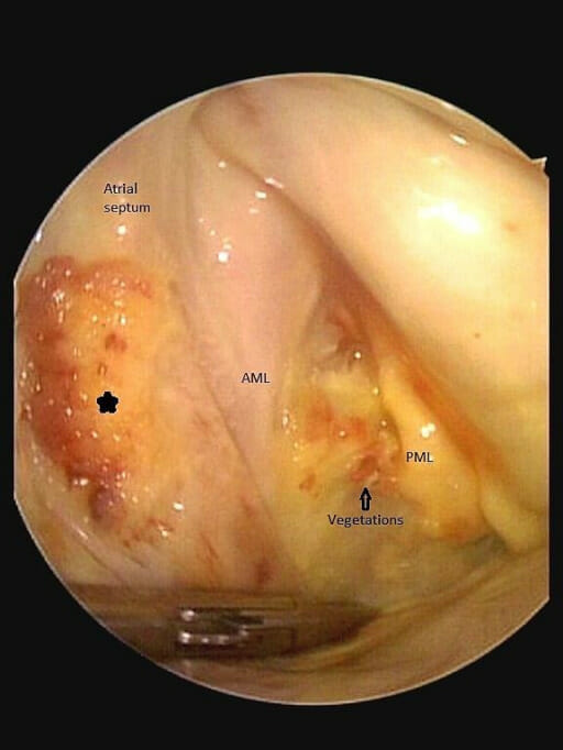
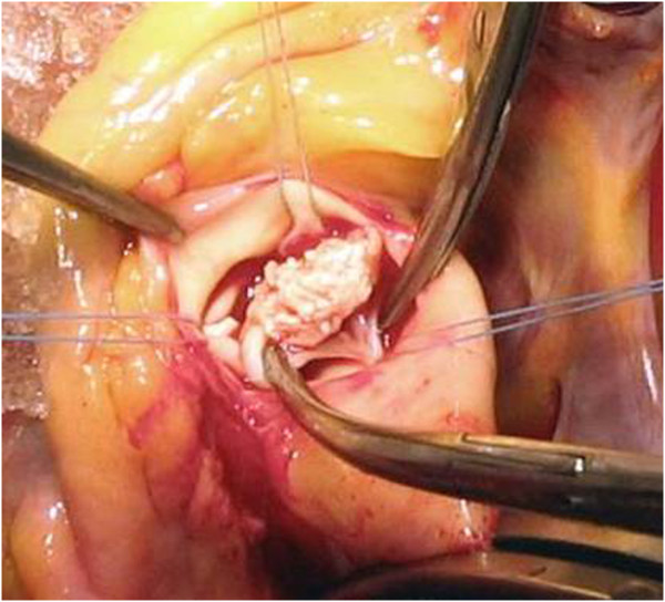
Intraoperative findings of a patient with left atrial endocarditis as a rare complication of mitral valve endocarditis. The patient presented with the peripheral stigmata of IE in addition to holosystolic murmur over the left sternal border. The asterisk indicates the atrial vegetation, the “footprints” of mitral valve endocarditis. AML: anterior mitral leaflet PML: posterior mitral leaflet.
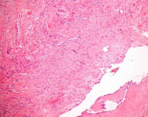
Histologic findings from the resected atrial septum showing an old focal subendocardial bleed, a focal mild to moderate fibrosis, and some neutrophil granulocytes.
Noninfective endocarditisNoninfective endocarditisFormation of a non-infectious thrombus, referred to as vegetation, on previously undamaged endocardium. It usually occurs as a complication of connective-tissue diseases and cancers because of the associated hypercoagulable state.Endocarditis[10,11,20]
Endothelial injury to the valve leaflets due to:
Trauma
Circulating immune complexesImmune complexesThe complex formed by the binding of antigen and antibody molecules. The deposition of large antigen-antibody complexes leading to tissue damage causes immune complex diseases.C3 Deficiency
CytokinesCytokinesNon-antibody proteins secreted by inflammatory leukocytes and some non-leukocytic cells, that act as intercellular mediators. They differ from classical hormones in that they are produced by a number of tissue or cell types rather than by specialized glands. They generally act locally in a paracrine or autocrine rather than endocrine manner.Adaptive Immune Response
Antigen-antibody reactions
Platelet activationPlatelet activationA series of progressive, overlapping events, triggered by exposure of the platelets to subendothelial tissue. These events include shape change, adhesiveness, aggregation, and release reactions. When carried through to completion, these events lead to the formation of a stable hemostatic plug.Hemostasis and deposition occurs (often during a hypercoagulableHypercoagulableHypercoagulable states (also referred to as thrombophilias) are a group of hematologic diseases defined by an increased risk of clot formation (i.e., thrombosis) due to either an increase in procoagulants, a decrease in anticoagulants, or a decrease in fibrinolysis. Hypercoagulable States state).
Immune complexesImmune complexesThe complex formed by the binding of antigen and antibody molecules. The deposition of large antigen-antibody complexes leading to tissue damage causes immune complex diseases.C3 Deficiency
Mononuclear cells
Vegetations are easily dislodged → embolic complications
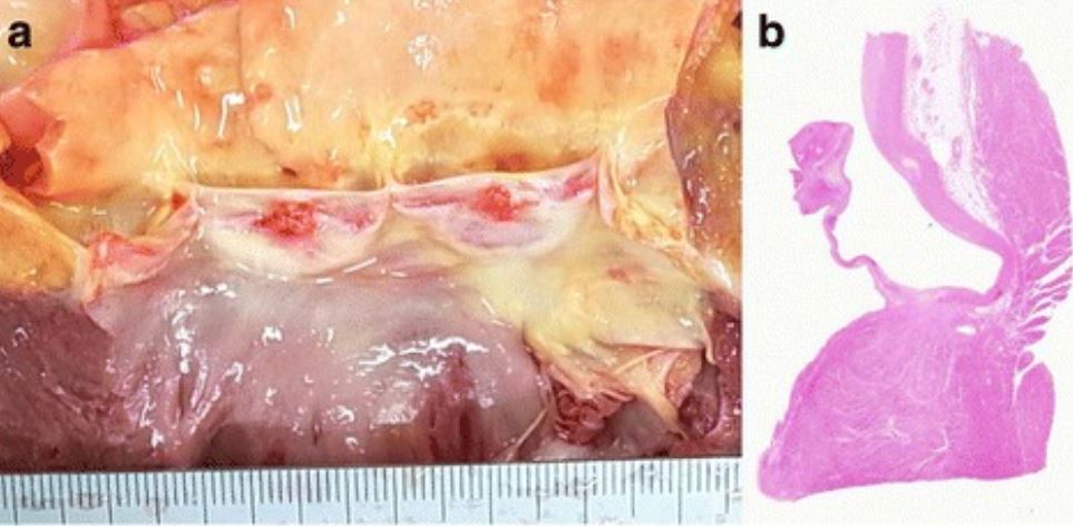
Vegetations of the aortic valve at autopsy: a: The macroscopic appearance of the aortic valve demonstrates 2 vegetations of 4 mm and 5 mm in diameter. b: Histologic evaluation shows that the vegetations consist of fibrin without bacterial colonies, consistent with noninfective endocarditis.
Image: “Newly recognized cerebral infarctions on postmortem imaging: a report of three cases with systemic infectious disease” by BMC Medical Imaging. License: CC BY 4.0
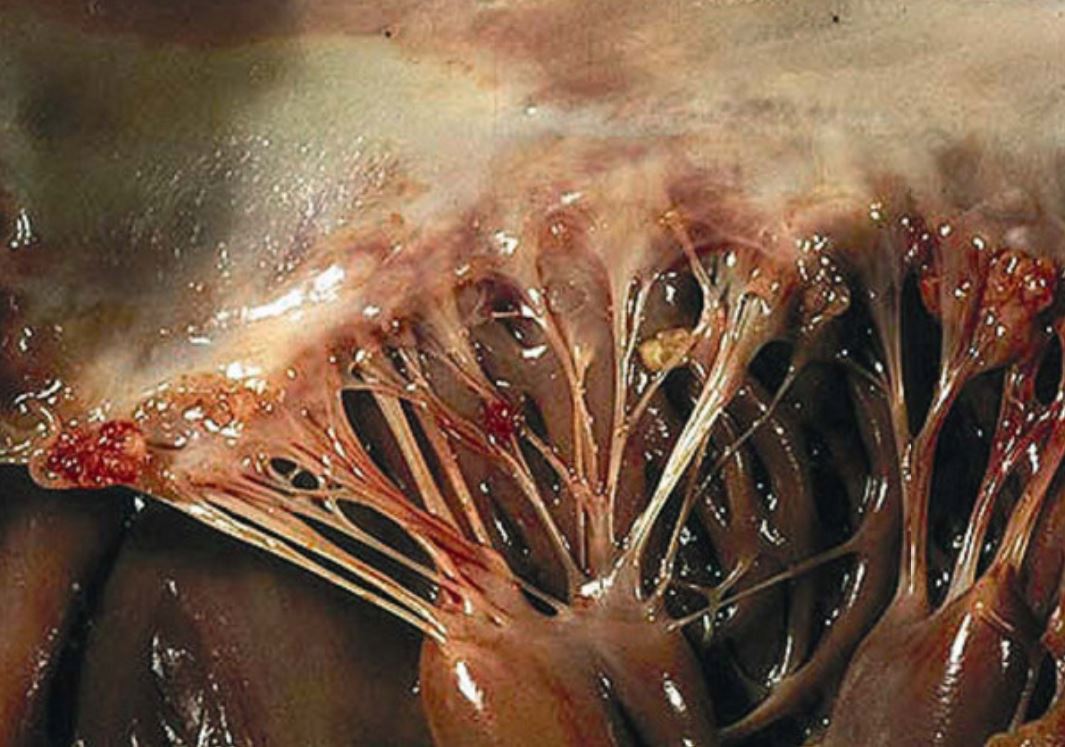
Verrucous vegetations seen in Libman-Sacks endocarditis of the mitral valve: The sterile vegetations typically have a wart-like morphology. The vegetations can be found near the edge of the leaflets along the line of closure, both on the atrial and ventricular sides of the leaflets, and can even be found on the chordae and the endocardium. In this case, several microthrombi are present on the free edge of the leaflet and on the chordae.
Image: “Mitral valve surgery for mitral regurgitation caused by Libman-Sacks endocarditis: a report of four cases and a systematic review of the literature” by Bouma, W., et al. License: CC BY 2.0
Infective endocarditisInfective endocarditisInfective endocarditis (IE) is caused by infection or inflammation of the inner lining of the heart (endocardium), most commonly affecting the heart valves.Endocarditis can be further classified based on the clinical course, type of valve, and location.
Most common cause is S. aureusS. aureusPotentially pathogenic bacteria found in nasal membranes, skin, hair follicles, and perineum of warm-blooded animals. They may cause a wide range of infections and intoxications.Staphylococcus.
Subacute infectious endocarditisSubacute infectious endocarditisEndocardium infection that is usually caused by Streptococcus. Subacute infective endocarditis evolves over weeks and months with modest toxicity and rare metastatic infection.Endocarditis (endocarditisEndocarditisEndocarditis is an inflammatory disease involving the inner lining (endometrium) of the heart, most commonly affecting the cardiac valves. Both infectious and noninfectious etiologies lead to vegetations on the valve leaflets. Patients may present with nonspecific symptoms such as fever and fatigue. Endocarditis lenta):
More gradual onset of symptoms
Progresses more slowly (weeks to months)
Smaller vegetations
More commonly affects congenitally abnormal or diseased valves
PatientsPatientsIndividuals participating in the health care system for the purpose of receiving therapeutic, diagnostic, or preventive procedures.Clinician–Patient Relationship may survive for months untreated.
StaphylococcusStaphylococcusStaphylococcus is a medically important genera of Gram-positive, aerobic cocci. These bacteria form clusters resembling grapes on culture plates. Staphylococci are ubiquitous for humans, and many strains compose the normal skin flora.Staphylococcus
StreptococcusStreptococcusStreptococcus is one of the two medically important genera of gram-positive cocci, the other being Staphylococcus. Streptococci are identified as different species on blood agar on the basis of their hemolytic pattern and sensitivity to optochin and bacitracin. There are many pathogenic species of streptococci, including S. pyogenes, S. agalactiae, S. pneumoniae, and the viridans streptococci.Streptococcus
HACEK organisms
Prosthetic valveProsthetic ValveSoft Tissue AbscessendocarditisEndocarditisEndocarditis is an inflammatory disease involving the inner lining (endometrium) of the heart, most commonly affecting the cardiac valves. Both infectious and noninfectious etiologies lead to vegetations on the valve leaflets. Patients may present with nonspecific symptoms such as fever and fatigue. Endocarditis:
Mitral valveMitral valveThe valve between the left atrium and left ventricle of the heart.Heart: Anatomy
Aortic valveAortic valveThe valve between the left ventricle and the ascending aorta which prevents backflow into the left ventricle.Heart: Anatomy
Right-sided endocarditisRight-sided endocarditisEndocarditis (most common in IV drug use or with a right-sided cardiac anomaly or intravascular or cardiac implantable device):
Tricuspid valveTricuspid valveThe valve consisting of three cusps situated between the right atrium and right ventricle of the heart.Heart: Anatomy
Presentation and course depend on the etiology, location of vegetations, and severity.
General signs and symptoms[2,4,10]
The following are more frequently seen in IEIEInfective endocarditis (IE) is caused by infection or inflammation of the inner lining of the heart (endocardium), most commonly affecting the heart valves.Endocarditis than NIE:
FeverFeverFever is defined as a measured body temperature of at least 38°C (100.4°F). Fever is caused by circulating endogenous and/or exogenous pyrogens that increase levels of prostaglandin E2 in the hypothalamus. Fever is commonly associated with chills, rigors, sweating, and flushing of the skin. Fever (endocarditisEndocarditisEndocarditis is an inflammatory disease involving the inner lining (endometrium) of the heart, most commonly affecting the cardiac valves. Both infectious and noninfectious etiologies lead to vegetations on the valve leaflets. Patients may present with nonspecific symptoms such as fever and fatigue. Endocarditis should be suspected in a patient with a feverFeverFever is defined as a measured body temperature of at least 38°C (100.4°F). Fever is caused by circulating endogenous and/or exogenous pyrogens that increase levels of prostaglandin E2 in the hypothalamus. Fever is commonly associated with chills, rigors, sweating, and flushing of the skin. Fever of unknown origin)
FatigueFatigueThe state of weariness following a period of exertion, mental or physical, characterized by a decreased capacity for work and reduced efficiency to respond to stimuli.Fibromyalgia
TachycardiaTachycardiaAbnormally rapid heartbeat, usually with a heart rate above 100 beats per minute for adults. Tachycardia accompanied by disturbance in the cardiac depolarization (cardiac arrhythmia) is called tachyarrhythmia.Sepsis in Children
Arrhythmia:
Potentially due to spread of the infection and myocardial abscessAbscessAccumulation of purulent material in tissues, organs, or circumscribed spaces, usually associated with signs of infection.Chronic Granulomatous Disease formation
Results in disruption of the atrioventricular conduction system → conduction delay or heart block
Extracardiac findings[2,12,13]
The following are potential findings in IEIEInfective endocarditis (IE) is caused by infection or inflammation of the inner lining of the heart (endocardium), most commonly affecting the heart valves.Endocarditis:
Due to microemboli in capillariesCapillariesCapillaries are the primary structures in the circulatory system that allow the exchange of gas, nutrients, and other materials between the blood and the extracellular fluid (ECF). Capillaries are the smallest of the blood vessels. Because a capillary diameter is so small, only 1 RBC may pass through at a time.Capillaries: Histology
Painful red nodules on pads of the fingers and toes
Due to immune complex deposition and inflammationInflammationInflammation is a complex set of responses to infection and injury involving leukocytes as the principal cellular mediators in the body’s defense against pathogenic organisms. Inflammation is also seen as a response to tissue injury in the process of wound healing. The 5 cardinal signs of inflammation are pain, heat, redness, swelling, and loss of function. Inflammation
ConjunctivaConjunctivaThe mucous membrane that covers the posterior surface of the eyelids and the anterior pericorneal surface of the eyeball.Eye: Anatomy hemorrhage
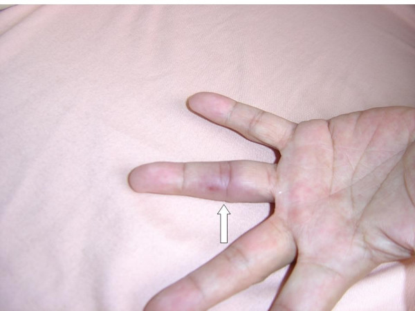
Osler node on the 4th digit of the left hand
Image: “Osler’s node on the fourth digit of the left hand” by Yang ML, Chen YH, Chen TC, Lin WR, Lin CY, Lu PL. License: CC BY 2.0
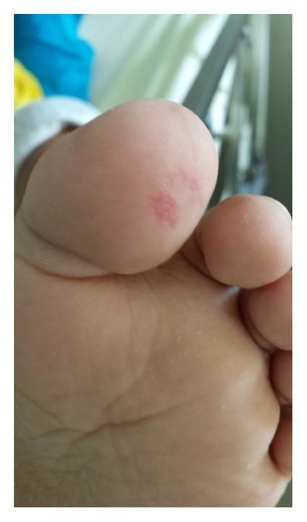
Janeway lesion on the toe in a patient with infective endocarditis
Image: “Janeway lesion on patient’s left toe” by Case Reports in Infectious Diseases. License: CC BY 4.0
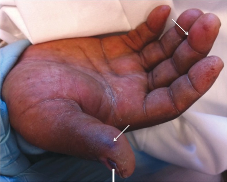
Janeway lesion showing as painless, macular, hemorrhagic, irregularly-shaped lesions on patient’s palm: Two pronounced lesions are seen at thumb and middle finger. Subungual splinter hemorrhages (arrowhead) are seen at the nail bed of thumb.
Image: “Acute endocarditis in intravenous drug users: a case report and literature review” by Ji Y, Kujtan L, Kershner D. License: CC BY 2.0
Mnemonic
Signs of IE can be remembered with the mnemonic “FROM JANE”:
Fever
Roth spots
Osler nodes
Murmur
Janeway lesions
Anemia
Nail bed hemorrhage
Emboli
Systemic embolizationEmbolizationA method of hemostasis utilizing various agents such as gelfoam, silastic, metal, glass, or plastic pellets, autologous clot, fat, and muscle as emboli. It has been used in the treatment of spinal cord and intracranial arteriovenous malformations, renal arteriovenous fistulas, gastrointestinal bleeding, epistaxis, hypersplenism, certain highly vascular tumors, traumatic rupture of blood vessels, and control of operative hemorrhage.Gastrointestinal Bleeding[2,4,7,10,18]
System embolizationEmbolizationA method of hemostasis utilizing various agents such as gelfoam, silastic, metal, glass, or plastic pellets, autologous clot, fat, and muscle as emboli. It has been used in the treatment of spinal cord and intracranial arteriovenous malformations, renal arteriovenous fistulas, gastrointestinal bleeding, epistaxis, hypersplenism, certain highly vascular tumors, traumatic rupture of blood vessels, and control of operative hemorrhage.Gastrointestinal Bleeding may occur in both IEIEInfective endocarditis (IE) is caused by infection or inflammation of the inner lining of the heart (endocardium), most commonly affecting the heart valves.Endocarditis and NIE. Often, an embolic event is the only presenting evidence of NIE.
Transient ischemic attackTransient ischemic attackTransient ischemic attack (TIA) is a temporary episode of neurologic dysfunction caused by ischemia without infarction that resolves completely when blood supply is restored. Transient ischemic attack is a neurologic emergency that warrants urgent medical attention. Transient Ischemic Attack (TIA) or stroke:
New focal deficit, seizuresSeizuresA seizure is abnormal electrical activity of the neurons in the cerebral cortex that can manifest in numerous ways depending on the region of the brain affected. Seizures consist of a sudden imbalance that occurs between the excitatory and inhibitory signals in cortical neurons, creating a net excitation. The 2 major classes of seizures are focal and generalized. Seizures, encephalopathyEncephalopathyHyper-IgM Syndrome, or change of consciousness
Can be a presenting symptom in 20% of IEIEInfective endocarditis (IE) is caused by infection or inflammation of the inner lining of the heart (endocardium), most commonly affecting the heart valves.Endocarditis cases
Multiple emboli are common.
Ischemic strokeIschemic StrokeAn ischemic stroke (also known as cerebrovascular accident) is an acute neurologic injury that occurs as a result of brain ischemia; this condition may be due to cerebral blood vessel occlusion by thrombosis or embolism, or rarely due to systemic hypoperfusion. Ischemic Stroke can undergo hemorrhagic transformationTransformationChange brought about to an organism’s genetic composition by unidirectional transfer (transfection; transduction, genetic; conjugation, genetic, etc.) and incorporation of foreign DNA into prokaryotic or eukaryotic cells by recombination of part or all of that DNA into the cell’s genome.Bacteriology in up to 50% of cases.
An abscessAbscessAccumulation of purulent material in tissues, organs, or circumscribed spaces, usually associated with signs of infection.Chronic Granulomatous Disease can form in the infarctInfarctArea of necrotic cells in an organ, arising mainly from hypoxia and ischemiaIschemic Cell Damage cavity.
Pulmonary embolismPulmonary EmbolismPulmonary embolism (PE) is a potentially fatal condition that occurs as a result of intraluminal obstruction of the main pulmonary artery or its branches. The causative factors include thrombi, air, amniotic fluid, and fat. In PE, gas exchange is impaired due to the decreased return of deoxygenated blood to the lungs. Pulmonary Embolism:
DyspneaDyspneaDyspnea is the subjective sensation of breathing discomfort. Dyspnea is a normal manifestation of heavy physical or psychological exertion, but also may be caused by underlying conditions (both pulmonary and extrapulmonary). Dyspnea
Pleuritic chest painPainAn unpleasant sensation induced by noxious stimuli which are detected by nerve endings of nociceptive neurons.Pain: Types and Pathways
Cough
HemoptysisHemoptysisHemoptysis is defined as the expectoration of blood originating in the lower respiratory tract. Hemoptysis is a consequence of another disease process and can be classified as either life threatening or non-life threatening. Hemoptysis can result in significant morbidity and mortality due to both drowning (reduced gas exchange as the lungs fill with blood) and hemorrhagic shock. Hemoptysis
Renal infarctInfarctArea of necrotic cells in an organ, arising mainly from hypoxia and ischemiaIschemic Cell Damage:
Splenic emboli: LUQ painPainAn unpleasant sensation induced by noxious stimuli which are detected by nerve endings of nociceptive neurons.Pain: Types and Pathways
Other complications[2,4,7,10]
Cardiac:
Perivalvular abscessAbscessAccumulation of purulent material in tissues, organs, or circumscribed spaces, usually associated with signs of infection.Chronic Granulomatous Disease
Valve insufficiency
Valve rupture
Heart failureHeart FailureA heterogeneous condition in which the heart is unable to pump out sufficient blood to meet the metabolic need of the body. Heart failure can be caused by structural defects, functional abnormalities (ventricular dysfunction), or a sudden overload beyond its capacity. Chronic heart failure is more common than acute heart failure which results from sudden insult to cardiac function, such as myocardial infarction.Total Anomalous Pulmonary Venous Return (TAPVR)
PericarditisPericarditisPericarditis is an inflammation of the pericardium, often with fluid accumulation. It can be caused by infection (often viral), myocardial infarction, drugs, malignancies, metabolic disorders, autoimmune disorders, or trauma. Acute, subacute, and chronic forms exist. Pericarditis → cardiac tamponadeTamponadePericardial effusion, usually of rapid onset, exceeding ventricular filling pressures and causing collapse of the heart with a markedly reduced cardiac output.Pericarditis
Metastatic infectionsInfectionsInvasion of the host organism by microorganisms or their toxins or by parasites that can cause pathological conditions or diseases.Chronic Granulomatous Disease:
BrainBrainThe part of central nervous system that is contained within the skull (cranium). Arising from the neural tube, the embryonic brain is comprised of three major parts including prosencephalon (the forebrain); mesencephalon (the midbrain); and rhombencephalon (the hindbrain). The developed brain consists of cerebrum; cerebellum; and other structures in the brain stem.Nervous System: Anatomy, Structure, and ClassificationabscessAbscessAccumulation of purulent material in tissues, organs, or circumscribed spaces, usually associated with signs of infection.Chronic Granulomatous Disease or meningitisMeningitisMeningitis is inflammation of the meninges, the protective membranes of the brain, and spinal cord. The causes of meningitis are varied, with the most common being bacterial or viral infection. The classic presentation of meningitis is a triad of fever, altered mental status, and nuchal rigidity. Meningitis
OsteomyelitisOsteomyelitisOsteomyelitis is an infection of the bone that results from the spread of microorganisms from the blood (hematogenous), nearby infected tissue, or open wounds (non-hematogenous). Infections are most commonly caused by Staphylococcus aureus.Osteomyelitis
Renal abscessAbscessAccumulation of purulent material in tissues, organs, or circumscribed spaces, usually associated with signs of infection.Chronic Granulomatous Disease
PneumoniaPneumoniaPneumonia or pulmonary inflammation is an acute or chronic inflammation of lung tissue. Causes include infection with bacteria, viruses, or fungi. In more rare cases, pneumonia can also be caused through toxic triggers through inhalation of toxic substances, immunological processes, or in the course of radiotherapy.Pneumonia or lung abscessAbscessAccumulation of purulent material in tissues, organs, or circumscribed spaces, usually associated with signs of infection.Chronic Granulomatous Disease
Obtain an echocardiogramEchocardiogramTransposition of the Great Arteries as soon as infective endocarditisInfective endocarditisInfective endocarditis (IE) is caused by infection or inflammation of the inner lining of the heart (endocardium), most commonly affecting the heart valves.Endocarditis is suspected (within 12 hours of evaluation).
Transesophageal echo (TEETEEUltrasonic recording of the size, motion, and composition of the heart and surrounding tissues using a transducer placed in the esophagus.Imaging of the Heart and Great Vessels):
More sensitive, but more invasive
Should be performed if TTETTEImaging of the Heart and Great Vessels is negative and suspicion for endocarditisEndocarditisEndocarditis is an inflammatory disease involving the inner lining (endometrium) of the heart, most commonly affecting the cardiac valves. Both infectious and noninfectious etiologies lead to vegetations on the valve leaflets. Patients may present with nonspecific symptoms such as fever and fatigue. Endocarditis is high
Should be performed for all cases of suspected prosthetic valveProsthetic ValveSoft Tissue Abscess or pacemakerPacemakerA device designed to stimulate, by electric impulses, contraction of the heart muscles. It may be temporary (external) or permanent (internal or internal-external).BradyarrhythmiasendocarditisEndocarditisEndocarditis is an inflammatory disease involving the inner lining (endometrium) of the heart, most commonly affecting the cardiac valves. Both infectious and noninfectious etiologies lead to vegetations on the valve leaflets. Patients may present with nonspecific symptoms such as fever and fatigue. Endocarditis and when an abscessAbscessAccumulation of purulent material in tissues, organs, or circumscribed spaces, usually associated with signs of infection.Chronic Granulomatous Disease is suspected
If negative, repeat TEETEEUltrasonic recording of the size, motion, and composition of the heart and surrounding tissues using a transducer placed in the esophagus.Imaging of the Heart and Great Vessels in 3–5 days after initial TEETEEUltrasonic recording of the size, motion, and composition of the heart and surrounding tissues using a transducer placed in the esophagus.Imaging of the Heart and Great Vessels (or earlier if clinical changes occur) if high suspicion persists.
Findings:
Valvular vegetations:
Size
Mobility
Calcifications
Complications:
Valvular insufficiency
AbscessAbscessAccumulation of purulent material in tissues, organs, or circumscribed spaces, usually associated with signs of infection.Chronic Granulomatous Disease
Cannot determine the cause of vegetations (IEIEInfective endocarditis (IE) is caused by infection or inflammation of the inner lining of the heart (endocardium), most commonly affecting the heart valves.Endocarditis versus NIE)
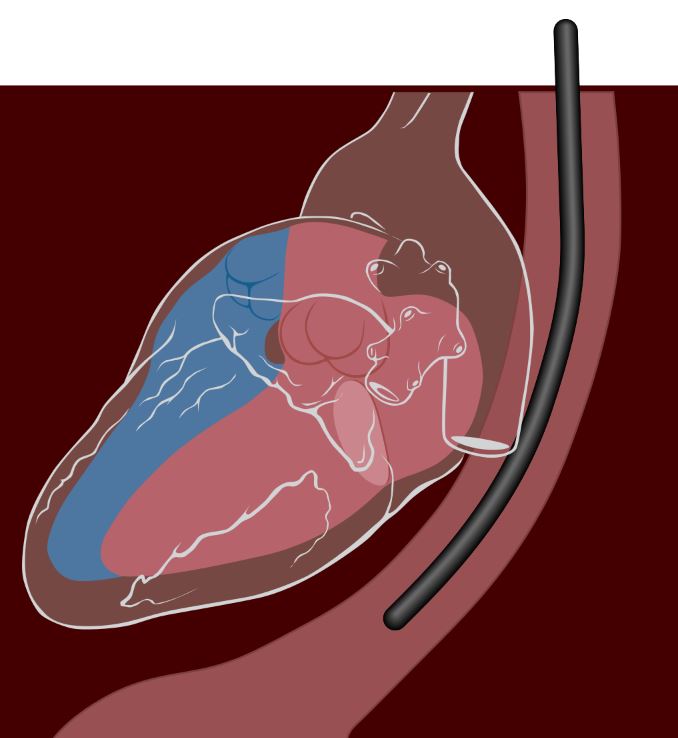
Depiction of transesophageal echocardiography: Patients are generally sedated for this procedure. The echo transducer is lowered into the esophagus, which places it right next to the left side of the heart.
Image: “Transesophageal echocardiography diagram” by Patrick J. Lynch. License: CC BY 2.5, edited by Lecturio.
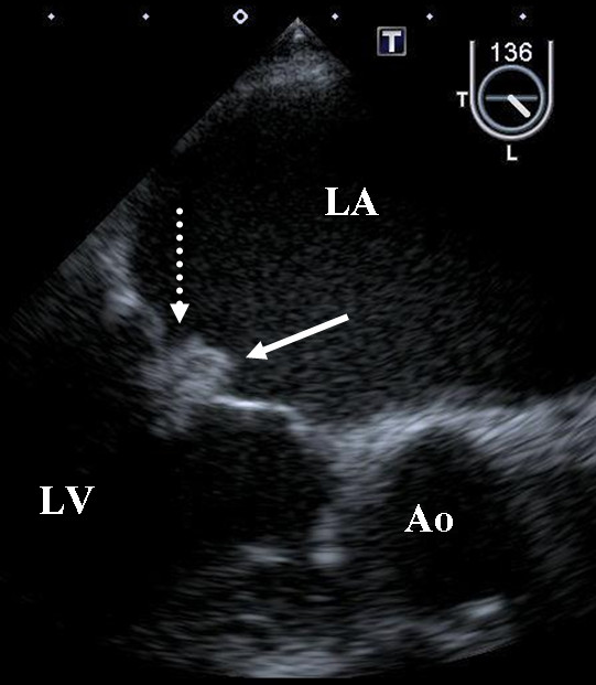
Transesophageal echocardiography images demonstrating vegetations on the mitral valve in a patient with noninfective endocarditis due to malignancy. The solid arrow is pointing to a vegetation on the anterior leaflet, and the dotted arrow is pointing to a vegetation on the posterior leaflet. Ao: aorta; LA: left atrium; LV: left ventricle
Image: “Nonbacterial thrombotic endocarditis associated with cancer of unknown origin complicated with thrombus in the left auricular appendage: case report” by Norisada, K., et al. License: CC BY 2.0
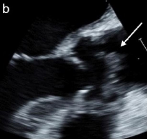
Transesophageal echocardiography image demonstrating a large pedunculated vegetation within the ascending aorta arising from the commissure of the right and noncoronary cusp of the aortic valve with a calcified base consistent with endocarditis
Image: “Lomentospora prolificans endocarditis–case report and literature review” by Kelly M, Stevens R, Konecny P. License: CC BY 4.0, cropped by Lecturio.
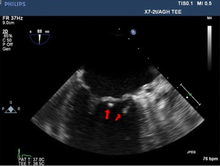
Transesophageal echocardiography image showing 2 vegetations (red arrows) on the mitral valve in a patient with endocarditis
Image: “Pseudomonas mendocina native valve infective endocarditis: a case report” by Journal of Medical Case Reports. License: CC BY 4.0
Supporting workup[2,12,13,15–17,20,23]
Laboratory findings:
The following are nonspecific but may signal IEIEInfective endocarditis (IE) is caused by infection or inflammation of the inner lining of the heart (endocardium), most commonly affecting the heart valves.Endocarditis:
Blood cultures:
3 sets should be obtained from different sites.
Must be obtained prior to starting antibiotics
Negative cultures do not rule out IEIEInfective endocarditis (IE) is caused by infection or inflammation of the inner lining of the heart (endocardium), most commonly affecting the heart valves.Endocarditis.
Blood cultures may be negative in approximately 10% of infective endocarditisInfective endocarditisInfective endocarditis (IE) is caused by infection or inflammation of the inner lining of the heart (endocardium), most commonly affecting the heart valves.Endocarditis cases.
Most common reason for negative cultures is prior antibiotic use.
SerologySerologyThe study of serum, especially of antigen-antibody reactions in vitro.Yellow Fever Virus and polymerase chain reactionPolymerase chain reactionPolymerase chain reaction (PCR) is a technique that amplifies DNA fragments exponentially for analysis. The process is highly specific, allowing for the targeting of specific genomic sequences, even with minuscule sample amounts. The PCR cycles multiple times through 3 phases: denaturation of the template DNA, annealing of a specific primer to the individual DNA strands, and synthesis/elongation of new DNA molecules.Polymerase Chain Reaction (PCR):
Consider pursuing if cultures are negative
Useful for fastidiousFastidiousBordetella organisms that are difficult to isolate (Coxiella, BartonellaBartonellaBartonella is a genus of gram-negative bacteria in the family Bartonellaceae. As a facultative intracellular parasite, Bartonella can infect healthy people as well as act as an opportunistic pathogen. Bartonella species are transmitted by vectors such as ticks, fleas, sandflies, and mosquitoes. B. henselae is the most common of the 3 species known to cause human disease.Bartonella, ChlamydiaChlamydiaChlamydiae are obligate intracellular gram-negative bacteria. They lack a peptidoglycan layer and are best visualized using Giemsa stain. The family of Chlamydiaceae comprises 3 pathogens that can infect humans: Chlamydia trachomatis, Chlamydia psittaci, and Chlamydia pneumoniae.Chlamydia, TropherymaTropherymaA genus of gram-positive bacteria in the family cellulomonadaceae.Whipple’s Disease, BrucellaBrucellaBrucellosis (also known as undulant fever, Mediterranean fever, or Malta fever) is a zoonotic infection that spreads predominantly through ingestion of unpasteurized dairy products or direct contact with infected animal products. Clinical manifestations include fever, arthralgias, malaise, lymphadenopathy, and hepatosplenomegaly. Brucella/Brucellosis, LegionellaLegionellaLegionella is a facultative intracellular, gram-negative bacilli. Legionella does not grow on common culture media because it requires certain supplementation (cysteine and iron). Legionella pneumophila (L. pneumophila) accounts for the majority of human infections.Legionella/Legionellosis, and AspergillusAspergillusA genus of mitosporic fungi containing about 100 species and eleven different teleomorphs in the family trichocomaceae.Echinocandinsspp.)
Antinuclear antibodiesAntibodiesImmunoglobulins (Igs), also known as antibodies, are glycoprotein molecules produced by plasma cells that act in immune responses by recognizing and binding particular antigens. The various Ig classes are IgG (the most abundant), IgM, IgE, IgD, and IgA, which differ in their biologic features, structure, target specificity, and distribution.Immunoglobulins: Types and Functions
Lupus anticoagulantLupus anticoagulantAn antiphospholipid antibody found in association with systemic lupus erythematosus, antiphospholipid syndrome; and in a variety of other diseases as well as in healthy individuals. In vitro, the antibody interferes with the conversion of prothrombin to thrombin and prolongs the partial thromboplastin time. In vivo, it exerts a procoagulant effect resulting in thrombosis mainly in the larger veins and arteries. It further causes obstetrical complications, including fetal death and spontaneous abortion, as well as a variety of hematologic and neurologic complications.Antiphospholipid Syndrome
Antiphospholipid antibodiesAntiphospholipid antibodiesAutoantibodies directed against phospholipids. These antibodies are characteristically found in patients with systemic lupus erythematosus, antiphospholipid syndrome; related autoimmune diseases, some non-autoimmune diseases, and also in healthy individuals.Antiphospholipid Syndrome
Coagulation studiesCoagulation studiesCoagulation studies are a group of hematologic laboratory studies that reflect the function of blood vessels, platelets, and coagulation factors, which all interact with one another to achieve hemostasis. Coagulation studies are usually ordered to evaluate patients with bleeding or hypercoagulation disorders.Coagulation Studies to identify DICDICDisseminated intravascular coagulation (DIC) is a condition characterized by systemic bodywide activation of the coagulation cascade. This cascade results in both widespread microvascular thrombi contributing to multiple organ dysfunction and consumption of clotting factors and platelets, leading to hemorrhage. Disseminated Intravascular Coagulation (disseminated intravascular coagulationDisseminated intravascular coagulationDisseminated intravascular coagulation (DIC) is a condition characterized by systemic bodywide activation of the coagulation cascade. This cascade results in both widespread microvascular thrombi contributing to multiple organ dysfunction and consumption of clotting factors and platelets, leading to hemorrhage. Disseminated Intravascular Coagulation)
Imaging:
ECGECGAn electrocardiogram (ECG) is a graphic representation of the electrical activity of the heart plotted against time. Adhesive electrodes are affixed to the skin surface allowing measurement of cardiac impulses from many angles. The ECG provides 3-dimensional information about the conduction system of the heart, the myocardium, and other cardiac structures. Electrocardiogram (ECG):
A baseline tracing should be performed in all patientsPatientsIndividuals participating in the health care system for the purpose of receiving therapeutic, diagnostic, or preventive procedures.Clinician–Patient Relationship.
Bundle branch blockBundle branch blockA form of heart block in which the electrical stimulation of heart ventricles is interrupted at either one of the branches of bundle of His thus preventing the simultaneous depolarization of the two ventricles.Bundle Branch and Fascicular Blocks
Chest X-rayX-rayPenetrating electromagnetic radiation emitted when the inner orbital electrons of an atom are excited and release radiant energy. X-ray wavelengths range from 1 pm to 10 nm. Hard x-rays are the higher energy, shorter wavelength x-rays. Soft x-rays or grenz rays are less energetic and longer in wavelength. The short wavelength end of the x-ray spectrum overlaps the gamma rays wavelength range. The distinction between gamma rays and x-rays is based on their radiation source.Pulmonary Function Tests:
Can rule out other causes of symptoms
Potential findings in endocarditisEndocarditisEndocarditis is an inflammatory disease involving the inner lining (endometrium) of the heart, most commonly affecting the cardiac valves. Both infectious and noninfectious etiologies lead to vegetations on the valve leaflets. Patients may present with nonspecific symptoms such as fever and fatigue. Endocarditis:
Septic emboli to the lungsLungsLungs are the main organs of the respiratory system. Lungs are paired viscera located in the thoracic cavity and are composed of spongy tissue. The primary function of the lungs is to oxygenate blood and eliminate CO2. Lungs: Anatomy
Pulmonary edemaPulmonary edemaPulmonary edema is a condition caused by excess fluid within the lung parenchyma and alveoli as a consequence of a disease process. Based on etiology, pulmonary edema is classified as cardiogenic or noncardiogenic. Patients may present with progressive dyspnea, orthopnea, cough, or respiratory failure.Pulmonary Edema and cardiomegalyCardiomegalyEnlargement of the heart, usually indicated by a cardiothoracic ratio above 0. 50. Heart enlargement may involve the right, the left, or both heart ventricles or heart atria. Cardiomegaly is a nonspecific symptom seen in patients with chronic systolic heart failure (heart failure) or several forms of cardiomyopathies.Ebstein’s Anomaly
CT:
Can be used to assess for sites of metastatic infection
May be useful if an underlying malignancyMalignancyHemothorax is suspected (for NIE)
Cardiac CT can be used to better delineate valvular anatomy as well as to evaluate coronary arteriesArteriesArteries are tubular collections of cells that transport oxygenated blood and nutrients from the heart to the tissues of the body. The blood passes through the arteries in order of decreasing luminal diameter, starting in the largest artery (the aorta) and ending in the small arterioles. Arteries are classified into 3 types: large elastic arteries, medium muscular arteries, and small arteries and arterioles. Arteries: Histology if surgical intervention is planned.
A good alternative to coronary angiographyAngiographyRadiography of blood vessels after injection of a contrast medium.Cardiac Surgery (which may risk vegetation dislodgement and embolizationEmbolizationA method of hemostasis utilizing various agents such as gelfoam, silastic, metal, glass, or plastic pellets, autologous clot, fat, and muscle as emboli. It has been used in the treatment of spinal cord and intracranial arteriovenous malformations, renal arteriovenous fistulas, gastrointestinal bleeding, epistaxis, hypersplenism, certain highly vascular tumors, traumatic rupture of blood vessels, and control of operative hemorrhage.Gastrointestinal Bleeding)
May be especially useful for NIE, in which vegetations may be small and friable and not picked up by echocardiographyEchocardiographyUltrasonic recording of the size, motion, and composition of the heart and surrounding tissues. The standard approach is transthoracic.Tricuspid Valve Atresia (TVA)
May be able to differentiate IEIEInfective endocarditis (IE) is caused by infection or inflammation of the inner lining of the heart (endocardium), most commonly affecting the heart valves.Endocarditis from NIE based on the pattern of emboli distribution
BrainBrainThe part of central nervous system that is contained within the skull (cranium). Arising from the neural tube, the embryonic brain is comprised of three major parts including prosencephalon (the forebrain); mesencephalon (the midbrain); and rhombencephalon (the hindbrain). The developed brain consists of cerebrum; cerebellum; and other structures in the brain stem.Nervous System: Anatomy, Structure, and Classification MRI: performed if cerebral embolic events are suspected (obtain with vascular imaging or MRAMRAImaging of the Heart and Great Vessels to check for mycotic aneurysmMycotic aneurysmAspergillus/Aspergillosis)
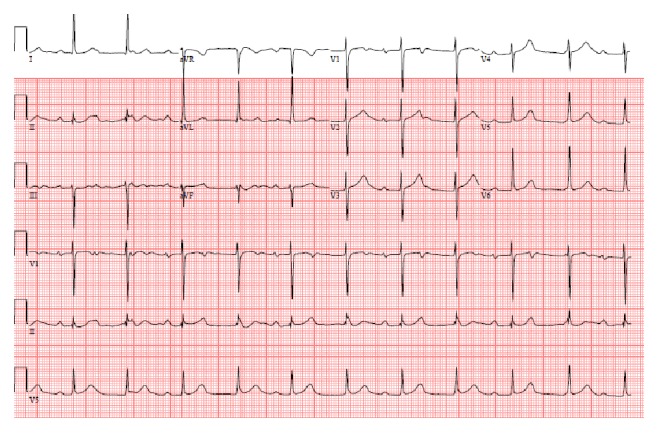
An ECG showing atrioventricular dissociation in a patient with S. viridans endocarditis
Image: “Timing for pacing after acquired conduction disease in the setting of endocarditis” by Brancheau D, Degheim G, Machado C. License: CC BY 3.0
Duke diagnostic criteria[16,22,23]
The Duke diagnostic criteria is a set of clinical criteria that can aid in the diagnosis of IEIEInfective endocarditis (IE) is caused by infection or inflammation of the inner lining of the heart (endocardium), most commonly affecting the heart valves.Endocarditis.
Must meet 1 of the following for a definitive diagnosis of IEIEInfective endocarditis (IE) is caused by infection or inflammation of the inner lining of the heart (endocardium), most commonly affecting the heart valves.Endocarditis:
2 major criteria
1 major plus 3 minor criteria
5 minor criteria
Major criteria:
Positive blood cultures (1 of the following):
Typical organism for IEIEInfective endocarditis (IE) is caused by infection or inflammation of the inner lining of the heart (endocardium), most commonly affecting the heart valves.Endocarditis in 2 separate blood cultures
Persistently positive cultures
Single positive culture for Coxiella burnetiiCoxiella burnetiiA species of gram-negative bacteria that grows preferentially in the vacuoles of the host cell. It is the etiological agent of q fever.Coxiella/Q Fever
Findings of endocardial involvement by echocardiographyEchocardiographyUltrasonic recording of the size, motion, and composition of the heart and surrounding tissues. The standard approach is transthoracic.Tricuspid Valve Atresia (TVA) (1 of the following):
Mobile echodense masses attached to valvular leaflets or endocardiumEndocardiumThe innermost layer of the heart, comprised of endothelial cells.Heart: Anatomy
FeverFeverFever is defined as a measured body temperature of at least 38°C (100.4°F). Fever is caused by circulating endogenous and/or exogenous pyrogens that increase levels of prostaglandin E2 in the hypothalamus. Fever is commonly associated with chills, rigors, sweating, and flushing of the skin. Fever > 38°C (100.4°F)
Microbiologic findings by culture that do not meet major criteria
Cases that may not be reliably diagnosed by Duke criteria:
PacemakerPacemakerA device designed to stimulate, by electric impulses, contraction of the heart muscles. It may be temporary (external) or permanent (internal or internal-external).BradyarrhythmiasendocarditisEndocarditisEndocarditis is an inflammatory disease involving the inner lining (endometrium) of the heart, most commonly affecting the cardiac valves. Both infectious and noninfectious etiologies lead to vegetations on the valve leaflets. Patients may present with nonspecific symptoms such as fever and fatigue. Endocarditis
Prosthetic valveProsthetic ValveSoft Tissue AbscessendocarditisEndocarditisEndocarditis is an inflammatory disease involving the inner lining (endometrium) of the heart, most commonly affecting the cardiac valves. Both infectious and noninfectious etiologies lead to vegetations on the valve leaflets. Patients may present with nonspecific symptoms such as fever and fatigue. Endocarditis
Blood-culture–negative infective endocarditisInfective endocarditisInfective endocarditis (IE) is caused by infection or inflammation of the inner lining of the heart (endocardium), most commonly affecting the heart valves.Endocarditis
Medical management of IEIEInfective endocarditis (IE) is caused by infection or inflammation of the inner lining of the heart (endocardium), most commonly affecting the heart valves.Endocarditis[14–17,23]
Prompt initiation of IV antibiotics is necessary if the patient is acutely ill. However, blood cultures should be obtained prior to the start of antibiotic therapy. The recovery rate of bacteriaBacteriaBacteria are prokaryotic single-celled microorganisms that are metabolically active and divide by binary fission. Some of these organisms play a significant role in the pathogenesis of diseases. Bacteriology is diminished by 40% when antibiotics are administered prior to obtaining blood cultures.
Recommended consultations:
Infectious disease
Cardiology
Cardiothoracic surgery
Empiric antibiotic therapy options: generally cover staphylococci (MSSA and MRSAMRSAA strain of Staphylococcus aureus that is non-susceptible to the action of methicillin. The mechanism of resistance usually involves modification of normal or the presence of acquired penicillin binding proteins.Staphylococcus), streptococci, and enterococci[15]
AmpicillinAmpicillinSemi-synthetic derivative of penicillin that functions as an orally active broad-spectrum antibiotic.Penicillins 2 g IV every 4 hours
VancomycinVancomycinAntibacterial obtained from streptomyces orientalis. It is a glycopeptide related to ristocetin that inhibits bacterial cell wall assembly and is toxic to kidneys and the inner ear.Glycopeptides + gentamicinGentamicinAminoglycosides (if allergic to penicillinPenicillinRheumatic Fever)
VancomycinVancomycinAntibacterial obtained from streptomyces orientalis. It is a glycopeptide related to ristocetin that inhibits bacterial cell wall assembly and is toxic to kidneys and the inner ear.Glycopeptides 30 mg/kg/day IV in 2 divided doses (dosing adjusted per protocol to achieve trough concentration of 10–20 μg/mL)
GentamicinGentamicinAminoglycosides 3 mg/kg/dose IV (in 1 dose or 3 divided doses (dosing adjusted to achieve trough concentration of < 1 µg/mL)
VancomycinVancomycinAntibacterial obtained from streptomyces orientalis. It is a glycopeptide related to ristocetin that inhibits bacterial cell wall assembly and is toxic to kidneys and the inner ear.Glycopeptides + gentamicinGentamicinAminoglycosides + rifampinRifampinA semisynthetic antibiotic produced from streptomyces mediterranei. It has a broad antibacterial spectrum, including activity against several forms of Mycobacterium. In susceptible organisms it inhibits dna-dependent RNA polymerase activity by forming a stable complex with the enzyme. It thus suppresses the initiation of RNA synthesis. Rifampin is bactericidal, and acts on both intracellular and extracellular organisms.Epiglottitis for early (< 1 year) prosthetic valveProsthetic ValveSoft Tissue Abscess or healthcare-associated endocarditisEndocarditisEndocarditis is an inflammatory disease involving the inner lining (endometrium) of the heart, most commonly affecting the cardiac valves. Both infectious and noninfectious etiologies lead to vegetations on the valve leaflets. Patients may present with nonspecific symptoms such as fever and fatigue. Endocarditis:
VancomycinVancomycinAntibacterial obtained from streptomyces orientalis. It is a glycopeptide related to ristocetin that inhibits bacterial cell wall assembly and is toxic to kidneys and the inner ear.Glycopeptides 30 mg/kg/day IV in 2 divided doses (dosing adjusted per protocol to achieve trough concentration of 10–20 μg/mL)
GentamicinGentamicinAminoglycosides 3 mg/kg/dose IV (dosing adjusted to achieve trough concentration of < 1 µg/mL)
RifampinRifampinA semisynthetic antibiotic produced from streptomyces mediterranei. It has a broad antibacterial spectrum, including activity against several forms of Mycobacterium. In susceptible organisms it inhibits dna-dependent RNA polymerase activity by forming a stable complex with the enzyme. It thus suppresses the initiation of RNA synthesis. Rifampin is bactericidal, and acts on both intracellular and extracellular organisms.Epiglottitis 900–1200 mg/day IV or orally in 3 divided doses
Tailor antibiotics or antifungals based on:
Identified pathogen
Sensitivities
Repeat blood cultures every 24–48 hours until negative.
> 2 weeks: Outpatient parenteral antibiotic therapy can be considered, except in cases of heart failureHeart FailureA heterogeneous condition in which the heart is unable to pump out sufficient blood to meet the metabolic need of the body. Heart failure can be caused by structural defects, functional abnormalities (ventricular dysfunction), or a sudden overload beyond its capacity. Chronic heart failure is more common than acute heart failure which results from sudden insult to cardiac function, such as myocardial infarction.Total Anomalous Pulmonary Venous Return (TAPVR), worrisome echocardiographic findings, renal impairment, or neurologic signs.
Table: Antibiotic therapy in adults for (left-sided) native IEIEInfective endocarditis (IE) is caused by infection or inflammation of the inner lining of the heart (endocardium), most commonly affecting the heart valves.Endocarditis caused by most common pathogens[15-17,23]
CefazolinCefazolinA semisynthetic cephalosporin analog with broad-spectrum antibiotic action due to inhibition of bacterial cell wall synthesis. It attains high serum levels and is excreted quickly via the urine.Cephalosporins or cefotaximeCefotaximeSemisynthetic broad-spectrum cephalosporin.Cephalosporins 2 g IV every 8 hours for 6 weeks
MRSAMRSAA strain of Staphylococcus aureus that is non-susceptible to the action of methicillin. The mechanism of resistance usually involves modification of normal or the presence of acquired penicillin binding proteins.Staphylococcus (or MSSA with penicillinPenicillinRheumatic FeveranaphylaxisAnaphylaxisAn acute hypersensitivity reaction due to exposure to a previously encountered antigen. The reaction may include rapidly progressing urticaria, respiratory distress, vascular collapse, systemic shock, and death.Type I Hypersensitivity Reaction)
VancomycinVancomycinAntibacterial obtained from streptomyces orientalis. It is a glycopeptide related to ristocetin that inhibits bacterial cell wall assembly and is toxic to kidneys and the inner ear.Glycopeptidesa for 6 weeks, OR
StreptococcusStreptococcusStreptococcus is one of the two medically important genera of gram-positive cocci, the other being Staphylococcus. Streptococci are identified as different species on blood agar on the basis of their hemolytic pattern and sensitivity to optochin and bacitracin. There are many pathogenic species of streptococci, including S. pyogenes, S. agalactiae, S. pneumoniae, and the viridans streptococci.Streptococcus (penicillin-sensitive)
PenicillinPenicillinRheumatic Fever G 12 million–18 million units/day continuously or in 4–6 divided doses for 4 weeks, OR
CeftriaxoneCeftriaxoneA broad-spectrum cephalosporin antibiotic and cefotaxime derivative with a very long half-life and high penetrability to meninges, eyes and inner ears.Cephalosporins 2 g IV daily for 4 weeks, OR
In Europeb: amoxicillinAmoxicillinA broad-spectrum semisynthetic antibiotic similar to ampicillin except that its resistance to gastric acid permits higher serum levels with oral administration.Penicillins 100–200 mg/kg/day IV in 4–6 divided doses for 4 weeks
PenicillinPenicillinRheumatic Fever G 12 million–18 million units/day continuously or in 4–6 divided doses for 2 weeks, OR
CeftriaxoneCeftriaxoneA broad-spectrum cephalosporin antibiotic and cefotaxime derivative with a very long half-life and high penetrability to meninges, eyes and inner ears.Cephalosporins 2 g IV daily for 2 weeks, PLUS
VancomycinVancomycinAntibacterial obtained from streptomyces orientalis. It is a glycopeptide related to ristocetin that inhibits bacterial cell wall assembly and is toxic to kidneys and the inner ear.Glycopeptidesa for 4 weeks if penicillin-allergic
StreptococcusStreptococcusStreptococcus is one of the two medically important genera of gram-positive cocci, the other being Staphylococcus. Streptococci are identified as different species on blood agar on the basis of their hemolytic pattern and sensitivity to optochin and bacitracin. There are many pathogenic species of streptococci, including S. pyogenes, S. agalactiae, S. pneumoniae, and the viridans streptococci.Streptococcus (relatively penicillin-resistant)
Combination therapy:
PenicillinPenicillinRheumatic Fever G 24 million units/day continuously or in 4–6 divided doses or ampicillinAmpicillinSemi-synthetic derivative of penicillin that functions as an orally active broad-spectrum antibiotic.Penicillins 2 g IV every 4 hours for 4 weeks, OR
In Europeb:
CeftriaxoneCeftriaxoneA broad-spectrum cephalosporin antibiotic and cefotaxime derivative with a very long half-life and high penetrability to meninges, eyes and inner ears.Cephalosporins 2 g IV/day for 4 weeks, OR
AmoxicillinAmoxicillinA broad-spectrum semisynthetic antibiotic similar to ampicillin except that its resistance to gastric acid permits higher serum levels with oral administration.Penicillins 200 mg/kg/day IV in 4–6 divided doses for 4 weeks, PLUS
VancomycinVancomycinAntibacterial obtained from streptomyces orientalis. It is a glycopeptide related to ristocetin that inhibits bacterial cell wall assembly and is toxic to kidneys and the inner ear.Glycopeptidesa for 4 weeks if penicillin-allergic, OR
In Europeb: vancomycinVancomycinAntibacterial obtained from streptomyces orientalis. It is a glycopeptide related to ristocetin that inhibits bacterial cell wall assembly and is toxic to kidneys and the inner ear.Glycopeptidesa for 4 weeks + gentamicinGentamicinAminoglycosidesc for 2 weeks if penicillin-allergic
EnterococcusEnterococcusEnterococcus is a genus of oval-shaped gram-positive cocci that are arranged in pairs or short chains. Distinguishing factors include optochin resistance and the presence of pyrrolidonyl arylamidase (PYR) and Lancefield D antigen. Enterococcus is part of the normal flora of the human GI tract.Enterococcus (primarily EnterococcusEnterococcusEnterococcus is a genus of oval-shaped gram-positive cocci that are arranged in pairs or short chains. Distinguishing factors include optochin resistance and the presence of pyrrolidonyl arylamidase (PYR) and Lancefield D antigen. Enterococcus is part of the normal flora of the human GI tract.Enterococcus faecalis (approximately 90% of cases))
AmpicillinAmpicillinSemi-synthetic derivative of penicillin that functions as an orally active broad-spectrum antibiotic.Penicillins 2 g IV every 4 hours for 6 weeks, PLUS
CeftriaxoneCeftriaxoneA broad-spectrum cephalosporin antibiotic and cefotaxime derivative with a very long half-life and high penetrability to meninges, eyes and inner ears.Cephalosporins 2 g IV every 12 hours for 6 weeks
PenicillinPenicillinRheumatic Fever G 18 million–30 million units/day IV continuously or in 6 divided doses or ampicillinAmpicillinSemi-synthetic derivative of penicillin that functions as an orally active broad-spectrum antibiotic.Penicillins 2 g IV every 4 hours for 4–6 weeks, PLUS
AmoxicillinAmoxicillinA broad-spectrum semisynthetic antibiotic similar to ampicillin except that its resistance to gastric acid permits higher serum levels with oral administration.Penicillins 200 mg/kg/day IV in 4–6 doses for 4–6 weeks, PLUS
In Europeb: vancomycinVancomycinAntibacterial obtained from streptomyces orientalis. It is a glycopeptide related to ristocetin that inhibits bacterial cell wall assembly and is toxic to kidneys and the inner ear.Glycopeptidesa PLUS gentamicinGentamicinAminoglycosidesc for 6 weeks if penicillin-allergic
MRSA: methicillin-resistant Staphylococcus aureus MSSA: methicillin-sensitive Staphylococcus aureus aVancomycin: 30 mg/kg/day IV in 2 divided doses (dosing adjusted per protocol to achieve trough concentration of 10–20 μg/mL)[23] bEuropean Society of Cardiology (ESC)[15] cGentamicin 3 mg/kg/day IV or IM in 1 dose or 3 divided doses (dosing adjusted to reach a peak serum concentration of 3–4 µg/mL and a trough concentration of < 1 µg/mL)[23]
Table: Antibiotic therapy in adults for (left-sided) prosthetic valveProsthetic ValveSoft Tissue AbscessIEIEInfective endocarditis (IE) is caused by infection or inflammation of the inner lining of the heart (endocardium), most commonly affecting the heart valves.Endocarditis caused by most common pathogens[15-17,23]
Organism
Antibiotic options
MSSA
NafcillinNafcillinA semi-synthetic antibiotic related to penicillin.Staphylococcal Scalded Skin Syndrome (SSSS) or oxacillin 2 g IV every 4 hours (if penicillin-allergic: cefazolinCefazolinA semisynthetic cephalosporin analog with broad-spectrum antibiotic action due to inhibition of bacterial cell wall synthesis. It attains high serum levels and is excreted quickly via the urine.Cephalosporins 2 g IV every 8 hours), PLUS
RifampinRifampinA semisynthetic antibiotic produced from streptomyces mediterranei. It has a broad antibacterial spectrum, including activity against several forms of Mycobacterium. In susceptible organisms it inhibits dna-dependent RNA polymerase activity by forming a stable complex with the enzyme. It thus suppresses the initiation of RNA synthesis. Rifampin is bactericidal, and acts on both intracellular and extracellular organisms.Epiglottitis 900–1200 mg/day IV or orally in 3 divided doses for ≥ 6 weeks, PLUS
MRSAMRSAA strain of Staphylococcus aureus that is non-susceptible to the action of methicillin. The mechanism of resistance usually involves modification of normal or the presence of acquired penicillin binding proteins.Staphylococcus (or MSSA with penicillinPenicillinRheumatic FeveranaphylaxisAnaphylaxisAn acute hypersensitivity reaction due to exposure to a previously encountered antigen. The reaction may include rapidly progressing urticaria, respiratory distress, vascular collapse, systemic shock, and death.Type I Hypersensitivity Reaction)
VancomycinVancomycinAntibacterial obtained from streptomyces orientalis. It is a glycopeptide related to ristocetin that inhibits bacterial cell wall assembly and is toxic to kidneys and the inner ear.Glycopeptidesa, PLUS
RifampinRifampinA semisynthetic antibiotic produced from streptomyces mediterranei. It has a broad antibacterial spectrum, including activity against several forms of Mycobacterium. In susceptible organisms it inhibits dna-dependent RNA polymerase activity by forming a stable complex with the enzyme. It thus suppresses the initiation of RNA synthesis. Rifampin is bactericidal, and acts on both intracellular and extracellular organisms.Epiglottitis for at least 6 weeks, PLUS
StreptococcusStreptococcusStreptococcus is one of the two medically important genera of gram-positive cocci, the other being Staphylococcus. Streptococci are identified as different species on blood agar on the basis of their hemolytic pattern and sensitivity to optochin and bacitracin. There are many pathogenic species of streptococci, including S. pyogenes, S. agalactiae, S. pneumoniae, and the viridans streptococci.Streptococcus (penicillin-sensitive)
PenicillinPenicillinRheumatic Fever G 24 million units/day continuously or in 4–6 divided doses or ampicillinAmpicillinSemi-synthetic derivative of penicillin that functions as an orally active broad-spectrum antibiotic.Penicillins 2 g IV every 4 hours for 6 weeks, OR
CeftriaxoneCeftriaxoneA broad-spectrum cephalosporin antibiotic and cefotaxime derivative with a very long half-life and high penetrability to meninges, eyes and inner ears.Cephalosporins 2 g IV daily for 6 weeks, OR
In Europeb: amoxicillinAmoxicillinA broad-spectrum semisynthetic antibiotic similar to ampicillin except that its resistance to gastric acid permits higher serum levels with oral administration.Penicillins 100–200 mg/kg/day IV in 4–6 divided doses for 6 weeks
(With or without gentamicinGentamicinAminoglycosidesc for 2 weeks)
VancomycinVancomycinAntibacterial obtained from streptomyces orientalis. It is a glycopeptide related to ristocetin that inhibits bacterial cell wall assembly and is toxic to kidneys and the inner ear.Glycopeptidesa for 6 weeks if penicillin-allergic
StreptococcusStreptococcusStreptococcus is one of the two medically important genera of gram-positive cocci, the other being Staphylococcus. Streptococci are identified as different species on blood agar on the basis of their hemolytic pattern and sensitivity to optochin and bacitracin. There are many pathogenic species of streptococci, including S. pyogenes, S. agalactiae, S. pneumoniae, and the viridans streptococci.Streptococcus (relatively penicillin-resistant)
PenicillinPenicillinRheumatic Fever G OR ampicillinAmpicillinSemi-synthetic derivative of penicillin that functions as an orally active broad-spectrum antibiotic.Penicillins OR ceftriaxoneCeftriaxoneA broad-spectrum cephalosporin antibiotic and cefotaxime derivative with a very long half-life and high penetrability to meninges, eyes and inner ears.Cephalosporins for 6 weeks, PLUS
VancomycinVancomycinAntibacterial obtained from streptomyces orientalis. It is a glycopeptide related to ristocetin that inhibits bacterial cell wall assembly and is toxic to kidneys and the inner ear.Glycopeptidesa for 6 weeks if penicillin-allergic
In Europeb: vancomycinVancomycinAntibacterial obtained from streptomyces orientalis. It is a glycopeptide related to ristocetin that inhibits bacterial cell wall assembly and is toxic to kidneys and the inner ear.Glycopeptides 6 weeks PLUS gentamicinGentamicinAminoglycosides 2 weeks if penicillin-allergic
EnterococcusEnterococcusEnterococcus is a genus of oval-shaped gram-positive cocci that are arranged in pairs or short chains. Distinguishing factors include optochin resistance and the presence of pyrrolidonyl arylamidase (PYR) and Lancefield D antigen. Enterococcus is part of the normal flora of the human GI tract.Enterococcus (similar regimen as native-valve IEIEInfective endocarditis (IE) is caused by infection or inflammation of the inner lining of the heart (endocardium), most commonly affecting the heart valves.Endocarditis)
AmpicillinAmpicillinSemi-synthetic derivative of penicillin that functions as an orally active broad-spectrum antibiotic.Penicillins PLUS ceftriaxoneCeftriaxoneA broad-spectrum cephalosporin antibiotic and cefotaxime derivative with a very long half-life and high penetrability to meninges, eyes and inner ears.Cephalosporins for 6 weeks
AmoxicillinAmoxicillinA broad-spectrum semisynthetic antibiotic similar to ampicillin except that its resistance to gastric acid permits higher serum levels with oral administration.Penicillins for 4–6 weeks, PLUS
VancomycinVancomycinAntibacterial obtained from streptomyces orientalis. It is a glycopeptide related to ristocetin that inhibits bacterial cell wall assembly and is toxic to kidneys and the inner ear.Glycopeptidesa, PLUS
MRSA: methicillin-resistant Staphylococcus aureus MSSA: methicillin-sensitive Staphylococcus aureus aVancomycin: 30 mg/kg/day IV in 2 divided doses (dosing adjusted per protocol to achieve trough concentration of 10–20 μg/mL)[23] bEuropean Society of Cardiology (ESC)[15] cGentamicin 3 mg/kg/day IV or IM in 1 dose or 3 divided doses (dosing adjusted to reach a peak serum concentration of 3–4 µg/mL and a trough concentration of < 1 µg/mL)[23]
Heart failureHeart FailureA heterogeneous condition in which the heart is unable to pump out sufficient blood to meet the metabolic need of the body. Heart failure can be caused by structural defects, functional abnormalities (ventricular dysfunction), or a sudden overload beyond its capacity. Chronic heart failure is more common than acute heart failure which results from sudden insult to cardiac function, such as myocardial infarction.Total Anomalous Pulmonary Venous Return (TAPVR)
Perivalvular abscessAbscessAccumulation of purulent material in tissues, organs, or circumscribed spaces, usually associated with signs of infection.Chronic Granulomatous Disease
Conduction abnormalities
Implanted hardware removal (e.g., pacemakerPacemakerA device designed to stimulate, by electric impulses, contraction of the heart muscles. It may be temporary (external) or permanent (internal or internal-external).Bradyarrhythmias) is indicated if:
Definite lead or hardware infection
SepsisSepsisSystemic inflammatory response syndrome with a proven or suspected infectious etiology. When sepsis is associated with organ dysfunction distant from the site of infection, it is called severe sepsis. When sepsis is accompanied by hypotension despite adequate fluid infusion, it is called septic shock.Sepsis and Septic Shock
Pocket infection
Persistent bacteremiaBacteremiaThe presence of viable bacteria circulating in the blood. Fever, chills, tachycardia, and tachypnea are common acute manifestations of bacteremia. The majority of cases are seen in already hospitalized patients, most of whom have underlying diseases or procedures which render their bloodstreams susceptible to invasion.Glycopeptides
StaphylococcusStaphylococcusStaphylococcus is a medically important genera of Gram-positive, aerobic cocci. These bacteria form clusters resembling grapes on culture plates. Staphylococci are ubiquitous for humans, and many strains compose the normal skin flora.StaphylococcusbacteremiaBacteremiaThe presence of viable bacteria circulating in the blood. Fever, chills, tachycardia, and tachypnea are common acute manifestations of bacteremia. The majority of cases are seen in already hospitalized patients, most of whom have underlying diseases or procedures which render their bloodstreams susceptible to invasion.Glycopeptides
Mycotic aneurysms:
To rule out a mycotic aneurysmMycotic aneurysmAspergillus/Aspergillosis, catheter angiographyAngiographyRadiography of blood vessels after injection of a contrast medium.Cardiac Surgery should be performed if there is intracranial hemorrhageIntracranial hemorrhageSubarachnoid hemorrhage (SAH) is a type of cerebrovascular accident (stroke) resulting from intracranial hemorrhage into the subarachnoid space between the arachnoid and the pia mater layers of the meninges surrounding the brain. Most sahs originate from a saccular aneurysm in the circle of willis but may also occur as a result of trauma, uncontrolled hypertension, vasculitis, anticoagulant use, or stimulant use.Subarachnoid Hemorrhage in the setting of IEIEInfective endocarditis (IE) is caused by infection or inflammation of the inner lining of the heart (endocardium), most commonly affecting the heart valves.Endocarditis.
Mycotic aneurysms should be repaired if they expand despite antibiotic therapy.
Timing:
> 50% of patientsPatientsIndividuals participating in the health care system for the purpose of receiving therapeutic, diagnostic, or preventive procedures.Clinician–Patient Relationship with endocarditisEndocarditisEndocarditis is an inflammatory disease involving the inner lining (endometrium) of the heart, most commonly affecting the cardiac valves. Both infectious and noninfectious etiologies lead to vegetations on the valve leaflets. Patients may present with nonspecific symptoms such as fever and fatigue. Endocarditis will need valve surgery.
The majority of patientsPatientsIndividuals participating in the health care system for the purpose of receiving therapeutic, diagnostic, or preventive procedures.Clinician–Patient Relationship will require it within the active phase (while still on antibiotics).
Some may require it emergently (1st 24–48 hours).
In the presence of ischemic or hemorrhagic strokeHemorrhagic strokeStroke due to rupture of a weakened blood vessel in the brain (e.g., cerebral hemispheres; cerebellum; subarachnoid space).Subarachnoid Hemorrhage, valve replacement/repair should ideally be delayed for 4 weeks.
If there is a decline in cardiac function, recurrent stroke, systemic embolizationEmbolizationA method of hemostasis utilizing various agents such as gelfoam, silastic, metal, glass, or plastic pellets, autologous clot, fat, and muscle as emboli. It has been used in the treatment of spinal cord and intracranial arteriovenous malformations, renal arteriovenous fistulas, gastrointestinal bleeding, epistaxis, hypersplenism, certain highly vascular tumors, traumatic rupture of blood vessels, and control of operative hemorrhage.Gastrointestinal Bleeding, or uncontrolled infection despite antibiotics, delay may not be feasible.
Intraoperative vegetation findings on the aortic valve in a patient with endocarditis
Image: “A rare case of Candida parapsilosis endocarditis in a young healthy woman” by Pelemiš, M., et al. License: CC BY 2.0
With cardiac conditions that have increased adverse outcomes from IEIEInfective endocarditis (IE) is caused by infection or inflammation of the inner lining of the heart (endocardium), most commonly affecting the heart valves.Endocarditis, AND
Are undergoing procedures that highly predispose them to bacteremiaBacteremiaThe presence of viable bacteria circulating in the blood. Fever, chills, tachycardia, and tachypnea are common acute manifestations of bacteremia. The majority of cases are seen in already hospitalized patients, most of whom have underlying diseases or procedures which render their bloodstreams susceptible to invasion.Glycopeptides
Pre-procedure antibiotics:
Routine IEIEInfective endocarditis (IE) is caused by infection or inflammation of the inner lining of the heart (endocardium), most commonly affecting the heart valves.EndocarditisprophylaxisProphylaxisCephalosporins is not recommended in all oral/dental, respiratory tract, gastrointestinal (GI), genitourinary (GU), or dermatologic procedures.
Assess the risk associated with the procedure and review the cardiac condition(s) of the patient.
High-risk procedures:
Dental procedures involving manipulation of the gingiva or mucosa (e.g., tooth extraction, periodontal surgery)
Respiratory tract procedures involving manipulation of mucosa (e.g., invasive procedure to treat or drain an infection, such as an abscessAbscessAccumulation of purulent material in tissues, organs, or circumscribed spaces, usually associated with signs of infection.Chronic Granulomatous Disease or empyemaEmpyemaPresence of pus in a hollow organ or body cavity.Pneumonia)
Procedures involving infected or colonized sites (e.g., infected skinSkinThe skin, also referred to as the integumentary system, is the largest organ of the body. The skin is primarily composed of the epidermis (outer layer) and dermis (deep layer). The epidermis is primarily composed of keratinocytes that undergo rapid turnover, while the dermis contains dense layers of connective tissue.Skin: Structure and Functions structure, urgent invasive intervention in an infected site)
Repaired congenital heart defect with prosthetic material
Prior episode of endocarditisEndocarditisEndocarditis is an inflammatory disease involving the inner lining (endometrium) of the heart, most commonly affecting the cardiac valves. Both infectious and noninfectious etiologies lead to vegetations on the valve leaflets. Patients may present with nonspecific symptoms such as fever and fatigue. Endocarditis
Transplanted heart with valvulopathy
Complete treatment for active infectionsInfectionsInvasion of the host organism by microorganisms or their toxins or by parasites that can cause pathological conditions or diseases.Chronic Granulomatous Disease
Proper dental hygiene
Avoid IV drug abuse
Table: Antibiotics for prophylaxisProphylaxisCephalosporins of bacterial endocarditisBacterial endocarditisInflammation of the endocardium caused by bacteria that entered the bloodstream. The strains of bacteria vary with predisposing factors, such as congenital heart defects; heart valve diseases; heart valve prosthesis implantation; or intravenous drug use.Endocarditis in adults[15m21,24,25]
Procedure
Related organism
Antibiotic choices
Dental
Viridans group StreptococcusStreptococcusStreptococcus is one of the two medically important genera of gram-positive cocci, the other being Staphylococcus. Streptococci are identified as different species on blood agar on the basis of their hemolytic pattern and sensitivity to optochin and bacitracin. There are many pathogenic species of streptococci, including S. pyogenes, S. agalactiae, S. pneumoniae, and the viridans streptococci.Streptococcus
AmoxicillinAmoxicillinA broad-spectrum semisynthetic antibiotic similar to ampicillin except that its resistance to gastric acid permits higher serum levels with oral administration.Penicillins 2 g by mouth or IV (1st choice)
Alternatives*:
AmpicillinAmpicillinSemi-synthetic derivative of penicillin that functions as an orally active broad-spectrum antibiotic.Penicillins 2 g IM or IV
Cephalexin 2 g by mouth
CefazolinCefazolinA semisynthetic cephalosporin analog with broad-spectrum antibiotic action due to inhibition of bacterial cell wall synthesis. It attains high serum levels and is excreted quickly via the urine.Cephalosporins/ceftriaxoneCeftriaxoneA broad-spectrum cephalosporin antibiotic and cefotaxime derivative with a very long half-life and high penetrability to meninges, eyes and inner ears.Cephalosporins 1 g IM or IV
StaphylococcusStaphylococcusStaphylococcus is a medically important genera of Gram-positive, aerobic cocci. These bacteria form clusters resembling grapes on culture plates. Staphylococci are ubiquitous for humans, and many strains compose the normal skin flora.Staphylococcus (MSSA or MRSAMRSAA strain of Staphylococcus aureus that is non-susceptible to the action of methicillin. The mechanism of resistance usually involves modification of normal or the presence of acquired penicillin binding proteins.Staphylococcus)
CefazolinCefazolinA semisynthetic cephalosporin analog with broad-spectrum antibiotic action due to inhibition of bacterial cell wall synthesis. It attains high serum levels and is excreted quickly via the urine.Cephalosporins 2 g IV (3 g IV if ≥ 120 kg) OR
VancomycinVancomycinAntibacterial obtained from streptomyces orientalis. It is a glycopeptide related to ristocetin that inhibits bacterial cell wall assembly and is toxic to kidneys and the inner ear.Glycopeptides 1–1.5 g IV (maximum, 2 g IV)
StaphylococcusStaphylococcusStaphylococcus is a medically important genera of Gram-positive, aerobic cocci. These bacteria form clusters resembling grapes on culture plates. Staphylococci are ubiquitous for humans, and many strains compose the normal skin flora.Staphylococcus
CefazolinCefazolinA semisynthetic cephalosporin analog with broad-spectrum antibiotic action due to inhibition of bacterial cell wall synthesis. It attains high serum levels and is excreted quickly via the urine.Cephalosporins 1 g IV
GI or GU
EnterococcusEnterococcusEnterococcus is a genus of oval-shaped gram-positive cocci that are arranged in pairs or short chains. Distinguishing factors include optochin resistance and the presence of pyrrolidonyl arylamidase (PYR) and Lancefield D antigen. Enterococcus is part of the normal flora of the human GI tract.Enterococcus
AmoxicillinAmoxicillinA broad-spectrum semisynthetic antibiotic similar to ampicillin except that its resistance to gastric acid permits higher serum levels with oral administration.Penicillins/ampicillinAmpicillinSemi-synthetic derivative of penicillin that functions as an orally active broad-spectrum antibiotic.Penicillins 2 g IV
SkinSkinThe skin, also referred to as the integumentary system, is the largest organ of the body. The skin is primarily composed of the epidermis (outer layer) and dermis (deep layer). The epidermis is primarily composed of keratinocytes that undergo rapid turnover, while the dermis contains dense layers of connective tissue.Skin: Structure and Functions
StreptococcusStreptococcusStreptococcus is one of the two medically important genera of gram-positive cocci, the other being Staphylococcus. Streptococci are identified as different species on blood agar on the basis of their hemolytic pattern and sensitivity to optochin and bacitracin. There are many pathogenic species of streptococci, including S. pyogenes, S. agalactiae, S. pneumoniae, and the viridans streptococci.Streptococcus and StaphylococcusStaphylococcusStaphylococcus is a medically important genera of Gram-positive, aerobic cocci. These bacteria form clusters resembling grapes on culture plates. Staphylococci are ubiquitous for humans, and many strains compose the normal skin flora.Staphylococcus (MSSA and MRSAMRSAA strain of Staphylococcus aureus that is non-susceptible to the action of methicillin. The mechanism of resistance usually involves modification of normal or the presence of acquired penicillin binding proteins.Staphylococcus)
AmoxicillinAmoxicillinA broad-spectrum semisynthetic antibiotic similar to ampicillin except that its resistance to gastric acid permits higher serum levels with oral administration.Penicillins 2 g by mouth or IV
Cephalexin 2 g by mouth
CefazolinCefazolinA semisynthetic cephalosporin analog with broad-spectrum antibiotic action due to inhibition of bacterial cell wall synthesis. It attains high serum levels and is excreted quickly via the urine.Cephalosporins/ceftriaxoneCeftriaxoneA broad-spectrum cephalosporin antibiotic and cefotaxime derivative with a very long half-life and high penetrability to meninges, eyes and inner ears.Cephalosporins 1 g IV
VancomycinVancomycinAntibacterial obtained from streptomyces orientalis. It is a glycopeptide related to ristocetin that inhibits bacterial cell wall assembly and is toxic to kidneys and the inner ear.Glycopeptides 1–1.5 g IV
*Clindamycin is recommended by the European Society of Cardiology as an alternative in patients with penicillin allergy, but it is not endorsed by the American Heart Association (AHA). Azithromycin/clarithromycin 500 mg by mouth, doxycycline 200 mg by mouth, and vancomycin (in adults) are considered alternatives by the AHA in cases of penicillin allergy.
Cardiac myxomaMyxomaA benign neoplasm derived from connective tissue, consisting chiefly of polyhedral and stellate cells that are loosely embedded in a soft mucoid matrix, thereby resembling primitive mesenchymal tissue. It occurs frequently intramuscularly where it may be mistaken for a sarcoma. It appears also in the jaws and the skin.Cardiac Myxoma: a benignBenignFibroadenomatumorTumorInflammation and the most common of the primary tumors of the adult heart. PatientsPatientsIndividuals participating in the health care system for the purpose of receiving therapeutic, diagnostic, or preventive procedures.Clinician–Patient Relationship may develop signs and symptoms of valvular obstruction, thromboembolismThromboembolismObstruction of a blood vessel (embolism) by a blood clot (thrombus) in the blood stream.Systemic Lupus Erythematosus, and arrhythmias. Diagnosis is made by echocardiographyEchocardiographyUltrasonic recording of the size, motion, and composition of the heart and surrounding tissues. The standard approach is transthoracic.Tricuspid Valve Atresia (TVA), cardiac MRICardiac MRIImaging of the Heart and Great Vessels, or cardiac CT. Complete surgical excision is required because of the substantial risk of embolizationEmbolizationA method of hemostasis utilizing various agents such as gelfoam, silastic, metal, glass, or plastic pellets, autologous clot, fat, and muscle as emboli. It has been used in the treatment of spinal cord and intracranial arteriovenous malformations, renal arteriovenous fistulas, gastrointestinal bleeding, epistaxis, hypersplenism, certain highly vascular tumors, traumatic rupture of blood vessels, and control of operative hemorrhage.Gastrointestinal Bleeding and cardiovascular complications, including sudden death.
Prosthetic valveProsthetic ValveSoft Tissue AbscessthrombosisThrombosisFormation and development of a thrombus or blood clot in the blood vessel.Epidemic Typhus: insufficient anticoagulationAnticoagulationPulmonary Hypertension Drugs after a prosthetic valveProsthetic ValveSoft Tissue Abscess is implanted can lead to thrombosisThrombosisFormation and development of a thrombus or blood clot in the blood vessel.Epidemic Typhus of the valve, which places the patient at risk of thromboembolismThromboembolismObstruction of a blood vessel (embolism) by a blood clot (thrombus) in the blood stream.Systemic Lupus Erythematosus and valvular stenosisStenosisHypoplastic Left Heart Syndrome (HLHS) or regurgitationRegurgitationGastroesophageal Reflux Disease (GERD), leading to heart failureHeart FailureA heterogeneous condition in which the heart is unable to pump out sufficient blood to meet the metabolic need of the body. Heart failure can be caused by structural defects, functional abnormalities (ventricular dysfunction), or a sudden overload beyond its capacity. Chronic heart failure is more common than acute heart failure which results from sudden insult to cardiac function, such as myocardial infarction.Total Anomalous Pulmonary Venous Return (TAPVR). The diagnosis is made based on the clinical history and echocardiographyEchocardiographyUltrasonic recording of the size, motion, and composition of the heart and surrounding tissues. The standard approach is transthoracic.Tricuspid Valve Atresia (TVA). Management varies, but may include thrombolytic therapy, appropriate anticoagulationAnticoagulationPulmonary Hypertension Drugs, and potential cardiac surgeryCardiac surgeryCardiac surgery is the surgical management of cardiac abnormalities and of the great vessels of the thorax. In general terms, surgical intervention of the heart is performed to directly restore adequate pump function, correct inherent structural issues, and reestablish proper blood supply via the coronary circulation.Cardiac Surgery.
Pulmonary embolismPulmonary EmbolismPulmonary embolism (PE) is a potentially fatal condition that occurs as a result of intraluminal obstruction of the main pulmonary artery or its branches. The causative factors include thrombi, air, amniotic fluid, and fat. In PE, gas exchange is impaired due to the decreased return of deoxygenated blood to the lungs. Pulmonary Embolism: obstruction of the pulmonary arteriesArteriesArteries are tubular collections of cells that transport oxygenated blood and nutrients from the heart to the tissues of the body. The blood passes through the arteries in order of decreasing luminal diameter, starting in the largest artery (the aorta) and ending in the small arterioles. Arteries are classified into 3 types: large elastic arteries, medium muscular arteries, and small arteries and arterioles. Arteries: Histology, most often due to thrombus migration from the deep venous system. Signs and symptoms include pleuritic chest painPainAn unpleasant sensation induced by noxious stimuli which are detected by nerve endings of nociceptive neurons.Pain: Types and Pathways, dyspneaDyspneaDyspnea is the subjective sensation of breathing discomfort. Dyspnea is a normal manifestation of heavy physical or psychological exertion, but also may be caused by underlying conditions (both pulmonary and extrapulmonary). Dyspnea, tachypneaTachypneaIncreased respiratory rate.Pulmonary Examination, and tachycardiaTachycardiaAbnormally rapid heartbeat, usually with a heart rate above 100 beats per minute for adults. Tachycardia accompanied by disturbance in the cardiac depolarization (cardiac arrhythmia) is called tachyarrhythmia.Sepsis in Children. Severe cases can result in hemodynamic instability or cardiopulmonary arrestCardiopulmonary arrestCardiac arrest is the sudden, complete cessation of cardiac output with hemodynamic collapse. Patients present as pulseless, unresponsive, and apneic. Rhythms associated with cardiac arrest are ventricular fibrillation/tachycardia, asystole, or pulseless electrical activity.Cardiac Arrest. A chest CT with angiographyAngiographyRadiography of blood vessels after injection of a contrast medium.Cardiac Surgery is the primary method of diagnosis. Management includes oxygenation, anticoagulationAnticoagulationPulmonary Hypertension Drugs, and thrombolytic therapy for unstable patientsUnstable PatientsBlunt Chest Trauma.
MyocarditisMyocarditisMyocarditis is an inflammatory disease of the myocardium, which may occur alone or in association with a systemic process. There are numerous etiologies of myocarditis, but all lead to inflammation and myocyte injury, most often leading to signs and symptoms of heart failure. Myocarditis:inflammationInflammationInflammation is a complex set of responses to infection and injury involving leukocytes as the principal cellular mediators in the body’s defense against pathogenic organisms. Inflammation is also seen as a response to tissue injury in the process of wound healing. The 5 cardinal signs of inflammation are pain, heat, redness, swelling, and loss of function. Inflammation of the heart muscle that can be infectious or noninfectious in etiology. The presentation varies but can include signs and symptoms of heart failureHeart FailureA heterogeneous condition in which the heart is unable to pump out sufficient blood to meet the metabolic need of the body. Heart failure can be caused by structural defects, functional abnormalities (ventricular dysfunction), or a sudden overload beyond its capacity. Chronic heart failure is more common than acute heart failure which results from sudden insult to cardiac function, such as myocardial infarction.Total Anomalous Pulmonary Venous Return (TAPVR) and cardiogenic shockCardiogenic shockShock resulting from diminution of cardiac output in heart disease.Types of Shock. EchocardiogramEchocardiogramTransposition of the Great Arteries may show global systolic dysfunctionSystolic dysfunctionDilated Cardiomyopathy, cardiac MRICardiac MRIImaging of the Heart and Great Vessels will show edemaEdemaEdema is a condition in which excess serous fluid accumulates in the body cavity or interstitial space of connective tissues. Edema is a symptom observed in several medical conditions. It can be categorized into 2 types, namely, peripheral (in the extremities) and internal (in an organ or body cavity). Edema, and endomyocardial biopsyEndomyocardial biopsyMyocarditis can give a definitive diagnosis. Treatment focuses on management of heart failureHeart FailureA heterogeneous condition in which the heart is unable to pump out sufficient blood to meet the metabolic need of the body. Heart failure can be caused by structural defects, functional abnormalities (ventricular dysfunction), or a sudden overload beyond its capacity. Chronic heart failure is more common than acute heart failure which results from sudden insult to cardiac function, such as myocardial infarction.Total Anomalous Pulmonary Venous Return (TAPVR) and the underlying cause.
PericarditisPericarditisPericarditis is an inflammation of the pericardium, often with fluid accumulation. It can be caused by infection (often viral), myocardial infarction, drugs, malignancies, metabolic disorders, autoimmune disorders, or trauma. Acute, subacute, and chronic forms exist. Pericarditis: inflammationInflammationInflammation is a complex set of responses to infection and injury involving leukocytes as the principal cellular mediators in the body’s defense against pathogenic organisms. Inflammation is also seen as a response to tissue injury in the process of wound healing. The 5 cardinal signs of inflammation are pain, heat, redness, swelling, and loss of function. Inflammation of the outer lining of the heart resulting from infection, autoimmune disease, radiationRadiationEmission or propagation of acoustic waves (sound), electromagnetic energy waves (such as light; radio waves; gamma rays; or x-rays), or a stream of subatomic particles (such as electrons; neutrons; protons; or alpha particles).Osteosarcoma, surgery, or myocardial infarctionMyocardial infarctionMI is ischemia and death of an area of myocardial tissue due to insufficient blood flow and oxygenation, usually from thrombus formation on a ruptured atherosclerotic plaque in the epicardial arteries. Clinical presentation is most commonly with chest pain, but women and patients with diabetes may have atypical symptoms.Myocardial Infarction. PericarditisPericarditisPericarditis is an inflammation of the pericardium, often with fluid accumulation. It can be caused by infection (often viral), myocardial infarction, drugs, malignancies, metabolic disorders, autoimmune disorders, or trauma. Acute, subacute, and chronic forms exist. Pericarditis clinically presents with feverFeverFever is defined as a measured body temperature of at least 38°C (100.4°F). Fever is caused by circulating endogenous and/or exogenous pyrogens that increase levels of prostaglandin E2 in the hypothalamus. Fever is commonly associated with chills, rigors, sweating, and flushing of the skin. Fever, pleuritic chest painPainAn unpleasant sensation induced by noxious stimuli which are detected by nerve endings of nociceptive neurons.Pain: Types and Pathways that increases with lying supine, and a pericardial rub on auscultation. An ECGECGAn electrocardiogram (ECG) is a graphic representation of the electrical activity of the heart plotted against time. Adhesive electrodes are affixed to the skin surface allowing measurement of cardiac impulses from many angles. The ECG provides 3-dimensional information about the conduction system of the heart, the myocardium, and other cardiac structures. Electrocardiogram (ECG) with diffuse ST-segment elevation and an echo showing a pericardial effusionPericardial effusionFluid accumulation within the pericardium. Serous effusions are associated with pericardial diseases. Hemopericardium is associated with trauma. Lipid-containing effusion (chylopericardium) results from leakage of thoracic duct. Severe cases can lead to cardiac tamponade.Pericardial Effusion and Cardiac Tamponade can confirm the diagnosis. Management is supportive.
Billing and Coding
Diagnosis Codes:
These codes are used to diagnose infective endocarditisInfective endocarditisInfective endocarditis (IE) is caused by infection or inflammation of the inner lining of the heart (endocardium), most commonly affecting the heart valves.Endocarditis, an infection of the endocardiumEndocardiumThe innermost layer of the heart, comprised of endothelial cells.Heart: Anatomy, typically involving the heart valves. It is crucial to specify the acuity and if the causative organism is known.
Coding System
Code
Description
ICD-10-CM
I33.0
Acute and subacute infective endocarditisInfective endocarditisInfective endocarditis (IE) is caused by infection or inflammation of the inner lining of the heart (endocardium), most commonly affecting the heart valves.Endocarditis
ICD-10-CM
I38
EndocarditisEndocarditisEndocarditis is an inflammatory disease involving the inner lining (endometrium) of the heart, most commonly affecting the cardiac valves. Both infectious and noninfectious etiologies lead to vegetations on the valve leaflets. Patients may present with nonspecific symptoms such as fever and fatigue. Endocarditis, valve unspecified
Evaluation & Workup:
These codes are for the two key components of the Duke criteria for diagnosing endocarditisEndocarditisEndocarditis is an inflammatory disease involving the inner lining (endometrium) of the heart, most commonly affecting the cardiac valves. Both infectious and noninfectious etiologies lead to vegetations on the valve leaflets. Patients may present with nonspecific symptoms such as fever and fatigue. Endocarditis: multiple sets of blood cultures to isolate the causative organism, and an echocardiogramEchocardiogramTransposition of the Great Arteries to visualize vegetations on the heart valves.
EchocardiographyEchocardiographyUltrasonic recording of the size, motion, and composition of the heart and surrounding tissues. The standard approach is transthoracic.Tricuspid Valve Atresia (TVA), transthoracic, real-time with image documentationDocumentationSystematic organization, storage, retrieval, and dissemination of specialized information, especially of a scientific or technical nature. It often involves authenticating or validating information.Advance Directives
Medications:
Treatment requires a long course of high-dose intravenous antibiotics, such as vancomycinVancomycinAntibacterial obtained from streptomyces orientalis. It is a glycopeptide related to ristocetin that inhibits bacterial cell wall assembly and is toxic to kidneys and the inner ear.Glycopeptides and ceftriaxoneCeftriaxoneA broad-spectrum cephalosporin antibiotic and cefotaxime derivative with a very long half-life and high penetrability to meninges, eyes and inner ears.Cephalosporins, chosen empirically based on likely pathogens and then tailored to culture results.
Coding System
Code
Description
RxNorm
11124
VancomycinVancomycinAntibacterial obtained from streptomyces orientalis. It is a glycopeptide related to ristocetin that inhibits bacterial cell wall assembly and is toxic to kidneys and the inner ear.Glycopeptides (ingredient)
RxNorm
2240
CeftriaxoneCeftriaxoneA broad-spectrum cephalosporin antibiotic and cefotaxime derivative with a very long half-life and high penetrability to meninges, eyes and inner ears.Cephalosporins (ingredient)
Procedures/Interventions:
This code is for valve replacement surgery, which is indicated for severe endocarditisEndocarditisEndocarditis is an inflammatory disease involving the inner lining (endometrium) of the heart, most commonly affecting the cardiac valves. Both infectious and noninfectious etiologies lead to vegetations on the valve leaflets. Patients may present with nonspecific symptoms such as fever and fatigue. Endocarditis complicated by heart failureHeart FailureA heterogeneous condition in which the heart is unable to pump out sufficient blood to meet the metabolic need of the body. Heart failure can be caused by structural defects, functional abnormalities (ventricular dysfunction), or a sudden overload beyond its capacity. Chronic heart failure is more common than acute heart failure which results from sudden insult to cardiac function, such as myocardial infarction.Total Anomalous Pulmonary Venous Return (TAPVR), persistent infection, or large vegetations at high risk of embolizationEmbolizationA method of hemostasis utilizing various agents such as gelfoam, silastic, metal, glass, or plastic pellets, autologous clot, fat, and muscle as emboli. It has been used in the treatment of spinal cord and intracranial arteriovenous malformations, renal arteriovenous fistulas, gastrointestinal bleeding, epistaxis, hypersplenism, certain highly vascular tumors, traumatic rupture of blood vessels, and control of operative hemorrhage.Gastrointestinal Bleeding.
Coding System
Code
Description
CPT
33405
Replacement, aortic valveAortic valveThe valve between the left ventricle and the ascending aorta which prevents backflow into the left ventricle.Heart: Anatomy, with cardiopulmonary bypass; with prosthetic valveProsthetic ValveSoft Tissue Abscess other than homograft or stentless valve
Habib, G., Lancellotti, P., Antunes, M. J., Bongiorni, M. G., Casalta, J. P., Del Zotti, F., Dulgheru, R., El Khoury, G., Erba, P. A., Iung, B., Miro, J. M., Mulder, B. J., Plonska-Gosciniak, E., Price, S., Roos-Hesselink, J., Snygg-Martin, U., Thuny, F., Tornos Mas, P., Vilacosta, I., Zamorano, J. L., … ESC Scientific Document Group. (2015). 2015 ESC guidelines for the management of infective endocarditis: the Task Force for the Management of Infective Endocarditis of the European Society of Cardiology (ESC). Endorsed by: European Association for Cardio-Thoracic Surgery (EACTS), the European Association of Nuclear Medicine (EANM). European Heart Journal, 36(44), 3075–3128. https://doi.org/10.1093/eurheartj/ehv319
Bayer, A. S., Bolger, A. F., Taubert, K. A. (1998). Diagnosis and management of infective endocarditis and its complications. Circulation, 98(25), 2936–2948. https://doi.org/10.1161/01.CIR.98.25.2936
Cuervo, G., Escrihuela-Vidal, F., Gudiol, C., Carratalà, J. (2021). Current challenges in the management of infective endocarditis. Frontiers in Medicine, 8, 641243. doi: 10.3389/fmed.2021.641243
Byrne, J. G., Rezai, K., Sanchez, J. A. (2011). Surgical management of endocarditis: the Society of Thoracic Surgeons clinical practice guideline. Annals of Thoracic Surgery,91(6), P2012–P2019. https://doi.org/10.1016/j.athoracsur.2011.01.106
Asopa S., Patel A., Khan O. A., Sharma R.,Ohri S. K. (2007). Non-bacterial thrombotic endocarditis. European Journal of Cardio-Thoracic Surgery, 32(5), 696–701. https://doi.org/10.1016/j.ejcts.2007.07.029
Hurrell H., Roberts-Thompson R., Prendergast B. D. (2020). Non-infective endocarditis. Heart, 106, 1023–1029. doi:10.1136/heartjnl-2019-315204
Kaya C. T., Erol C. (2018). How to achieve infective endocarditis prophylaxis.European Society of Cardiology, 16(33).
Otto, C. M., Nishimura, R. A., Bonow, R. O., Carabello, B. A., Erwin, J. P., 3rd, Gentile, F., Jneid, H., Krieger, E. V., Mack, M., McLeod, C., O’Gara, P. T., Rigolin, V. H., Sundt, T. M., 3rd, Thompson, A., Toly, C. (2021). 2020 ACC/AHA guideline for the management of patients with valvular heart disease: executive summary: a report of the American College of Cardiology/American Heart Association joint committee on clinical practice guidelines. Circulation, 143(5), e35–e71. https://doi.org/10.1161/CIR.0000000000000932
Baddour, L. M., Wilson, W. R., Bayer, A. S., Fowler, V. G., Jr., Tleyjeh, I. M., Rybak, M. J., Barsic, B., Lockhart, P. B., Gewitz, M. H., Levison, M. E., Bolger, A. F., Steckelberg, J. M., Baltimore, R. S., Fink, A. M., O’Gara, P., Taubert, K. A., American Heart Association Committee on Rheumatic Fever, Endocarditis, and Kawasaki Disease of the Council on Cardiovascular Disease in the Young, Council on Clinical Cardiology, Council on Cardiovascular Surgery and Anesthesia, and Stroke Council. (2015). Infective endocarditis in adults: diagnosis, antimicrobial therapy, and management of complications: a scientific statement for healthcare professionals from the American Heart Association. Circulation, 132(15), 1435–1486. https://doi.org/10.1161/CIR.0000000000000296
Wilson, W., Taubert, K. A., Gewitz, M., Lockhart, P. B., Baddour, L. M., Levison, M., Bolger, A., Cabell, C. H., Takahashi, M., Baltimore, R. S., Newburger, J. W., Strom, B. L., Tani, L. Y., Gerber, M., Bonow, R. O., Pallasch, T., Shulman, S. T., Rowley, A. H., Burns, J. C., Ferrieri, P., … American Heart Association. (2008). Prevention of infective endocarditis: guidelines from the American Heart Association: a guideline from the American Heart Association Rheumatic Fever, Endocarditis and Kawasaki Disease Committee, Council on Cardiovascular Disease in the Young, and the Council on Clinical Cardiology, Council on Cardiovascular Surgery and Anesthesia, and the Quality of Care and Outcomes Research Interdisciplinary Working Group. Journal of the American Dental Association, 139(Suppl), 3S–24S. https://doi.org/10.14219/jada.archive.2008.0346
Wilson, W. R., Gewitz, M., Lockhart, P. B., Bolger, A. F., DeSimone, D. C., Kazi, D. S., Couper, D. J., Beaton, A., Kilmartin, C., Miro, J. M., Sable, C., Jackson, M. A., Baddour, L. M., American Heart Association Young Hearts Rheumatic Fever, Endocarditis and Kawasaki Disease Committee of the Council on Lifelong Congenital Heart Disease and Heart Health in the Young; Council on Cardiovascular and Stroke Nursing; and the Council on Quality of Care and Outcomes Research. (2021). Prevention of viridans group streptococcal infective endocarditis: a scientific statement from the American Heart Association. Circulation, 143(20), e963–e978. https://doi.org/10.1161/CIR.0000000000000969
Create your free account or log in to continue reading!