Achieve Mastery of Medical Concepts

Study for medical school and boards with Lecturio

Chronic Eosinophilic Leukemia

Chronic eosinophilic leukemia (CEL) is a chronic myeloproliferative neoplasm caused by autonomous clonal proliferation of normal-appearing eosinophils Eosinophils Granular leukocytes with a nucleus that usually has two lobes connected by a slender thread of chromatin, and cytoplasm containing coarse, round granules that are uniform in size and stainable by eosin. Innate Immunity: Phagocytes and Antigen Presentation, resulting in increased eosinophils Eosinophils Granular leukocytes with a nucleus that usually has two lobes connected by a slender thread of chromatin, and cytoplasm containing coarse, round granules that are uniform in size and stainable by eosin. Innate Immunity: Phagocytes and Antigen Presentation in the peripheral blood and bone marrow Bone marrow The soft tissue filling the cavities of bones. Bone marrow exists in two types, yellow and red. Yellow marrow is found in the large cavities of large bones and consists mostly of fat cells and a few primitive blood cells. Red marrow is a hematopoietic tissue and is the site of production of erythrocytes and granular leukocytes. Bone marrow is made up of a framework of connective tissue containing branching fibers with the frame being filled with marrow cells. Bone Marrow: Composition and Hematopoiesis. The disorder is a myeloid variant of hypereosinophilic syndrome (HES) and is associated with tissue infiltration leading to end-organ damage. Patients Patients Individuals participating in the health care system for the purpose of receiving therapeutic, diagnostic, or preventive procedures. Clinician–Patient Relationship present with constitutional symptoms Constitutional Symptoms Antineutrophil Cytoplasmic Antibody (ANCA)-Associated Vasculitis along with signs and symptoms of anemia Anemia Anemia is a condition in which individuals have low Hb levels, which can arise from various causes. Anemia is accompanied by a reduced number of RBCs and may manifest with fatigue, shortness of breath, pallor, and weakness. Subtypes are classified by the size of RBCs, chronicity, and etiology. Anemia: Overview and Types and thrombocytopenia Thrombocytopenia Thrombocytopenia occurs when the platelet count is < 150,000 per microliter. The normal range for platelets is usually 150,000-450,000/µL of whole blood. Thrombocytopenia can be a result of decreased production, increased destruction, or splenic sequestration of platelets. Patients are often asymptomatic until platelet counts are < 50,000/µL. Thrombocytopenia. Studies show absolute eosinophilic count ≥ 1.5 x 10⁹/L, with bone marrow Bone marrow The soft tissue filling the cavities of bones. Bone marrow exists in two types, yellow and red. Yellow marrow is found in the large cavities of large bones and consists mostly of fat cells and a few primitive blood cells. Red marrow is a hematopoietic tissue and is the site of production of erythrocytes and granular leukocytes. Bone marrow is made up of a framework of connective tissue containing branching fibers with the frame being filled with marrow cells. Bone Marrow: Composition and Hematopoiesis blasts Blasts Injuries resulting when a person is struck by particles impelled with violent force from an explosion. Blast causes pulmonary contusion and hemorrhage, laceration of other thoracic and abdominal viscera, ruptured ear drums, and minor effects in the central nervous system. Blunt Chest Trauma (5%–19%). Evidence of clonal abnormality or elevated blasts Blasts Injuries resulting when a person is struck by particles impelled with violent force from an explosion. Blast causes pulmonary contusion and hemorrhage, laceration of other thoracic and abdominal viscera, ruptured ear drums, and minor effects in the central nervous system. Blunt Chest Trauma are needed; without either, idiopathic Idiopathic Dermatomyositis HES is the appropriate diagnosis. Nevertheless, both have a similar treatment approach. Management aims to reduce the burden of hypercellularity to prevent end-organ damage. Treatment options include corticosteroids Corticosteroids Chorioretinitis, chemotherapeutic agents, and interferon-α.

Last updated: Dec 29, 2023

Editorial responsibility: Stanley Oiseth, Lindsay Jones, Evelin Maza

Overview

Definition

Chronic eosinophilic leukemia, not otherwise specified (CEL, NOS) is a rare chronic myeloproliferative neoplasm typified by clonal eosinophilic expansion in the bone marrow Bone marrow The soft tissue filling the cavities of bones. Bone marrow exists in two types, yellow and red. Yellow marrow is found in the large cavities of large bones and consists mostly of fat cells and a few primitive blood cells. Red marrow is a hematopoietic tissue and is the site of production of erythrocytes and granular leukocytes. Bone marrow is made up of a framework of connective tissue containing branching fibers with the frame being filled with marrow cells. Bone Marrow: Composition and Hematopoiesis with increased blasts Blasts Injuries resulting when a person is struck by particles impelled with violent force from an explosion. Blast causes pulmonary contusion and hemorrhage, laceration of other thoracic and abdominal viscera, ruptured ear drums, and minor effects in the central nervous system. Blunt Chest Trauma (< 20%).

Classifying CEL, NOS

Based on eosinophilias:

  • Hypereosinophilia: absolute eosinophilic count of ≥ 1.5 x 10⁹/L
  • Hypereosinophilic syndrome (HES): hypereosinophilia with associated organ damage
    • HES myeloid variants:
    • Idiopathic Idiopathic Dermatomyositis HES is the appropriate term if HES has:
      • No proven clonality or not associated with elevated blasts Blasts Injuries resulting when a person is struck by particles impelled with violent force from an explosion. Blast causes pulmonary contusion and hemorrhage, laceration of other thoracic and abdominal viscera, ruptured ear drums, and minor effects in the central nervous system. Blunt Chest Trauma
      • No secondary causes
    • Other clinically relevant variants (e.g., lymphocytic variants, familial variants)

CEL, NOS is classified under chronic myeloproliferative neoplasms Neoplasms New abnormal growth of tissue. Malignant neoplasms show a greater degree of anaplasia and have the properties of invasion and metastasis, compared to benign neoplasms. Benign Bone Tumors (WHO classification):

  • CML CML Chronic myeloid leukemia is a malignant proliferation of the granulocytic cell line characterized by a fairly normal differentiation. The underlying genetic abnormality is the Philadelphia chromosome, an abbreviated chromosome 22, resulting from reciprocal (9;22)(q34;q11) translocation. Chronic Myeloid Leukemia
  • Polycythemia Polycythemia An increase in the total red cell mass of the blood. Renal Cell Carcinoma vera (PCV)
  • Primary myelofibrosis Primary myelofibrosis Primary myelofibrosis (PMF) is a myeloproliferative neoplasm characterized by chronic myeloproliferation with nonclonal fibroblastic deposition, resulting in bone marrow fibrosis. The abnormality stems from genetic mutations of the hematopoietic stem cells (typically, JAK2 mutation). Primary symptoms are anemia and extramedullary hematopoiesis,. Primary Myelofibrosis (PMF)
  • Essential thrombocythemia Essential thrombocythemia Essential thrombocythemia (ET) is a type of myeloproliferative neoplasm characterized by the clonal thrombocytosis linked to somatic mutations involving Janus kinase 2 (JAK2), calreticulin (CALR), and myeloproliferative leukemia virus oncogene (MPL). Patients can be asymptomatic or present with vasomotor symptoms. Essential Thrombocythemia (ET)
  • Chronic neutrophilic leukemia (CNL)
  • CEL, NOS
  • Myeloproliferative neoplasm, unclassified

Epidemiology and etiology

Pathophysiology

Hematopoiesis Hematopoiesis The development and formation of various types of blood cells. Hematopoiesis can take place in the bone marrow (medullary) or outside the bone marrow (extramedullary hematopoiesis). Bone Marrow: Composition and Hematopoiesis

Hematopoiesis Hematopoiesis The development and formation of various types of blood cells. Hematopoiesis can take place in the bone marrow (medullary) or outside the bone marrow (extramedullary hematopoiesis). Bone Marrow: Composition and Hematopoiesis starts with a hematopoietic stem cell, which is prompted to divide and differentiate with appropriate chemical stimuli (hemopoietic growth factors).

  • Lymphoid stem cell: gives rise to lymphocytes Lymphocytes Lymphocytes are heterogeneous WBCs involved in immune response. Lymphocytes develop from the bone marrow, starting from hematopoietic stem cells (HSCs) and progressing to common lymphoid progenitors (CLPs). B and T lymphocytes and natural killer (NK) cells arise from the lineage. Lymphocytes: Histology
  • Myeloid stem cell: eventually differentiates into platelets Platelets Platelets are small cell fragments involved in hemostasis. Thrombopoiesis takes place primarily in the bone marrow through a series of cell differentiation and is influenced by several cytokines. Platelets are formed after fragmentation of the megakaryocyte cytoplasm. Platelets: Histology, erythrocytes Erythrocytes Erythrocytes, or red blood cells (RBCs), are the most abundant cells in the blood. While erythrocytes in the fetus are initially produced in the yolk sac then the liver, the bone marrow eventually becomes the main site of production. Erythrocytes: Histology, granulocytes Granulocytes Leukocytes with abundant granules in the cytoplasm. They are divided into three groups according to the staining properties of the granules: neutrophilic, eosinophilic, and basophilic. Mature granulocytes are the neutrophils; eosinophils; and basophils. White Myeloid Cells: Histology ( neutrophils Neutrophils Granular leukocytes having a nucleus with three to five lobes connected by slender threads of chromatin, and cytoplasm containing fine inconspicuous granules and stainable by neutral dyes. Innate Immunity: Phagocytes and Antigen Presentation, basophils Basophils Granular leukocytes characterized by a relatively pale-staining, lobate nucleus and cytoplasm containing coarse dark-staining granules of variable size and stainable by basic dyes. Innate Immunity: Phagocytes and Antigen Presentation, eosinophils Eosinophils Granular leukocytes with a nucleus that usually has two lobes connected by a slender thread of chromatin, and cytoplasm containing coarse, round granules that are uniform in size and stainable by eosin. Innate Immunity: Phagocytes and Antigen Presentation), and monocytes Monocytes Large, phagocytic mononuclear leukocytes produced in the vertebrate bone marrow and released into the blood; contain a large, oval or somewhat indented nucleus surrounded by voluminous cytoplasm and numerous organelles. Innate Immunity: Phagocytes and Antigen Presentation
    • IL-3 stimulates the differentiation of multipotent hematopoietic stem cells Hematopoietic stem cells Progenitor cells from which all blood cells derived. They are found primarily in the bone marrow and also in small numbers in the peripheral blood. Bone Marrow: Composition and Hematopoiesis into myeloid progenitor cells Myeloid progenitor cells Stem cells derived from hematopoietic stem cells. Derived from these myeloid progenitor cells are the megakaryocytes; erythroid cells; myeloid cells; and some dendritic cells. Acute Myeloid Leukemia.
    • Granulocyte-macrophage colony-stimulating factor ( GM-CSF GM-CSF An acidic glycoprotein of mw 23 kda with internal disulfide bonds. The protein is produced in response to a number of inflammatory mediators by mesenchymal cells present in the hemopoietic environment and at peripheral sites of inflammation. GM-CSF is able to stimulate the production of neutrophilic granulocytes, macrophages, and mixed granulocyte-macrophage colonies from bone marrow cells and can stimulate the formation of eosinophil colonies from fetal liver progenitor cells. GM-CSF can also stimulate some functional activities in mature granulocytes and macrophages. White Myeloid Cells: Histology) → differentiation from myeloid progenitors to granulocytes Granulocytes Leukocytes with abundant granules in the cytoplasm. They are divided into three groups according to the staining properties of the granules: neutrophilic, eosinophilic, and basophilic. Mature granulocytes are the neutrophils; eosinophils; and basophils. White Myeloid Cells: Histology ( neutrophils Neutrophils Granular leukocytes having a nucleus with three to five lobes connected by slender threads of chromatin, and cytoplasm containing fine inconspicuous granules and stainable by neutral dyes. Innate Immunity: Phagocytes and Antigen Presentation) and monocytes Monocytes Large, phagocytic mononuclear leukocytes produced in the vertebrate bone marrow and released into the blood; contain a large, oval or somewhat indented nucleus surrounded by voluminous cytoplasm and numerous organelles. Innate Immunity: Phagocytes and Antigen Presentation 
    • IL-5 → differentiation to eosinophils Eosinophils Granular leukocytes with a nucleus that usually has two lobes connected by a slender thread of chromatin, and cytoplasm containing coarse, round granules that are uniform in size and stainable by eosin. Innate Immunity: Phagocytes and Antigen Presentation
    • Thrombopoietin Thrombopoietin A humoral factor that stimulates the production of thrombocytes (blood platelets). Thrombopoietin stimulates the proliferation of bone marrow megakaryocytes and their release of blood platelets. The process is called thrombopoiesis. Platelets: Histology (TPO) → differentiation to thrombocytes ( platelets Platelets Platelets are small cell fragments involved in hemostasis. Thrombopoiesis takes place primarily in the bone marrow through a series of cell differentiation and is influenced by several cytokines. Platelets are formed after fragmentation of the megakaryocyte cytoplasm. Platelets: Histology)
    • Erythropoietin Erythropoietin Glycoprotein hormone, secreted chiefly by the kidney in the adult and the liver in the fetus, that acts on erythroid stem cells of the bone marrow to stimulate proliferation and differentiation. Erythrocytes: Histology ( EPO EPO Glycoprotein hormone, secreted chiefly by the kidney in the adult and the liver in the fetus, that acts on erythroid stem cells of the bone marrow to stimulate proliferation and differentiation. Erythrocytes: Histology) → differentiation to erythrocytes Erythrocytes Erythrocytes, or red blood cells (RBCs), are the most abundant cells in the blood. While erythrocytes in the fetus are initially produced in the yolk sac then the liver, the bone marrow eventually becomes the main site of production. Erythrocytes: Histology ( RBCs RBCs Erythrocytes, or red blood cells (RBCs), are the most abundant cells in the blood. While erythrocytes in the fetus are initially produced in the yolk sac then the liver, the bone marrow eventually becomes the main site of production. Erythrocytes: Histology)
Bone marrow hematopoiesis

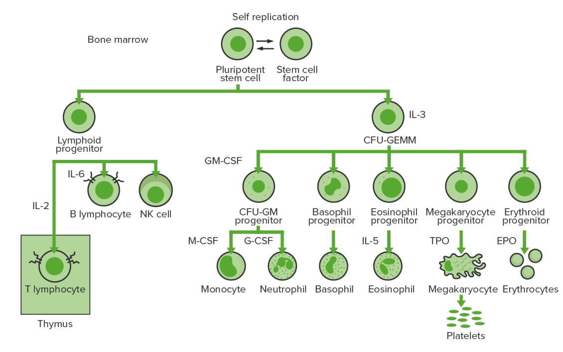
Bone-marrow hematopoiesis: proliferation and differentiation of the formed elements of blood.
CFU-GEMM: colony-forming unit–granulocyte, erythrocyte, monocyte, megakaryocyte
CFU-GM: colony-forming unit–granulocyte-macrophage
GM-CSF: granulocyte-macrophage colony-stimulating factor
M-CSF: macrophage colony-stimulating factor
G-CSF: granulocyte colony-stimulating factor
NK: natural killer
TPO: thrombopoietin

Image by Lecturio. License: CC BY-NC-SA 4.0

Eosinophilia Eosinophilia Abnormal increase of eosinophils in the blood, tissues or organs. Autosomal Dominant Hyperimmunoglobulin E Syndrome

  • Overproduction of eosinophils Eosinophils Granular leukocytes with a nucleus that usually has two lobes connected by a slender thread of chromatin, and cytoplasm containing coarse, round granules that are uniform in size and stainable by eosin. Innate Immunity: Phagocytes and Antigen Presentation in bone marrow Bone marrow The soft tissue filling the cavities of bones. Bone marrow exists in two types, yellow and red. Yellow marrow is found in the large cavities of large bones and consists mostly of fat cells and a few primitive blood cells. Red marrow is a hematopoietic tissue and is the site of production of erythrocytes and granular leukocytes. Bone marrow is made up of a framework of connective tissue containing branching fibers with the frame being filled with marrow cells. Bone Marrow: Composition and Hematopoiesis in CEL, NOS occurs but the underlying mechanism that facilitates this process is not clear.
  • Other variants associated with HES have chromosomal aberrations chromosomal aberrations Abnormal number or structure of chromosomes. Chromosome aberrations may result in chromosome disorders. Chromosome Testing (e.g., PDGFRA fusion with other genes Genes A category of nucleic acid sequences that function as units of heredity and which code for the basic instructions for the development, reproduction, and maintenance of organisms. DNA Types and Structure, rearrangements involving PDGFRB).
  • Theoretically, enhanced activity or defective regulation of eosinophil synthesis Synthesis Polymerase Chain Reaction (PCR) ia suspected.
  • With ↑ eosinophils Eosinophils Granular leukocytes with a nucleus that usually has two lobes connected by a slender thread of chromatin, and cytoplasm containing coarse, round granules that are uniform in size and stainable by eosin. Innate Immunity: Phagocytes and Antigen Presentation, organ infiltration follows → enhanced fibrosis Fibrosis Any pathological condition where fibrous connective tissue invades any organ, usually as a consequence of inflammation or other injury. Bronchiolitis Obliterans → tissue and organ damage and dysfunction

Clinical Presentation

  • Insidious onset
  • Constitutional symptoms Constitutional Symptoms Antineutrophil Cytoplasmic Antibody (ANCA)-Associated Vasculitis ( fever Fever Fever is defined as a measured body temperature of at least 38°C (100.4°F). Fever is caused by circulating endogenous and/or exogenous pyrogens that increase levels of prostaglandin E2 in the hypothalamus. Fever is commonly associated with chills, rigors, sweating, and flushing of the skin. Fever, weight loss Weight loss Decrease in existing body weight. Bariatric Surgery, night sweats Night sweats Tuberculosis)
  • Symptoms related to:
    • Anemia Anemia Anemia is a condition in which individuals have low Hb levels, which can arise from various causes. Anemia is accompanied by a reduced number of RBCs and may manifest with fatigue, shortness of breath, pallor, and weakness. Subtypes are classified by the size of RBCs, chronicity, and etiology. Anemia: Overview and Types: fatigue Fatigue The state of weariness following a period of exertion, mental or physical, characterized by a decreased capacity for work and reduced efficiency to respond to stimuli. Fibromyalgia, palpitations Palpitations Ebstein’s Anomaly
    • Thrombocytopenia Thrombocytopenia Thrombocytopenia occurs when the platelet count is < 150,000 per microliter. The normal range for platelets is usually 150,000-450,000/µL of whole blood. Thrombocytopenia can be a result of decreased production, increased destruction, or splenic sequestration of platelets. Patients are often asymptomatic until platelet counts are < 50,000/µL. Thrombocytopenia: bruising, petechiae Petechiae Primary Skin Lesions 
  • Hepatosplenomegaly Hepatosplenomegaly Cytomegalovirus
  • Can have overlapping symptoms (associated with eosinophilic activation):
    • Angioedema Angioedema Angioedema is a localized, self-limited (but potentially life-threatening), nonpitting, asymmetrical edema occurring in the deep layers of the skin and mucosal tissue. The common underlying pathophysiology involves inflammatory mediators triggering significant vasodilation and increased capillary permeability. Angioedema
    • Dermatologic: cutaneous lesions, rashes Rashes Rashes are a group of diseases that cause abnormal coloration and texture to the skin. The etiologies are numerous but can include irritation, allergens, infections, or inflammatory conditions. Rashes that present in only 1 area of the body are called localized rashes. Generalized rashes occur diffusely throughout the body. Generalized and Localized Rashes
    • Pulmonary/respiratory: cough, asthma Asthma Asthma is a chronic inflammatory respiratory condition characterized by bronchial hyperresponsiveness and airflow obstruction. The disease is believed to result from the complex interaction of host and environmental factors that increase disease predisposition, with inflammation causing symptoms and structural changes. Patients typically present with wheezing, cough, and dyspnea. Asthma
    • GI: gastroenteritis Gastroenteritis Gastroenteritis is inflammation of the stomach and intestines, commonly caused by infections from bacteria, viruses, or parasites. Transmission may be foodborne, fecal-oral, or through animal contact. Common clinical features include abdominal pain, diarrhea, vomiting, fever, and dehydration. Gastroenteritis/ diarrhea Diarrhea Diarrhea is defined as ≥ 3 watery or loose stools in a 24-hour period. There are a multitude of etiologies, which can be classified based on the underlying mechanism of disease. The duration of symptoms (acute or chronic) and characteristics of the stools (e.g., watery, bloody, steatorrheic, mucoid) can help guide further diagnostic evaluation. Diarrhea
    • Rheumatologic: vasculitis Vasculitis Inflammation of any one of the blood vessels, including the arteries; veins; and rest of the vasculature system in the body. Systemic Lupus Erythematosus, arthralgia Arthralgia Pain in the joint. Rheumatic Fever
    • Cardiac:
      • Cardiomegaly Cardiomegaly Enlargement of the heart, usually indicated by a cardiothoracic ratio above 0. 50. Heart enlargement may involve the right, the left, or both heart ventricles or heart atria. Cardiomegaly is a nonspecific symptom seen in patients with chronic systolic heart failure (heart failure) or several forms of cardiomyopathies. Ebstein’s Anomaly, cardiomyopathy Cardiomyopathy Cardiomyopathy refers to a group of myocardial diseases associated with structural changes of the heart muscles (myocardium) and impaired systolic and/or diastolic function in the absence of other heart disorders (coronary artery disease, hypertension, valvular disease, and congenital heart disease). Cardiomyopathy: Overview and Types (endomyocardial fibrosis Fibrosis Any pathological condition where fibrous connective tissue invades any organ, usually as a consequence of inflammation or other injury. Bronchiolitis Obliterans is most serious complication)
      • Cardiac involvement is unpredictable (not correlated to level of eosinophilia Eosinophilia Abnormal increase of eosinophils in the blood, tissues or organs. Autosomal Dominant Hyperimmunoglobulin E Syndrome).
      • Found also in a different variant with FIP1L1-PDGFRA fusion
    • Neurologic: neuropathy Neuropathy Leprosy, memory Memory Complex mental function having four distinct phases: (1) memorizing or learning, (2) retention, (3) recall, and (4) recognition. Clinically, it is usually subdivided into immediate, recent, and remote memory. Psychiatric Assessment loss

Diagnosis

Diagnosis

Chronic eosinophilic leukemia, not otherwise specified is a diagnosis of exclusion (requires ruling out other eosinophilic conditions) and is defined according to the WHO diagnostic criteria:

  • Meets the following criteria:
    • Eosinophil count: absolute eosinophilic count ≥ 1.5 x 10⁹/L
    • Peripheral blood blast count: > 2% 
    • Bone marrow Bone marrow The soft tissue filling the cavities of bones. Bone marrow exists in two types, yellow and red. Yellow marrow is found in the large cavities of large bones and consists mostly of fat cells and a few primitive blood cells. Red marrow is a hematopoietic tissue and is the site of production of erythrocytes and granular leukocytes. Bone marrow is made up of a framework of connective tissue containing branching fibers with the frame being filled with marrow cells. Bone Marrow: Composition and Hematopoiesis blast cell count: 5%–19% of all nucleated cells
    • Evidence of clonality:
      • Detection of clonal cytogenetic abnormality OR
      • By demonstration of a very skewed expression of X chromosome X chromosome The female sex chromosome, being the differential sex chromosome carried by half the male gametes and all female gametes in human and other male-heterogametic species. Basic Terms of Genetics genes Genes A category of nucleic acid sequences that function as units of heredity and which code for the basic instructions for the development, reproduction, and maintenance of organisms. DNA Types and Structure
  • Does not meet the WHO diagnostic criteria for:
    • Other myeloproliferative neoplasms Neoplasms New abnormal growth of tissue. Malignant neoplasms show a greater degree of anaplasia and have the properties of invasion and metastasis, compared to benign neoplasms. Benign Bone Tumors:
      • CML CML Chronic myeloid leukemia is a malignant proliferation of the granulocytic cell line characterized by a fairly normal differentiation. The underlying genetic abnormality is the Philadelphia chromosome, an abbreviated chromosome 22, resulting from reciprocal (9;22)(q34;q11) translocation. Chronic Myeloid Leukemia
      • PCV
      • ET
      • PMF
      • CNL
    • inv(16)(p13q22) or t(16;16)(p13;q22)
    • Myelodysplastic syndrome/myeloproliferative neoplasm (chronic myelomonocytic leukemia or atypical CML CML Chronic myeloid leukemia is a malignant proliferation of the granulocytic cell line characterized by a fairly normal differentiation. The underlying genetic abnormality is the Philadelphia chromosome, an abbreviated chromosome 22, resulting from reciprocal (9;22)(q34;q11) translocation. Chronic Myeloid Leukemia)
    • Lymphocytic variant of hypereosinophilia 
  • No rearrangement of PDGFRA, PDGFRB, FGFR1, or with PCM1-JAK2
Hypereosinophilia

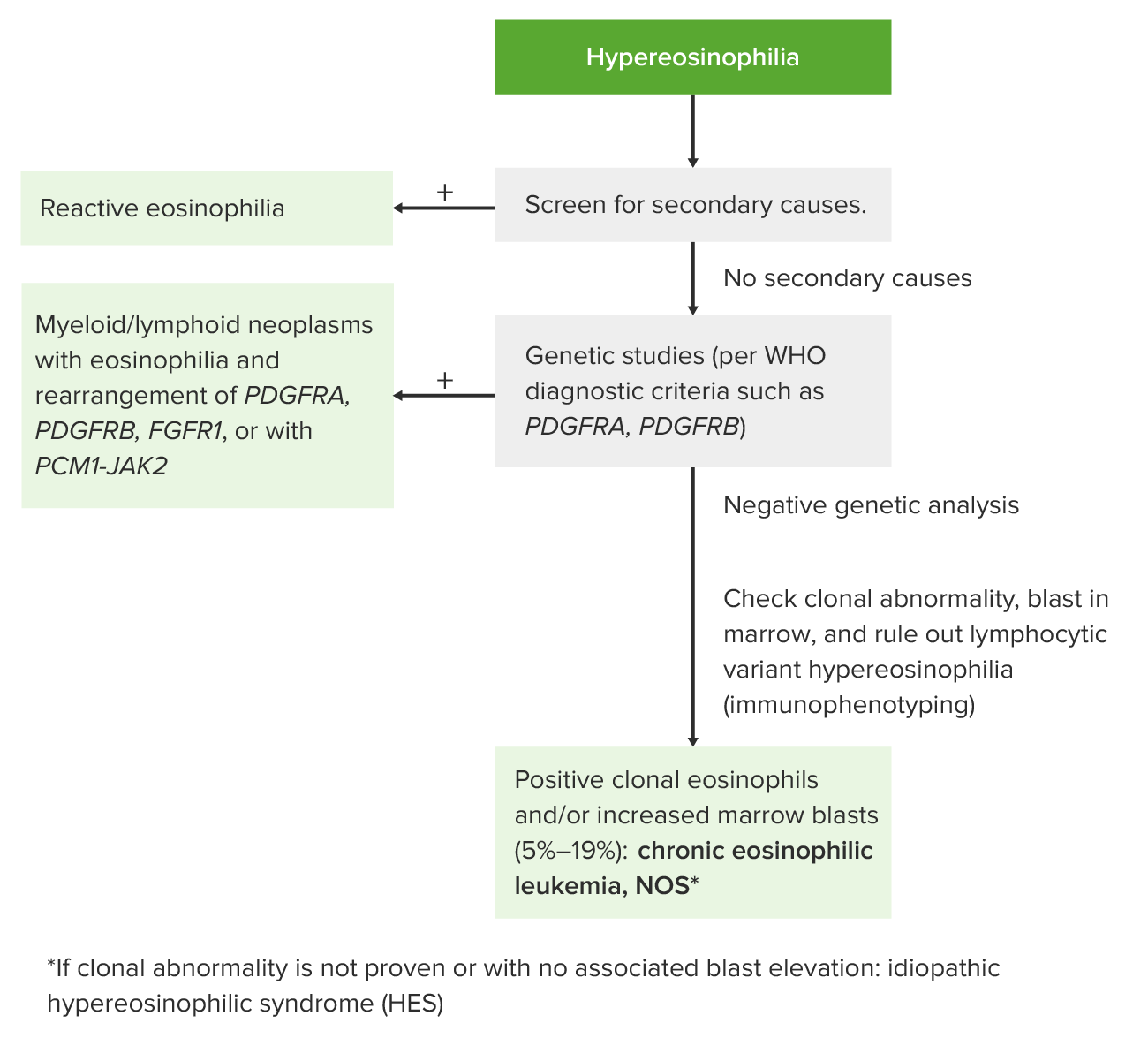
Approach to hypereosinophilia

Image by Lecturio.

Tests

  • Laboratory features found in CEL, NOS (which are also found in other myeloid variants):
    • CBC and peripheral blood smear Peripheral Blood Smear Anemia: Overview and Types:
      • Eosinophils Eosinophils Granular leukocytes with a nucleus that usually has two lobes connected by a slender thread of chromatin, and cytoplasm containing coarse, round granules that are uniform in size and stainable by eosin. Innate Immunity: Phagocytes and Antigen Presentation: absolute eosinophilic count ≥ 1.5 x 10⁹/L
      • Anemia Anemia Anemia is a condition in which individuals have low Hb levels, which can arise from various causes. Anemia is accompanied by a reduced number of RBCs and may manifest with fatigue, shortness of breath, pallor, and weakness. Subtypes are classified by the size of RBCs, chronicity, and etiology. Anemia: Overview and Types
      • Thrombocytopenia Thrombocytopenia Thrombocytopenia occurs when the platelet count is < 150,000 per microliter. The normal range for platelets is usually 150,000-450,000/µL of whole blood. Thrombocytopenia can be a result of decreased production, increased destruction, or splenic sequestration of platelets. Patients are often asymptomatic until platelet counts are < 50,000/µL. Thrombocytopenia
      • Circulating leukocyte precursors
    • Serum vitamin B12 (elevated)
    • Bone marrow Bone marrow The soft tissue filling the cavities of bones. Bone marrow exists in two types, yellow and red. Yellow marrow is found in the large cavities of large bones and consists mostly of fat cells and a few primitive blood cells. Red marrow is a hematopoietic tissue and is the site of production of erythrocytes and granular leukocytes. Bone marrow is made up of a framework of connective tissue containing branching fibers with the frame being filled with marrow cells. Bone Marrow: Composition and Hematopoiesis biopsy Biopsy Removal and pathologic examination of specimens from the living body. Ewing Sarcoma: hypercellular marrow with markedly increased eosinophil precursors and eosinophils Eosinophils Granular leukocytes with a nucleus that usually has two lobes connected by a slender thread of chromatin, and cytoplasm containing coarse, round granules that are uniform in size and stainable by eosin. Innate Immunity: Phagocytes and Antigen Presentation (orderly maturation)
  • Check studies for chromosomal or gene Gene A category of nucleic acid sequences that function as units of heredity and which code for the basic instructions for the development, reproduction, and maintenance of organisms. Basic Terms of Genetics abnormalities to rule out other disorders:
    • BCR-ABL/ Philadelphia chromosome Philadelphia chromosome An aberrant form of human chromosome 22 characterized by translocation of the distal end of chromosome 9 from 9q34, to the long arm of chromosome 22 at 22q11. It is present in the bone marrow cells of 80 to 90 percent of patients with chronic myelocytic leukemia. Acute Lymphoblastic Leukemia
    • PDGFRA 
    • PDGFRB
    • PCM1-JAK2 
    • FGFR1
  • Assess end-organ involvement depending on presentation:
    • Metabolic panel
    • Liver Liver The liver is the largest gland in the human body. The liver is found in the superior right quadrant of the abdomen and weighs approximately 1.5 kilograms. Its main functions are detoxification, metabolism, nutrient storage (e.g., iron and vitamins), synthesis of coagulation factors, formation of bile, filtration, and storage of blood. Liver: Anatomy profile
    • Echocardiogram Echocardiogram Transposition of the Great Vessels
    • Imaging:
      • Echocardiogram Echocardiogram Transposition of the Great Vessels
      • Chest X-ray X-ray Penetrating electromagnetic radiation emitted when the inner orbital electrons of an atom are excited and release radiant energy. X-ray wavelengths range from 1 pm to 10 nm. Hard x-rays are the higher energy, shorter wavelength x-rays. Soft x-rays or grenz rays are less energetic and longer in wavelength. The short wavelength end of the x-ray spectrum overlaps the gamma rays wavelength range. The distinction between gamma rays and x-rays is based on their radiation source. Pulmonary Function Tests/CT
      • Abdominal CT
Activated eosinophils in hypereosinophilic syndrome

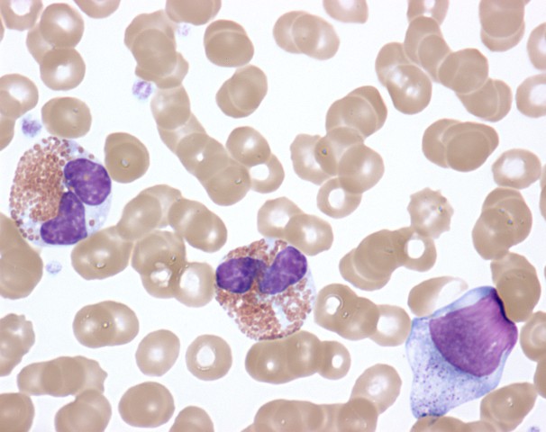
Activated eosinophils in the peripheral blood of a patient with hypereosinophilic syndrome showing cytoplasmic clearing, nuclear dysplasia, and the presence of immature forms

Image: “Activated Eosinophils in Idiopathic Hypereosinophilic Syndrome (9125007255)” by NIAID. License: CC BY 2.0

Management

  • Goals of therapy:
    • Reduction of absolute eosinophil count (AEC): maintain below 1,500 cells/μL (to prevent end-organ damage)
    • Improvement of signs and symptoms
    • Prevention of disease progression
  • CEL, NOS clinical course:
    • Course is variable Variable Variables represent information about something that can change. The design of the measurement scales, or of the methods for obtaining information, will determine the data gathered and the characteristics of that data. As a result, a variable can be qualitative or quantitative, and may be further classified into subgroups. Types of Variables.
    • Transformation Transformation Change brought about to an organism’s genetic composition by unidirectional transfer (transfection; transduction, genetic; conjugation, genetic, etc.) and incorporation of foreign DNA into prokaryotic or eukaryotic cells by recombination of part or all of that DNA into the cell’s genome. Bacteriology to acute leukemia seen
  • Treatment:
    • Due to rarity and variability in the course of disease, optimal treatment is not clear.
    • Initial approach: corticosteroids Corticosteroids Chorioretinitis (1st-line therapy, as with idiopathic Idiopathic Dermatomyositis HES)
    • Steroid-refractory cases:
      • Chemotherapeutic agents: hydroxyurea Hydroxyurea An antineoplastic agent that inhibits DNA synthesis through the inhibition of ribonucleoside diphosphate reductase. Antimetabolite Chemotherapy, cyclophosphamide Cyclophosphamide Precursor of an alkylating nitrogen mustard antineoplastic and immunosuppressive agent that must be activated in the liver to form the active aldophosphamide. It has been used in the treatment of lymphoma and leukemia. Its side effect, alopecia, has been used for defleecing sheep. Cyclophosphamide may also cause sterility, birth defects, mutations, and cancer. Immunosuppressants, vincristine Vincristine An antitumor alkaloid isolated from vinca rosea. Microtubule and Topoisomerase Inhibitors
      • Interferon-α
    • Some patients Patients Individuals participating in the health care system for the purpose of receiving therapeutic, diagnostic, or preventive procedures. Clinician–Patient Relationship may benefit from a trial of imatinib Imatinib A tyrosine kinase inhibitor and antineoplastic agent that inhibits the bcr-abl kinase created by chromosome rearrangements in chronic myeloid leukemia and acute lymphoblastic leukemia, as well as pdg-derived tyrosine kinases that are overexpressed in gastrointestinal stromal tumors. Targeted and Other Nontraditional Antineoplastic Therapy, a tyrosine Tyrosine A non-essential amino acid. In animals it is synthesized from phenylalanine. It is also the precursor of epinephrine; thyroid hormones; and melanin. Synthesis of Nonessential Amino Acids kinase inhibitor (which is effective for PDGFRA-positive HES variants).
    • Other therapies that have shown some efficacy:

Differential Diagnosis

Other eosinophilias

  • Secondary eosinophilia Eosinophilia Abnormal increase of eosinophils in the blood, tissues or organs. Autosomal Dominant Hyperimmunoglobulin E Syndrome: a cytokine-derived (IL-5) reactive phenomenon. Worldwide, parasitic diseases are the predominant cause, but in developed countries, allergic diseases are the most common etiology. Signs and symptoms are constitutional and end-organ-involvement presentations can be seen. Diagnostic tests Diagnostic tests Diagnostic tests are important aspects in making a diagnosis. Some of the most important epidemiological values of diagnostic tests include sensitivity and specificity, false positives and false negatives, positive and negative predictive values, likelihood ratios, and pre-test and post-test probabilities. Epidemiological Values of Diagnostic Tests are CBC with differential, blood chemistries, and potential bone marrow Bone marrow The soft tissue filling the cavities of bones. Bone marrow exists in two types, yellow and red. Yellow marrow is found in the large cavities of large bones and consists mostly of fat cells and a few primitive blood cells. Red marrow is a hematopoietic tissue and is the site of production of erythrocytes and granular leukocytes. Bone marrow is made up of a framework of connective tissue containing branching fibers with the frame being filled with marrow cells. Bone Marrow: Composition and Hematopoiesis aspirate evaluation. Management is with steroids Steroids A group of polycyclic compounds closely related biochemically to terpenes. They include cholesterol, numerous hormones, precursors of certain vitamins, bile acids, alcohols (sterols), and certain natural drugs and poisons. Steroids have a common nucleus, a fused, reduced 17-carbon atom ring system, cyclopentanoperhydrophenanthrene. Most steroids also have two methyl groups and an aliphatic side-chain attached to the nucleus. Benign Liver Tumors, avoidance of the underlying inciting agent, and supportive interventions.
  • Acute eosinophilic leukemia: a neoplastic disease characterized by a marked increase in the number of immature eosinophils Eosinophils Granular leukocytes with a nucleus that usually has two lobes connected by a slender thread of chromatin, and cytoplasm containing coarse, round granules that are uniform in size and stainable by eosin. Innate Immunity: Phagocytes and Antigen Presentation (unlike CEL) in the blood and/or marrow, > 10% blast forms in bone marrow Bone marrow The soft tissue filling the cavities of bones. Bone marrow exists in two types, yellow and red. Yellow marrow is found in the large cavities of large bones and consists mostly of fat cells and a few primitive blood cells. Red marrow is a hematopoietic tissue and is the site of production of erythrocytes and granular leukocytes. Bone marrow is made up of a framework of connective tissue containing branching fibers with the frame being filled with marrow cells. Bone Marrow: Composition and Hematopoiesis, and infiltration of tissues ( nervous system Nervous system The nervous system is a small and complex system that consists of an intricate network of neural cells (or neurons) and even more glial cells (for support and insulation). It is divided according to its anatomical components as well as its functional characteristics. The brain and spinal cord are referred to as the central nervous system, and the branches of nerves from these structures are referred to as the peripheral nervous system. Nervous System: Anatomy, Structure, and Classification and bone Bone Bone is a compact type of hardened connective tissue composed of bone cells, membranes, an extracellular mineralized matrix, and central bone marrow. The 2 primary types of bone are compact and spongy. Bones: Structure and Types) with immature eosinophil forms. Anemia Anemia Anemia is a condition in which individuals have low Hb levels, which can arise from various causes. Anemia is accompanied by a reduced number of RBCs and may manifest with fatigue, shortness of breath, pallor, and weakness. Subtypes are classified by the size of RBCs, chronicity, and etiology. Anemia: Overview and Types, thrombocytopenia Thrombocytopenia Thrombocytopenia occurs when the platelet count is < 150,000 per microliter. The normal range for platelets is usually 150,000-450,000/µL of whole blood. Thrombocytopenia can be a result of decreased production, increased destruction, or splenic sequestration of platelets. Patients are often asymptomatic until platelet counts are < 50,000/µL. Thrombocytopenia, hepatosplenomegaly Hepatosplenomegaly Cytomegalovirus, and susceptibility to infection are also seen. Diagnosis is made by history and physical examination findings along with blood and marrow evaluations. Management is with chemotherapy Chemotherapy Osteosarcoma and hematopoietic stem cell transplantation Hematopoietic stem cell transplantation Transfer of hematopoietic stem cells from bone marrow or blood between individuals within the same species (homologous transplantation) or transfer within the same individual (autologous transplantation). Hematopoietic stem cell transplantation has been used as an alternative to bone marrow transplantation in the treatment of a variety of neoplasms. Organ Transplantation.
  • Systemic mastocytosis with eosinophilia Eosinophilia Abnormal increase of eosinophils in the blood, tissues or organs. Autosomal Dominant Hyperimmunoglobulin E Syndrome: a type of mast cell Mast cell Granulated cells that are found in almost all tissues, most abundantly in the skin and the gastrointestinal tract. Like the basophils, mast cells contain large amounts of histamine and heparin. Unlike basophils, mast cells normally remain in the tissues and do not circulate in the blood. Mast cells, derived from the bone marrow stem cells, are regulated by the stem cell factor. Angioedema disease caused by the accumulation of functionally defective mast cells Mast cells Granulated cells that are found in almost all tissues, most abundantly in the skin and the gastrointestinal tract. Like the basophils, mast cells contain large amounts of histamine and heparin. Unlike basophils, mast cells normally remain in the tissues and do not circulate in the blood. Mast cells, derived from the bone marrow stem cells, are regulated by the stem cell factor. Innate Immunity: Phagocytes and Antigen Presentation, associated with KIT mutations (often KIT D816V) in most patients Patients Individuals participating in the health care system for the purpose of receiving therapeutic, diagnostic, or preventive procedures. Clinician–Patient Relationship. A characteristic skin Skin The skin, also referred to as the integumentary system, is the largest organ of the body. The skin is primarily composed of the epidermis (outer layer) and dermis (deep layer). The epidermis is primarily composed of keratinocytes that undergo rapid turnover, while the dermis contains dense layers of connective tissue. Skin: Structure and Functions rash Rash Rocky Mountain Spotted Fever ( urticaria Urticaria Urticaria is raised, well-circumscribed areas (wheals) of edema (swelling) and erythema (redness) involving the dermis and epidermis with associated pruritus (itch). Urticaria is not a single disease but rather is a reaction pattern representing cutaneous mast cell degranulation. Urticaria (Hives) pigmentosa) and GI symptoms are noted along with hepatosplenomegaly Hepatosplenomegaly Cytomegalovirus. Diagnosis is made by the characteristic skin Skin The skin, also referred to as the integumentary system, is the largest organ of the body. The skin is primarily composed of the epidermis (outer layer) and dermis (deep layer). The epidermis is primarily composed of keratinocytes that undergo rapid turnover, while the dermis contains dense layers of connective tissue. Skin: Structure and Functions lesions and dense infiltrates of mast cells Mast cells Granulated cells that are found in almost all tissues, most abundantly in the skin and the gastrointestinal tract. Like the basophils, mast cells contain large amounts of histamine and heparin. Unlike basophils, mast cells normally remain in the tissues and do not circulate in the blood. Mast cells, derived from the bone marrow stem cells, are regulated by the stem cell factor. Innate Immunity: Phagocytes and Antigen Presentation in marrow or in extracutaneous organs. Tests show elevated serum tryptase Tryptase A family of neutral serine proteases with trypsin-like activity. Tryptases are primarily found in the secretory granules of mast cells and are released during mast cell degranulation. Exocrine Pancreatic Cancer and positive mutation Mutation Genetic mutations are errors in DNA that can cause protein misfolding and dysfunction. There are various types of mutations, including chromosomal, point, frameshift, and expansion mutations. Types of Mutations screening Screening Preoperative Care for KIT D816V. Management is with corticosteroids Corticosteroids Chorioretinitis, symptomatic interventions, and different agents ( midostaurin Midostaurin Acute Myeloid Leukemia, hydroxyurea Hydroxyurea An antineoplastic agent that inhibits DNA synthesis through the inhibition of ribonucleoside diphosphate reductase. Antimetabolite Chemotherapy, and others in different settings).
  • PDGFRA-mutated eosinophilia Eosinophilia Abnormal increase of eosinophils in the blood, tissues or organs. Autosomal Dominant Hyperimmunoglobulin E Syndrome: prominent eosinophilia Eosinophilia Abnormal increase of eosinophils in the blood, tissues or organs. Autosomal Dominant Hyperimmunoglobulin E Syndrome with platelet-derived growth factor Platelet-derived growth factor Mitogenic peptide growth hormone carried in the alpha-granules of platelets. It is released when platelets adhere to traumatized tissues. Connective tissue cells near the traumatized region respond by initiating the process of replication. Hypertrophic and Keloid Scars receptors Receptors Receptors are proteins located either on the surface of or within a cell that can bind to signaling molecules known as ligands (e.g., hormones) and cause some type of response within the cell. Receptors α (PDGFRA on chromosome Chromosome In a prokaryotic cell or in the nucleus of a eukaryotic cell, a structure consisting of or containing DNA which carries the genetic information essential to the cell. Basic Terms of Genetics 4q12) and β (PDGFRB on chromosome Chromosome In a prokaryotic cell or in the nucleus of a eukaryotic cell, a structure consisting of or containing DNA which carries the genetic information essential to the cell. Basic Terms of Genetics 5q31-q32) are affected in activating mutations. Genetic studies in cases with hypereosinophilia help distinguish these conditions. Both PDGFRA– and PDGFRB-related disorders are responsive to imatinib Imatinib A tyrosine kinase inhibitor and antineoplastic agent that inhibits the bcr-abl kinase created by chromosome rearrangements in chronic myeloid leukemia and acute lymphoblastic leukemia, as well as pdg-derived tyrosine kinases that are overexpressed in gastrointestinal stromal tumors. Targeted and Other Nontraditional Antineoplastic Therapy.

Other chronic myeloproliferative neoplasms Neoplasms New abnormal growth of tissue. Malignant neoplasms show a greater degree of anaplasia and have the properties of invasion and metastasis, compared to benign neoplasms. Benign Bone Tumors

  • Chronic myeloid leukemia Chronic myeloid leukemia Chronic myeloid leukemia is a malignant proliferation of the granulocytic cell line characterized by a fairly normal differentiation. The underlying genetic abnormality is the Philadelphia chromosome, an abbreviated chromosome 22, resulting from reciprocal (9;22)(q34;q11) translocation. Chronic Myeloid Leukemia: a malignant proliferation of WBCs, a myeloproliferative disorder with fairly normal differentiation. The underlying pathology involves translocation of genetic material resulting in a Philadelphia chromosome Philadelphia chromosome An aberrant form of human chromosome 22 characterized by translocation of the distal end of chromosome 9 from 9q34, to the long arm of chromosome 22 at 22q11. It is present in the bone marrow cells of 80 to 90 percent of patients with chronic myelocytic leukemia. Acute Lymphoblastic Leukemia with deregulated tyrosine Tyrosine A non-essential amino acid. In animals it is synthesized from phenylalanine. It is also the precursor of epinephrine; thyroid hormones; and melanin. Synthesis of Nonessential Amino Acids kinase production. Symptoms include constitutional complaints, early satiety Early Satiety Bariatric Surgery, sternal tenderness, and hepatosplenomegaly Hepatosplenomegaly Cytomegalovirus. Laboratory studies show elevated WBCs and a peripheral smear with increased numbers of immature cells. Management focuses on tyrosine Tyrosine A non-essential amino acid. In animals it is synthesized from phenylalanine. It is also the precursor of epinephrine; thyroid hormones; and melanin. Synthesis of Nonessential Amino Acids kinase inhibition.
  • Primary myelofibrosis Primary myelofibrosis Primary myelofibrosis (PMF) is a myeloproliferative neoplasm characterized by chronic myeloproliferation with nonclonal fibroblastic deposition, resulting in bone marrow fibrosis. The abnormality stems from genetic mutations of the hematopoietic stem cells (typically, JAK2 mutation). Primary symptoms are anemia and extramedullary hematopoiesis,. Primary Myelofibrosis: a chronic myeloproliferative neoplasm characterized by fibrosis Fibrosis Any pathological condition where fibrous connective tissue invades any organ, usually as a consequence of inflammation or other injury. Bronchiolitis Obliterans of the bone marrow Bone marrow The soft tissue filling the cavities of bones. Bone marrow exists in two types, yellow and red. Yellow marrow is found in the large cavities of large bones and consists mostly of fat cells and a few primitive blood cells. Red marrow is a hematopoietic tissue and is the site of production of erythrocytes and granular leukocytes. Bone marrow is made up of a framework of connective tissue containing branching fibers with the frame being filled with marrow cells. Bone Marrow: Composition and Hematopoiesis and extramedullary hematopoiesis Extramedullary Hematopoiesis Primary Myelofibrosis in the spleen Spleen The spleen is the largest lymphoid organ in the body, located in the LUQ of the abdomen, superior to the left kidney and posterior to the stomach at the level of the 9th-11th ribs just below the diaphragm. The spleen is highly vascular and acts as an important blood filter, cleansing the blood of pathogens and damaged erythrocytes. Spleen: Anatomy and liver Liver The liver is the largest gland in the human body. The liver is found in the superior right quadrant of the abdomen and weighs approximately 1.5 kilograms. Its main functions are detoxification, metabolism, nutrient storage (e.g., iron and vitamins), synthesis of coagulation factors, formation of bile, filtration, and storage of blood. Liver: Anatomy. The condition is linked to mutations in JAK2, CALR, and MPL. Clinical findings are severe fatigue Fatigue The state of weariness following a period of exertion, mental or physical, characterized by a decreased capacity for work and reduced efficiency to respond to stimuli. Fibromyalgia, splenomegaly Splenomegaly Splenomegaly is pathologic enlargement of the spleen that is attributable to numerous causes, including infections, hemoglobinopathies, infiltrative processes, and outflow obstruction of the portal vein. Splenomegaly, hepatomegaly, and anemia Anemia Anemia is a condition in which individuals have low Hb levels, which can arise from various causes. Anemia is accompanied by a reduced number of RBCs and may manifest with fatigue, shortness of breath, pallor, and weakness. Subtypes are classified by the size of RBCs, chronicity, and etiology. Anemia: Overview and Types. The peripheral smear is leukoerythroblastic and contains precursors of WBCs and RBCs RBCs Erythrocytes, or red blood cells (RBCs), are the most abundant cells in the blood. While erythrocytes in the fetus are initially produced in the yolk sac then the liver, the bone marrow eventually becomes the main site of production. Erythrocytes: Histology, nucleated RBCs RBCs Erythrocytes, or red blood cells (RBCs), are the most abundant cells in the blood. While erythrocytes in the fetus are initially produced in the yolk sac then the liver, the bone marrow eventually becomes the main site of production. Erythrocytes: Histology, and teardrop cells Teardrop Cells Primary Myelofibrosis. Diagnosis is made by bone marrow Bone marrow The soft tissue filling the cavities of bones. Bone marrow exists in two types, yellow and red. Yellow marrow is found in the large cavities of large bones and consists mostly of fat cells and a few primitive blood cells. Red marrow is a hematopoietic tissue and is the site of production of erythrocytes and granular leukocytes. Bone marrow is made up of a framework of connective tissue containing branching fibers with the frame being filled with marrow cells. Bone Marrow: Composition and Hematopoiesis examination and molecular testing. Management includes allogeneic hematopoietic cell transplantation, and the medications ruxolitinib Ruxolitinib Targeted and Other Nontraditional Antineoplastic Therapy and fedratinib.
  • PCV: an unregulated overproduction of hematopoietic stem cells Hematopoietic stem cells Progenitor cells from which all blood cells derived. They are found primarily in the bone marrow and also in small numbers in the peripheral blood. Bone Marrow: Composition and Hematopoiesis, mainly the RBCs RBCs Erythrocytes, or red blood cells (RBCs), are the most abundant cells in the blood. While erythrocytes in the fetus are initially produced in the yolk sac then the liver, the bone marrow eventually becomes the main site of production. Erythrocytes: Histology, despite low levels of EPO EPO Glycoprotein hormone, secreted chiefly by the kidney in the adult and the liver in the fetus, that acts on erythroid stem cells of the bone marrow to stimulate proliferation and differentiation. Erythrocytes: Histology. There is a gain-of-function mutation Mutation Genetic mutations are errors in DNA that can cause protein misfolding and dysfunction. There are various types of mutations, including chromosomal, point, frameshift, and expansion mutations. Types of Mutations in the JAK2 gene Gene A category of nucleic acid sequences that function as units of heredity and which code for the basic instructions for the development, reproduction, and maintenance of organisms. Basic Terms of Genetics. The majority of patients Patients Individuals participating in the health care system for the purpose of receiving therapeutic, diagnostic, or preventive procedures. Clinician–Patient Relationship present with hyperviscosity Hyperviscosity Hypercoagulable States symptoms. Management includes phlebotomy Phlebotomy The techniques used to draw blood from a vein for diagnostic purposes or for treatment of certain blood disorders such as erythrocytosis, hemochromatosis, polycythemia vera, and porphyria cutanea tarda. Hereditary Hemochromatosis, low-dose aspirin Aspirin The prototypical analgesic used in the treatment of mild to moderate pain. It has anti-inflammatory and antipyretic properties and acts as an inhibitor of cyclooxygenase which results in the inhibition of the biosynthesis of prostaglandins. Aspirin also inhibits platelet aggregation and is used in the prevention of arterial and venous thrombosis. Nonsteroidal Antiinflammatory Drugs (NSAIDs), and myelosuppressive therapies.
  • Essential thrombocythemia Essential thrombocythemia Essential thrombocythemia (ET) is a type of myeloproliferative neoplasm characterized by the clonal thrombocytosis linked to somatic mutations involving Janus kinase 2 (JAK2), calreticulin (CALR), and myeloproliferative leukemia virus oncogene (MPL). Patients can be asymptomatic or present with vasomotor symptoms. Essential Thrombocythemia: a proliferation of megakaryocytes usually due to JAK2, CALR, and MPL mutations resulting in thrombocytosis. Clinical manifestations are headaches, visual disturbances, and erythromelalgia Erythromelalgia A peripheral arterial disease that is characterized by the triad of erythema, burning pain, and increased skin temperature of the extremities (or red, painful extremities). Erythromelalgia may be classified as primary or idiopathic, familial or non-familial. Secondary erythromelalgia is associated with other diseases, the most common being myeloproliferative disorders. Polycythemia Vera. Excessive counts of platelets Platelets Platelets are small cell fragments involved in hemostasis. Thrombopoiesis takes place primarily in the bone marrow through a series of cell differentiation and is influenced by several cytokines. Platelets are formed after fragmentation of the megakaryocyte cytoplasm. Platelets: Histology result in both thrombosis Thrombosis Formation and development of a thrombus or blood clot in the blood vessel. Epidemic Typhus and bleeding. Management aims to reduce platelet count (with medications such as hydroxyurea Hydroxyurea An antineoplastic agent that inhibits DNA synthesis through the inhibition of ribonucleoside diphosphate reductase. Antimetabolite Chemotherapy) and decrease the risk of thrombosis Thrombosis Formation and development of a thrombus or blood clot in the blood vessel. Epidemic Typhus (by systemic anticoagulation Anticoagulation Pulmonary Hypertension Drugs and/or antiplatelet agents Antiplatelet agents Antiplatelet agents are medications that inhibit platelet aggregation, a critical step in the formation of the initial platelet plug. Abnormal, or inappropriate, platelet aggregation is a key step in the pathophysiology of arterial ischemic events. The primary categories of antiplatelet agents include aspirin, ADP inhibitors, phosphodiesterase/adenosine uptake inhibitors, and glycoprotein IIb/IIIa inhibitors. Antiplatelet Drugs).

References

  1. Cheah, C.Y., Burbury, K., Apperley, J.F., et al. (2014). Patients with myeloid malignancies bearing PDGFRB fusion genes achieve durable long-term remissions with imatinib. Blood. 123(23),3574–3577. https://pubmed.ncbi.nlm.nih.gov/24687085/
  2. Crane, M.M., et al. (2010). Incidence of myeloproliferative hypereosinophilic syndrome in the United States and an estimate of all hypereosinophilic syndrome incidence. J Allergy Clin Immunol. 126(1),179–181. https://pubmed.ncbi.nlm.nih.gov/20639012/
  3. Gotlib, J. (2017). World Health Organization‐defined eosinophilic disorders: 2017 update on diagnosis, risk stratification, and management. Am J Hematol. 92(11),1243– 1259. https://doi.org/10.1002/ajh.24880
  4. Helbig, G., et al. (2012). Chronic eosinophilic leukemia-not otherwise specified has a poor prognosis with unresponsiveness to conventional treatment and high risk of acute transformation. Am J Hematol. 87(6),643–645. https://pubmed.ncbi.nlm.nih.gov/22473587/
  5. National Cancer institute. (2020). Chronic Myeloproliferative Neoplasms Treatment (PDQ®)–Health Professional Version. Retrieved May 2, 2021, from https://www.cancer.gov/types/myeloproliferative/hp/chronic-treatment-pdq#_101
  6. Nguyen, L., Zhang, L. (2021). Myeloproliferative neoplasms (MPN) Chronic eosinophilic leukemia. Pathologyoutlines. Retrieved May 2, 2021, from https://www.pathologyoutlines.com/topic/myeloproliferativeCEL.html
  7. Ogbogu, P.U., Bochner, B.S., Butterfield, J.H., et al. (2009). Hypereosinophilic syndrome: a multicenter, retrospective analysis of clinical characteristics and response to therapy. J Allergy Clin Immunol. 124(6),1319–1325.e3. https://pubmed.ncbi.nlm.nih.gov/19910029/
  8. Roufosse, F., Klion, A., and Weller, P. (2021). Hypereosinophilic syndromes: Clinical manifestations, pathophysiology, and diagnosis. In Larson, R. (Ed.), UpToDate. Retrieved April 17, 2021, from https://www.uptodate.com/contents/hypereosinophilic-syndromes-clinical-manifestations-pathophysiology-and-diagnosis
  9. Samavedi, A.V., (2020). Hypereosinophilic Syndrome. Medscape. Retrieved April 17, 2021, from https://reference.medscape.com/article/202030-overview
  10. Thakral, B., Wang, S. (2018). Myeloproliferative and “Overlap” Myelodysplastic/Myeloproliferative Neoplasm. In Hematopathology (Third Edition), Elsevier. https://www.sciencedirect.com/science/article/pii/B9780323479134000173
  11. Tefferi, A., Longo D.L. (2018). Less common hematologic malignancies. In Jameson, J., et al. (Eds.), Harrison’s Principles of Internal Medicine, 20e. McGraw-Hill. 
  12. Valent, P., Klion, A.D., Horny, H.P., et al. (2012). Contemporary consensus proposal on criteria and classification of eosinophilic disorders and related syndromes. J Allergy Clin Immunol. 130(3),607–612.e9. https://pubmed.ncbi.nlm.nih.gov/22460074/
  13. Weller, P., and Klion, A. (2021). Eosinophil biology and causes of eosinophilia. In Mahoney, D, and Newburger, P. (Ed.), UpToDate. Retrieved April 17, 2021, from https://www.uptodate.com/contents/eosinophil-biology-and-causes-of-eosinophilia

Create your free account or log in to continue reading!

Sign up now and get free access to Lecturio with concept pages, medical videos, and questions for your medical education.

User Reviews

Details