Achieve Mastery of Medical Concepts

Study for medical school and boards with Lecturio

Essential Thrombocythemia

Essential thrombocythemia (ET) is a type of myeloproliferative neoplasm characterized by the clonal thrombocytosis linked to somatic mutations involving Janus kinase 2 (JAK2), calreticulin (CALR), and myeloproliferative leukemia virus Virus Viruses are infectious, obligate intracellular parasites composed of a nucleic acid core surrounded by a protein capsid. Viruses can be either naked (non-enveloped) or enveloped. The classification of viruses is complex and based on many factors, including type and structure of the nucleoid and capsid, the presence of an envelope, the replication cycle, and the host range. Virology oncogene (MPL). Patients Patients Individuals participating in the health care system for the purpose of receiving therapeutic, diagnostic, or preventive procedures. Clinician–Patient Relationship can be asymptomatic or present with vasomotor symptoms such as headaches, erythromelalgia Erythromelalgia A peripheral arterial disease that is characterized by the triad of erythema, burning pain, and increased skin temperature of the extremities (or red, painful extremities). Erythromelalgia may be classified as primary or idiopathic, familial or non-familial. Secondary erythromelalgia is associated with other diseases, the most common being myeloproliferative disorders. Polycythemia Vera, and transient visual disturbances. The clinical course can be complicated by thrombohemorrhagic events as well as progression to myelofibrosis and AML AML Acute myeloid leukemia (AML) is a hematologic malignancy characterized by the uncontrolled proliferation of myeloid precursor cells. Seen predominantly in older adults, AML includes an accumulation of myeloblasts and a replacement of normal marrow by malignant cells, which leads to impaired hematopoiesis. Acute Myeloid Leukemia. The diagnosis is based on a laboratory finding of thrombocytosis, bone marrow Bone marrow The soft tissue filling the cavities of bones. Bone marrow exists in two types, yellow and red. Yellow marrow is found in the large cavities of large bones and consists mostly of fat cells and a few primitive blood cells. Red marrow is a hematopoietic tissue and is the site of production of erythrocytes and granular leukocytes. Bone marrow is made up of a framework of connective tissue containing branching fibers with the frame being filled with marrow cells. Bone Marrow: Composition and Hematopoiesis biopsy Biopsy Removal and pathologic examination of specimens from the living body. Ewing Sarcoma, and genetic studies. Treatment aims to reduce platelet count by cytoreductive agents ( hydroxyurea Hydroxyurea An antineoplastic agent that inhibits DNA synthesis through the inhibition of ribonucleoside diphosphate reductase. Antimetabolite Chemotherapy) and to decrease thrombosis Thrombosis Formation and development of a thrombus or blood clot in the blood vessel. Epidemic Typhus with aspirin Aspirin The prototypical analgesic used in the treatment of mild to moderate pain. It has anti-inflammatory and antipyretic properties and acts as an inhibitor of cyclooxygenase which results in the inhibition of the biosynthesis of prostaglandins. Aspirin also inhibits platelet aggregation and is used in the prevention of arterial and venous thrombosis. Nonsteroidal Antiinflammatory Drugs (NSAIDs) and systemic anticoagulation Anticoagulation Pulmonary Hypertension Drugs based on thrombosis Thrombosis Formation and development of a thrombus or blood clot in the blood vessel. Epidemic Typhus risk stratification.

Last updated: Oct 24, 2022

Editorial responsibility: Stanley Oiseth, Lindsay Jones, Evelin Maza

Overview

Definition

Essential thrombocythemia (ET) is a chronic myeloproliferative neoplasm characterized by excessive platelet production and increased thrombotic and hemorrhagic tendency. Other names are essential thrombocytosis and primary thrombocytosis.

Epidemiology

  • Incidence Incidence The number of new cases of a given disease during a given period in a specified population. It also is used for the rate at which new events occur in a defined population. It is differentiated from prevalence, which refers to all cases in the population at a given time. Measures of Disease Frequency: 1–2.5 cases per 100,000 in the United States
  • Incidence Incidence The number of new cases of a given disease during a given period in a specified population. It also is used for the rate at which new events occur in a defined population. It is differentiated from prevalence, which refers to all cases in the population at a given time. Measures of Disease Frequency increases with age (median age at diagnosis: 60 years)
  • Female:male ratio of 2:1

Etiology

  • Proliferation of bone marrow Bone marrow The soft tissue filling the cavities of bones. Bone marrow exists in two types, yellow and red. Yellow marrow is found in the large cavities of large bones and consists mostly of fat cells and a few primitive blood cells. Red marrow is a hematopoietic tissue and is the site of production of erythrocytes and granular leukocytes. Bone marrow is made up of a framework of connective tissue containing branching fibers with the frame being filled with marrow cells. Bone Marrow: Composition and Hematopoiesis megakaryocytes due to presence of 1 of 3 driver mutations involving:
    • Janus kinase 2 (JAK2): 50%–60% of patients Patients Individuals participating in the health care system for the purpose of receiving therapeutic, diagnostic, or preventive procedures. Clinician–Patient Relationship
    • Calreticulin (CALR): 25% of patients Patients Individuals participating in the health care system for the purpose of receiving therapeutic, diagnostic, or preventive procedures. Clinician–Patient Relationship
    • Myeloproliferative leukemia virus Virus Viruses are infectious, obligate intracellular parasites composed of a nucleic acid core surrounded by a protein capsid. Viruses can be either naked (non-enveloped) or enveloped. The classification of viruses is complex and based on many factors, including type and structure of the nucleoid and capsid, the presence of an envelope, the replication cycle, and the host range. Virology oncogene (MPL): 5%–10% of patients Patients Individuals participating in the health care system for the purpose of receiving therapeutic, diagnostic, or preventive procedures. Clinician–Patient Relationship
  • Predisposing conditions in patients Patients Individuals participating in the health care system for the purpose of receiving therapeutic, diagnostic, or preventive procedures. Clinician–Patient Relationship with above mutations:

Pathophysiology

Hematopoiesis Hematopoiesis The development and formation of various types of blood cells. Hematopoiesis can take place in the bone marrow (medullary) or outside the bone marrow (extramedullary hematopoiesis). Bone Marrow: Composition and Hematopoiesis

Hematopoiesis Hematopoiesis The development and formation of various types of blood cells. Hematopoiesis can take place in the bone marrow (medullary) or outside the bone marrow (extramedullary hematopoiesis). Bone Marrow: Composition and Hematopoiesis starts with the hematopoietic stem cell, which is prompted to divide and differentiate with appropriate chemical stimuli (hemopoietic growth factors).

  • Lymphoid stem cells: give rise to lymphocytes Lymphocytes Lymphocytes are heterogeneous WBCs involved in immune response. Lymphocytes develop from the bone marrow, starting from hematopoietic stem cells (HSCs) and progressing to common lymphoid progenitors (CLPs). B and T lymphocytes and natural killer (NK) cells arise from the lineage. Lymphocytes: Histology
  • Myeloid stem cell: eventually differentiate into platelets Platelets Platelets are small cell fragments involved in hemostasis. Thrombopoiesis takes place primarily in the bone marrow through a series of cell differentiation and is influenced by several cytokines. Platelets are formed after fragmentation of the megakaryocyte cytoplasm. Platelets: Histology, erythrocytes Erythrocytes Erythrocytes, or red blood cells (RBCs), are the most abundant cells in the blood. While erythrocytes in the fetus are initially produced in the yolk sac then the liver, the bone marrow eventually becomes the main site of production. Erythrocytes: Histology, granulocytes Granulocytes Leukocytes with abundant granules in the cytoplasm. They are divided into three groups according to the staining properties of the granules: neutrophilic, eosinophilic, and basophilic. Mature granulocytes are the neutrophils; eosinophils; and basophils. White Myeloid Cells: Histology ( neutrophils Neutrophils Granular leukocytes having a nucleus with three to five lobes connected by slender threads of chromatin, and cytoplasm containing fine inconspicuous granules and stainable by neutral dyes. Innate Immunity: Phagocytes and Antigen Presentation, basophils Basophils Granular leukocytes characterized by a relatively pale-staining, lobate nucleus and cytoplasm containing coarse dark-staining granules of variable size and stainable by basic dyes. Innate Immunity: Phagocytes and Antigen Presentation, eosinophils Eosinophils Granular leukocytes with a nucleus that usually has two lobes connected by a slender thread of chromatin, and cytoplasm containing coarse, round granules that are uniform in size and stainable by eosin. Innate Immunity: Phagocytes and Antigen Presentation) and monocytes Monocytes Large, phagocytic mononuclear leukocytes produced in the vertebrate bone marrow and released into the blood; contain a large, oval or somewhat indented nucleus surrounded by voluminous cytoplasm and numerous organelles. Innate Immunity: Phagocytes and Antigen Presentation
    • IL-3 stimulates the differentiation of multipotent hematopoietic stem cells Hematopoietic stem cells Progenitor cells from which all blood cells derived. They are found primarily in the bone marrow and also in small numbers in the peripheral blood. Bone Marrow: Composition and Hematopoiesis into myeloid progenitor cells Myeloid progenitor cells Stem cells derived from hematopoietic stem cells. Derived from these myeloid progenitor cells are the megakaryocytes; erythroid cells; myeloid cells; and some dendritic cells. Acute Myeloid Leukemia.
    • Granulocyte macrophage colony-stimulating factor Macrophage colony-stimulating factor A mononuclear phagocyte colony-stimulating factor (M-CSF) synthesized by mesenchymal cells. The compound stimulates the survival, proliferation, and differentiation of hematopoietic cells of the monocyte-macrophage series. M-CSF is a disulfide-bonded glycoprotein dimer with a mw of 70 kda. It binds to a specific high affinity receptor. White Myeloid Cells: Histology ( GM-CSF GM-CSF An acidic glycoprotein of mw 23 kda with internal disulfide bonds. The protein is produced in response to a number of inflammatory mediators by mesenchymal cells present in the hemopoietic environment and at peripheral sites of inflammation. GM-CSF is able to stimulate the production of neutrophilic granulocytes, macrophages, and mixed granulocyte-macrophage colonies from bone marrow cells and can stimulate the formation of eosinophil colonies from fetal liver progenitor cells. GM-CSF can also stimulate some functional activities in mature granulocytes and macrophages. White Myeloid Cells: Histology) → differentiation from myeloid progenitors to granulocytes Granulocytes Leukocytes with abundant granules in the cytoplasm. They are divided into three groups according to the staining properties of the granules: neutrophilic, eosinophilic, and basophilic. Mature granulocytes are the neutrophils; eosinophils; and basophils. White Myeloid Cells: Histology ( neutrophils Neutrophils Granular leukocytes having a nucleus with three to five lobes connected by slender threads of chromatin, and cytoplasm containing fine inconspicuous granules and stainable by neutral dyes. Innate Immunity: Phagocytes and Antigen Presentation) and monocytes Monocytes Large, phagocytic mononuclear leukocytes produced in the vertebrate bone marrow and released into the blood; contain a large, oval or somewhat indented nucleus surrounded by voluminous cytoplasm and numerous organelles. Innate Immunity: Phagocytes and Antigen Presentation 
    • IL-5 → differentiation to eosinophils Eosinophils Granular leukocytes with a nucleus that usually has two lobes connected by a slender thread of chromatin, and cytoplasm containing coarse, round granules that are uniform in size and stainable by eosin. Innate Immunity: Phagocytes and Antigen Presentation
    • Thrombopoietin Thrombopoietin A humoral factor that stimulates the production of thrombocytes (blood platelets). Thrombopoietin stimulates the proliferation of bone marrow megakaryocytes and their release of blood platelets. The process is called thrombopoiesis. Platelets: Histology (TPO) → differentiation to thrombocytes ( platelets Platelets Platelets are small cell fragments involved in hemostasis. Thrombopoiesis takes place primarily in the bone marrow through a series of cell differentiation and is influenced by several cytokines. Platelets are formed after fragmentation of the megakaryocyte cytoplasm. Platelets: Histology)
    • Erythropoietin Erythropoietin Glycoprotein hormone, secreted chiefly by the kidney in the adult and the liver in the fetus, that acts on erythroid stem cells of the bone marrow to stimulate proliferation and differentiation. Erythrocytes: Histology ( EPO EPO Glycoprotein hormone, secreted chiefly by the kidney in the adult and the liver in the fetus, that acts on erythroid stem cells of the bone marrow to stimulate proliferation and differentiation. Erythrocytes: Histology) → differentiation to erythrocytes Erythrocytes Erythrocytes, or red blood cells (RBCs), are the most abundant cells in the blood. While erythrocytes in the fetus are initially produced in the yolk sac then the liver, the bone marrow eventually becomes the main site of production. Erythrocytes: Histology ( RBCs RBCs Erythrocytes, or red blood cells (RBCs), are the most abundant cells in the blood. While erythrocytes in the fetus are initially produced in the yolk sac then the liver, the bone marrow eventually becomes the main site of production. Erythrocytes: Histology)

Production of platelets Platelets Platelets are small cell fragments involved in hemostasis. Thrombopoiesis takes place primarily in the bone marrow through a series of cell differentiation and is influenced by several cytokines. Platelets are formed after fragmentation of the megakaryocyte cytoplasm. Platelets: Histology:

  • Liver Liver The liver is the largest gland in the human body. The liver is found in the superior right quadrant of the abdomen and weighs approximately 1.5 kilograms. Its main functions are detoxification, metabolism, nutrient storage (e.g., iron and vitamins), synthesis of coagulation factors, formation of bile, filtration, and storage of blood. Liver: Anatomy and kidneys Kidneys The kidneys are a pair of bean-shaped organs located retroperitoneally against the posterior wall of the abdomen on either side of the spine. As part of the urinary tract, the kidneys are responsible for blood filtration and excretion of water-soluble waste in the urine. Kidneys: Anatomy produce thrombopoietin Thrombopoietin A humoral factor that stimulates the production of thrombocytes (blood platelets). Thrombopoietin stimulates the proliferation of bone marrow megakaryocytes and their release of blood platelets. The process is called thrombopoiesis. Platelets: Histology, which regulates production of platelets Platelets Platelets are small cell fragments involved in hemostasis. Thrombopoiesis takes place primarily in the bone marrow through a series of cell differentiation and is influenced by several cytokines. Platelets are formed after fragmentation of the megakaryocyte cytoplasm. Platelets: Histology.
  • Thrombopoietin Thrombopoietin A humoral factor that stimulates the production of thrombocytes (blood platelets). Thrombopoietin stimulates the proliferation of bone marrow megakaryocytes and their release of blood platelets. The process is called thrombopoiesis. Platelets: Histology binds to the thrombopoietin Thrombopoietin A humoral factor that stimulates the production of thrombocytes (blood platelets). Thrombopoietin stimulates the proliferation of bone marrow megakaryocytes and their release of blood platelets. The process is called thrombopoiesis. Platelets: Histology cell receptor Receptor Receptors are proteins located either on the surface of or within a cell that can bind to signaling molecules known as ligands (e.g., hormones) and cause some type of response within the cell. Receptors (TPO-R) (encoded by MPL gene Gene A category of nucleic acid sequences that function as units of heredity and which code for the basic instructions for the development, reproduction, and maintenance of organisms. Basic Terms of Genetics) in stem cells → JAK2 activated for signal transduction Transduction The transfer of bacterial DNA by phages from an infected bacterium to another bacterium. This also refers to the transfer of genes into eukaryotic cells by viruses. This naturally occurring process is routinely employed as a gene transfer technique. Bacteriology
  • Results in hematopoietic cell division Cell Division A type of cell nucleus division by means of which the two daughter nuclei normally receive identical complements of the number of chromosomes of the somatic cells of the species. Cell Cycle and maturation of megakaryocytes → platelets Platelets Platelets are small cell fragments involved in hemostasis. Thrombopoiesis takes place primarily in the bone marrow through a series of cell differentiation and is influenced by several cytokines. Platelets are formed after fragmentation of the megakaryocyte cytoplasm. Platelets: Histology
Bone marrow hematopoiesis

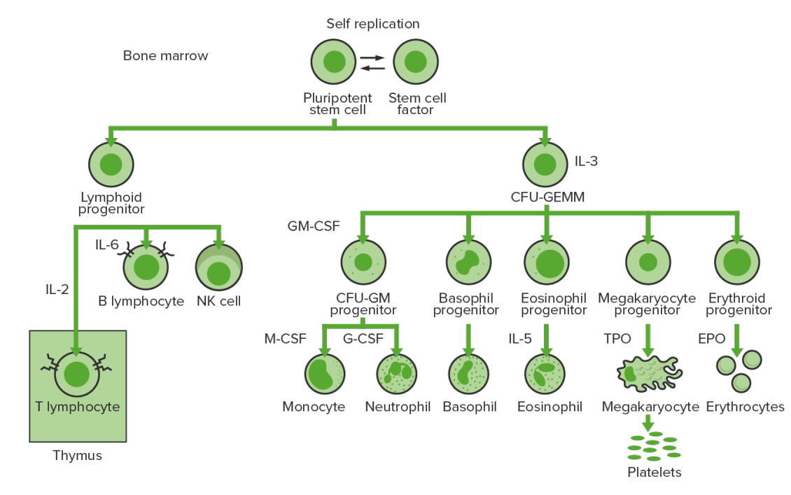
Bone-marrow hematopoiesis: proliferation and differentiation of the formed elements of blood.
CFU-GEMM: colony-forming unit–granulocyte, erythrocyte, monocyte, megakaryocyte
CFU-GM: colony-forming unit–granulocyte-macrophage
GM-CSF: granulocyte-macrophage colony-stimulating factor
M-CSF: macrophage colony-stimulating factor
G-CSF: granulocyte colony-stimulating factor
NK: natural killer
TPO: thrombopoietin

Image by Lecturio. License: CC BY-NC-SA 4.0

Driver mutations

  • Mutation Mutation Genetic mutations are errors in DNA that can cause protein misfolding and dysfunction. There are various types of mutations, including chromosomal, point, frameshift, and expansion mutations. Types of Mutations of JAK2 gene Gene A category of nucleic acid sequences that function as units of heredity and which code for the basic instructions for the development, reproduction, and maintenance of organisms. Basic Terms of Genetics:
    • Affects JAK2:
      • A nonreceptor tyrosine Tyrosine A non-essential amino acid. In animals it is synthesized from phenylalanine. It is also the precursor of epinephrine; thyroid hormones; and melanin. Synthesis of Nonessential Amino Acids kinase (encoded by the JAK2 gene Gene A category of nucleic acid sequences that function as units of heredity and which code for the basic instructions for the development, reproduction, and maintenance of organisms. Basic Terms of Genetics) in the cytoplasm involved in signal transduction Transduction The transfer of bacterial DNA by phages from an infected bacterium to another bacterium. This also refers to the transfer of genes into eukaryotic cells by viruses. This naturally occurring process is routinely employed as a gene transfer technique. Bacteriology (JAK–signal transducer Transducer A device placed on the patient’s body to visualize a target Ultrasound (Sonography) of activators of transcription Transcription Transcription of genetic information is the first step in gene expression. Transcription is the process by which DNA is used as a template to make mRNA. This process is divided into 3 stages: initiation, elongation, and termination. Stages of Transcription (STAT) pathway) 
      • Mediates the cellular response to growth factors and cytokines Cytokines Non-antibody proteins secreted by inflammatory leukocytes and some non-leukocytic cells, that act as intercellular mediators. They differ from classical hormones in that they are produced by a number of tissue or cell types rather than by specialized glands. They generally act locally in a paracrine or autocrine rather than endocrine manner. Adaptive Immune Response
      • Allows extracellular signals to activate genes Genes A category of nucleic acid sequences that function as units of heredity and which code for the basic instructions for the development, reproduction, and maintenance of organisms. DNA Types and Structure 
      • Most common mutation Mutation Genetic mutations are errors in DNA that can cause protein misfolding and dysfunction. There are various types of mutations, including chromosomal, point, frameshift, and expansion mutations. Types of Mutations: valine to phenylalanine Phenylalanine An essential aromatic amino acid that is a precursor of melanin; dopamine; noradrenalin (norepinephrine), and thyroxine. Synthesis of Nonessential Amino Acids at codon Codon A set of three nucleotides in a protein coding sequence that specifies individual amino acids or a termination signal. Most codons are universal, but some organisms do not produce the transfer RNAs complementary to all codons. These codons are referred to as unassigned codons. Basic Terms of Genetics 617: JAK2 V617F 
    • Effect:
      • Gain of function mutation Mutation Genetic mutations are errors in DNA that can cause protein misfolding and dysfunction. There are various types of mutations, including chromosomal, point, frameshift, and expansion mutations. Types of Mutations → constitutive activation of JAK–STAT resulting in cytokine-independent cell proliferation
      • Leads to activation of TPO-R → ↑ receptivity to thrombopoietin Thrombopoietin A humoral factor that stimulates the production of thrombocytes (blood platelets). Thrombopoietin stimulates the proliferation of bone marrow megakaryocytes and their release of blood platelets. The process is called thrombopoiesis. Platelets: Histology → overproduction of megakaryocytes
  • Mutation Mutation Genetic mutations are errors in DNA that can cause protein misfolding and dysfunction. There are various types of mutations, including chromosomal, point, frameshift, and expansion mutations. Types of Mutations of CALR gene Gene A category of nucleic acid sequences that function as units of heredity and which code for the basic instructions for the development, reproduction, and maintenance of organisms. Basic Terms of Genetics:
    • Affects calreticulin, a Ca CA Condylomata acuminata are a clinical manifestation of genital HPV infection. Condylomata acuminata are described as raised, pearly, flesh-colored, papular, cauliflower-like lesions seen in the anogenital region that may cause itching, pain, or bleeding. Condylomata Acuminata (Genital Warts)2+-binding protein involved in cellular proliferation, differentiation, and apoptosis Apoptosis A regulated cell death mechanism characterized by distinctive morphologic changes in the nucleus and cytoplasm, including the endonucleolytic cleavage of genomic DNA, at regularly spaced, internucleosomal sites, I.e., DNA fragmentation. It is genetically-programmed and serves as a balance to mitosis in regulating the size of animal tissues and in mediating pathologic processes associated with tumor growth. Ischemic Cell Damage
    • Effect:
      • Mutant CALR interacts directly with the TPO-R, causing its constitutive activation
      • Platelets Platelets Platelets are small cell fragments involved in hemostasis. Thrombopoiesis takes place primarily in the bone marrow through a series of cell differentiation and is influenced by several cytokines. Platelets are formed after fragmentation of the megakaryocyte cytoplasm. Platelets: Histology, even without thrombopoietin Thrombopoietin A humoral factor that stimulates the production of thrombocytes (blood platelets). Thrombopoietin stimulates the proliferation of bone marrow megakaryocytes and their release of blood platelets. The process is called thrombopoiesis. Platelets: Histology
  • Mutation Mutation Genetic mutations are errors in DNA that can cause protein misfolding and dysfunction. There are various types of mutations, including chromosomal, point, frameshift, and expansion mutations. Types of Mutations of MPL gene Gene A category of nucleic acid sequences that function as units of heredity and which code for the basic instructions for the development, reproduction, and maintenance of organisms. Basic Terms of Genetics:
    • The TPO-R is encoded by MPL.
    • Gain of function point mutation Point Mutation A mutation caused by the substitution of one nucleotide for another. This results in the DNA molecule having a change in a single base pair. Types of Mutations
    • Effect: constitutive activation of thrombopoietin Thrombopoietin A humoral factor that stimulates the production of thrombocytes (blood platelets). Thrombopoietin stimulates the proliferation of bone marrow megakaryocytes and their release of blood platelets. The process is called thrombopoiesis. Platelets: Histology receptor Receptor Receptors are proteins located either on the surface of or within a cell that can bind to signaling molecules known as ligands (e.g., hormones) and cause some type of response within the cell. Receptors

Thrombosis Thrombosis Formation and development of a thrombus or blood clot in the blood vessel. Epidemic Typhus in essential thrombocythemia

Increased incidence Incidence The number of new cases of a given disease during a given period in a specified population. It also is used for the rate at which new events occur in a defined population. It is differentiated from prevalence, which refers to all cases in the population at a given time. Measures of Disease Frequency due to:

  • ↑ Platelet chemotaxis Chemotaxis The movement of leukocytes in response to a chemical concentration gradient or to products formed in an immunologic reaction. Leukocyte Adhesion Deficiency Type 1 in response to procoagulation signals
  • Thrombus formation from misshapen, abnormally large platelets Platelets Platelets are small cell fragments involved in hemostasis. Thrombopoiesis takes place primarily in the bone marrow through a series of cell differentiation and is influenced by several cytokines. Platelets are formed after fragmentation of the megakaryocyte cytoplasm. Platelets: Histology 
  • ↑ Levels of soluble markers of platelet activation Platelet activation A series of progressive, overlapping events, triggered by exposure of the platelets to subendothelial tissue. These events include shape change, adhesiveness, aggregation, and release reactions. When carried through to completion, these events lead to the formation of a stable hemostatic plug. Hemostasis (i.e., P-selectin P-selectin Cell adhesion molecule and cd antigen that mediates the adhesion of neutrophils and monocytes to activated platelets and endothelial cells. Tumor Necrosis Factor (TNF))
  • Concurrent activation of leukocytes Leukocytes White blood cells. These include granular leukocytes (basophils; eosinophils; and neutrophils) as well as non-granular leukocytes (lymphocytes and monocytes). White Myeloid Cells: Histology and endothelial cells → platelet aggregation Platelet aggregation The attachment of platelets to one another. This clumping together can be induced by a number of agents (e.g., thrombin; collagen) and is part of the mechanism leading to the formation of a thrombus. Hemostasis

Hemorrhagic diathesis in essential thrombocythemia

Increased hemorrhagic diathesis in ET is due to:

  • Severe thrombocythemia (i.e., platelet counts Platelet counts The number of platelets per unit volume in a sample of venous blood. Coagulation Studies > 1000 × 10⁹/L) → ↑ platelet metalloproteinases → ↑ proteases Proteases Proteins and Peptides causing proteolysis
  • Proteolysis of large von Willebrand Factor von Willebrand factor A high-molecular-weight plasma protein, produced by endothelial cells and megakaryocytes, that is part of the factor VIII/von Willebrand factor complex. The von Willebrand factor has receptors for collagen, platelets, and ristocetin activity as well as the immunologically distinct antigenic determinants. It functions in adhesion of platelets to collagen and hemostatic plug formation. The prolonged bleeding time in von Willebrand diseases is due to the deficiency of this factor. Hemostasis (VWF) multimers leads to functional VWF deficiency → acquired type 2 Type 2 Spinal Muscular Atrophy von Willebrand syndrome

Clinical Presentation

Signs and symptoms

  • Asymptomatic (60%): diagnosis made incidentally
  • Weight loss Weight loss Decrease in existing body weight. Bariatric Surgery, fever Fever Fever is defined as a measured body temperature of at least 38°C (100.4°F). Fever is caused by circulating endogenous and/or exogenous pyrogens that increase levels of prostaglandin E2 in the hypothalamus. Fever is commonly associated with chills, rigors, sweating, and flushing of the skin. Fever
  • Gout Gout Gout is a heterogeneous metabolic disease associated with elevated serum uric acid levels (> 6.8 mg/dL) and abnormal deposits of monosodium urate in tissues. The condition is often familial and is initially characterized by painful, recurring, and usually monoarticular acute arthritis, or “gout flare,” followed later by chronic deforming arthritis. Gout
  • Pruritus Pruritus An intense itching sensation that produces the urge to rub or scratch the skin to obtain relief. Atopic Dermatitis (Eczema) (not commonly seen, unlike in polycythemia Polycythemia An increase in the total red cell mass of the blood. Renal Cell Carcinoma vera)
  • Vasomotor:
    • Headache Headache The symptom of pain in the cranial region. It may be an isolated benign occurrence or manifestation of a wide variety of headache disorders. Brain Abscess, dizziness Dizziness An imprecise term which may refer to a sense of spatial disorientation, motion of the environment, or lightheadedness. Lateral Medullary Syndrome (Wallenberg Syndrome), syncope Syncope Syncope is a short-term loss of consciousness and loss of postural stability followed by spontaneous return of consciousness to the previous neurologic baseline without the need for resuscitation. The condition is caused by transient interruption of cerebral blood flow that may be benign or related to a underlying life-threatening condition. Syncope, chest pain Pain An unpleasant sensation induced by noxious stimuli which are detected by nerve endings of nociceptive neurons. Pain: Types and Pathways
    • Erythromelalgia Erythromelalgia A peripheral arterial disease that is characterized by the triad of erythema, burning pain, and increased skin temperature of the extremities (or red, painful extremities). Erythromelalgia may be classified as primary or idiopathic, familial or non-familial. Secondary erythromelalgia is associated with other diseases, the most common being myeloproliferative disorders. Polycythemia Vera: red warm congestion and burning sensation of digits and toes due to microvascular thrombosis Thrombosis Formation and development of a thrombus or blood clot in the blood vessel. Epidemic Typhus
    • Livedo reticularis Livedo reticularis A condition characterized by a reticular or fishnet pattern on the skin of lower extremities and other parts of the body. This red and blue pattern is due to deoxygenated blood in unstable dermal blood vessels. The condition is intensified by cold exposure and relieved by rewarming. Chronic Kidney Disease: mottled reticulated purplish discoloration of the skin Skin The skin, also referred to as the integumentary system, is the largest organ of the body. The skin is primarily composed of the epidermis (outer layer) and dermis (deep layer). The epidermis is primarily composed of keratinocytes that undergo rapid turnover, while the dermis contains dense layers of connective tissue. Skin: Structure and Functions
    • Transient visual problems: scintillating scotoma Scotoma A localized defect in the visual field bordered by an area of normal vision. This occurs with a variety of eye diseases (e.g., retinal diseases and glaucoma); optic nerve diseases, and other conditions. Cranial Nerve Palsies, amaurosis fugax Amaurosis fugax Transient complete or partial monocular blindness due to retinal ischemia. This may be caused by emboli from the carotid artery (usually in association with carotid stenosis) and other locations that enter the central retinal artery. Carotid Artery Stenosis, ophthalmic migraine Migraine Migraine headache is a primary headache disorder and is among the most prevalent disorders in the world. Migraine is characterized by episodic, moderate to severe headaches that may be associated with increased sensitivity to light and sound, as well as nausea and/or vomiting. Migraine Headache
  • Splenomegaly Splenomegaly Splenomegaly is pathologic enlargement of the spleen that is attributable to numerous causes, including infections, hemoglobinopathies, infiltrative processes, and outflow obstruction of the portal vein. Splenomegaly
Physical findings of essential thrombocythemia

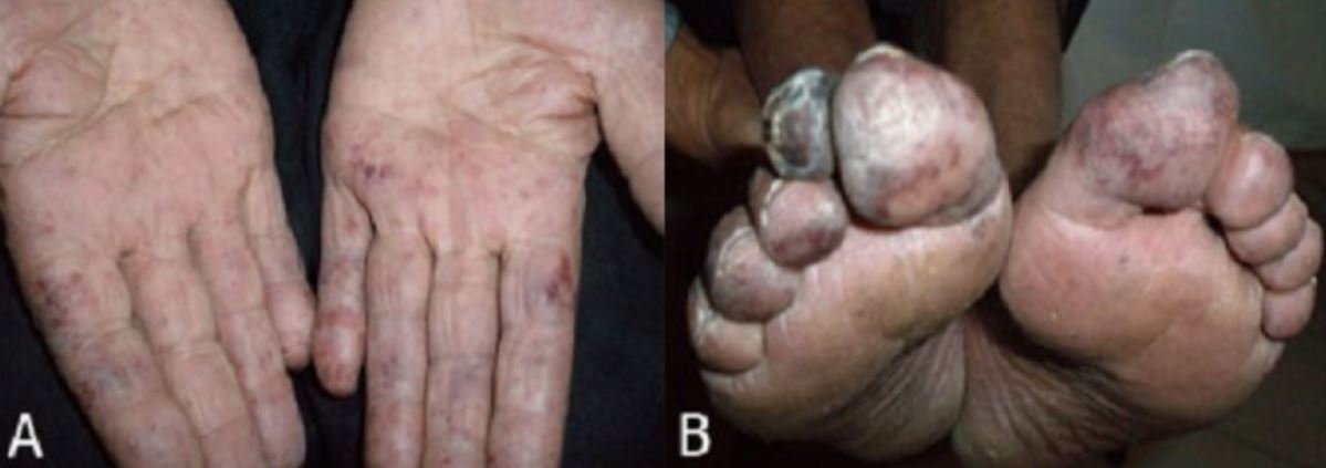
Physical findings of essential thrombocythemia:
Livedo reticularis and areas of mottled bluish ischemia of fingers (A) and toe gangrene (B)

Image: “Thromboembolic complication in essential thrombocythemia” by Mozaheb, Z. License: CC BY 2.0

Complications

  • Thrombosis Thrombosis Formation and development of a thrombus or blood clot in the blood vessel. Epidemic Typhus:
    • Deep venous thrombosis Venous thrombosis The formation or presence of a blood clot (thrombus) within a vein. Budd-Chiari Syndrome ( DVT DVT Deep vein thrombosis (DVT) usually occurs in the deep veins of the lower extremities. The affected veins include the femoral, popliteal, iliofemoral, and pelvic veins. Proximal DVT is more likely to cause a pulmonary embolism (PE) and is generally considered more serious. Deep Vein Thrombosis) often involving unusual anatomic sites:
      • Portal vein Portal vein A short thick vein formed by union of the superior mesenteric vein and the splenic vein. Liver: Anatomy 
      • Hepatic venous system ( Budd-Chiari syndrome Budd-Chiari syndrome Budd-Chiari syndrome is a condition resulting from the interruption of the normal outflow of blood from the liver. The primary type arises from a venous process (affecting the hepatic veins or inferior vena cava) such as thrombosis, but can also be from a lesion compressing or invading the veins (secondary type). The patient typically presents with hepatomegaly, ascites, and abdominal discomfort. Budd-Chiari Syndrome)
      • Splenic vein
      • Mesenteric vein
      • Retinal artery or vein
    • Cerebrovascular events, transient ischemic attacks
    • Pulmonary embolism Pulmonary Embolism Pulmonary embolism (PE) is a potentially fatal condition that occurs as a result of intraluminal obstruction of the main pulmonary artery or its branches. The causative factors include thrombi, air, amniotic fluid, and fat. In PE, gas exchange is impaired due to the decreased return of deoxygenated blood to the lungs. Pulmonary Embolism
    • MI MI MI is ischemia and death of an area of myocardial tissue due to insufficient blood flow and oxygenation, usually from thrombus formation on a ruptured atherosclerotic plaque in the epicardial arteries. Clinical presentation is most commonly with chest pain, but women and patients with diabetes may have atypical symptoms. Myocardial Infarction
    • Peripheral vascular disease (leading to gangrene Gangrene Death and putrefaction of tissue usually due to a loss of blood supply. Small Bowel Obstruction)
    • Superficial thrombophlebitis Superficial Thrombophlebitis Thromboangiitis Obliterans (Buerger’s Disease)
  • Bleeding:
    • GI or genitourinary bleeding
    • Bruises, ecchymosis Ecchymosis Extravasation of blood into the skin, resulting in a nonelevated, rounded or irregular, blue or purplish patch, larger than a petechia. Orbital Fractures
    • Gingival bleeding
    • Excessive bleeding after trauma/surgery
  • Recurrent first-trimester pregnancy Pregnancy The status during which female mammals carry their developing young (embryos or fetuses) in utero before birth, beginning from fertilization to birth. Pregnancy: Diagnosis, Physiology, and Care loss

Diagnosis

Medical history

WHO criteria (2016)

Diagnosis requires all 4 major criteria or first 3 major criteria and 1 minor criterion.

  • Major criteria:
    • Platelet count ≥ 450 × 10⁹/L
    • Bone marrow Bone marrow The soft tissue filling the cavities of bones. Bone marrow exists in two types, yellow and red. Yellow marrow is found in the large cavities of large bones and consists mostly of fat cells and a few primitive blood cells. Red marrow is a hematopoietic tissue and is the site of production of erythrocytes and granular leukocytes. Bone marrow is made up of a framework of connective tissue containing branching fibers with the frame being filled with marrow cells. Bone Marrow: Composition and Hematopoiesis biopsy Biopsy Removal and pathologic examination of specimens from the living body. Ewing Sarcoma: megakaryocyte Megakaryocyte Very large bone marrow cells which release mature blood platelets. Platelets: Histology proliferation with large and mature morphologic features, without significant erythropoiesis Erythropoiesis The production of red blood cells (erythrocytes). In humans, erythrocytes are produced by the yolk sac in the first trimester; by the liver in the second trimester; by the bone marrow in the third trimester and after birth. In normal individuals, the erythrocyte count in the peripheral blood remains relatively constant implying a balance between the rate of erythrocyte production and rate of destruction. Erythrocytes: Histology or granulopoiesis
    • Does not fit the criteria for other myeloproliferative disorders (i.e., polycythemia Polycythemia An increase in the total red cell mass of the blood. Renal Cell Carcinoma vera, CML CML Chronic myeloid leukemia is a malignant proliferation of the granulocytic cell line characterized by a fairly normal differentiation. The underlying genetic abnormality is the Philadelphia chromosome, an abbreviated chromosome 22, resulting from reciprocal (9;22)(q34;q11) translocation. Chronic Myeloid Leukemia)
    • Presence of JAK2, CALR, or MPL mutation Mutation Genetic mutations are errors in DNA that can cause protein misfolding and dysfunction. There are various types of mutations, including chromosomal, point, frameshift, and expansion mutations. Types of Mutations
  • Minor criterion: presence of other clonal marker or reactive thrombocytosis ruled out

Laboratory findings

  • CBC:
    • ↑↑ Platelets Platelets Platelets are small cell fragments involved in hemostasis. Thrombopoiesis takes place primarily in the bone marrow through a series of cell differentiation and is influenced by several cytokines. Platelets are formed after fragmentation of the megakaryocyte cytoplasm. Platelets: Histology (≥ 450 × 10⁹/L)
    • Bleeding time Bleeding time Duration of blood flow after skin puncture. This test is used as a measure of capillary and platelet function. Hemostasis
    • Normal Hb Hb The oxygen-carrying proteins of erythrocytes. They are found in all vertebrates and some invertebrates. The number of globin subunits in the hemoglobin quaternary structure differs between species. Structures range from monomeric to a variety of multimeric arrangements. Gas Exchange
    • Normal WBC
  • Biochemistry (all due to increased cell turnover):
    • Potassium Potassium An element in the alkali group of metals with an atomic symbol k, atomic number 19, and atomic weight 39. 10. It is the chief cation in the intracellular fluid of muscle and other cells. Potassium ion is a strong electrolyte that plays a significant role in the regulation of fluid volume and maintenance of the water-electrolyte balance. Hyperkalemia
    • Uric acid Uric acid An oxidation product, via xanthine oxidase, of oxypurines such as xanthine and hypoxanthine. It is the final oxidation product of purine catabolism in humans and primates, whereas in most other mammals urate oxidase further oxidizes it to allantoin. Nephrolithiasis
    • Phosphate Phosphate Inorganic salts of phosphoric acid. Electrolytes
  • Peripheral blood smear Peripheral Blood Smear Anemia: Overview and Types:
    • Increased platelets Platelets Platelets are small cell fragments involved in hemostasis. Thrombopoiesis takes place primarily in the bone marrow through a series of cell differentiation and is influenced by several cytokines. Platelets are formed after fragmentation of the megakaryocyte cytoplasm. Platelets: Histology
    • Platelet anisocytosis
  • Bone marrow Bone marrow The soft tissue filling the cavities of bones. Bone marrow exists in two types, yellow and red. Yellow marrow is found in the large cavities of large bones and consists mostly of fat cells and a few primitive blood cells. Red marrow is a hematopoietic tissue and is the site of production of erythrocytes and granular leukocytes. Bone marrow is made up of a framework of connective tissue containing branching fibers with the frame being filled with marrow cells. Bone Marrow: Composition and Hematopoiesis biopsy Biopsy Removal and pathologic examination of specimens from the living body. Ewing Sarcoma:
    • Megakaryocytic hyperplasia Hyperplasia An increase in the number of cells in a tissue or organ without tumor formation. It differs from hypertrophy, which is an increase in bulk without an increase in the number of cells. Cellular Adaptation
    • Giant megakaryocytes
    • Lobulated and/or hyperlobulated nuclei
  • Genetic testing Genetic Testing Detection of a mutation; genotype; karyotype; or specific alleles associated with genetic traits, heritable diseases, or predisposition to a disease, or that may lead to the disease in descendants. It includes prenatal genetic testing. Myotonic Dystrophies:
    • Screen for genetic mutations Genetic Mutations Carcinogenesis: JAK2 (most common), CALR, MPL
    • Strengthens diagnosis and also gives information regarding complications (CALR mutation Mutation Genetic mutations are errors in DNA that can cause protein misfolding and dysfunction. There are various types of mutations, including chromosomal, point, frameshift, and expansion mutations. Types of Mutations has lower risk of thrombosis Thrombosis Formation and development of a thrombus or blood clot in the blood vessel. Epidemic Typhus than JAK2 mutation Mutation Genetic mutations are errors in DNA that can cause protein misfolding and dysfunction. There are various types of mutations, including chromosomal, point, frameshift, and expansion mutations. Types of Mutations)
Bone marrow biopsy in essential thrombocythemia

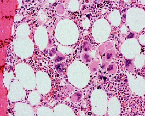
Bone marrow biopsy in essential thrombocythemia:
Increased megakaryocytes can be seen.

Image: “Sequential occurrence of thrombotic thrombocytopenic purpura, essential thrombocythemia, and idiopathic thrombocytopenic purpura in a 42-year-old African-American woman: a case report and review of the literature” by Farhat MH, Kuriakose P, Jawad M, Hanbali A. License: CC BY 2.0

Comparison with other myeloproliferative neoplasms Neoplasms New abnormal growth of tissue. Malignant neoplasms show a greater degree of anaplasia and have the properties of invasion and metastasis, compared to benign neoplasms. Benign Bone Tumors

Myeloproliferative neoplasms Neoplasms New abnormal growth of tissue. Malignant neoplasms show a greater degree of anaplasia and have the properties of invasion and metastasis, compared to benign neoplasms. Benign Bone Tumors can be compared with the following WHO classification:

Table: Classic types of myeloproliferative neoplasms Neoplasms New abnormal growth of tissue. Malignant neoplasms show a greater degree of anaplasia and have the properties of invasion and metastasis, compared to benign neoplasms. Benign Bone Tumors
Disease Mutations Key points
CML CML Chronic myeloid leukemia is a malignant proliferation of the granulocytic cell line characterized by a fairly normal differentiation. The underlying genetic abnormality is the Philadelphia chromosome, an abbreviated chromosome 22, resulting from reciprocal (9;22)(q34;q11) translocation. Chronic Myeloid Leukemia BCR-ABL1 ( Philadelphia chromosome Philadelphia chromosome An aberrant form of human chromosome 22 characterized by translocation of the distal end of chromosome 9 from 9q34, to the long arm of chromosome 22 at 22q11. It is present in the bone marrow cells of 80 to 90 percent of patients with chronic myelocytic leukemia. Acute Lymphoblastic Leukemia) Proliferation of mature and maturing granulocytes Granulocytes Leukocytes with abundant granules in the cytoplasm. They are divided into three groups according to the staining properties of the granules: neutrophilic, eosinophilic, and basophilic. Mature granulocytes are the neutrophils; eosinophils; and basophils. White Myeloid Cells: Histology
ET JAK2, CALR, or MPL Excessive clonal platelet production
Polycythemia Polycythemia An increase in the total red cell mass of the blood. Renal Cell Carcinoma vera ( PV PV Polycythemia vera (PV) is a chronic myeloproliferative neoplasm characterized by the overproduction of rbcs. In addition, the wbc and platelet counts are also increased, which differentiate pv from erythrocytosis seen with chronic hypoxia and other chronic conditions. Polycythemia Vera) JAK2 Elevated RBC mass Mass Three-dimensional lesion that occupies a space within the breast Imaging of the Breast
Primary myelofibrosis Primary myelofibrosis Primary myelofibrosis (PMF) is a myeloproliferative neoplasm characterized by chronic myeloproliferation with nonclonal fibroblastic deposition, resulting in bone marrow fibrosis. The abnormality stems from genetic mutations of the hematopoietic stem cells (typically, JAK2 mutation). Primary symptoms are anemia and extramedullary hematopoiesis,. Primary Myelofibrosis ( PMF PMF Primary myelofibrosis (PMF) is a myeloproliferative neoplasm characterized by chronic myeloproliferation with nonclonal fibroblastic deposition, resulting in bone marrow fibrosis. The abnormality stems from genetic mutations of the hematopoietic stem cells (typically, jak2 mutation). Primary symptoms are anemia and extramedullary hematopoiesis,. Primary Myelofibrosis) JAK2, CALR, or MPL Obliterative bone marrow fibrosis Bone Marrow Fibrosis Primary Myelofibrosis

Other types:

  • Chronic neutrophilic leukemia (CNL)
  • Chronic eosinophilic leukemia Chronic eosinophilic leukemia Chronic eosinophilic leukemia (CEL) is a chronic myeloproliferative neoplasm caused by autonomous clonal proliferation of normal-appearing eosinophils, resulting in increased eosinophils in the peripheral blood and bone marrow. The disorder is a myeloid variant of hypereosinophilic syndrome (HES) and is associated with tissue infiltration leading to end-organ damage. Chronic Eosinophilic Leukemia ( CEL CEL Chronic eosinophilic leukemia (CEL) is a chronic myeloproliferative neoplasm caused by autonomous clonal proliferation of normal-appearing eosinophils, resulting in increased eosinophils in the peripheral blood and bone marrow. The disorder is a myeloid variant of hypereosinophilic syndrome (HES) and is associated with tissue infiltration leading to end-organ damage. Chronic Eosinophilic Leukemia), not otherwise specified
  • Myeloproliferative neoplasm, unclassifiable

Management

Management approach

  • Factors considered in treating patients Patients Individuals participating in the health care system for the purpose of receiving therapeutic, diagnostic, or preventive procedures. Clinician–Patient Relationship with ET:
    • Presence and severity of symptoms
    • Presence of thrombotic events
    • Comorbidities Comorbidities The presence of co-existing or additional diseases with reference to an initial diagnosis or with reference to the index condition that is the subject of study. Comorbidity may affect the ability of affected individuals to function and also their survival; it may be used as a prognostic indicator for length of hospital stay, cost factors, and outcome or survival. St. Louis Encephalitis Virus ( diabetes Diabetes Diabetes mellitus (DM) is a metabolic disease characterized by hyperglycemia and dysfunction of the regulation of glucose metabolism by insulin. Type 1 DM is diagnosed mostly in children and young adults as the result of autoimmune destruction of β cells in the pancreas and the resulting lack of insulin. Type 2 DM has a significant association with obesity and is characterized by insulin resistance. Diabetes Mellitus and other factors increase cardiovascular risk)
    • The risk for developing post-ET myelofibrosis and acute myeloid leukemia Acute Myeloid Leukemia Acute myeloid leukemia (AML) is a hematologic malignancy characterized by the uncontrolled proliferation of myeloid precursor cells. Seen predominantly in older adults, AML includes an accumulation of myeloblasts and a replacement of normal marrow by malignant cells, which leads to impaired hematopoiesis. Acute Myeloid Leukemia is lower than for developing thrombosis Thrombosis Formation and development of a thrombus or blood clot in the blood vessel. Epidemic Typhus.
  • Goals:
    • ↓ Risk of thrombosis Thrombosis Formation and development of a thrombus or blood clot in the blood vessel. Epidemic Typhus and bleeding
    • ↓ Associated symptoms
  • Therapeutic options:
    • Aspirin Aspirin The prototypical analgesic used in the treatment of mild to moderate pain. It has anti-inflammatory and antipyretic properties and acts as an inhibitor of cyclooxygenase which results in the inhibition of the biosynthesis of prostaglandins. Aspirin also inhibits platelet aggregation and is used in the prevention of arterial and venous thrombosis. Nonsteroidal Antiinflammatory Drugs (NSAIDs):
      • 40–100 mg once or twice daily
      • Screen for acquired VWF in patients Patients Individuals participating in the health care system for the purpose of receiving therapeutic, diagnostic, or preventive procedures. Clinician–Patient Relationship with platelets Platelets Platelets are small cell fragments involved in hemostasis. Thrombopoiesis takes place primarily in the bone marrow through a series of cell differentiation and is influenced by several cytokines. Platelets are formed after fragmentation of the megakaryocyte cytoplasm. Platelets: Histology > 1 million/µL (cannot give aspirin Aspirin The prototypical analgesic used in the treatment of mild to moderate pain. It has anti-inflammatory and antipyretic properties and acts as an inhibitor of cyclooxygenase which results in the inhibition of the biosynthesis of prostaglandins. Aspirin also inhibits platelet aggregation and is used in the prevention of arterial and venous thrombosis. Nonsteroidal Antiinflammatory Drugs (NSAIDs) if VWF present owing to ↑ bleeding risk).
    • Hydroxyurea Hydroxyurea An antineoplastic agent that inhibits DNA synthesis through the inhibition of ribonucleoside diphosphate reductase. Antimetabolite Chemotherapy:
      • Cytoreductive agent of choice
      • Goal is to maintain platelet count at 100,000–400,000/µL.
      • Teratogenic
    • Anagrelide:
      • ↓ Platelet count and aggregation Aggregation The attachment of platelets to one another. This clumping together can be induced by a number of agents (e.g., thrombin; collagen) and is part of the mechanism leading to the formation of a thrombus. Coagulation Studies
      • Possible cardiac toxicity Toxicity Dosage Calculation
    • Pegylated interferon-ɑ:
      • Immunomodulatory antiproliferative agent
      • Can be given to pregnant women

Risk stratification

  • The choice of treatment is based on thrombosis Thrombosis Formation and development of a thrombus or blood clot in the blood vessel. Epidemic Typhus risk stratification.
  • Use of aspirin Aspirin The prototypical analgesic used in the treatment of mild to moderate pain. It has anti-inflammatory and antipyretic properties and acts as an inhibitor of cyclooxygenase which results in the inhibition of the biosynthesis of prostaglandins. Aspirin also inhibits platelet aggregation and is used in the prevention of arterial and venous thrombosis. Nonsteroidal Antiinflammatory Drugs (NSAIDs) or cytoreductive therapy does not cure ET but can decrease risk of complications.
Table: Thrombosis Thrombosis Formation and development of a thrombus or blood clot in the blood vessel. Epidemic Typhus risk stratification
Risk for thrombosis Thrombosis Formation and development of a thrombus or blood clot in the blood vessel. Epidemic Typhus Features Treatment
Very low risk
  • No history of thrombosis Thrombosis Formation and development of a thrombus or blood clot in the blood vessel. Epidemic Typhus
  • Age ≤ 60 years
  • No JAK2 V617F mutation Mutation Genetic mutations are errors in DNA that can cause protein misfolding and dysfunction. There are various types of mutations, including chromosomal, point, frameshift, and expansion mutations. Types of Mutations
  • Observation alone
  • Vasomotor symptoms: aspirin Aspirin The prototypical analgesic used in the treatment of mild to moderate pain. It has anti-inflammatory and antipyretic properties and acts as an inhibitor of cyclooxygenase which results in the inhibition of the biosynthesis of prostaglandins. Aspirin also inhibits platelet aggregation and is used in the prevention of arterial and venous thrombosis. Nonsteroidal Antiinflammatory Drugs (NSAIDs) daily
  • Cardiovascular risk factors: aspirin Aspirin The prototypical analgesic used in the treatment of mild to moderate pain. It has anti-inflammatory and antipyretic properties and acts as an inhibitor of cyclooxygenase which results in the inhibition of the biosynthesis of prostaglandins. Aspirin also inhibits platelet aggregation and is used in the prevention of arterial and venous thrombosis. Nonsteroidal Antiinflammatory Drugs (NSAIDs) daily
Low risk
  • No history of thrombosis Thrombosis Formation and development of a thrombus or blood clot in the blood vessel. Epidemic Typhus
  • Age ≤ 60 years
  • JAK2 V617F mutation Mutation Genetic mutations are errors in DNA that can cause protein misfolding and dysfunction. There are various types of mutations, including chromosomal, point, frameshift, and expansion mutations. Types of Mutations
  • Aspirin Aspirin The prototypical analgesic used in the treatment of mild to moderate pain. It has anti-inflammatory and antipyretic properties and acts as an inhibitor of cyclooxygenase which results in the inhibition of the biosynthesis of prostaglandins. Aspirin also inhibits platelet aggregation and is used in the prevention of arterial and venous thrombosis. Nonsteroidal Antiinflammatory Drugs (NSAIDs) once daily
  • JAK2 mutation Mutation Genetic mutations are errors in DNA that can cause protein misfolding and dysfunction. There are various types of mutations, including chromosomal, point, frameshift, and expansion mutations. Types of Mutations and cardiovascular risk factors: aspirin Aspirin The prototypical analgesic used in the treatment of mild to moderate pain. It has anti-inflammatory and antipyretic properties and acts as an inhibitor of cyclooxygenase which results in the inhibition of the biosynthesis of prostaglandins. Aspirin also inhibits platelet aggregation and is used in the prevention of arterial and venous thrombosis. Nonsteroidal Antiinflammatory Drugs (NSAIDs) twice daily
Intermediate risk
  • No history of thrombosis Thrombosis Formation and development of a thrombus or blood clot in the blood vessel. Epidemic Typhus
  • Age > 60 years
  • No JAK2 V617F mutation Mutation Genetic mutations are errors in DNA that can cause protein misfolding and dysfunction. There are various types of mutations, including chromosomal, point, frameshift, and expansion mutations. Types of Mutations
  • Aspirin Aspirin The prototypical analgesic used in the treatment of mild to moderate pain. It has anti-inflammatory and antipyretic properties and acts as an inhibitor of cyclooxygenase which results in the inhibition of the biosynthesis of prostaglandins. Aspirin also inhibits platelet aggregation and is used in the prevention of arterial and venous thrombosis. Nonsteroidal Antiinflammatory Drugs (NSAIDs) once or twice daily (especially if with cardiovascular risk factors)
  • Hydroxyurea Hydroxyurea An antineoplastic agent that inhibits DNA synthesis through the inhibition of ribonucleoside diphosphate reductase. Antimetabolite Chemotherapy (may be given but with less clear benefit)
High risk
  • History of thrombosis Thrombosis Formation and development of a thrombus or blood clot in the blood vessel. Epidemic Typhus
  • Age > 60 years, especially with JAK2 V617F mutation Mutation Genetic mutations are errors in DNA that can cause protein misfolding and dysfunction. There are various types of mutations, including chromosomal, point, frameshift, and expansion mutations. Types of Mutations
  • Options:
    • Hydroxyurea Hydroxyurea An antineoplastic agent that inhibits DNA synthesis through the inhibition of ribonucleoside diphosphate reductase. Antimetabolite Chemotherapy
    • Anagrelide
    • Interferon-α
  • Aspirin Aspirin The prototypical analgesic used in the treatment of mild to moderate pain. It has anti-inflammatory and antipyretic properties and acts as an inhibitor of cyclooxygenase which results in the inhibition of the biosynthesis of prostaglandins. Aspirin also inhibits platelet aggregation and is used in the prevention of arterial and venous thrombosis. Nonsteroidal Antiinflammatory Drugs (NSAIDs) once or twice daily
  • Anticoagulation Anticoagulation Pulmonary Hypertension Drugs if with history of DVT DVT Deep vein thrombosis (DVT) usually occurs in the deep veins of the lower extremities. The affected veins include the femoral, popliteal, iliofemoral, and pelvic veins. Proximal DVT is more likely to cause a pulmonary embolism (PE) and is generally considered more serious. Deep Vein Thrombosis

Differential Diagnosis

Other chronic myeloproliferative neoplasms Neoplasms New abnormal growth of tissue. Malignant neoplasms show a greater degree of anaplasia and have the properties of invasion and metastasis, compared to benign neoplasms. Benign Bone Tumors

  • Primary myelofibrosis Primary myelofibrosis Primary myelofibrosis (PMF) is a myeloproliferative neoplasm characterized by chronic myeloproliferation with nonclonal fibroblastic deposition, resulting in bone marrow fibrosis. The abnormality stems from genetic mutations of the hematopoietic stem cells (typically, JAK2 mutation). Primary symptoms are anemia and extramedullary hematopoiesis,. Primary Myelofibrosis ( PMF PMF Primary myelofibrosis (PMF) is a myeloproliferative neoplasm characterized by chronic myeloproliferation with nonclonal fibroblastic deposition, resulting in bone marrow fibrosis. The abnormality stems from genetic mutations of the hematopoietic stem cells (typically, jak2 mutation). Primary symptoms are anemia and extramedullary hematopoiesis,. Primary Myelofibrosis): chronic myeloproliferative neoplasm characterized by fibrosis Fibrosis Any pathological condition where fibrous connective tissue invades any organ, usually as a consequence of inflammation or other injury. Bronchiolitis Obliterans of the bone marrow Bone marrow The soft tissue filling the cavities of bones. Bone marrow exists in two types, yellow and red. Yellow marrow is found in the large cavities of large bones and consists mostly of fat cells and a few primitive blood cells. Red marrow is a hematopoietic tissue and is the site of production of erythrocytes and granular leukocytes. Bone marrow is made up of a framework of connective tissue containing branching fibers with the frame being filled with marrow cells. Bone Marrow: Composition and Hematopoiesis and extramedullary hematopoiesis Extramedullary Hematopoiesis Primary Myelofibrosis in the spleen Spleen The spleen is the largest lymphoid organ in the body, located in the LUQ of the abdomen, superior to the left kidney and posterior to the stomach at the level of the 9th-11th ribs just below the diaphragm. The spleen is highly vascular and acts as an important blood filter, cleansing the blood of pathogens and damaged erythrocytes. Spleen: Anatomy and liver Liver The liver is the largest gland in the human body. The liver is found in the superior right quadrant of the abdomen and weighs approximately 1.5 kilograms. Its main functions are detoxification, metabolism, nutrient storage (e.g., iron and vitamins), synthesis of coagulation factors, formation of bile, filtration, and storage of blood. Liver: Anatomy. Primary myelofibrosis Primary myelofibrosis Primary myelofibrosis (PMF) is a myeloproliferative neoplasm characterized by chronic myeloproliferation with nonclonal fibroblastic deposition, resulting in bone marrow fibrosis. The abnormality stems from genetic mutations of the hematopoietic stem cells (typically, JAK2 mutation). Primary symptoms are anemia and extramedullary hematopoiesis,. Primary Myelofibrosis is linked to the same somatic mutations as ET. Clinical findings are severe fatigue Fatigue The state of weariness following a period of exertion, mental or physical, characterized by a decreased capacity for work and reduced efficiency to respond to stimuli. Fibromyalgia, splenomegaly Splenomegaly Splenomegaly is pathologic enlargement of the spleen that is attributable to numerous causes, including infections, hemoglobinopathies, infiltrative processes, and outflow obstruction of the portal vein. Splenomegaly, hepatomegaly, and anemia Anemia Anemia is a condition in which individuals have low Hb levels, which can arise from various causes. Anemia is accompanied by a reduced number of RBCs and may manifest with fatigue, shortness of breath, pallor, and weakness. Subtypes are classified by the size of RBCs, chronicity, and etiology. Anemia: Overview and Types. The peripheral blood smear Peripheral Blood Smear Anemia: Overview and Types shows leukoerythroblastosis and contains precursors of WBCs and RBCs RBCs Erythrocytes, or red blood cells (RBCs), are the most abundant cells in the blood. While erythrocytes in the fetus are initially produced in the yolk sac then the liver, the bone marrow eventually becomes the main site of production. Erythrocytes: Histology, nucleated RBCs RBCs Erythrocytes, or red blood cells (RBCs), are the most abundant cells in the blood. While erythrocytes in the fetus are initially produced in the yolk sac then the liver, the bone marrow eventually becomes the main site of production. Erythrocytes: Histology, and teardrop cells Teardrop Cells Primary Myelofibrosis. Diagnosis is made by bone marrow Bone marrow The soft tissue filling the cavities of bones. Bone marrow exists in two types, yellow and red. Yellow marrow is found in the large cavities of large bones and consists mostly of fat cells and a few primitive blood cells. Red marrow is a hematopoietic tissue and is the site of production of erythrocytes and granular leukocytes. Bone marrow is made up of a framework of connective tissue containing branching fibers with the frame being filled with marrow cells. Bone Marrow: Composition and Hematopoiesis examination and molecular testing. In contrast to ET, PMF PMF Primary myelofibrosis (PMF) is a myeloproliferative neoplasm characterized by chronic myeloproliferation with nonclonal fibroblastic deposition, resulting in bone marrow fibrosis. The abnormality stems from genetic mutations of the hematopoietic stem cells (typically, jak2 mutation). Primary symptoms are anemia and extramedullary hematopoiesis,. Primary Myelofibrosis is associated with obliterative bone marrow fibrosis Bone Marrow Fibrosis Primary Myelofibrosis. Management includes allogeneic hematopoietic stem cell transplantation Hematopoietic stem cell transplantation Transfer of hematopoietic stem cells from bone marrow or blood between individuals within the same species (homologous transplantation) or transfer within the same individual (autologous transplantation). Hematopoietic stem cell transplantation has been used as an alternative to bone marrow transplantation in the treatment of a variety of neoplasms. Organ Transplantation and the medications ruxolitinib Ruxolitinib Targeted and Other Nontraditional Antineoplastic Therapy and fedratinib.
  • PV PV Polycythemia vera (PV) is a chronic myeloproliferative neoplasm characterized by the overproduction of rbcs. In addition, the wbc and platelet counts are also increased, which differentiate pv from erythrocytosis seen with chronic hypoxia and other chronic conditions. Polycythemia Vera: chronic myeloproliferative neoplasm characterized by overproduction of RBCs RBCs Erythrocytes, or red blood cells (RBCs), are the most abundant cells in the blood. While erythrocytes in the fetus are initially produced in the yolk sac then the liver, the bone marrow eventually becomes the main site of production. Erythrocytes: Histology, distinguishing it from ET. In addition, WBCs and platelets Platelets Platelets are small cell fragments involved in hemostasis. Thrombopoiesis takes place primarily in the bone marrow through a series of cell differentiation and is influenced by several cytokines. Platelets are formed after fragmentation of the megakaryocyte cytoplasm. Platelets: Histology are also increased, which differentiates PV PV Polycythemia vera (PV) is a chronic myeloproliferative neoplasm characterized by the overproduction of rbcs. In addition, the wbc and platelet counts are also increased, which differentiate pv from erythrocytosis seen with chronic hypoxia and other chronic conditions. Polycythemia Vera from the erythrocytosis seen with chronic hypoxia Hypoxia Sub-optimal oxygen levels in the ambient air of living organisms. Ischemic Cell Damage and other conditions. Similar to ET, this disease has a genetic basis due to mutation Mutation Genetic mutations are errors in DNA that can cause protein misfolding and dysfunction. There are various types of mutations, including chromosomal, point, frameshift, and expansion mutations. Types of Mutations in the JAK2 gene Gene A category of nucleic acid sequences that function as units of heredity and which code for the basic instructions for the development, reproduction, and maintenance of organisms. Basic Terms of Genetics. Management includes phlebotomy Phlebotomy The techniques used to draw blood from a vein for diagnostic purposes or for treatment of certain blood disorders such as erythrocytosis, hemochromatosis, polycythemia vera, and porphyria cutanea tarda. Hereditary Hemochromatosis, low-dose aspirin Aspirin The prototypical analgesic used in the treatment of mild to moderate pain. It has anti-inflammatory and antipyretic properties and acts as an inhibitor of cyclooxygenase which results in the inhibition of the biosynthesis of prostaglandins. Aspirin also inhibits platelet aggregation and is used in the prevention of arterial and venous thrombosis. Nonsteroidal Antiinflammatory Drugs (NSAIDs), and myelosuppressive therapies.
  • CML CML Chronic myeloid leukemia is a malignant proliferation of the granulocytic cell line characterized by a fairly normal differentiation. The underlying genetic abnormality is the Philadelphia chromosome, an abbreviated chromosome 22, resulting from reciprocal (9;22)(q34;q11) translocation. Chronic Myeloid Leukemia: malignant proliferation of the granulocytic cell line, with a fairly normal differentiation. Chronic myeloid leukemia Chronic myeloid leukemia Chronic myeloid leukemia is a malignant proliferation of the granulocytic cell line characterized by a fairly normal differentiation. The underlying genetic abnormality is the Philadelphia chromosome, an abbreviated chromosome 22, resulting from reciprocal (9;22)(q34;q11) translocation. Chronic Myeloid Leukemia has the Philadelphia chromosome Philadelphia chromosome An aberrant form of human chromosome 22 characterized by translocation of the distal end of chromosome 9 from 9q34, to the long arm of chromosome 22 at 22q11. It is present in the bone marrow cells of 80 to 90 percent of patients with chronic myelocytic leukemia. Acute Lymphoblastic Leukemia, which contains the BCR-ABL1 fusion gene Gene A category of nucleic acid sequences that function as units of heredity and which code for the basic instructions for the development, reproduction, and maintenance of organisms. Basic Terms of Genetics. The effect is constitutive tyrosine Tyrosine A non-essential amino acid. In animals it is synthesized from phenylalanine. It is also the precursor of epinephrine; thyroid hormones; and melanin. Synthesis of Nonessential Amino Acids kinase activation leading to uncontrolled granulocyte production. Patients Patients Individuals participating in the health care system for the purpose of receiving therapeutic, diagnostic, or preventive procedures. Clinician–Patient Relationship can have constitutional symptoms Constitutional Symptoms Antineutrophil Cytoplasmic Antibody (ANCA)-Associated Vasculitis, sternal pain Pain An unpleasant sensation induced by noxious stimuli which are detected by nerve endings of nociceptive neurons. Pain: Types and Pathways, and splenomegaly Splenomegaly Splenomegaly is pathologic enlargement of the spleen that is attributable to numerous causes, including infections, hemoglobinopathies, infiltrative processes, and outflow obstruction of the portal vein. Splenomegaly. Studies show elevated WBCs, increased immature cells on peripheral blood smear Peripheral Blood Smear Anemia: Overview and Types, and Philadelphia chromosomes Chromosomes In a prokaryotic cell or in the nucleus of a eukaryotic cell, a structure consisting of or containing DNA which carries the genetic information essential to the cell. DNA Types and Structure demonstrated by cytogenetic techniques. Management includes tyrosine Tyrosine A non-essential amino acid. In animals it is synthesized from phenylalanine. It is also the precursor of epinephrine; thyroid hormones; and melanin. Synthesis of Nonessential Amino Acids kinase inhibitors (TKIs), allogeneic hematopoietic stem cell transplantation Hematopoietic stem cell transplantation Transfer of hematopoietic stem cells from bone marrow or blood between individuals within the same species (homologous transplantation) or transfer within the same individual (autologous transplantation). Hematopoietic stem cell transplantation has been used as an alternative to bone marrow transplantation in the treatment of a variety of neoplasms. Organ Transplantation, and palliative agents.

Other disorders that resemble essential thrombocythemia

  • Secondary thrombocythemia: reactive thrombocythemia is by far the most frequent cause of thrombocytosis encountered in routine clinical practice. This disorder can be due to chronic infection, malignancy Malignancy Hemothorax, iron Iron A metallic element with atomic symbol fe, atomic number 26, and atomic weight 55. 85. It is an essential constituent of hemoglobins; cytochromes; and iron-binding proteins. It plays a role in cellular redox reactions and in the transport of oxygen. Trace Elements deficiency, having undergone splenectomy Splenectomy Surgical procedure involving either partial or entire removal of the spleen. Rupture of the Spleen (or in cases of functional asplenia Functional Asplenia Asplenia, after blood loss), and autoimmune and chronic inflammatory conditions. Medical history, absence of specific mutations associated with ET and bone marrow Bone marrow The soft tissue filling the cavities of bones. Bone marrow exists in two types, yellow and red. Yellow marrow is found in the large cavities of large bones and consists mostly of fat cells and a few primitive blood cells. Red marrow is a hematopoietic tissue and is the site of production of erythrocytes and granular leukocytes. Bone marrow is made up of a framework of connective tissue containing branching fibers with the frame being filled with marrow cells. Bone Marrow: Composition and Hematopoiesis biopsy Biopsy Removal and pathologic examination of specimens from the living body. Ewing Sarcoma without hypercellularity and megakaryocyte Megakaryocyte Very large bone marrow cells which release mature blood platelets. Platelets: Histology cluster formation help differentiate this condition from ET.
  • Myelodysplastic syndrome: group of malignant stem cell disorders characterized by hypercellularity of the bone marrow Bone marrow The soft tissue filling the cavities of bones. Bone marrow exists in two types, yellow and red. Yellow marrow is found in the large cavities of large bones and consists mostly of fat cells and a few primitive blood cells. Red marrow is a hematopoietic tissue and is the site of production of erythrocytes and granular leukocytes. Bone marrow is made up of a framework of connective tissue containing branching fibers with the frame being filled with marrow cells. Bone Marrow: Composition and Hematopoiesis yet with maturation defects. Notable findings are blast cells (< 20%), peripheral blood cytopenias Cytopenias IPEX Syndrome, and dysplasia. There is a 30% risk of transformation Transformation Change brought about to an organism’s genetic composition by unidirectional transfer (transfection; transduction, genetic; conjugation, genetic, etc.) and incorporation of foreign DNA into prokaryotic or eukaryotic cells by recombination of part or all of that DNA into the cell’s genome. Bacteriology into AML AML Acute myeloid leukemia (AML) is a hematologic malignancy characterized by the uncontrolled proliferation of myeloid precursor cells. Seen predominantly in older adults, AML includes an accumulation of myeloblasts and a replacement of normal marrow by malignant cells, which leads to impaired hematopoiesis. Acute Myeloid Leukemia. Patients Patients Individuals participating in the health care system for the purpose of receiving therapeutic, diagnostic, or preventive procedures. Clinician–Patient Relationship are older adults (> 60 years of age) and present with fatigue Fatigue The state of weariness following a period of exertion, mental or physical, characterized by a decreased capacity for work and reduced efficiency to respond to stimuli. Fibromyalgia and symptoms of anemia Anemia Anemia is a condition in which individuals have low Hb levels, which can arise from various causes. Anemia is accompanied by a reduced number of RBCs and may manifest with fatigue, shortness of breath, pallor, and weakness. Subtypes are classified by the size of RBCs, chronicity, and etiology. Anemia: Overview and Types, neutropenia Neutropenia Neutrophils are an important component of the immune system and play a significant role in the eradication of infections. Low numbers of circulating neutrophils, referred to as neutropenia, predispose the body to recurrent infections or sepsis, though patients can also be asymptomatic. Neutropenia (predisposing to infections Infections Invasion of the host organism by microorganisms or their toxins or by parasites that can cause pathological conditions or diseases. Chronic Granulomatous Disease), or thrombocytopenia Thrombocytopenia Thrombocytopenia occurs when the platelet count is < 150,000 per microliter. The normal range for platelets is usually 150,000-450,000/µL of whole blood. Thrombocytopenia can be a result of decreased production, increased destruction, or splenic sequestration of platelets. Patients are often asymptomatic until platelet counts are < 50,000/µL. Thrombocytopenia (bleeding).

References

  1. Michiels, J.J., et al. (2006). The paradox of platelet activation and impaired function: platelet–von Willebrand factor interactions, and the etiology of thrombotic and hemorrhagic manifestations in essential thrombocythemia and polycythemia vera. Semin Thromb Hemost 32:589–604. https://doi.org/10.1055/s-2006-949664
  2. Rick, M. (2021). Acquired von Willebrand syndrome. UpToDate. Retrieved April 23, 2021, from https://www.uptodate.com/contents/acquired-von-willebrand-syndrome
  3. Tefferi, A. (2021). Diagnosis and clinical manifestations of essential thrombocythemia. UpToDate. Retrieved April 20, 2021, from https://www.uptodate.com/contents/diagnosis-and-clinical-manifestations-of-essential-thrombocythemia
  4. Tefferi A. (2021). Prognosis and treatment of essential thrombocythemia. UpToDate. Retrieved April 20, 2021, from https://www.uptodate.com/contents/prognosis-and-treatment-of-essential-thrombocythemia
  5. Tefferi, A., Barbui, T. (2015). Essential thrombocythemia and polycythemia vera: focus on clinical practice. Mayo Clin Proc 90:1283–1293. https://doi.org/10.1016/j.mayocp.2015.05.014

Create your free account or log in to continue reading!

Sign up now and get free access to Lecturio with concept pages, medical videos, and questions for your medical education.

User Reviews

Details