Langerhans cell histiocytosis (LCH) is a rare, idiopathic Idiopathic Dermatomyositis neoplastic disorder of dendritic cells Dendritic cells Specialized cells of the hematopoietic system that have branch-like extensions. They are found throughout the lymphatic system, and in non-lymphoid tissues such as skin and the epithelia of the intestinal, respiratory, and reproductive tracts. They trap and process antigens, and present them to T-cells, thereby stimulating cell-mediated immunity. They are different from the non-hematopoietic follicular dendritic cells, which have a similar morphology and immune system function, but with respect to humoral immunity (antibody production). Skin: Structure and Functions caused by somatic mutations of BRAF, MAP2K1, RAS RAS Renal artery stenosis (RAS) is the narrowing of one or both renal arteries, usually caused by atherosclerotic disease or by fibromuscular dysplasia. If the stenosis is severe enough, the stenosis causes decreased renal blood flow, which activates the renin-angiotensin-aldosterone system (RAAS) and leads to renovascular hypertension (RVH). Renal Artery Stenosis, and ARAF genes Genes A category of nucleic acid sequences that function as units of heredity and which code for the basic instructions for the development, reproduction, and maintenance of organisms. DNA Types and Structure. Generalized symptoms may include fever Fever Fever is defined as a measured body temperature of at least 38°C (100.4°F). Fever is caused by circulating endogenous and/or exogenous pyrogens that increase levels of prostaglandin E2 in the hypothalamus. Fever is commonly associated with chills, rigors, sweating, and flushing of the skin. Fever, fatigue Fatigue The state of weariness following a period of exertion, mental or physical, characterized by a decreased capacity for work and reduced efficiency to respond to stimuli. Fibromyalgia, and weight loss Weight loss Decrease in existing body weight. Bariatric Surgery. Pulmonary LCH presents with dyspnea Dyspnea Dyspnea is the subjective sensation of breathing discomfort. Dyspnea is a normal manifestation of heavy physical or psychological exertion, but also may be caused by underlying conditions (both pulmonary and extrapulmonary). Dyspnea, pleuritic chest pain Pain An unpleasant sensation induced by noxious stimuli which are detected by nerve endings of nociceptive neurons. Pain: Types and Pathways, and a nonproductive cough. Nonpulmonary LCH manifestations depend on the organ involved (e.g., bone Bone Bone is a compact type of hardened connective tissue composed of bone cells, membranes, an extracellular mineralized matrix, and central bone marrow. The 2 primary types of bone are compact and spongy. Bones: Structure and Types pain Pain An unpleasant sensation induced by noxious stimuli which are detected by nerve endings of nociceptive neurons. Pain: Types and Pathways, endocrinopathies). The diagnostic approach involves a thorough history and physical examination, with baseline laboratory tests and imaging (e.g., X-ray X-ray Penetrating electromagnetic radiation emitted when the inner orbital electrons of an atom are excited and release radiant energy. X-ray wavelengths range from 1 pm to 10 nm. Hard x-rays are the higher energy, shorter wavelength x-rays. Soft x-rays or grenz rays are less energetic and longer in wavelength. The short wavelength end of the x-ray spectrum overlaps the gamma rays wavelength range. The distinction between gamma rays and x-rays is based on their radiation source. Pulmonary Function Tests). Whole-body positron emission tomography ( PET PET An imaging technique that combines a positron-emission tomography (PET) scanner and a ct X ray scanner. This establishes a precise anatomic localization in the same session. Nuclear Imaging)–CT scanning detects disease activity, and biopsy Biopsy Removal and pathologic examination of specimens from the living body. Ewing Sarcoma of lesions confirms the diagnosis. Management is dependent on the extent of the disease. Treatment methods include localized therapy (e.g., curettage Curettage A scraping, usually of the interior of a cavity or tract, for removal of new growth or other abnormal tissue, or to obtain material for tissue diagnosis. It is performed with a curet (curette), a spoon-shaped instrument designed for that purpose. Benign Bone Tumors of the lesion for bone Bone Bone is a compact type of hardened connective tissue composed of bone cells, membranes, an extracellular mineralized matrix, and central bone marrow. The 2 primary types of bone are compact and spongy. Bones: Structure and Types disease, radiotherapy) and systemic therapy (e.g., chemotherapeutic agents, targeted therapy Targeted Therapy Targeted therapy exerts antineoplastic activity against cancer cells by interfering with unique properties found in tumors or malignancies. The types of drugs can be small molecules, which are able to enter cells, or monoclonal antibodies, which have targets outside of or on the surface of cells. Targeted and Other Nontraditional Antineoplastic Therapy).
Last updated: Apr 3, 2023
Langerhans cell histiocytosis (LCH) is a rare, idiopathic Idiopathic Dermatomyositis neoplastic disorder of dendritic cells Dendritic cells Specialized cells of the hematopoietic system that have branch-like extensions. They are found throughout the lymphatic system, and in non-lymphoid tissues such as skin and the epithelia of the intestinal, respiratory, and reproductive tracts. They trap and process antigens, and present them to T-cells, thereby stimulating cell-mediated immunity. They are different from the non-hematopoietic follicular dendritic cells, which have a similar morphology and immune system function, but with respect to humoral immunity (antibody production). Skin: Structure and Functions (Langerhans cells), which are involved in antigen Antigen Substances that are recognized by the immune system and induce an immune reaction. Vaccination presentation to T cells T cells Lymphocytes responsible for cell-mediated immunity. Two types have been identified – cytotoxic (t-lymphocytes, cytotoxic) and helper T-lymphocytes (t-lymphocytes, helper-inducer). They are formed when lymphocytes circulate through the thymus gland and differentiate to thymocytes. When exposed to an antigen, they divide rapidly and produce large numbers of new T cells sensitized to that antigen. T cells: Types and Functions.
The Histiocyte Society and WHO both have classifications for histiocyte disorders.
The Histiocyte Society divides the histiocytic disorders into 5 categories based on the following:
| Histiocytic disorder group | Disorders |
|---|---|
| Langerhans (L) group |
|
| Cutaneous and mucocutaneous (C) group |
|
| Rosai-Dorfman disease (R) group |
|
| Malignant histiocytosis (M) group |
|
| Hemophagocytic lymphohistiocytosis Hemophagocytic lymphohistiocytosis A group of related disorders characterized by lymphocytosis; histiocytosis; and hemophagocytosis. The two major forms are familial and reactive. Epstein-Barr Virus (H) group |
|
LCH is caused by somatic mutations of genes Genes A category of nucleic acid sequences that function as units of heredity and which code for the basic instructions for the development, reproduction, and maintenance of organisms. DNA Types and Structure regulating the MAPK/ERK signaling pathway, such as:
LCH can present as a single-system or multisystem disorder.
Single-system LCH:
Multisystem LCH:
Signs and symptoms depend on the number and location of involved sites.
| System | Manifestations (may include any of the following): |
|---|---|
| General |
|
| Pulmonary |
|
| Bone Bone Bone is a compact type of hardened connective tissue composed of bone cells, membranes, an extracellular mineralized matrix, and central bone marrow. The 2 primary types of bone are compact and spongy. Bones: Structure and Types |
|
| Cutaneous |
|
| Oral |
|
| Hematology |
|
| Gastrointestinal tract |
|
| Liver Liver The liver is the largest gland in the human body. The liver is found in the superior right quadrant of the abdomen and weighs approximately 1.5 kilograms. Its main functions are detoxification, metabolism, nutrient storage (e.g., iron and vitamins), synthesis of coagulation factors, formation of bile, filtration, and storage of blood. Liver: Anatomy/ spleen Spleen The spleen is the largest lymphoid organ in the body, located in the LUQ of the abdomen, superior to the left kidney and posterior to the stomach at the level of the 9th-11th ribs just below the diaphragm. The spleen is highly vascular and acts as an important blood filter, cleansing the blood of pathogens and damaged erythrocytes. Spleen: Anatomy |
|
| Endocrine |
|
| Central nervous system Central nervous system The main information-processing organs of the nervous system, consisting of the brain, spinal cord, and meninges. Nervous System: Anatomy, Structure, and Classification |
|
| Auricular |
|
| Ocular |
|
| Lymphatics |
|

Right-sided exophthalmos as a result of involvement of the orbital wall by Langerhans cell histiocytosis
Image: “Proptosis due to Langerhans Cell” by Turkish Journal of Ophthalmology. License: CC BY 2.5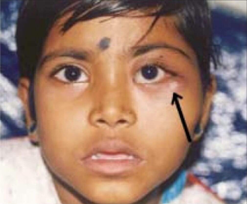
An ill-defined mass (arrow) on the lateral orbital wall due to Langerhans cell histiocytosis
Image: “LCH presented with ill-defined mass in lateral orbital wall” by Department of Ophthalmology, Sri Sankaradeva Nethralaya, Guwahati. License: CC BY 2.0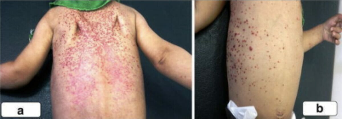
A scaly, erythematous rash on the back, shoulders, and chest of a boy with Langerhans cell histiocytosis
Image: “A scaly, erythematous rash on the back of the second boy spread to his shoulders (a) and upper chest wall (b)” by Unit of Pediatric Surgery, Al Diwaniya General Teaching Hospital, Al Qadisiya, Iraq. License: CC BY 4.0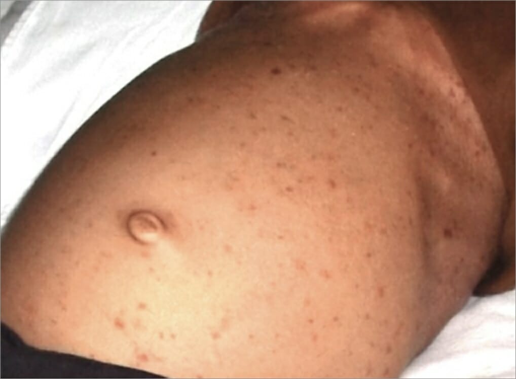
An eruption of papules on an individual’s stomach as a manifestation of Langerhans cell histiocytosis
Image: “After hospitalization” by Department of Oral Medicine and Radiology, Jaipur Dental College, Jaipur, Rajasthan, India. License: CC BY 3.0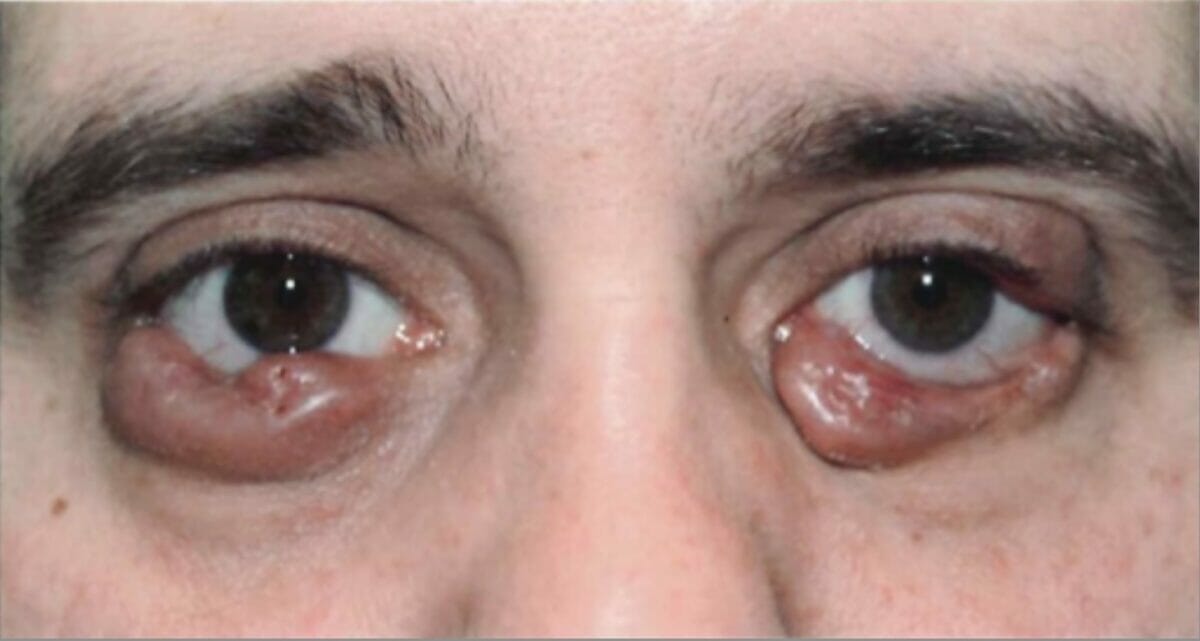
Bilateral lower eyelid growths due to Langerhans cell histiocytosis
Image: “Before radiation treatment” by King’s College London. License: CC BY 3.0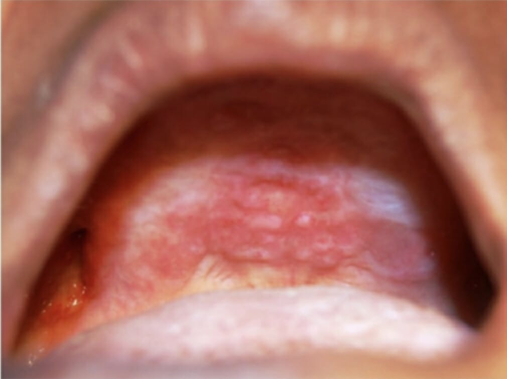
Oral lesions on the hard palate and maxillary alveolar ridge as a manifestation of Langerhans cell histiocytosis
Image: “Oral lesions in the hard palate and maxillary alveolar ridge” by Department of Oral Medicine and Radiology, M S Ramaiah Dental College and Hospital, Bangaluru. License: CC BY 2.5Complications depend on the organ system involved, but may include:
Laboratory studies generally demonstrate findings consistent with the organ systems involved and help rule out other diagnoses (list is not exhaustive):
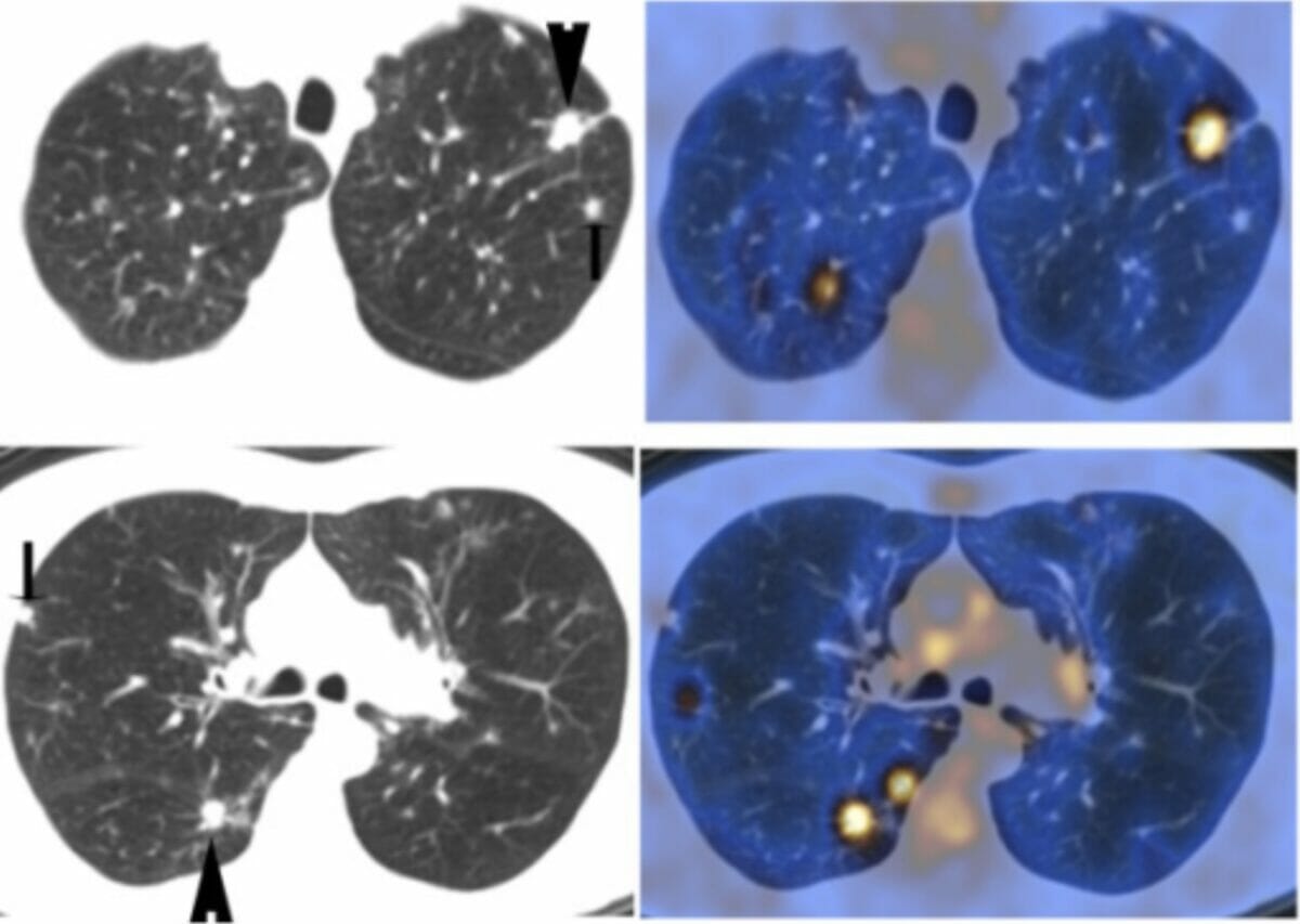
PET findings in nodular pulmonary Langerhans cell histiocytosis:
The chest CT images on the left upper and lower panels show multiple lung nodules. The corresponding PET images on the right upper and lower panels show PET uptake. The larger pulmonary nodules (arrowheads in the CT images) demonstrate intense PET uptake, while other nodules (smaller arrows in the CT images) are PET-negative.
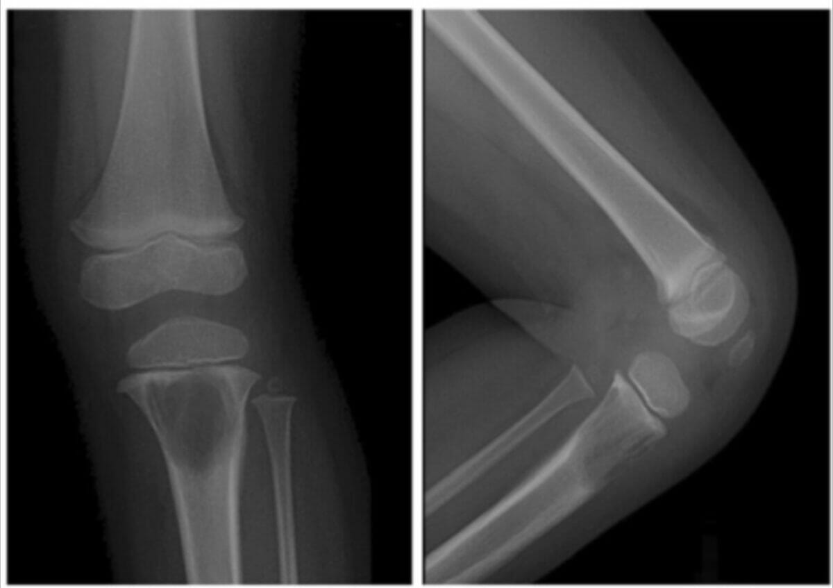
Radiographs of a child’s left knee demonstrating a large lytic lesion in the proximal tibial metaphysis due to Langerhans cell histiocytosis
Image: “A 2-year-old girl with left knee pain and a medullary lytic lesion in the proximal tibial metaphysis” by Department of Radiology, Tri-Service General Hospital, National Defense Medical Center, Taipei, Taiwan. License: CC BY 4.0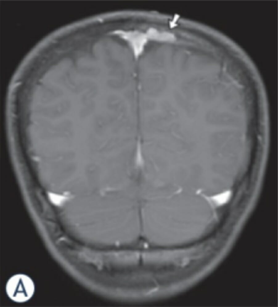
MRI images in an 11-year-old boy with LCH:
Coronal enhanced T1-MR image reveals an osseous enhancing mass (arrow) combined with epidural and subdural involvement along the left side of the superior sagittal sinus.
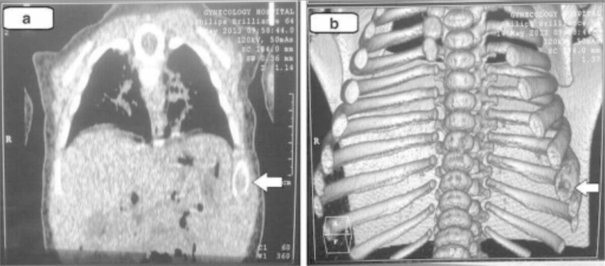
Chest computed tomography (and 3-dimensional imaging) findings in Langerhans cell histiocytosis: These images demonstrate lytic changes of the left 8th rib (arrows).
Image: “Chest computed tomography” by Unit of Pediatric Surgery, Al Diwaniya General Teaching Hospital, Al Qadisiya, Iraq. License: CC BY 4.0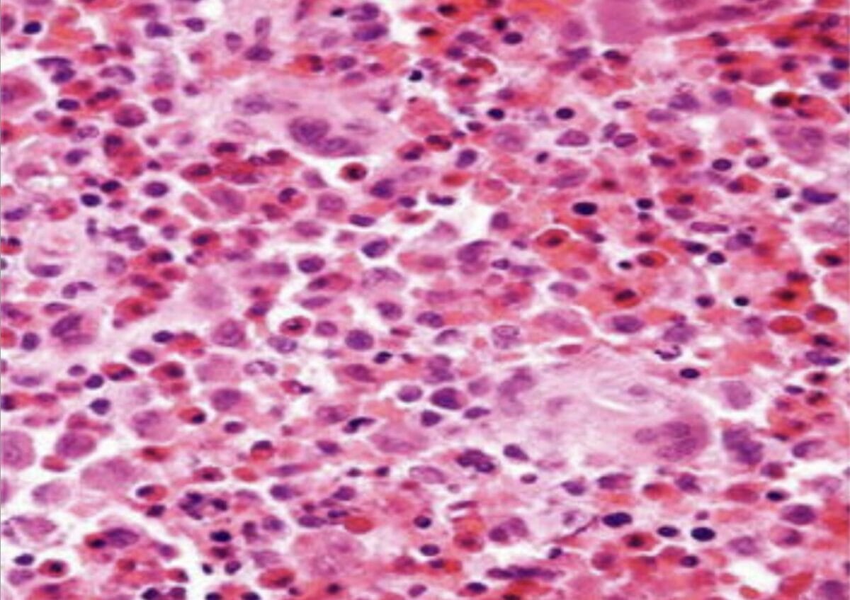
Skin biopsy histopathology (hematoxylin and eosin × 100) showing aggregates of histiocytic cells with an abundant eosinophilic and granular cytoplasm
Image: “Skin biopsy histopathology” by Unit of Pediatric Surgery, Al Diwaniya General Teaching Hospital, Al Qadisiya, Iraq. License: CC BY 4.0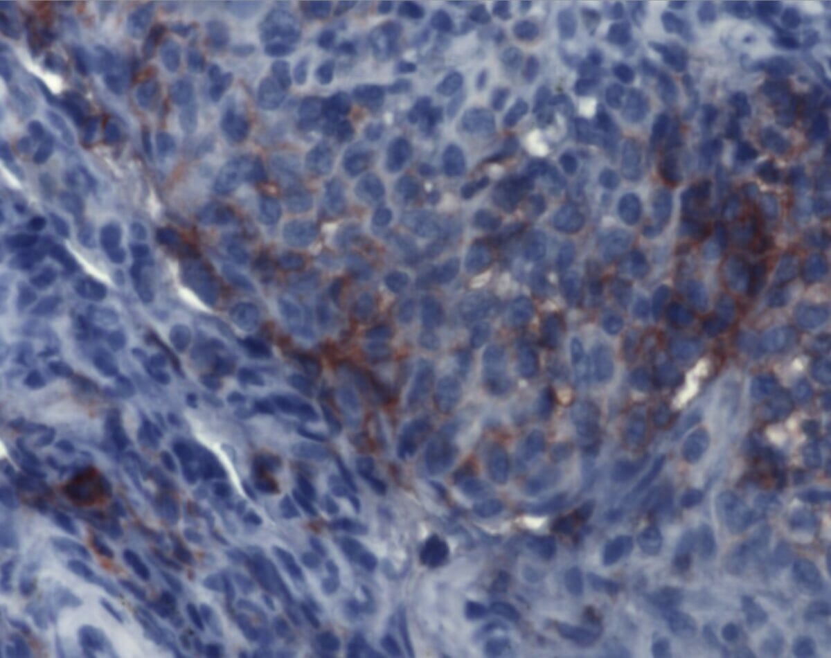
Immunohistochemistry demonstrates findings consistent with Langerhans cell histiocytosis. Aggregates of Langerhans cells are detectable by monoclonal antibodies against CD1a.
Image: “Immunohistochemistry of Langerhans cell histiocytosis” by Institute of Pathology and Neuropathology, University Hospital Essen, University of Duisburg-Essen, Germany. License: CC BY 2.0The choice in management for LCH is based on the type of LCH, clinical presentation, organs involved, extent of involvement, and organ function. Treatment is usually guided by an oncologist (and potentially other specialists).
Approach:
Multisystem involvement in children:
Multisystem involvement in adults:
Single-system involvement: