Necrotizing fasciitis (NF) is a life-threatening type of necrotizing soft tissue infection (NSTI) that causes rapid destruction of the deep soft tissues, including subcutaneous tissue and muscle fascia. The lay press often refers to organisms that cause NSTI as "flesh-eating bacteria." Patients may present with significant pain out of proportion to the presenting symptoms and rapidly progressive erythema of the affected area. Most patients will also have systemic signs of infection, including fever, hypotension, altered mental status, and multisystem organ failure. The diagnosis is primarily clinical since patients can quickly progress to septic shock without source control. This type of infection is a surgical emergency and requires emergent surgical debridement, parenteral antibiotics, and close hemodynamic monitoring.
IncidenceIncidenceThe number of new cases of a given disease during a given period in a specified population. It also is used for the rate at which new events occur in a defined population. It is differentiated from prevalence, which refers to all cases in the population at a given time.Measures of Disease Frequency: 0.3–15 cases per 100,000/year
20%–40% of affected individuals have diabetesDiabetesDiabetes mellitus (DM) is a metabolic disease characterized by hyperglycemia and dysfunction of the regulation of glucose metabolism by insulin. Type 1 DM is diagnosed mostly in children and young adults as the result of autoimmune destruction of β cells in the pancreas and the resulting lack of insulin. Type 2 DM has a significant association with obesity and is characterized by insulin resistance.Diabetes Mellitus.
Areas most commonly affected: anterior abdominal wallAbdominal wallThe outer margins of the abdomen, extending from the osteocartilaginous thoracic cage to the pelvis. Though its major part is muscular, the abdominal wall consists of at least seven layers: the skin, subcutaneous fat, deep fascia; abdominal muscles, transversalis fascia, extraperitoneal fat, and the parietal peritoneum.Surgical Anatomy of the Abdomen, perineumPerineumThe body region lying between the genital area and the anus on the surface of the trunk, and to the shallow compartment lying deep to this area that is inferior to the pelvic diaphragm. The surface area is between the vulva and the anus in the female, and between the scrotum and the anus in the male.Vagina, Vulva, and Pelvic Floor: Anatomy, scrotumScrotumA cutaneous pouch of skin containing the testicles and spermatic cords.Testicles: Anatomy, extremities
Etiology[1,3,4, 8,11,13]
Necrotizing fasciitisNecrotizing fasciitisNecrotizing fasciitis is a life-threatening infection that causes rapid destruction and necrosis of the fascia and subcutaneous tissues. Patients may present with significant pain out of proportion to the presenting symptoms and rapidly progressive erythema of the affected area. Necrotizing Fasciitis is divided into microbiologic categories based on the causative organism(s):
StreptococcusStreptococcusStreptococcus is one of the two medically important genera of gram-positive cocci, the other being Staphylococcus. Streptococci are identified as different species on blood agar on the basis of their hemolytic pattern and sensitivity to optochin and bacitracin. There are many pathogenic species of streptococci, including S. pyogenes, S. agalactiae, S. pneumoniae, and the viridans streptococci.Streptococcus pyogenes
BacteroidesBacteroidesBacteroides is a genus of opportunistic, anaerobic, gram-negative bacilli. Bacteroides fragilis is the most common species involved in human disease and is part of the normal flora of the large intestine.Bacteroides
PeptostreptococcusPeptostreptococcusA genus of gram-positive, anaerobic, coccoid bacteria that is part of the normal flora of humans. Its organisms are opportunistic pathogens causing bacteremias and soft tissue infections.Perianal and Perirectal Abscess
Escherichia coliEscherichia coliThe gram-negative bacterium Escherichia coli is a key component of the human gut microbiota. Most strains of E. coli are avirulent, but occasionally they escape the GI tract, infecting the urinary tract and other sites. Less common strains of E. coli are able to cause disease within the GI tract, most commonly presenting as abdominal pain and diarrhea. Escherichia coli
KlebsiellaKlebsiellaKlebsiella are encapsulated gram-negative, lactose-fermenting bacilli. They form pink colonies on MacConkey agar due to lactose fermentation. The main virulence factor is a polysaccharide capsule. Klebsiella pneumoniae is the most important pathogenic species.Klebsiella
ProteusProteusProteus spp. are gram-negative, facultatively anaerobic bacilli. Different types of infection result from Proteus, but the urinary tract is the most common site. The majority of cases are caused by Proteus mirabilis (P. mirabilis). The bacteria are part of the normal intestinal flora and are also found in the environment. Proteus
Often seen in older adults with comorbiditiesComorbiditiesThe presence of co-existing or additional diseases with reference to an initial diagnosis or with reference to the index condition that is the subject of study. Comorbidity may affect the ability of affected individuals to function and also their survival; it may be used as a prognostic indicator for length of hospital stay, cost factors, and outcome or survival.St. Louis Encephalitis Virus, particularly diabetesDiabetesDiabetes mellitus (DM) is a metabolic disease characterized by hyperglycemia and dysfunction of the regulation of glucose metabolism by insulin. Type 1 DM is diagnosed mostly in children and young adults as the result of autoimmune destruction of β cells in the pancreas and the resulting lack of insulin. Type 2 DM has a significant association with obesity and is characterized by insulin resistance.Diabetes Mellitus mellitus
Most common type
Type II:
Monomicrobial infection:
Group A StreptococcusStreptococcusStreptococcus is one of the two medically important genera of gram-positive cocci, the other being Staphylococcus. Streptococci are identified as different species on blood agar on the basis of their hemolytic pattern and sensitivity to optochin and bacitracin. There are many pathogenic species of streptococci, including S. pyogenes, S. agalactiae, S. pneumoniae, and the viridans streptococci.Streptococcus (most common)
Staphylococcus aureusStaphylococcus aureusPotentially pathogenic bacteria found in nasal membranes, skin, hair follicles, and perineum of warm-blooded animals. They may cause a wide range of infections and intoxications.Brain Abscess (including MRSAMRSAA strain of Staphylococcus aureus that is non-susceptible to the action of methicillin. The mechanism of resistance usually involves modification of normal or the presence of acquired penicillin binding proteins.Staphylococcus)
Occurs in any age group
Frequently found in individuals with no significant risk factors
Type III:
Marine/aquatic gram-negative organisms
Vibrio vulnificusVibrio vulnificusA species of halophilic bacteria in the genus vibrio, which lives in warm seawater. It can cause infections in those who eat raw contaminated seafood or have open wounds exposed to seawater.Vibrio
From aquatic injuries or consumption of raw seafood
More often seen in patientsPatientsIndividuals participating in the health care system for the purpose of receiving therapeutic, diagnostic, or preventive procedures.Clinician–Patient Relationship with chronic liverLiverThe liver is the largest gland in the human body. The liver is found in the superior right quadrant of the abdomen and weighs approximately 1.5 kilograms. Its main functions are detoxification, metabolism, nutrient storage (e.g., iron and vitamins), synthesis of coagulation factors, formation of bile, filtration, and storage of blood. Liver: Anatomy disease
Aeromonas hydrophilaAeromonas hydrophilaA species of gram-negative, facultatively anaerobic, rod-shaped bacteria that may be pathogenic for frogs, fish, and mammals, including man. In humans, cellulitis and diarrhea can result from infection with this organism.Cellulitis: from freshwater exposure through a lacerationLacerationTorn, ragged, mangled wounds.Blunt Chest Trauma
Gas gangreneGangreneDeath and putrefaction of tissue usually due to a loss of blood supply.Small Bowel Obstruction (treated as a distinct entity by others)
Most commonly from traumatic inoculation with Clostridium perfringensClostridium perfringensThe most common etiologic agent of gas gangrene. It is differentiable into several distinct types based on the distribution of twelve different toxins.Gas Gangrene
Clostridium septicumClostridium septicumA species of gram-positive bacteria in the family clostridiaceae. Infections have a strong association with malignancies and also with gas gangrene.Gas Gangrene is linked with colonColonThe large intestines constitute the last portion of the digestive system. The large intestine consists of the cecum, appendix, colon (with ascending, transverse, descending, and sigmoid segments), rectum, and anal canal. The primary function of the colon is to remove water and compact the stool prior to expulsion from the body via the rectum and anal canal. Colon, Cecum, and Appendix: Anatomy cancer and leukemia.
Type IV
Fungal infectionsInfectionsInvasion of the host organism by microorganisms or their toxins or by parasites that can cause pathological conditions or diseases.Chronic Granulomatous Disease, mainly CandidaCandidaCandida is a genus of dimorphic, opportunistic fungi. Candida albicans is part of the normal human flora and is the most common cause of candidiasis. The clinical presentation varies and can include localized mucocutaneous infections (e.g., oropharyngeal, esophageal, intertriginous, and vulvovaginal candidiasis) and invasive disease (e.g., candidemia, intraabdominal abscess, pericarditis, and meningitis). Candida/Candidiasis and Zygomycetes/mucormycetes
Seen in immunocompromisedimmunocompromisedA human or animal whose immunologic mechanism is deficient because of an immunodeficiency disorder or other disease or as the result of the administration of immunosuppressive drugs or radiation.Gastroenteritis hosts
Risk factors[1,2,4,8,9,11,14]
Immunosuppression:
DiabetesDiabetesDiabetes mellitus (DM) is a metabolic disease characterized by hyperglycemia and dysfunction of the regulation of glucose metabolism by insulin. Type 1 DM is diagnosed mostly in children and young adults as the result of autoimmune destruction of β cells in the pancreas and the resulting lack of insulin. Type 2 DM has a significant association with obesity and is characterized by insulin resistance.Diabetes Mellitus mellitus (most common predisposing condition)
NeutropeniaNeutropeniaNeutrophils are an important component of the immune system and play a significant role in the eradication of infections. Low numbers of circulating neutrophils, referred to as neutropenia, predispose the body to recurrent infections or sepsis, though patients can also be asymptomatic. Neutropenia
SkinSkinThe skin, also referred to as the integumentary system, is the largest organ of the body. The skin is primarily composed of the epidermis (outer layer) and dermis (deep layer). The epidermis is primarily composed of keratinocytes that undergo rapid turnover, while the dermis contains dense layers of connective tissue.Skin: Structure and Functions damage:
ObesityObesityObesity is a condition associated with excess body weight, specifically with the deposition of excessive adipose tissue. Obesity is considered a global epidemic. Major influences come from the western diet and sedentary lifestyles, but the exact mechanisms likely include a mixture of genetic and environmental factors. Obesity
SmokingSmokingWillful or deliberate act of inhaling and exhaling smoke from burning substances or agents held by hand.Interstitial Lung Diseases
AlcoholismAlcoholismA primary, chronic disease with genetic, psychosocial, and environmental factors influencing its development and manifestations. The disease is often progressive and fatal. It is characterized by impaired control over drinking, preoccupation with the drug alcohol, use of alcohol despite adverse consequences, and distortions in thinking, most notably denial. Each of these symptoms may be continuous or periodic.Wernicke Encephalopathy and Korsakoff Syndrome
LiverLiverThe liver is the largest gland in the human body. The liver is found in the superior right quadrant of the abdomen and weighs approximately 1.5 kilograms. Its main functions are detoxification, metabolism, nutrient storage (e.g., iron and vitamins), synthesis of coagulation factors, formation of bile, filtration, and storage of blood. Liver: AnatomycirrhosisCirrhosisCirrhosis is a late stage of hepatic parenchymal necrosis and scarring (fibrosis) most commonly due to hepatitis C infection and alcoholic liver disease. Patients may present with jaundice, ascites, and hepatosplenomegaly. Cirrhosis can also cause complications such as hepatic encephalopathy, portal hypertension, portal vein thrombosis, and hepatorenal syndrome. Cirrhosis
Chronic cardiac disease
Chronic renal failureRenal failureConditions in which the kidneys perform below the normal level in the ability to remove wastes, concentrate urine, and maintain electrolyte balance; blood pressure; and calcium metabolism. Renal insufficiency can be classified by the degree of kidney damage (as measured by the level of proteinuria) and reduction in glomerular filtration rate.Crush Syndrome
Peripheral vascular disease
Advanced age
MalnutritionMalnutritionMalnutrition is a clinical state caused by an imbalance or deficiency of calories and/or micronutrients and macronutrients. The 2 main manifestations of acute severe malnutrition are marasmus (total caloric insufficiency) and kwashiorkor (protein malnutrition with characteristic edema).Malnutrition in children in resource-limited countries
Pathophysiology[1,2,7,8,11,14]
Routes:
BacteriaBacteriaBacteria are prokaryotic single-celled microorganisms that are metabolically active and divide by binary fission. Some of these organisms play a significant role in the pathogenesis of diseases. Bacteriology extend into the subcutaneous tissueSubcutaneous tissueLoose connective tissue lying under the dermis, which binds skin loosely to subjacent tissues. It may contain a pad of adipocytes, which vary in number according to the area of the body and vary in size according to the nutritional state.Soft Tissue Abscess from:
Defined portal of entry:
Nearby ulcer or superficial infection
Trauma
Bloodstream (most often S. pyogenes, seedingSeedingThe local implantation of tumor cells by contamination of instruments and surgical equipment during and after surgical resection, resulting in local growth of the cells and tumor formation.Grading, Staging, and Metastasis a site of deep, non-penetrating injury via transient bacteremiaBacteremiaThe presence of viable bacteria circulating in the blood. Fever, chills, tachycardia, and tachypnea are common acute manifestations of bacteremia. The majority of cases are seen in already hospitalized patients, most of whom have underlying diseases or procedures which render their bloodstreams susceptible to invasion.Glycopeptides, frequently from asymptomatic pharyngeal carriage)
Even in monomicrobial infectionsInfectionsInvasion of the host organism by microorganisms or their toxins or by parasites that can cause pathological conditions or diseases.Chronic Granulomatous Disease, other aerobic and anaerobic pathogens may play a role.
Toxin-mediated destruction:
Fulminant inflammatory response occurs, affecting tissues and vessels, causing microvascular thrombosisThrombosisFormation and development of a thrombus or blood clot in the blood vessel.Epidemic Typhus then tissue destruction follows.
In Group A streptococcal disease: streptolysin O leads to platelet-neutrophil aggregrates obstructing vessels.
Infection causes occlusion of subcutaneous capillariesCapillariesCapillaries are the primary structures in the circulatory system that allow the exchange of gas, nutrients, and other materials between the blood and the extracellular fluid (ECF). Capillaries are the smallest of the blood vessels. Because a capillary diameter is so small, only 1 RBC may pass through at a time.Capillaries: Histology, venulesVenulesThe minute vessels that collect blood from the capillary plexuses and join together to form veins.Veins: Histology, and arteriolesArteriolesThe smallest divisions of the arteries located between the muscular arteries and the capillaries.Arteries: Histology → tissue and fascial ischemiaIschemiaA hypoperfusion of the blood through an organ or tissue caused by a pathologic constriction or obstruction of its blood vessels, or an absence of blood circulation.Ischemic Cell Damage → progressive thrombosisThrombosisFormation and development of a thrombus or blood clot in the blood vessel.Epidemic Typhus → necrosisNecrosisThe death of cells in an organ or tissue due to disease, injury or failure of the blood supply.Ischemic Cell Damage
Damage occurs to superficial nerves → localized anesthesiaAnesthesiaA state characterized by loss of feeling or sensation. This depression of nerve function is usually the result of pharmacologic action and is induced to allow performance of surgery or other painful procedures.Anesthesiology: History and Basic Concepts
Hypoxic conditions → ↓ neutrophil function → proliferation of bacteriaBacteriaBacteria are prokaryotic single-celled microorganisms that are metabolically active and divide by binary fission. Some of these organisms play a significant role in the pathogenesis of diseases. Bacteriology
Fascial spread:
Infection and necrosisNecrosisThe death of cells in an organ or tissue due to disease, injury or failure of the blood supply.Ischemic Cell Damage can rapidly travel along fascial planes, possibly due to bacterial enzymesEnzymesEnzymes are complex protein biocatalysts that accelerate chemical reactions without being consumed by them. Due to the body’s constant metabolic needs, the absence of enzymes would make life unsustainable, as reactions would occur too slowly without these molecules. Basics of Enzymes and toxins.
Streptococcal enzymesEnzymesEnzymes are complex protein biocatalysts that accelerate chemical reactions without being consumed by them. Due to the body’s constant metabolic needs, the absence of enzymes would make life unsustainable, as reactions would occur too slowly without these molecules. Basics of Enzymes/toxins that drive fascial spread:
Streptococcal pyrogenic exotoxinsPyrogenic ExotoxinsScarlet Fever (notably from M1/M3 serotypes): superantigensSuperantigensMicrobial antigens that have in common an extremely potent activating effect on T-cells that bear a specific variable region. Superantigens cross-link the variable region with class II mhc proteins regardless of the peptide binding in the t-cell receptor’s pocket. The result is a transient expansion and subsequent death and anergy of the T-cells with the appropriate variable regions.Staphylococcus that drive the cytokine cascade, leading to multiorgan failure, and the streptococcal toxic shock syndromeStreptococcal Toxic Shock SyndromeToxic Shock Syndrome
Gas formation:
Aerobic metabolism produces water-soluble CO2.
Anaerobic metabolism by facultative organisms → hydrogen, and poorly-soluble gases
Hydrogen and nitrogenNitrogenAn element with the atomic symbol n, atomic number 7, and atomic weight [14. 00643; 14. 00728]. Nitrogen exists as a diatomic gas and makes up about 78% of the earth’s atmosphere by volume. It is a constituent of proteins and nucleic acids and found in all living cells.Urea Cycle are insoluble and accumulate in the subcutaneous tissues.
Seen as gas in imaging
In clostridial gas gangreneGangreneDeath and putrefaction of tissue usually due to a loss of blood supply.Small Bowel Obstruction, C. perfringensC. perfringensThe most common etiologic agent of gas gangrene. It is differentiable into several distinct types based on the distribution of twelve different toxins.Gas Gangrenealpha-toxinAlpha-ToxinGas Gangrene (phospholipase CPhospholipase CA subclass of phospholipases that hydrolyze the phosphoester bond found in the third position of glycerophospholipids. Although the singular term phospholipase C specifically refers to an enzyme that catalyzes the hydrolysis of phosphatidylcholine, it is commonly used in the literature to refer to broad variety of enzymes that specifically catalyze the hydrolysis of phosphatidylinositols.Pseudomonas) and other exotoxinsExotoxinsToxins produced, especially by bacterial or fungal cells, and released into the culture medium or environment.Bacteriology drives gas production leading to myonecrosisMyonecrosisA severe condition resulting from bacteria invading healthy muscle from adjacent traumatized muscle or soft tissue. The infection originates in a wound contaminated with bacteria of the genus Clostridium. C. perfringens accounts for the majority of cases (over eighty percent), while C. novyi, C. septicum, and C. histolyticum cause most of the other cases.Clostridia.
SkinSkinThe skin, also referred to as the integumentary system, is the largest organ of the body. The skin is primarily composed of the epidermis (outer layer) and dermis (deep layer). The epidermis is primarily composed of keratinocytes that undergo rapid turnover, while the dermis contains dense layers of connective tissue.Skin: Structure and Functions and soft tissueSoft TissueSoft Tissue Abscess findings[1,8,11]
Be sure to mark skinSkinThe skin, also referred to as the integumentary system, is the largest organ of the body. The skin is primarily composed of the epidermis (outer layer) and dermis (deep layer). The epidermis is primarily composed of keratinocytes that undergo rapid turnover, while the dermis contains dense layers of connective tissue.Skin: Structure and Functions lesions during the physical examination.
Early signs:
Acute, severe painPainAn unpleasant sensation induced by noxious stimuli which are detected by nerve endings of nociceptive neurons.Pain: Types and Pathways out of proportion with physical findings
ErythemaErythemaRedness of the skin produced by congestion of the capillaries. This condition may result from a variety of disease processes.Chalazion that quickly spreads over hours to days.
Warmth
Tense, indurated skinSkinThe skin, also referred to as the integumentary system, is the largest organ of the body. The skin is primarily composed of the epidermis (outer layer) and dermis (deep layer). The epidermis is primarily composed of keratinocytes that undergo rapid turnover, while the dermis contains dense layers of connective tissue.Skin: Structure and Functions
AnesthesiaAnesthesiaA state characterized by loss of feeling or sensation. This depression of nerve function is usually the result of pharmacologic action and is induced to allow performance of surgery or other painful procedures.Anesthesiology: History and Basic Concepts or paresthesia
BullaeBullaeErythema Multiforme, skinSkinThe skin, also referred to as the integumentary system, is the largest organ of the body. The skin is primarily composed of the epidermis (outer layer) and dermis (deep layer). The epidermis is primarily composed of keratinocytes that undergo rapid turnover, while the dermis contains dense layers of connective tissue.Skin: Structure and FunctionsecchymosisEcchymosisExtravasation of blood into the skin, resulting in a nonelevated, rounded or irregular, blue or purplish patch, larger than a petechia.Orbital Fractures, edemaEdemaEdema is a condition in which excess serous fluid accumulates in the body cavity or interstitial space of connective tissues. Edema is a symptom observed in several medical conditions. It can be categorized into 2 types, namely, peripheral (in the extremities) and internal (in an organ or body cavity). Edema outside of the area of erythemaErythemaRedness of the skin produced by congestion of the capillaries. This condition may result from a variety of disease processes.Chalazion, skin necrosisSkin NecrosisRocky Mountain Spotted Fever, or ulcerationUlcerationCorneal Abrasions, Erosion, and Ulcers
Common sites of infection:
Extremities (most common) > trunk > perineumPerineumThe body region lying between the genital area and the anus on the surface of the trunk, and to the shallow compartment lying deep to this area that is inferior to the pelvic diaphragm. The surface area is between the vulva and the anus in the female, and between the scrotum and the anus in the male.Vagina, Vulva, and Pelvic Floor: Anatomy > head/neckNeckThe part of a human or animal body connecting the head to the rest of the body.Peritonsillar Abscess
Fournier gangreneFournier gangreneAn acute necrotic infection of the scrotum; penis; or perineum. It is characterized by scrotum pain and redness with rapid progression to gangrene and sloughing of tissue. Fournier gangrene is usually secondary to perirectal or periurethral infections associated with local trauma, operative procedures, or urinary tract disease.Necrotizing Fasciitis (necrotizing fasciitis of the perineumNecrotizing fasciitis of the perineumAn acute necrotic infection of the scrotum; penis; or perineum. It is characterized by scrotum pain and redness with rapid progression to gangrene and sloughing of tissue. Fournier gangrene is usually secondary to perirectal or periurethral infections associated with local trauma, operative procedures, or urinary tract disease.Necrotizing Fasciitis):
Rapidly spreads to the anterior abdominal wallAbdominal wallThe outer margins of the abdomen, extending from the osteocartilaginous thoracic cage to the pelvis. Though its major part is muscular, the abdominal wall consists of at least seven layers: the skin, subcutaneous fat, deep fascia; abdominal muscles, transversalis fascia, extraperitoneal fat, and the parietal peritoneum.Surgical Anatomy of the Abdomen and gluteal muscles
Head and neckNeckThe part of a human or animal body connecting the head to the rest of the body.Peritonsillar Abscess:
Often has a dental or pharyngeal origin from procedures or trauma
May spread to the face, lower neckNeckThe part of a human or animal body connecting the head to the rest of the body.Peritonsillar Abscess, and mediastinumMediastinumThe mediastinum is the thoracic area between the 2 pleural cavities. The mediastinum contains vital structures of the circulatory, respiratory, digestive, and nervous systems including the heart and esophagus, and major thoracic vessels.Mediastinum and Great Vessels: Anatomy
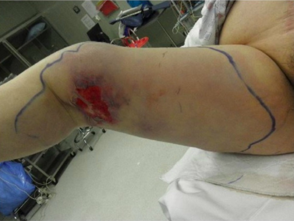
Rapidly spreading erythema, ulceration, and edema of the right leg due to necrotizing fasciitis
Image: “Preoperative photograph” by Department of Family Medicine, Morehouse School of Medicine, 1513 East Cleveland Avenue, Building 100, Suite 300A, Atlanta, GA 30344, USA. License: CC BY 3.0
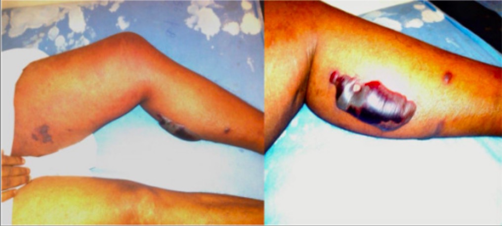
Bullae and erythema resulting from necrotizing fasciitis
Image: “Superficial skin manifestations of necrotizing fasciitis” by National Hospital of Sri Lanka, Regent Street, Colombo 10, Sri Lanka. License: CC BY 4.0
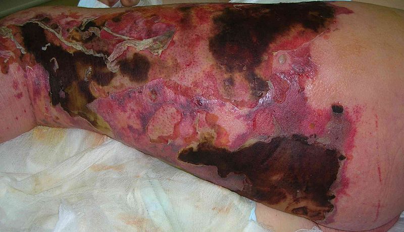
Cutaneous necrosis, erythema, and bullous changes due to necrotizing fasciitis of the leg
Image: “Necrotizing fasciitis” by Piotr Smuszkiewicz et al. License: CC BY 2.0
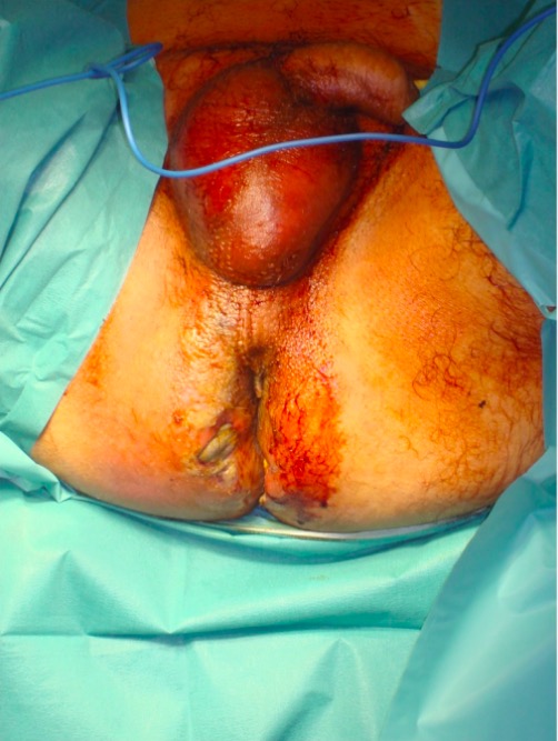
Fournier gangrene: Significant erythema and edema is noted throughout the scrotum and gluteal regions.
Image: “Fournier’s gangrene” by 3rd Department of Surgery, Attikon University Hospital, University of Athens School of Medicine, Athens, Greece. License: CC BY 4.0
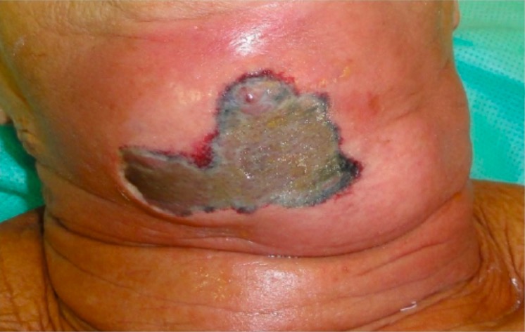
Erythema and necrotic tissue due to necrotizing fasciitis of the neck:
This developed after a dental extraction.
Image: “Necrotic tissue” by Faculdade de Odontologia de Pernambuco, Universidade de Pernambuco, Avenida General Newton Cavalcante, 1650 Aldeia dos Camarás, 54753-020 Camaragibe, PE, Brazil. License: CC BY 4.0
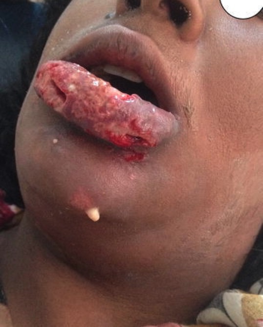
Necrotizing fasciitis of the lip and face:
Significant swelling, erythema, exfoliation, and purulent drainage is noted.
Image: “Patient with painful lower lip swelling” by Khartoum Teaching Dental Hospital, P.O box 122, Khartoum, Sudan. License: CC BY 4.0
High feverFeverFever is defined as a measured body temperature of at least 38°C (100.4°F). Fever is caused by circulating endogenous and/or exogenous pyrogens that increase levels of prostaglandin E2 in the hypothalamus. Fever is commonly associated with chills, rigors, sweating, and flushing of the skin. Fever
TachycardiaTachycardiaAbnormally rapid heartbeat, usually with a heart rate above 100 beats per minute for adults. Tachycardia accompanied by disturbance in the cardiac depolarization (cardiac arrhythmia) is called tachyarrhythmia.Sepsis in Children
HypotensionHypotensionHypotension is defined as low blood pressure, specifically < 90/60 mm Hg, and is most commonly a physiologic response. Hypotension may be mild, serious, or life threatening, depending on the cause. Hypotension
Altered mentation:
Confusion
Obtundation
Diagnosis
A definitive diagnosis of necrotizing fasciitisNecrotizing fasciitisNecrotizing fasciitis is a life-threatening infection that causes rapid destruction and necrosis of the fascia and subcutaneous tissues. Patients may present with significant pain out of proportion to the presenting symptoms and rapidly progressive erythema of the affected area. Necrotizing Fasciitis is made by surgical exploration and debridementDebridementThe removal of foreign material and devitalized or contaminated tissue from or adjacent to a traumatic or infected lesion until surrounding healthy tissue is exposed.Stevens-Johnson Syndrome. These processes should not be delayed to obtain diagnostic information, if the clinical suspicion is high.
However, the following may be helpful:
Laboratory evaluation[5,6,10,14]
Laboratory studies may demonstrate evidence of infection and organ damage.
↑ Erythrocyte sedimentation rateErythrocyte Sedimentation RateSoft Tissue Abscess and c-reactive proteinsProteinsLinear polypeptides that are synthesized on ribosomes and may be further modified, crosslinked, cleaved, or assembled into complex proteins with several subunits. The specific sequence of amino acids determines the shape the polypeptide will take, during protein folding, and the function of the protein.Energy Homeostasis
↓ HCO3 → metabolic acidosisAcidosisA pathologic condition of acid accumulation or depletion of base in the body. The two main types are respiratory acidosis and metabolic acidosis, due to metabolic acid build up.Respiratory Acidosis
↑ Lactic acid
↑ Blood ureaUreaA compound formed in the liver from ammonia produced by the deamination of amino acids. It is the principal end product of protein catabolism and constitutes about one half of the total urinary solids.Urea CyclenitrogenNitrogenAn element with the atomic symbol n, atomic number 7, and atomic weight [14. 00643; 14. 00728]. Nitrogen exists as a diatomic gas and makes up about 78% of the earth’s atmosphere by volume. It is a constituent of proteins and nucleic acids and found in all living cells.Urea Cycle and creatinine → acute renal failureRenal failureConditions in which the kidneys perform below the normal level in the ability to remove wastes, concentrate urine, and maintain electrolyte balance; blood pressure; and calcium metabolism. Renal insufficiency can be classified by the degree of kidney damage (as measured by the level of proteinuria) and reduction in glomerular filtration rate.Crush Syndrome
↑ GlucoseGlucoseA primary source of energy for living organisms. It is naturally occurring and is found in fruits and other parts of plants in its free state. It is used therapeutically in fluid and nutrient replacement.Lactose Intolerance
↓ Na
↑ CK, and/or ASTASTEnzymes of the transferase class that catalyze the conversion of l-aspartate and 2-ketoglutarate to oxaloacetate and l-glutamate.Liver Function Tests → indicative of deep infection involving muscle/fasciaFasciaLayers of connective tissue of variable thickness. The superficial fascia is found immediately below the skin; the deep fascia invests muscles, nerves, and other organs.Cellulitis (vs cellulitisCellulitisCellulitis is a common infection caused by bacteria that affects the dermis and subcutaneous tissue of the skin. It is frequently caused by Staphylococcus aureus and Streptococcus pyogenes. The skin infection presents as an erythematous and edematous area with warmth and tenderness. Cellulitis)
↑ ProcalcitoninProcalcitoninNeutropenic Fever on admission → increased risk of septic shockSeptic shockSepsis associated with hypotension or hypoperfusion despite adequate fluid resuscitation. Perfusion abnormalities may include, but are not limited to lactic acidosis; oliguria; or acute alteration in mental status.Sepsis and Septic Shock
Note: lactate >6 mmol/L and a serum Na <135 mEq/L on admission are independent predictors of in-hospital mortalityMortalityAll deaths reported in a given population.Measures of Health Status (in patientsPatientsIndividuals participating in the health care system for the purpose of receiving therapeutic, diagnostic, or preventive procedures.Clinician–Patient Relationship presenting with necrotizing soft tissueSoft TissueSoft Tissue Abscess infection).
Laboratory Risk IndicatorIndicatorMethods for assessing flow through a system by injection of a known quantity of an indicator, such as a dye, radionuclide, or chilled liquid, into the system and monitoring its concentration over time at a specific point in the system.Body Fluid Compartments for Necrotizing FasciitisNecrotizing fasciitisNecrotizing fasciitis is a life-threatening infection that causes rapid destruction and necrosis of the fascia and subcutaneous tissues. Patients may present with significant pain out of proportion to the presenting symptoms and rapidly progressive erythema of the affected area. Necrotizing Fasciitis (LRINEC) Score[5,6,8,9]
Calculates a score using WBC, c-reactive protein, hemoglobin, creatinine, sodiumSodiumA member of the alkali group of metals. It has the atomic symbol na, atomic number 11, and atomic weight 23.Hyponatremia, and glucoseGlucoseA primary source of energy for living organisms. It is naturally occurring and is found in fruits and other parts of plants in its free state. It is used therapeutically in fluid and nutrient replacement.Lactose Intolerance
Has a high specificity but low sensitivity
Should not be used alone to rule out infection; if clinical suspicion is high, consult surgery regardless of the score.
Score ≥ 6 is highly suggestive of necrotizing fasciitisNecrotizing fasciitisNecrotizing fasciitis is a life-threatening infection that causes rapid destruction and necrosis of the fascia and subcutaneous tissues. Patients may present with significant pain out of proportion to the presenting symptoms and rapidly progressive erythema of the affected area. Necrotizing Fasciitis.
Table: LRINEC scoring system for necrotizing fasciitisNecrotizing fasciitisNecrotizing fasciitis is a life-threatening infection that causes rapid destruction and necrosis of the fascia and subcutaneous tissues. Patients may present with significant pain out of proportion to the presenting symptoms and rapidly progressive erythema of the affected area. Necrotizing Fasciitis[5]
Laboratory test (units)
Value
Score
CRP (mg/L)
< 150
0
≥ 150
4
WBC (g/L)
< 15
0
15‒25
1
> 25
2
Hemoglobin (g/dL)
> 13.5
0
11‒13.5
1
< 11
2
SodiumSodiumA member of the alkali group of metals. It has the atomic symbol na, atomic number 11, and atomic weight 23.Hyponatremia (mmol/L)
≥ 135
0
< 135
2
Creatinine (μmol/L)
≤ 141
0
>141
2
GlucoseGlucoseA primary source of energy for living organisms. It is naturally occurring and is found in fruits and other parts of plants in its free state. It is used therapeutically in fluid and nutrient replacement.Lactose Intolerance (mmol/L)
≤ 10
0
> 10
1
Imaging[6,8,9,14]
Imaging may be needed to assist in the diagnosis, especially if there is uncertainty.
CT scan is the best imaging modality and may show:
Subcutaneous gas
Inflammatory changes along fascial planes
Fluid collections
In contrast-enahnced CT: no fascial enhancement in thickened soft-tissue fasciaFasciaLayers of connective tissue of variable thickness. The superficial fascia is found immediately below the skin; the deep fascia invests muscles, nerves, and other organs.Cellulitis(most specific finding)
MRI is an alternative:
Most sensitive test overall
Costly and time-consuming (not recommended if clinical suspicion is high, as this could delay surgical intervention)
Finding: T2 hyperintensity of deep fasciaFasciaLayers of connective tissue of variable thickness. The superficial fascia is found immediately below the skin; the deep fascia invests muscles, nerves, and other organs.Cellulitis
Can reveal gas in the tissues, particularly in the extremities
Cannot rule out necrotizing fasciitisNecrotizing fasciitisNecrotizing fasciitis is a life-threatening infection that causes rapid destruction and necrosis of the fascia and subcutaneous tissues. Patients may present with significant pain out of proportion to the presenting symptoms and rapidly progressive erythema of the affected area. Necrotizing Fasciitis
CT findings in necrotizing fasciitis:
Gas is noted in the subcutaneous space.
Image: “Abnormal air accumulation” by Department of Surgery, Division of Frontier Medical Science, Programs for Biomedical Research, Graduate School of Biomedical Science, Hiroshima University, Minami-ku, Hiroshima, Japan. License: CC BY 2.0
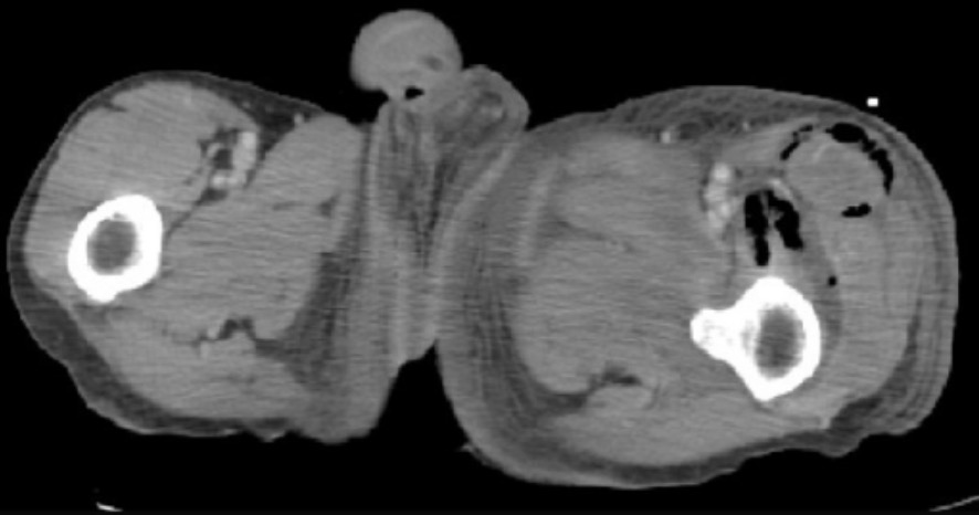
CT findings in necrotizing fasciitis:
This scan demonstrates fascial edema, subcutaneous fat stranding, and gas in the muscle compartments.
Image: “Computed tomography” by Poole Hospital NHS Foundation Trust, Longfleet Road, Poole, BH15 2JB, UK. License: CC BY 2.0
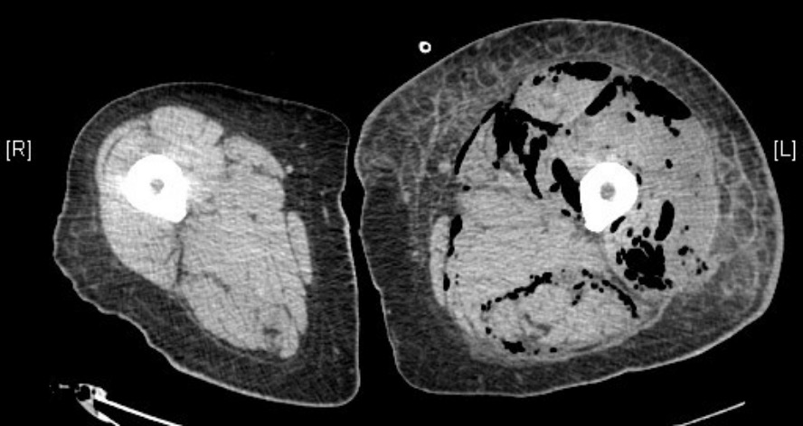
CT findings in necrotizing fasciitis:
This scan demonstrates subcutaneous gas tracking along fascial planes of the left leg and pelvis.
Image: “Frontal view of CT” by Henrik Galust et al. License: CC BY 3.0
Other tests[6,8]
The following may be used as complementary exams in cases where the diagnosis is uncertain:
Finger test:
2-cm excision made at bedside under local anesthesiaAnesthesiaA state characterized by loss of feeling or sensation. This depression of nerve function is usually the result of pharmacologic action and is induced to allow performance of surgery or other painful procedures.Anesthesiology: History and Basic Concepts
Wound explored bluntly with a gloved finger
Findings of necrotizing fasciitisNecrotizing fasciitisNecrotizing fasciitis is a life-threatening infection that causes rapid destruction and necrosis of the fascia and subcutaneous tissues. Patients may present with significant pain out of proportion to the presenting symptoms and rapidly progressive erythema of the affected area. Necrotizing Fasciitis:
“Dishwater pus” (thin grayish fluid)
Absence of bleeding
No tissue resistanceResistancePhysiologically, the opposition to flow of air caused by the forces of friction. As a part of pulmonary function testing, it is the ratio of driving pressure to the rate of air flow.Ventilation: Mechanics of Breathing to finger dissection
Frozen-section biopsyBiopsyRemoval and pathologic examination of specimens from the living body.Ewing Sarcoma:
Obtained during finger test or operative exploration
Can provide quick diagnosis confirmation, if in doubt
Management
Management of necrotizing fasciitisNecrotizing fasciitisNecrotizing fasciitis is a life-threatening infection that causes rapid destruction and necrosis of the fascia and subcutaneous tissues. Patients may present with significant pain out of proportion to the presenting symptoms and rapidly progressive erythema of the affected area. Necrotizing Fasciitis has slight variations depending on practice location. The following information was derived from US and UK literature and clinical guidelines. Overall, therapy should be guided by a multidisciplinary team, including surgery, intensive care, and infectious disease specialists.[8]
Surgery
Emergent surgical debridementDebridementThe removal of foreign material and devitalized or contaminated tissue from or adjacent to a traumatic or infected lesion until surrounding healthy tissue is exposed.Stevens-Johnson Syndrome is the mainstay of treatment.
Timing:[4,6‒9,14]
Surgery must be performed as early as possible, preferably in the 1st 6 hours.
Delay beyond the 1st 24 hours or restricted primary debridementDebridementThe removal of foreign material and devitalized or contaminated tissue from or adjacent to a traumatic or infected lesion until surrounding healthy tissue is exposed.Stevens-Johnson Syndrome are associated with significantly increased mortalityMortalityAll deaths reported in a given population.Measures of Health Status.
DebridementDebridementThe removal of foreign material and devitalized or contaminated tissue from or adjacent to a traumatic or infected lesion until surrounding healthy tissue is exposed.Stevens-Johnson Syndrome should aim to remove devitalized tissue as completely as possible; should extend to healthy, bleeding, contractile tissue
Microbiology/tissue samples are taken during the procedure.
Surgical exploration is also the diagnostic gold standard:
Warranted whenever clinical suspicion is high
Intraoperative findings include:
Dishwater-gray pus/exudateExudateExudates are fluids, cells, or other cellular substances that are slowly discharged from blood vessels usually from inflamed tissues.Pleural Effusion
Non-contracting muscle (dull and opaque)
Lack of bleeding due to microvascular thrombosisThrombosisFormation and development of a thrombus or blood clot in the blood vessel.Epidemic Typhus
Positive “finger test” (no tissue resistanceResistancePhysiologically, the opposition to flow of air caused by the forces of friction. As a part of pulmonary function testing, it is the ratio of driving pressure to the rate of air flow.Ventilation: Mechanics of Breathing; tissues easily separated by a gloved finger)
Surgical debridementDebridementThe removal of foreign material and devitalized or contaminated tissue from or adjacent to a traumatic or infected lesion until surrounding healthy tissue is exposed.Stevens-Johnson Syndrome frequently needs to be repeated over the next 24–48 hours:
“2nd-look operation” to reevaluate tissue viability and spread of infection
Sometimes, multiple serial debridements need to be performed.
In case of wide or disfiguring debridements, involve additional teams:
Urology for wounds involving the perineumPerineumThe body region lying between the genital area and the anus on the surface of the trunk, and to the shallow compartment lying deep to this area that is inferior to the pelvic diaphragm. The surface area is between the vulva and the anus in the female, and between the scrotum and the anus in the male.Vagina, Vulva, and Pelvic Floor: Anatomy, penisPenisThe penis is the male organ of copulation and micturition. The organ is composed of a root, body, and glans. The root is attached to the pubic bone by the crura penis. The body consists of the 2 parallel corpora cavernosa and the corpus spongiosum. The glans is ensheathed by the prepuce or foreskin. Penis: Anatomy or scrotumScrotumA cutaneous pouch of skin containing the testicles and spermatic cords.Testicles: Anatomy
Plastic surgery for complex reconstruction or muscle flap reconstruction
Orthopedics for infectionsInfectionsInvasion of the host organism by microorganisms or their toxins or by parasites that can cause pathological conditions or diseases.Chronic Granulomatous Disease with bony involvement
Technical considerations:[6]
Incisions performed parallel to Langer’s lines to promote healing and minimize scarringScarringInflammation
Wounds left open to heal by secondary intentionSecondary intentionWhen there are significant tissue losses and the wound surface cannot be brought together (e.g., lacerations, burns, and ulcers)Wound Healing and/or drains inserted to promote continuous drainage of infection
Wound Vac therapy frequently used to promote wound healingWound healingWound healing is a physiological process involving tissue repair in response to injury. It involves a complex interaction of various cell types, cytokines, and inflammatory mediators. Wound healing stages include hemostasis, inflammation, granulation, and remodeling. Wound Healing
Important to note:
Wound Vac is delayed until the wound is stable and debridements are done.
Use with extreme caution in anaerobic infectionsInfectionsInvasion of the host organism by microorganisms or their toxins or by parasites that can cause pathological conditions or diseases.Chronic Granulomatous Disease; the closed, oxygen-deprived environment may exacerbate anaerobic growth.
Anatomical considerations:[6]
Abdominal wallAbdominal wallThe outer margins of the abdomen, extending from the osteocartilaginous thoracic cage to the pelvis. Though its major part is muscular, the abdominal wall consists of at least seven layers: the skin, subcutaneous fat, deep fascia; abdominal muscles, transversalis fascia, extraperitoneal fat, and the parietal peritoneum.Surgical Anatomy of the Abdomen:
Explored in a longitudinal direction
Infection may spread to bowels and cause peritonitisPeritonitisInflammation of the peritoneum lining the abdominal cavity as the result of infectious, autoimmune, or chemical processes. Primary peritonitis is due to infection of the peritoneal cavity via hematogenous or lymphatic spread and without intra-abdominal source. Secondary peritonitis arises from the abdominal cavity itself through rupture or abscess of intra-abdominal organs.Penetrating Abdominal Injury and require exploratory laparotomyExploratory LaparotomyLaparotomy and Laparoscopy
May involve scrotumScrotumA cutaneous pouch of skin containing the testicles and spermatic cords.Testicles: Anatomy, inguinal regions, and lower abdominal wallAbdominal wallThe outer margins of the abdomen, extending from the osteocartilaginous thoracic cage to the pelvis. Though its major part is muscular, the abdominal wall consists of at least seven layers: the skin, subcutaneous fat, deep fascia; abdominal muscles, transversalis fascia, extraperitoneal fat, and the parietal peritoneum.Surgical Anatomy of the Abdomen
May require orchiectomy, cystectomy, and diverting colostomy depending on extent
Extremities:
Fasciotomies may be required.
AmputationAmputationAn amputation is the separation of a portion of the limb or the entire limb from the body, along with the bone. Amputations are generally indicated for conditions that compromise the viability of the limb or promote the spread of a local process that could manifest systemically. Amputation may be necessary with muscle involvement, rapidly spreading infection, vascular compromise, or severe shockShockShock is a life-threatening condition associated with impaired circulation that results in tissue hypoxia. The different types of shock are based on the underlying cause: distributive (↑ cardiac output (CO), ↓ systemic vascular resistance (SVR)), cardiogenic (↓ CO, ↑ SVR), hypovolemic (↓ CO, ↑ SVR), obstructive (↓ CO), and mixed. Types of Shock.
AxillaAxillaThe axilla is a pyramid-shaped space located between the upper thorax and the arm. The axilla has a base, an apex, and 4 walls (anterior, medial, lateral, posterior). The base of the pyramid is made up of the axillary skin. The apex is the axillary inlet, located between the 1st rib, superior border of the scapula, and clavicle. Axilla and Brachial Plexus: Anatomy:
Infection may spread rapidly along lymphatics and blood vessels
Early debridementDebridementThe removal of foreign material and devitalized or contaminated tissue from or adjacent to a traumatic or infected lesion until surrounding healthy tissue is exposed.Stevens-Johnson Syndrome is paramount
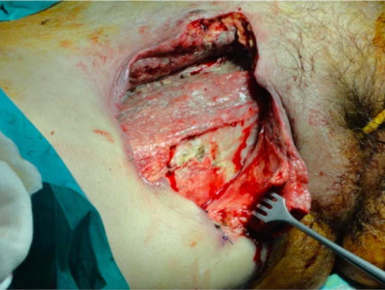
Surgical excision of necrotic tissues in necrotizing fasciitis
Image: “Management of necrotizing fasciitis” by 3rd Department of Surgery, Attikon University Hospital, University of Athens School of Medicine, Athens, Greece. License: CC BY 4.0
Antibiotic therapy
In the US, an initial therapy regimen should include each of the following categories:[4,6–9,14]
CarbapenemsCarbapenemsA group of beta-lactam antibiotics in which the sulfur atom in the thiazolidine ring of the penicillin molecule is replaced by a carbon atom. Thienamycins are a subgroup of carbapenems which have a sulfur atom as the first constituent of the side chain.Carbapenems and Aztreonam
PiperacillinPiperacillinSemisynthetic, broad-spectrum, ampicillin derived ureidopenicillin antibiotic proposed for pseudomonas infections. It is also used in combination with other antibiotics.Penicillins–tazobactamTazobactamA penicillanic acid and sulfone derivative and potent beta-lactamase inhibitor that enhances the activity of other anti-bacterial agents against beta-lactamase producing bacteria.Cephalosporins
CeftriaxoneCeftriaxoneA broad-spectrum cephalosporin antibiotic and cefotaxime derivative with a very long half-life and high penetrability to meninges, eyes and inner ears.Cephalosporins (or cefotaximeCefotaximeSemisynthetic broad-spectrum cephalosporin.Cephalosporins) plus metronidazoleMetronidazoleA nitroimidazole used to treat amebiasis; vaginitis; trichomonas infections; giardiasis; anaerobic bacteria; and treponemal infections.Pyogenic Liver Abscess
MRSAMRSAA strain of Staphylococcus aureus that is non-susceptible to the action of methicillin. The mechanism of resistance usually involves modification of normal or the presence of acquired penicillin binding proteins.Staphylococcus coverage (1 of the following):
VancomycinVancomycinAntibacterial obtained from streptomyces orientalis. It is a glycopeptide related to ristocetin that inhibits bacterial cell wall assembly and is toxic to kidneys and the inner ear.Glycopeptides
LinezolidLinezolidAn oxazolidinone and acetamide derived anti-bacterial agent and protein synthesis inhibitor that is used in the treatment of gram-positive bacterial infections of the skin and respiratory tract.Oxazolidinones (also has antitoxin effect)
For antitoxin effects against toxin-producing streptococci or staphylococci: clindamycinClindamycinAn antibacterial agent that is a semisynthetic analog of lincomycin.Lincosamides (if linezolidLinezolidAn oxazolidinone and acetamide derived anti-bacterial agent and protein synthesis inhibitor that is used in the treatment of gram-positive bacterial infections of the skin and respiratory tract.Oxazolidinones is given, clindamycinClindamycinAn antibacterial agent that is a semisynthetic analog of lincomycin.Lincosamides would not be needed)
In the UK:[11,12]
Regimens may vary greatly depending on the National Health Service (NHS) trustTrustConfidence in or reliance on a person or thing.Conflict of Interest hospital (see local guidelines).
An example of an initial therapy regimen for broad coverage may include:
Flucloxacillin
Benzylpenicillin
MetronidazoleMetronidazoleA nitroimidazole used to treat amebiasis; vaginitis; trichomonas infections; giardiasis; anaerobic bacteria; and treponemal infections.Pyogenic Liver Abscess
ClindamycinClindamycinAn antibacterial agent that is a semisynthetic analog of lincomycin.Lincosamides
If MRSAMRSAA strain of Staphylococcus aureus that is non-susceptible to the action of methicillin. The mechanism of resistance usually involves modification of normal or the presence of acquired penicillin binding proteins.Staphylococcus suspected (or beta-lactamBeta-LactamPenicillinsallergyAllergyAn abnormal adaptive immune response that may or may not involve antigen-specific IgEType I Hypersensitivity Reaction) → replace flucloxacillin and benzylpenicillin with vancomycinVancomycinAntibacterial obtained from streptomyces orientalis. It is a glycopeptide related to ristocetin that inhibits bacterial cell wall assembly and is toxic to kidneys and the inner ear.Glycopeptides or linezolidLinezolidAn oxazolidinone and acetamide derived anti-bacterial agent and protein synthesis inhibitor that is used in the treatment of gram-positive bacterial infections of the skin and respiratory tract.Oxazolidinones
Other example regimens by type:[13]
Type I: piperacillinPiperacillinSemisynthetic, broad-spectrum, ampicillin derived ureidopenicillin antibiotic proposed for pseudomonas infections. It is also used in combination with other antibiotics.Penicillins–tazobactamTazobactamA penicillanic acid and sulfone derivative and potent beta-lactamase inhibitor that enhances the activity of other anti-bacterial agents against beta-lactamase producing bacteria.Cephalosporins (or meropenemMeropenemA thienamycin derivative antibacterial agent that is more stable to renal dehydropeptidase I than imipenem, but does not need to be given with an enzyme inhibitor such as cilastatin. It is used in the treatment of bacterial infections, including infections in immunocompromised patients.Carbapenems and Aztreonam) + clindamycinClindamycinAn antibacterial agent that is a semisynthetic analog of lincomycin.Lincosamides
Type II: benzylpenicillin (or ceftriaxoneCeftriaxoneA broad-spectrum cephalosporin antibiotic and cefotaxime derivative with a very long half-life and high penetrability to meninges, eyes and inner ears.Cephalosporins or ciprofloxacinCiprofloxacinA broad-spectrum antimicrobial carboxyfluoroquinoline.Fluoroquinolones) + clindamycinClindamycinAn antibacterial agent that is a semisynthetic analog of lincomycin.Lincosamides
Therapy should eventually be tailored to culture data and local resistanceResistancePhysiologically, the opposition to flow of air caused by the forces of friction. As a part of pulmonary function testing, it is the ratio of driving pressure to the rate of air flow.Ventilation: Mechanics of Breathing patterns (when available) → examples:[6,7,9]
StreptococcusStreptococcusStreptococcus is one of the two medically important genera of gram-positive cocci, the other being Staphylococcus. Streptococci are identified as different species on blood agar on the basis of their hemolytic pattern and sensitivity to optochin and bacitracin. There are many pathogenic species of streptococci, including S. pyogenes, S. agalactiae, S. pneumoniae, and the viridans streptococci.Streptococcus: penicillinPenicillinRheumatic Fever + clindamycinClindamycinAn antibacterial agent that is a semisynthetic analog of lincomycin.Lincosamides
MSSA: penicillinsPenicillinsBeta-lactam antibiotics contain a beta-lactam ring as a part of their chemical structure. Drugs in this class include penicillin G and V, penicillinase-sensitive and penicillinase-resistant penicillins, cephalosporins, carbapenems, and aztreonam. Penicillins or cephalosporinsCephalosporinsCephalosporins are a group of bactericidal beta-lactam antibiotics (similar to penicillins) that exert their effects by preventing bacteria from producing their cell walls, ultimately leading to cell death. Cephalosporins are categorized by generation and all drug names begin with “cef-” or “ceph-.” Cephalosporins
MRSAMRSAA strain of Staphylococcus aureus that is non-susceptible to the action of methicillin. The mechanism of resistance usually involves modification of normal or the presence of acquired penicillin binding proteins.Staphylococcus: vancomycinVancomycinAntibacterial obtained from streptomyces orientalis. It is a glycopeptide related to ristocetin that inhibits bacterial cell wall assembly and is toxic to kidneys and the inner ear.Glycopeptides
Vibrio vulnificusVibrio vulnificusA species of halophilic bacteria in the genus vibrio, which lives in warm seawater. It can cause infections in those who eat raw contaminated seafood or have open wounds exposed to seawater.Vibrio→ treat with both:
TetracyclinesTetracyclinesTetracyclines are a class of broad-spectrum antibiotics indicated for a wide variety of bacterial infections. These medications bind the 30S ribosomal subunit to inhibit protein synthesis of bacteria. Tetracyclines cover gram-positive and gram-negative organisms, as well as atypical bacteria such as chlamydia, mycoplasma, spirochetes, and even protozoa. Tetracyclines (e.g., doxycycline, minocyclineMinocyclineA tetracycline analog, having a 7-dimethylamino and lacking the 5 methyl and hydroxyl groups, which is effective against tetracycline-resistant staphylococcus infections.Tetracyclines)
Duration recommendations are variableVariableVariables represent information about something that can change. The design of the measurement scales, or of the methods for obtaining information, will determine the data gathered and the characteristics of that data. As a result, a variable can be qualitative or quantitative, and may be further classified into subgroups.Types of Variables.
Consider continuing antibiotics until:
No further surgical debridementDebridementThe removal of foreign material and devitalized or contaminated tissue from or adjacent to a traumatic or infected lesion until surrounding healthy tissue is exposed.Stevens-Johnson Syndrome is needed
There is clinical improvement
There no feverFeverFever is defined as a measured body temperature of at least 38°C (100.4°F). Fever is caused by circulating endogenous and/or exogenous pyrogens that increase levels of prostaglandin E2 in the hypothalamus. Fever is commonly associated with chills, rigors, sweating, and flushing of the skin. Fever for 48–72 hours
Table: Initial antibiotic options for necrotizing fasciitisNecrotizing fasciitisNecrotizing fasciitis is a life-threatening infection that causes rapid destruction and necrosis of the fascia and subcutaneous tissues. Patients may present with significant pain out of proportion to the presenting symptoms and rapidly progressive erythema of the affected area. Necrotizing Fasciitis
Use
Medication
Typical dosing (adults)
US[7,8]
UK[11‒14]
General coverage options
ErtapenemErtapenemA carbapenem derivative antibacterial agent that is more stable to renal dehydropeptidase I than imipenem, but does not need to be given with an enzyme inhibitor such as cilastatin. It is used in the treatment of gram-positive and gram-negative bacterial infections including intra-abdominal infections, acute gynecological infections, complicated urinary tract infections, skin infections, and respiratory tract infections. It is also used to prevent infection in colorectal surgery.Carbapenems and Aztreonam
1 g daily
Not listed in guidelines
ImipenemImipenemSemisynthetic thienamycin that has a wide spectrum of antibacterial activity against gram-negative and gram-positive aerobic and anaerobic bacteria, including many multiresistant strains. It is stable to beta-lactamases. Clinical studies have demonstrated high efficacy in the treatment of infections of various body systems. Its effectiveness is enhanced when it is administered in combination with cilastatin, a renal dipeptidase inhibitor.Carbapenems and Aztreonam
1 g every 6–8 hours
MeropenemMeropenemA thienamycin derivative antibacterial agent that is more stable to renal dehydropeptidase I than imipenem, but does not need to be given with an enzyme inhibitor such as cilastatin. It is used in the treatment of bacterial infections, including infections in immunocompromised patients.Carbapenems and Aztreonam
1 g every 8 hours
1 g every 8 hours
PiperacillinPiperacillinSemisynthetic, broad-spectrum, ampicillin derived ureidopenicillin antibiotic proposed for pseudomonas infections. It is also used in combination with other antibiotics.Penicillins–tazobactamTazobactamA penicillanic acid and sulfone derivative and potent beta-lactamase inhibitor that enhances the activity of other anti-bacterial agents against beta-lactamase producing bacteria.Cephalosporins
3.375 g every 6 hours
4.5 g every 8 hours
CeftriaxoneCeftriaxoneA broad-spectrum cephalosporin antibiotic and cefotaxime derivative with a very long half-life and high penetrability to meninges, eyes and inner ears.Cephalosporins
MetronidazoleMetronidazoleA nitroimidazole used to treat amebiasis; vaginitis; trichomonas infections; giardiasis; anaerobic bacteria; and treponemal infections.Pyogenic Liver Abscess
500 mg every 6 hours
500 mg every 8 hours
MRSAMRSAA strain of Staphylococcus aureus that is non-susceptible to the action of methicillin. The mechanism of resistance usually involves modification of normal or the presence of acquired penicillin binding proteins.Staphylococcus coverage options
VancomycinVancomycinAntibacterial obtained from streptomyces orientalis. It is a glycopeptide related to ristocetin that inhibits bacterial cell wall assembly and is toxic to kidneys and the inner ear.Glycopeptides
LinezolidLinezolidAn oxazolidinone and acetamide derived anti-bacterial agent and protein synthesis inhibitor that is used in the treatment of gram-positive bacterial infections of the skin and respiratory tract.Oxazolidinones*
ClindamycinClindamycinAn antibacterial agent that is a semisynthetic analog of lincomycin.Lincosamides
600–900 mg every 6 hours
1.2 g every 6 hours
*Linezolid also has an antitoxin effect and is preferred over vancomycin in some guidelines.
Postoperative carePostoperative careAfter any procedure performed in the operating room, all patients must undergo close observation at least in the recovery room. After larger procedures and for patients who require hospitalization, observation must continue on the surgical ward. The primary intent of this practice is the early detection of postoperative complications. Postoperative Care[6–9]
The majority of patientsPatientsIndividuals participating in the health care system for the purpose of receiving therapeutic, diagnostic, or preventive procedures.Clinician–Patient Relationship will require monitoring in an intensive care unit for close monitoring and management.
Hemodynamic support:
Aggressive resuscitationResuscitationThe restoration to life or consciousness of one apparently dead. .Neonatal Respiratory Distress Syndrome with intravenous crystalloidCrystalloidIsotonic solutions of mineral salts, such as ringer’s lactate and sodium chloride (saline solution), used in fluid therapy to rehydrate blood volume.Intravenous Fluids fluids
VasopressorVasopressorAcute Cholangitis support for septic shockSeptic shockSepsis associated with hypotension or hypoperfusion despite adequate fluid resuscitation. Perfusion abnormalities may include, but are not limited to lactic acidosis; oliguria; or acute alteration in mental status.Sepsis and Septic Shock with MAP target ≥65 mm Hg (norepinephrineNorepinephrinePrecursor of epinephrine that is secreted by the adrenal medulla and is a widespread central and autonomic neurotransmitter. Norepinephrine is the principal transmitter of most postganglionic sympathetic fibers, and of the diffuse projection system in the brain that arises from the locus ceruleus.Receptors and Neurotransmitters of the CNS is first line)
Monitor hemoglobin and transfuse for blood loss associated with repeated debridements.
Leave wounds open after debridementDebridementThe removal of foreign material and devitalized or contaminated tissue from or adjacent to a traumatic or infected lesion until surrounding healthy tissue is exposed.Stevens-Johnson Syndrome.
Once sthe wound is stable (not requiring repeated debridements), may use negative-pressure wound therapy (NPWT/wound vac).
Use wound vac cautiously in anaerobic infection.
Controversial therapies[6–9]
The following may be considered due to a potential benefit, but there are not much clinical data:
Hyperbaric oxygenHyperbaric oxygenThe therapeutic intermittent administration of oxygen in a chamber at greater than sea-level atmospheric pressures (three atmospheres). It is considered effective treatment for air and gas embolisms, smoke inhalation, acute carbon monoxide poisoning, caisson disease, clostridial gangrene, etc. The list of treatment modalities includes stroke.Decompression Sickness therapy (used as an adjuvantAdjuvantSubstances that augment, stimulate, activate, potentiate, or modulate the immune response at either the cellular or humoral level. The classical agents (freund’s adjuvant, bcg, corynebacterium parvum, et al.) contain bacterial antigens. Some are endogenous (e.g., histamine, interferon, transfer factor, tuftsin, interleukin-1). Their mode of action is either non-specific, resulting in increased immune responsiveness to a wide variety of antigens, or antigen-specific, i.e., affecting a restricted type of immune response to a narrow group of antigens. The therapeutic efficacy of many biological response modifiers is related to their antigen-specific immunoadjuvanticity.Vaccination for source controlSource ControlSurgical Infections): not consistently recommended
Septic shockSeptic shockSepsis associated with hypotension or hypoperfusion despite adequate fluid resuscitation. Perfusion abnormalities may include, but are not limited to lactic acidosis; oliguria; or acute alteration in mental status.Sepsis and Septic Shock
Toxic shock syndromeToxic Shock SyndromeToxic shock syndrome (TSS) is an acute, multi-systemic disease caused by the toxin-producing bacteria, Staphylococcus aureus and Streptococcus pyogenes. Staphylococcal TSS is more common and associated with tampons and nasal packing. Toxic Shock Syndrome: NFNFNecrotizing fasciitis is a life-threatening infection that causes rapid destruction and necrosis of the fascia and subcutaneous tissues. Patients may present with significant pain out of proportion to the presenting symptoms and rapidly progressive erythema of the affected area.Necrotizing Fasciitis caused by GAS strains with M proteinsProteinsLinear polypeptides that are synthesized on ribosomes and may be further modified, crosslinked, cleaved, or assembled into complex proteins with several subunits. The specific sequence of amino acids determines the shape the polypeptide will take, during protein folding, and the function of the protein.Energy Homeostasis types 1 and 3 is associated with streptococcal toxic shock syndromeStreptococcal Toxic Shock SyndromeToxic Shock Syndrome in ~ 50% of cases,
CellulitisCellulitisCellulitis is a common infection caused by bacteria that affects the dermis and subcutaneous tissue of the skin. It is frequently caused by Staphylococcus aureus and Streptococcus pyogenes. The skin infection presents as an erythematous and edematous area with warmth and tenderness. Cellulitis: a common bacterial skinSkinThe skin, also referred to as the integumentary system, is the largest organ of the body. The skin is primarily composed of the epidermis (outer layer) and dermis (deep layer). The epidermis is primarily composed of keratinocytes that undergo rapid turnover, while the dermis contains dense layers of connective tissue.Skin: Structure and Functions infection that affects the deeper layers of the dermisDermisA layer of vascularized connective tissue underneath the epidermis. The surface of the dermis contains innervated papillae. Embedded in or beneath the dermis are sweat glands; hair follicles; and sebaceous glands.Skin: Structure and Functions and subcutaneous tissueSubcutaneous tissueLoose connective tissue lying under the dermis, which binds skin loosely to subjacent tissues. It may contain a pad of adipocytes, which vary in number according to the area of the body and vary in size according to the nutritional state.Soft Tissue Abscess. This condition is most commonly caused by S. aureusS. aureusPotentially pathogenic bacteria found in nasal membranes, skin, hair follicles, and perineum of warm-blooded animals. They may cause a wide range of infections and intoxications.Staphylococcus and S. pyogenes. CellulitisCellulitisCellulitis is a common infection caused by bacteria that affects the dermis and subcutaneous tissue of the skin. It is frequently caused by Staphylococcus aureus and Streptococcus pyogenes. The skin infection presents as an erythematous and edematous area with warmth and tenderness. Cellulitis presents as an erythematous, edematous area that is warm and tender to the touch. CrepitusCrepitusOsteoarthritis, necrosisNecrosisThe death of cells in an organ or tissue due to disease, injury or failure of the blood supply.Ischemic Cell Damage, and systemic toxicityToxicityDosage Calculation are not usually present. Diagnosis is clinical, and management involves antibiotics tailored to the suspected organism.
Pyoderma gangrenosumPyoderma gangrenosumAn idiopathic, rapidly evolving, and severely debilitating disease occurring most commonly in association with chronic ulcerative colitis. It is characterized by the presence of boggy, purplish ulcers with undermined borders, appearing mostly on the legs. The majority of cases are in people between 40 and 60 years old. Its etiology is unknown.Crohn Disease:neutrophilic dermatosisNeutrophilic DermatosisRheumatoid Arthritis that presents with inflammationInflammationInflammation is a complex set of responses to infection and injury involving leukocytes as the principal cellular mediators in the body’s defense against pathogenic organisms. Inflammation is also seen as a response to tissue injury in the process of wound healing. The 5 cardinal signs of inflammation are pain, heat, redness, swelling, and loss of function. Inflammation and ulcerationUlcerationCorneal Abrasions, Erosion, and Ulcers of the skinSkinThe skin, also referred to as the integumentary system, is the largest organ of the body. The skin is primarily composed of the epidermis (outer layer) and dermis (deep layer). The epidermis is primarily composed of keratinocytes that undergo rapid turnover, while the dermis contains dense layers of connective tissue.Skin: Structure and Functions. PatientsPatientsIndividuals participating in the health care system for the purpose of receiving therapeutic, diagnostic, or preventive procedures.Clinician–Patient Relationship present with a painful, inflamed papulePapuleElevated lesion < 1 cm in diameterGeneralized and Localized Rashes that progresses to an ulcer with irregular borders and purulent drainage. Unlike necrotizing fasciitisNecrotizing fasciitisNecrotizing fasciitis is a life-threatening infection that causes rapid destruction and necrosis of the fascia and subcutaneous tissues. Patients may present with significant pain out of proportion to the presenting symptoms and rapidly progressive erythema of the affected area. Necrotizing Fasciitis, there is rarely systemic toxicityToxicityDosage Calculation, the involvement of the fascial planes, or improvement with antibiotics. This condition will worsen with debridementDebridementThe removal of foreign material and devitalized or contaminated tissue from or adjacent to a traumatic or infected lesion until surrounding healthy tissue is exposed.Stevens-Johnson Syndrome. Pyoderma gangrenosumPyoderma gangrenosumAn idiopathic, rapidly evolving, and severely debilitating disease occurring most commonly in association with chronic ulcerative colitis. It is characterized by the presence of boggy, purplish ulcers with undermined borders, appearing mostly on the legs. The majority of cases are in people between 40 and 60 years old. Its etiology is unknown.Crohn Disease is based on the clinical assessment and histologic findings. This is managed with immunosuppressive therapy.
Pyomyositis: infection of the skeletal muscle that results from hematogenousHematogenousHepatocellular Carcinoma (HCC) and Liver Metastases spread from a primary infected source. PatientsPatientsIndividuals participating in the health care system for the purpose of receiving therapeutic, diagnostic, or preventive procedures.Clinician–Patient Relationship present with localized painPainAn unpleasant sensation induced by noxious stimuli which are detected by nerve endings of nociceptive neurons.Pain: Types and Pathways, fevers, and a variableVariableVariables represent information about something that can change. The design of the measurement scales, or of the methods for obtaining information, will determine the data gathered and the characteristics of that data. As a result, a variable can be qualitative or quantitative, and may be further classified into subgroups.Types of Variables degree of skinSkinThe skin, also referred to as the integumentary system, is the largest organ of the body. The skin is primarily composed of the epidermis (outer layer) and dermis (deep layer). The epidermis is primarily composed of keratinocytes that undergo rapid turnover, while the dermis contains dense layers of connective tissue.Skin: Structure and Functions findings from minimal erythemaErythemaRedness of the skin produced by congestion of the capillaries. This condition may result from a variety of disease processes.Chalazion to severe tenderness, indurationIndurationDermatologic Examination, and edemaEdemaEdema is a condition in which excess serous fluid accumulates in the body cavity or interstitial space of connective tissues. Edema is a symptom observed in several medical conditions. It can be categorized into 2 types, namely, peripheral (in the extremities) and internal (in an organ or body cavity). Edema. The diagnosis is made with imaging and culture data. Management typically consists of percutaneous drainagePercutaneous DrainageEchinococcus/Echinococcosis or, in severe cases, open surgical drainage with antibiotic therapy.
Gas gangreneGangreneDeath and putrefaction of tissue usually due to a loss of blood supply.Small Bowel Obstruction (clostridial myonecrosisClostridial myonecrosisGas gangrene, also known as clostridial myonecrosis, is a life-threatening muscle and soft tissue infection that usually develops after traumatic inoculation with clostridium perfringens (C. perfringens), but can also develop spontaneously in association with other Clostridium species.Gas Gangrene): traumatic or spontaneous (C. perfringensC. perfringensThe most common etiologic agent of gas gangrene. It is differentiable into several distinct types based on the distribution of twelve different toxins.Gas Gangrene, C. septicumC. septicumA species of gram-positive bacteria in the family clostridiaceae. Infections have a strong association with malignancies and also with gas gangrene.Gas Gangrene), with prominent tissue gas on imaging and crepitusCrepitusOsteoarthritis, difficult to distinguish from type I NFNFNecrotizing fasciitis is a life-threatening infection that causes rapid destruction and necrosis of the fascia and subcutaneous tissues. Patients may present with significant pain out of proportion to the presenting symptoms and rapidly progressive erythema of the affected area.Necrotizing Fasciitis. Infection of the muscle presenting with increasing painPainAn unpleasant sensation induced by noxious stimuli which are detected by nerve endings of nociceptive neurons.Pain: Types and Pathways within 24 hours of injury. Imaging shows gas in tissue (also seen in type I NSTI); tissue Gram stainGram stainKlebsiella shows large gram-positiveGram-PositivePenicillins/gram-variable rods, and surgery reveals non-contracting, non-bleeding muscle. Management includes urgent debridementDebridementThe removal of foreign material and devitalized or contaminated tissue from or adjacent to a traumatic or infected lesion until surrounding healthy tissue is exposed.Stevens-Johnson Syndrome and penicillinPenicillinRheumatic Fever plus clindamycinClindamycinAn antibacterial agent that is a semisynthetic analog of lincomycin.Lincosamides for 10–14 days.
Acute compartment syndromeCompartment SyndromeCompartment syndrome is a surgical emergency usually occurring secondary to trauma. The condition is marked by increased pressure within a compartment that compromises the circulation and function of the tissues within that space.Compartment Syndrome: increased pressure in the fascial compartment (form trauma, rebleeding, etcETCThe electron transport chain (ETC) sends electrons through a series of proteins, which generate an electrochemical proton gradient that produces energy in the form of adenosine triphosphate (ATP).Electron Transport Chain (ETC)). Condition can coexist with or mimic deep NSTI (painPainAn unpleasant sensation induced by noxious stimuli which are detected by nerve endings of nociceptive neurons.Pain: Types and Pathways out of proportion, with tense/swollen extremity, painPainAn unpleasant sensation induced by noxious stimuli which are detected by nerve endings of nociceptive neurons.Pain: Types and Pathways on passive stretch); requires compartment pressure measurement for diagnosis and treatment is emergent fasciotomyFasciotomySurgical incision on the fascia. It is used to decompress compartment pressure (e.g. in compartment syndromes; circumferential burns and extremity injuries) or to release contractures (e.g. in dupuytren’s contracture).Compartment Syndrome.
Billing and Coding
Diagnosis Codes:
This code is used to diagnose necrotizing fasciitis, a rare but life-threatening, rapidly progressing bacterial infection of the soft tissue and fascia (“flesh-eating disease”).
Coding System
Code
Description
ICD-10-CM
M72.6
Necrotizing fasciitisNecrotizing fasciitisNecrotizing fasciitis is a life-threatening infection that causes rapid destruction and necrosis of the fascia and subcutaneous tissues. Patients may present with significant pain out of proportion to the presenting symptoms and rapidly progressive erythema of the affected area. Necrotizing Fasciitis
SNOMED CT
110360009
Necrotizing fasciitisNecrotizing fasciitisNecrotizing fasciitis is a life-threatening infection that causes rapid destruction and necrosis of the fascia and subcutaneous tissues. Patients may present with significant pain out of proportion to the presenting symptoms and rapidly progressive erythema of the affected area. Necrotizing Fasciitis (disorder)
Procedures/Interventions:
This CPT code is for emergent, aggressive surgical debridementDebridementThe removal of foreign material and devitalized or contaminated tissue from or adjacent to a traumatic or infected lesion until surrounding healthy tissue is exposed.Stevens-Johnson Syndrome, which is the most critical component of treatment. All necrotic tissue must be removed to control the infection, often requiring multiple operations.
Coding System
Code
Description
CPT
11043
DebridementDebridementThe removal of foreign material and devitalized or contaminated tissue from or adjacent to a traumatic or infected lesion until surrounding healthy tissue is exposed.Stevens-Johnson Syndrome, muscle and/or fasciaFasciaLayers of connective tissue of variable thickness. The superficial fascia is found immediately below the skin; the deep fascia invests muscles, nerves, and other organs.Cellulitis (includes epidermisEpidermisThe external, nonvascular layer of the skin. It is made up, from within outward, of five layers of epithelium: (1) basal layer (stratum basale epidermidis); (2) spinous layer (stratum spinosum epidermidis); (3) granular layer (stratum granulosum epidermidis); (4) clear layer (stratum lucidum epidermidis); and (5) horny layer (stratum corneum epidermidis).Skin: Structure and Functions, dermisDermisA layer of vascularized connective tissue underneath the epidermis. The surface of the dermis contains innervated papillae. Embedded in or beneath the dermis are sweat glands; hair follicles; and sebaceous glands.Skin: Structure and Functions, and subcutaneous tissueSubcutaneous tissueLoose connective tissue lying under the dermis, which binds skin loosely to subjacent tissues. It may contain a pad of adipocytes, which vary in number according to the area of the body and vary in size according to the nutritional state.Soft Tissue Abscess, if performed); first 20 sq cm or less
Medications:
These codes are for the empiric, broad-spectrumBroad-SpectrumFluoroquinolones intravenous antibiotics (e.g., piperacillinPiperacillinSemisynthetic, broad-spectrum, ampicillin derived ureidopenicillin antibiotic proposed for pseudomonas infections. It is also used in combination with other antibiotics.Penicillins/tazobactamTazobactamA penicillanic acid and sulfone derivative and potent beta-lactamase inhibitor that enhances the activity of other anti-bacterial agents against beta-lactamase producing bacteria.Cephalosporins, vancomycinVancomycinAntibacterial obtained from streptomyces orientalis. It is a glycopeptide related to ristocetin that inhibits bacterial cell wall assembly and is toxic to kidneys and the inner ear.Glycopeptides, clindamycinClindamycinAn antibacterial agent that is a semisynthetic analog of lincomycin.Lincosamides) that are started immediately to cover the polymicrobial nature of the infection.
Coding System
Code
Description
RxNorm
1148971
PiperacillinPiperacillinSemisynthetic, broad-spectrum, ampicillin derived ureidopenicillin antibiotic proposed for pseudomonas infections. It is also used in combination with other antibiotics.Penicillins / TazobactamTazobactamA penicillanic acid and sulfone derivative and potent beta-lactamase inhibitor that enhances the activity of other anti-bacterial agents against beta-lactamase producing bacteria.Cephalosporins (ingredient)
RxNorm
11124
VancomycinVancomycinAntibacterial obtained from streptomyces orientalis. It is a glycopeptide related to ristocetin that inhibits bacterial cell wall assembly and is toxic to kidneys and the inner ear.Glycopeptides (ingredient)
RxNorm
2592
ClindamycinClindamycinAn antibacterial agent that is a semisynthetic analog of lincomycin.Lincosamides (ingredient)
Complications:
This code is for septic shockSeptic shockSepsis associated with hypotension or hypoperfusion despite adequate fluid resuscitation. Perfusion abnormalities may include, but are not limited to lactic acidosis; oliguria; or acute alteration in mental status.Sepsis and Septic Shock, a common and often fatal complication of necrotizing fasciitisNecrotizing fasciitisNecrotizing fasciitis is a life-threatening infection that causes rapid destruction and necrosis of the fascia and subcutaneous tissues. Patients may present with significant pain out of proportion to the presenting symptoms and rapidly progressive erythema of the affected area. Necrotizing Fasciitis, resulting from the overwhelming systemic inflammatory response to the infection.
Coding System
Code
Description
ICD-10-CM
R65.21
Severe sepsisSevere SepsisSepsis in Children with septic shockSeptic shockSepsis associated with hypotension or hypoperfusion despite adequate fluid resuscitation. Perfusion abnormalities may include, but are not limited to lactic acidosis; oliguria; or acute alteration in mental status.Sepsis and Septic Shock
Hua, C., Urbina, T., Bosc, R., Parks, T., Sriskandan, S., de Prost, N., & Chosidow, O. (2023). Necrotising soft-tissue infections. The Lancet. Infectious diseases, 23(3), e81–e94. https://doi.org/10.1016/S1473-3099(22)00583-7
Wong, C. H., Khin, L. W., Heng, K. S., Tan, K. C., Low, C. O. (2004) The LRINEC (Laboratory Risk Indicator for Necrotizing Fasciitis) score: a tool for distinguishing necrotizing fasciitis from other soft tissue infections. Critical Care Medicine, 32, 1535–1541. https://pubmed.ncbi.nlm.nih.gov/15241098/
Misiakos E. P., Bagias G., Patapis P., Sotiropoulos D., Kanavidis P., Machairas A. (2014). Current concepts in the management of necrotizing fasciitis. Frontiers in Surgery, 1, 36. https://www.ncbi.nlm.nih.gov/pmc/articles/PMC4286984/
Executive summary: practice guidelines for the diagnosis and management of skin and soft tissue infections: 2014 update by the Infectious Diseases Society of America. Clinical Infectious Diseases, 59(2), e10–e52. https://academic.oup.com/cid/article/59/2/147/442347?login=false
Sartelli M., Guirao X., Hardcastle T. C., et al. (2018). 2018 WSES/SIS-E consensus conference: recommendations for the management of skin and soft-tissue infections. World Journal of Emergency Surgery, 13, 58. https://wjes.biomedcentral.com/articles/10.1186/s13017-018-0219-9
Duane, T. M., et al. (2021). Surgical Infection Society 2020 updated guidelines on the management of complicated skin and soft tissue infections. Surgical Infections, 22(4), 383–399. https://www.liebertpub.com/doi/full/10.1089/sur.2020.436
Miller, J. M., et al. (2018). A guide to the utilization of the microbiology laboratory for diagnosis of infectious diseases: 2018 update by the Infectious Diseases Society of America and the American Society for Microbiology. Clinical Infectious Diseases, 67(6):e1–e94. https://www.idsociety.org/practice-guideline/laboratory-diagnosis-of-infectious-diseases/
Bonne, S. L., & Kadri, S. S. (2017). Evaluation and Management of Necrotizing Soft Tissue Infections. Infectious disease clinics of North America, 31(3), 497–511. https://doi.org/10.1016/j.idc.2017.05.011
Detanac, D., Mujdragic, M., Detanac, D. A., Zogic, E., Ceranic, L., Alihodzic, K., Mulic, M., & Mujdragic, H. (2021). Necrotizing Soft Tissue Infection: A Single-Center Retrospective Study of Treatment and Outcomes. Cureus, 13(5), e15039. https://doi.org/10.7759/cureus.15039
Create your free account or log in to continue reading!