Gastritis is inflammation of the gastric mucosa and can be classified by time course (acute or chronic), histologic features, and etiology. It is usually due to an infection (usually by Helicobacter pylori) or an immune-mediated process, but it can also be caused by substances that are direct gastric irritants. Chronic, metaplastic atrophic gastritis may be due to autoimmune or environmental causes and is a risk factor for gastric cancer. Individuals with gastritis may be asymptomatic or may have abdominal pain, dyspepsia, and nausea. Treatment of acute and chronic gastritis due to H. pylori infection involves acid suppression and antibiotics. Other causes are managed by avoiding offending agents and replacing associated deficiencies.
GastritisGastritisGastritis refers to inflammation of the gastric mucosa. Gastritis may occur suddenly (acute gastritis) or slowly over time (chronic gastritis). Gastritis may be asymptomatic or with symptoms, including burning abdominal pain (which either worsens or improves with eating), dyspepsia, nausea, and vomiting. Gastritisis theinflammationInflammationInflammation is a complex set of responses to infection and injury involving leukocytes as the principal cellular mediators in the body’s defense against pathogenic organisms. Inflammation is also seen as a response to tissue injury in the process of wound healing. The 5 cardinal signs of inflammation are pain, heat, redness, swelling, and loss of function. Inflammation of gastric mucosaGastric mucosaLining of the stomach, consisting of an inner epithelium, a middle lamina propria, and an outer muscularis mucosae. The surface cells produce mucus that protects the stomach from attack by digestive acid and enzymes. When the epithelium invaginates into the lamina propria at various region of the stomach (cardia; gastric fundus; and pylorus), different tubular gastric glands are formed. These glands consist of cells that secrete mucus, enzymes, hydrochloric acid, or hormones.Stomach: Anatomy associated with mucosal injury.
GastropathygastropathyDamage to the epithelial lining with no or minimal associated inflammation and is technically a separate entity from gastritisGastritisrefers to a disease of the stomachStomachThe stomach is a muscular sac in the upper left portion of the abdomen that plays a critical role in digestion. The stomach develops from the foregut and connects the esophagus with the duodenum. Structurally, the stomach is C-shaped and forms a greater and lesser curvature and is divided grossly into regions: the cardia, fundus, body, and pylorus. Stomach: Anatomy associated with reactive or hyperplasticHyperplasticColon Polyps epithelial changes with only minimal or no inflammationInflammationInflammation is a complex set of responses to infection and injury involving leukocytes as the principal cellular mediators in the body’s defense against pathogenic organisms. Inflammation is also seen as a response to tissue injury in the process of wound healing. The 5 cardinal signs of inflammation are pain, heat, redness, swelling, and loss of function. Inflammation.
Etiology[10]
GastritisGastritisGastritis refers to inflammation of the gastric mucosa. Gastritis may occur suddenly (acute gastritis) or slowly over time (chronic gastritis). Gastritis may be asymptomatic or with symptoms, including burning abdominal pain (which either worsens or improves with eating), dyspepsia, nausea, and vomiting. Gastritis is usually due to an infection or an immune-mediated process, but it can also be caused by substances that are direct gastric irritants. Numerous infectious and noninfectious diseases can cause gastritisGastritisGastritis refers to inflammation of the gastric mucosa. Gastritis may occur suddenly (acute gastritis) or slowly over time (chronic gastritis). Gastritis may be asymptomatic or with symptoms, including burning abdominal pain (which either worsens or improves with eating), dyspepsia, nausea, and vomiting. Gastritis.
Infectious:
Helicobacter pyloriHelicobacter pyloriA spiral bacterium active as a human gastric pathogen. It is a gram-negative, urease-positive, curved or slightly spiral organism initially isolated in 1982 from patients with lesions of gastritis or peptic ulcers in Western Australia. Helicobacter pylori was originally classified in the genus campylobacter, but RNA sequencing, cellular fatty acid profiles, growth patterns, and other taxonomic characteristics indicate that the micro-organism should be included in the genus Helicobacter. It has been officially transferred to Helicobacter gen.Helicobacter:
Most common
Route of transmission is unclear, but person-to-person transmission most likely.
Rare infectious causes:
MycobacteriumMycobacteriumMycobacterium is a genus of the family Mycobacteriaceae in the phylum Actinobacteria. Mycobacteria comprise more than 150 species of facultative intracellular bacilli that are mostly obligate aerobes. Mycobacteria are responsible for multiple human infections including serious diseases, such as tuberculosis (M. tuberculosis), leprosy (M. leprae), and M. avium complex infections.Mycobacterium avium-intracellulare
EnterococcusEnterococcusEnterococcus is a genus of oval-shaped gram-positive cocci that are arranged in pairs or short chains. Distinguishing factors include optochin resistance and the presence of pyrrolidonyl arylamidase (PYR) and Lancefield D antigen. Enterococcus is part of the normal flora of the human GI tract.Enterococcus
Herpes simplexHerpes SimplexA group of acute infections caused by herpes simplex virus type 1 or type 2 that is characterized by the development of one or more small fluid-filled vesicles with a raised erythematous base on the skin or mucous membrane. It occurs as a primary infection or recurs due to a reactivation of a latent infection.Congenital TORCH InfectionsvirusesVirusesMinute infectious agents whose genomes are composed of DNA or RNA, but not both. They are characterized by a lack of independent metabolism and the inability to replicate outside living host cells.Virology
CytomegalovirusCytomegalovirusCMV is a ubiquitous double-stranded DNA virus belonging to the Herpesviridae family. CMV infections can be transmitted in bodily fluids, such as blood, saliva, urine, semen, and breast milk. The initial infection is usually asymptomatic in the immunocompetent host, or it can present with symptoms of mononucleosis. Cytomegalovirus
Parasites
Autoimmune (associated with anti-parietal and anti-intrinsic factor antibodiesAntibodiesImmunoglobulins (Igs), also known as antibodies, are glycoprotein molecules produced by plasma cells that act in immune responses by recognizing and binding particular antigens. The various Ig classes are IgG (the most abundant), IgM, IgE, IgD, and IgA, which differ in their biologic features, structure, target specificity, and distribution.Immunoglobulins: Types and Functions, which may progress to pernicious anemiaPernicious anemiaA megaloblastic anemia occurring in children but more commonly in later life, characterized by histamine-fast achlorhydria, in which the laboratory and clinical manifestations are based on malabsorption of vitamin B12 due to a failure of the gastric mucosa to secrete adequate and potent intrinsic factor.Megaloblastic Anemia)
Chemical/reactive gastritisGastritisGastritis refers to inflammation of the gastric mucosa. Gastritis may occur suddenly (acute gastritis) or slowly over time (chronic gastritis). Gastritis may be asymptomatic or with symptoms, including burning abdominal pain (which either worsens or improves with eating), dyspepsia, nausea, and vomiting. Gastritis(technically gastropathies because they cause mucosal injury without significant inflammationInflammationInflammation is a complex set of responses to infection and injury involving leukocytes as the principal cellular mediators in the body’s defense against pathogenic organisms. Inflammation is also seen as a response to tissue injury in the process of wound healing. The 5 cardinal signs of inflammation are pain, heat, redness, swelling, and loss of function. Inflammation):
Medication-induced gastritisGastritisGastritis refers to inflammation of the gastric mucosa. Gastritis may occur suddenly (acute gastritis) or slowly over time (chronic gastritis). Gastritis may be asymptomatic or with symptoms, including burning abdominal pain (which either worsens or improves with eating), dyspepsia, nausea, and vomiting. Gastritis:
NSAIDsNSAIDSPrimary vs Secondary Headaches and aspirinAspirinThe prototypical analgesic used in the treatment of mild to moderate pain. It has anti-inflammatory and antipyretic properties and acts as an inhibitor of cyclooxygenase which results in the inhibition of the biosynthesis of prostaglandins. Aspirin also inhibits platelet aggregation and is used in the prevention of arterial and venous thrombosis.Nonsteroidal Antiinflammatory Drugs (NSAIDs), even low dose
BisphosphonatesBisphosphonatesBisphosphonates are pyrophosphate analogs most well-known for treating osteoporosis by preventing bone loss. Bisphosphonates end in the suffix “-dronate” or “-dronic acid” (e.g., alendronate, risedronate, pamidronate) and bind to hydroxyapatite crystals in bone, inhibiting osteoclast-induced bone resorption.Bisphosphonates (e.g., alendronateAlendronateA nonhormonal medication for the treatment of postmenopausal osteoporosis in women. This drug builds healthy bone, restoring some of the bone loss as a result of osteoporosis.Bisphosphonates)
Gastric irritants
Alcohol
CaffeineCaffeineA methylxanthine naturally occurring in some beverages and also used as a pharmacological agent. Caffeine’s most notable pharmacological effect is as a central nervous system stimulant, increasing alertness and producing agitation. Several cellular actions of caffeine have been observed, but it is not entirely clear how each contributes to its pharmacological profile. Among the most important are inhibition of cyclic nucleotide phosphodiesterases, antagonism of adenosine receptors, and modulation of intracellular calcium handling.Stimulants
Spicy foods
Tobacco
BileBileAn emulsifying agent produced in the liver and secreted into the duodenum. Its composition includes bile acids and salts; cholesterol; and electrolytes. It aids digestion of fats in the duodenum.Gallbladder and Biliary Tract: Anatomy reflux after gastrectomy (postgastrectomy gastritisGastritisGastritis refers to inflammation of the gastric mucosa. Gastritis may occur suddenly (acute gastritis) or slowly over time (chronic gastritis). Gastritis may be asymptomatic or with symptoms, including burning abdominal pain (which either worsens or improves with eating), dyspepsia, nausea, and vomiting. Gastritis)
Uncommon causes:
Granulomatous diseasesGranulomatous diseasesA defect of leukocyte function in which phagocytic cells ingest but fail to digest bacteria, resulting in recurring bacterial infections with granuloma formation. When chronic granulomatous disease is caused by mutations in the cybb gene, the condition is inherited in an X-linked recessive pattern. When chronic granulomatous disease is caused by cyba, ncf1, ncf2, or ncf4 gene mutations, the condition is inherited in an autosomal recessive pattern.Type IV Hypersensitivity Reaction:
Crohn disease
SarcoidosisSarcoidosisSarcoidosis is a multisystem inflammatory disease that causes noncaseating granulomas. The exact etiology is unknown. Sarcoidosis usually affects the lungs and thoracic lymph nodes, but it can also affect almost every system in the body, including the skin, heart, and eyes, most commonly. Sarcoidosis
RadiationRadiationEmission or propagation of acoustic waves (sound), electromagnetic energy waves (such as light; radio waves; gamma rays; or x-rays), or a stream of subatomic particles (such as electrons; neutrons; protons; or alpha particles).Osteosarcoma
VasculitisVasculitisInflammation of any one of the blood vessels, including the arteries; veins; and rest of the vasculature system in the body.Systemic Lupus Erythematosus
Collagenous gastritisGastritisGastritis refers to inflammation of the gastric mucosa. Gastritis may occur suddenly (acute gastritis) or slowly over time (chronic gastritis). Gastritis may be asymptomatic or with symptoms, including burning abdominal pain (which either worsens or improves with eating), dyspepsia, nausea, and vomiting. Gastritis
Eosinophilic gastritisGastritisGastritis refers to inflammation of the gastric mucosa. Gastritis may occur suddenly (acute gastritis) or slowly over time (chronic gastritis). Gastritis may be asymptomatic or with symptoms, including burning abdominal pain (which either worsens or improves with eating), dyspepsia, nausea, and vomiting. Gastritis
Ischemic gastritisGastritisGastritis refers to inflammation of the gastric mucosa. Gastritis may occur suddenly (acute gastritis) or slowly over time (chronic gastritis). Gastritis may be asymptomatic or with symptoms, including burning abdominal pain (which either worsens or improves with eating), dyspepsia, nausea, and vomiting. Gastritis:
BurnsBurnsA burn is a type of injury to the skin and deeper tissues caused by exposure to heat, electricity, chemicals, friction, or radiation. Burns are classified according to their depth as superficial (1st-degree), partial-thickness (2nd-degree), full-thickness (3rd-degree), and 4th-degree burns. Burns
Trauma
SepsisSepsisSystemic inflammatory response syndrome with a proven or suspected infectious etiology. When sepsis is associated with organ dysfunction distant from the site of infection, it is called severe sepsis. When sepsis is accompanied by hypotension despite adequate fluid infusion, it is called septic shock.Sepsis and Septic Shock
Classification[8,10,12]
There are multiple ways to classify gastritisGastritisGastritis refers to inflammation of the gastric mucosa. Gastritis may occur suddenly (acute gastritis) or slowly over time (chronic gastritis). Gastritis may be asymptomatic or with symptoms, including burning abdominal pain (which either worsens or improves with eating), dyspepsia, nausea, and vomiting. Gastritis:
By etiology/pathogenesis
By time course:
Acute
Chronic (much more common; often subdivided based on etiology):
Autoimmune metaplastic atrophic gastritisAtrophic gastritisGastritis with atrophy of the gastric mucosa, the gastric parietal cells, and the mucosal glands leading to achlorhydria. Atrophic gastritis usually progresses from chronic gastritis.Gastritis (AMAG)
Environmental metaplastic atrophic gastritisAtrophic gastritisGastritis with atrophy of the gastric mucosa, the gastric parietal cells, and the mucosal glands leading to achlorhydria. Atrophic gastritis usually progresses from chronic gastritis.Gastritis (EMAG)
By severity of mucosal injury:
Erosive: visible mucosal damage
Nonerosive: histologic inflammationInflammationInflammation is a complex set of responses to infection and injury involving leukocytes as the principal cellular mediators in the body’s defense against pathogenic organisms. Inflammation is also seen as a response to tissue injury in the process of wound healing. The 5 cardinal signs of inflammation are pain, heat, redness, swelling, and loss of function. Inflammation, with no overt mucosal injury
By histology, based on the inflammatory cell type and duration:
Acute gastritisGastritisGastritis refers to inflammation of the gastric mucosa. Gastritis may occur suddenly (acute gastritis) or slowly over time (chronic gastritis). Gastritis may be asymptomatic or with symptoms, including burning abdominal pain (which either worsens or improves with eating), dyspepsia, nausea, and vomiting. Gastritis: polymorphonuclear leukocyte infiltration of the gastric antrum and body.
Rare to have only neutrophilsNeutrophilsGranular leukocytes having a nucleus with three to five lobes connected by slender threads of chromatin, and cytoplasm containing fine inconspicuous granules and stainable by neutral dyes.Innate Immunity: Phagocytes and Antigen Presentation present
NeutrophilsNeutrophilsGranular leukocytes having a nucleus with three to five lobes connected by slender threads of chromatin, and cytoplasm containing fine inconspicuous granules and stainable by neutral dyes.Innate Immunity: Phagocytes and Antigen Presentation are much more commonly seen in “chronic active gastritisGastritisGastritis refers to inflammation of the gastric mucosa. Gastritis may occur suddenly (acute gastritis) or slowly over time (chronic gastritis). Gastritis may be asymptomatic or with symptoms, including burning abdominal pain (which either worsens or improves with eating), dyspepsia, nausea, and vomiting. Gastritis,” where both neutrophilsNeutrophilsGranular leukocytes having a nucleus with three to five lobes connected by slender threads of chromatin, and cytoplasm containing fine inconspicuous granules and stainable by neutral dyes.Innate Immunity: Phagocytes and Antigen Presentation are present along with chronic inflammatory cells.
Chronic gastritisGastritisGastritis refers to inflammation of the gastric mucosa. Gastritis may occur suddenly (acute gastritis) or slowly over time (chronic gastritis). Gastritis may be asymptomatic or with symptoms, including burning abdominal pain (which either worsens or improves with eating), dyspepsia, nausea, and vomiting. Gastritis (much more common than pure acute gastritisGastritisGastritis refers to inflammation of the gastric mucosa. Gastritis may occur suddenly (acute gastritis) or slowly over time (chronic gastritis). Gastritis may be asymptomatic or with symptoms, including burning abdominal pain (which either worsens or improves with eating), dyspepsia, nausea, and vomiting. Gastritis) → 3 types:
Diffuse antral gastritisGastritisGastritis refers to inflammation of the gastric mucosa. Gastritis may occur suddenly (acute gastritis) or slowly over time (chronic gastritis). Gastritis may be asymptomatic or with symptoms, including burning abdominal pain (which either worsens or improves with eating), dyspepsia, nausea, and vomiting. Gastritis: usually due to H. pyloriH. pyloriA spiral bacterium active as a human gastric pathogen. It is a gram-negative, urease-positive, curved or slightly spiral organism initially isolated in 1982 from patients with lesions of gastritis or peptic ulcers in Western Australia. Helicobacter pylori was originally classified in the genus campylobacter, but RNA sequencing, cellular fatty acid profiles, growth patterns, and other taxonomic characteristics indicate that the micro-organism should be included in the genus Helicobacter. It has been officially transferred to Helicobacter gen.Helicobacter infection, showing chronic inflammatory cells in the lamina propriaLamina propriaWhipple’s Disease and neutrophilsNeutrophilsGranular leukocytes having a nucleus with three to five lobes connected by slender threads of chromatin, and cytoplasm containing fine inconspicuous granules and stainable by neutral dyes.Innate Immunity: Phagocytes and Antigen Presentation infiltrating the gastric pit epitheliumEpitheliumThe epithelium is a complex of specialized cellular organizations arranged into sheets and lining cavities and covering the surfaces of the body. The cells exhibit polarity, having an apical and a basal pole. Structures important for the epithelial integrity and function involve the basement membrane, the semipermeable sheet on which the cells rest, and interdigitations, as well as cellular junctions. Surface Epithelium: Histology.
Environmental metaplastic atrophic gastritisAtrophic gastritisGastritis with atrophy of the gastric mucosa, the gastric parietal cells, and the mucosal glands leading to achlorhydria. Atrophic gastritis usually progresses from chronic gastritis.Gastritis (EMAG, also called “multifocalMultifocalRetinoblastomaatrophic gastritisAtrophic gastritisGastritis with atrophy of the gastric mucosa, the gastric parietal cells, and the mucosal glands leading to achlorhydria. Atrophic gastritis usually progresses from chronic gastritis.Gastritis”), typified by involvement of both the gastric antrum and corpus with glandular atrophyAtrophyDecrease in the size of a cell, tissue, organ, or multiple organs, associated with a variety of pathological conditions such as abnormal cellular changes, ischemia, malnutrition, or hormonal changes.Cellular Adaptation and intestinal metaplasiaMetaplasiaA condition in which there is a change of one adult cell type to another similar adult cell type.Cellular Adaptation (IM)
Autoimmune metaplastic atrophic gastritisAtrophic gastritisGastritis with atrophy of the gastric mucosa, the gastric parietal cells, and the mucosal glands leading to achlorhydria. Atrophic gastritis usually progresses from chronic gastritis.Gastritis (AMAG, “corpus-predominant AGAGMetabolic Acidosis”): an autoimmune destruction of glands, characterized by atrophic glands with extensive IM and nests of enterochromaffin-like cellsEnterochromaffin-like cellsNeuroendocrine cells in the glands of the gastric mucosa. They produce histamine and peptides such as chromogranins. Ecl cells respond to gastrin by releasing histamine which acts as a paracrine stimulator of the release of hydrochloric acid from the gastric parietal cells.Gastrointestinal Secretions, confined to the corpus mucosa
Note: Atrophic gastritisAtrophic gastritisGastritis with atrophy of the gastric mucosa, the gastric parietal cells, and the mucosal glands leading to achlorhydria. Atrophic gastritis usually progresses from chronic gastritis.Gastritis has an annual incidenceIncidenceThe number of new cases of a given disease during a given period in a specified population. It also is used for the rate at which new events occur in a defined population. It is differentiated from prevalence, which refers to all cases in the population at a given time.Measures of Disease Frequency of progression to gastric cancerGastric cancerGastric cancer is the 3rd-most common cause of cancer-related deaths worldwide. The majority of cases are from adenocarcinoma. The modifiable risk factors include Helicobacter pylori infection, smoking, and nitrate-rich diets. Gastric Cancer of approximately 0.1% to 1.0%, with EMAG > AMAG.
There is no universal classification or gradingGradingMethods which attempt to express in replicable terms the level of cell differentiation in neoplasms as increasing anaplasia correlates with the aggressiveness of the neoplasm.Grading, Staging, and Metastasis system, but 2 common systems are listed below, for reference only:
Updated Sydney system:[12]
Requires endoscopyEndoscopyProcedures of applying endoscopes for disease diagnosis and treatment. Endoscopy involves passing an optical instrument through a small incision in the skin i.e., percutaneous; or through a natural orifice and along natural body pathways such as the digestive tract; and/or through an incision in the wall of a tubular structure or organ, i.e. Transluminal, to examine or perform surgery on the interior parts of the body.Gastroesophageal Reflux Disease (GERD) and biopsies from the antrum, corpus, and incisura angularis
Classifies gastritisGastritisGastritis refers to inflammation of the gastric mucosa. Gastritis may occur suddenly (acute gastritis) or slowly over time (chronic gastritis). Gastritis may be asymptomatic or with symptoms, including burning abdominal pain (which either worsens or improves with eating), dyspepsia, nausea, and vomiting. Gastritis based on:
Topography (where it occurs in the stomachStomachThe stomach is a muscular sac in the upper left portion of the abdomen that plays a critical role in digestion. The stomach develops from the foregut and connects the esophagus with the duodenum. Structurally, the stomach is C-shaped and forms a greater and lesser curvature and is divided grossly into regions: the cardia, fundus, body, and pylorus. Stomach: Anatomy) → provides the “core” for the classification:
Antrum (H. pyloriH. pyloriA spiral bacterium active as a human gastric pathogen. It is a gram-negative, urease-positive, curved or slightly spiral organism initially isolated in 1982 from patients with lesions of gastritis or peptic ulcers in Western Australia. Helicobacter pylori was originally classified in the genus campylobacter, but RNA sequencing, cellular fatty acid profiles, growth patterns, and other taxonomic characteristics indicate that the micro-organism should be included in the genus Helicobacter. It has been officially transferred to Helicobacter gen.Helicobactertypically starts in the antrum.)
Corpus (AMAG typically starts in the corpus.)
Pangastritis (throughout the entire stomachStomachThe stomach is a muscular sac in the upper left portion of the abdomen that plays a critical role in digestion. The stomach develops from the foregut and connects the esophagus with the duodenum. Structurally, the stomach is C-shaped and forms a greater and lesser curvature and is divided grossly into regions: the cardia, fundus, body, and pylorus. Stomach: Anatomy)
Likely etiology → provides the “prefix”; examples:
Infectious (specific agent should be noted)
Autoimmune
Environmental
Other gastropathies: chemical/reactive gastritisGastritisGastritis refers to inflammation of the gastric mucosa. Gastritis may occur suddenly (acute gastritis) or slowly over time (chronic gastritis). Gastritis may be asymptomatic or with symptoms, including burning abdominal pain (which either worsens or improves with eating), dyspepsia, nausea, and vomiting. Gastritis from bileBileAn emulsifying agent produced in the liver and secreted into the duodenum. Its composition includes bile acids and salts; cholesterol; and electrolytes. It aids digestion of fats in the duodenum.Gallbladder and Biliary Tract: Anatomy reflux or NSAIDsNSAIDSPrimary vs Secondary Headaches, radiationRadiationEmission or propagation of acoustic waves (sound), electromagnetic energy waves (such as light; radio waves; gamma rays; or x-rays), or a stream of subatomic particles (such as electrons; neutrons; protons; or alpha particles).Osteosarcoma injury, granulomatous diseaseGranulomatous diseaseA defect of leukocyte function in which phagocytic cells ingest but fail to digest bacteria, resulting in recurring bacterial infections with granuloma formation. When chronic granulomatous disease is caused by mutations in the cybb gene, the condition is inherited in an X-linked recessive pattern. When chronic granulomatous disease is caused by cyba, ncf1, ncf2, or ncf4 gene mutations, the condition is inherited in an autosomal recessive pattern.Common Variable Immunodeficiency (CVID), etcETCThe electron transport chain (ETC) sends electrons through a series of proteins, which generate an electrochemical proton gradient that produces energy in the form of adenosine triphosphate (ATP).Electron Transport Chain (ETC).
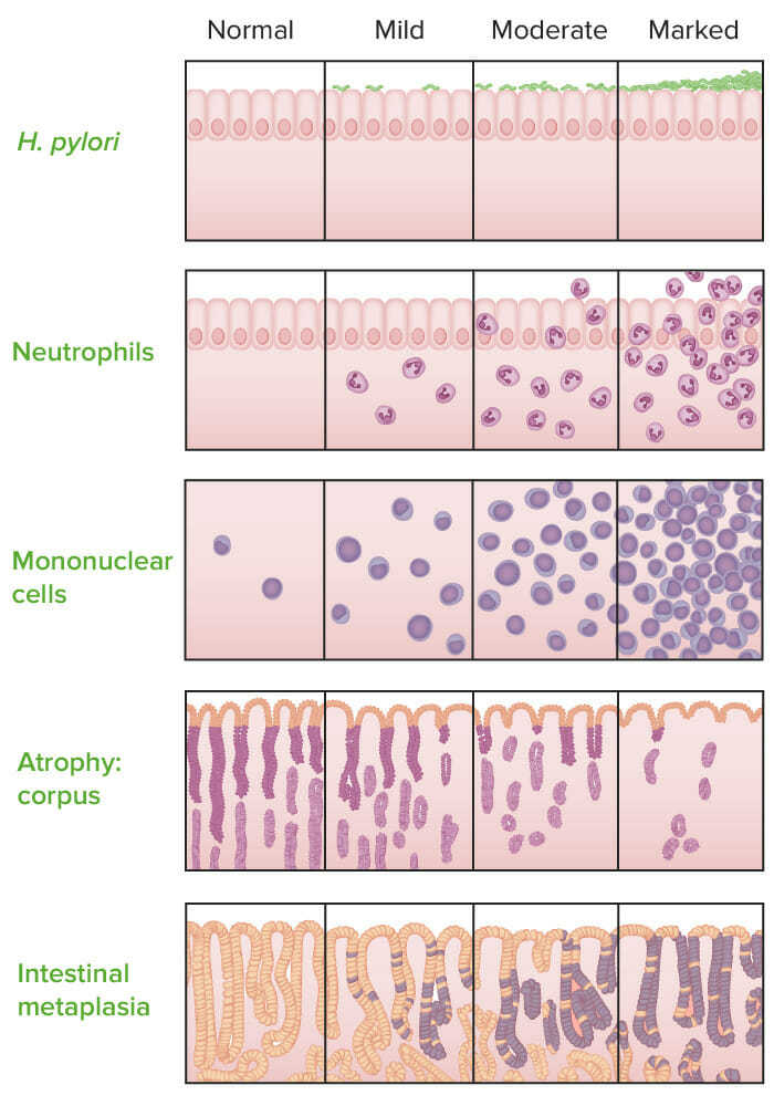
Morphology → provides possible “suffixes”; the system defines 5 specific morphologic variables that are graded from none → mild → moderate → severe/marked:
Chronic inflammationChronic InflammationInflammation (based on presence of lymphocytesLymphocytesLymphocytes are heterogeneous WBCs involved in immune response. Lymphocytes develop from the bone marrow, starting from hematopoietic stem cells (HSCs) and progressing to common lymphoid progenitors (CLPs). B and T lymphocytes and natural killer (NK) cells arise from the lineage.Lymphocytes: Histology and plasmaPlasmaThe residual portion of blood that is left after removal of blood cells by centrifugation without prior blood coagulation.Transfusion Products cells)
Activity (based on presence of neutrophilsNeutrophilsGranular leukocytes having a nucleus with three to five lobes connected by slender threads of chromatin, and cytoplasm containing fine inconspicuous granules and stainable by neutral dyes.Innate Immunity: Phagocytes and Antigen Presentation, indicating active inflammationInflammationInflammation is a complex set of responses to infection and injury involving leukocytes as the principal cellular mediators in the body’s defense against pathogenic organisms. Inflammation is also seen as a response to tissue injury in the process of wound healing. The 5 cardinal signs of inflammation are pain, heat, redness, swelling, and loss of function. Inflammation)
Intestinal metaplasiaMetaplasiaA condition in which there is a change of one adult cell type to another similar adult cell type.Cellular Adaptation (gastric epitheliumEpitheliumThe epithelium is a complex of specialized cellular organizations arranged into sheets and lining cavities and covering the surfaces of the body. The cells exhibit polarity, having an apical and a basal pole. Structures important for the epithelial integrity and function involve the basement membrane, the semipermeable sheet on which the cells rest, and interdigitations, as well as cellular junctions. Surface Epithelium: Histology is replaced with intestinal-type cells)
AtrophyAtrophyDecrease in the size of a cell, tissue, organ, or multiple organs, associated with a variety of pathological conditions such as abnormal cellular changes, ischemia, malnutrition, or hormonal changes.Cellular Adaptation (based on loss of normal mucosal glands)
Presence of H. pyloriH. pyloriA spiral bacterium active as a human gastric pathogen. It is a gram-negative, urease-positive, curved or slightly spiral organism initially isolated in 1982 from patients with lesions of gastritis or peptic ulcers in Western Australia. Helicobacter pylori was originally classified in the genus campylobacter, but RNA sequencing, cellular fatty acid profiles, growth patterns, and other taxonomic characteristics indicate that the micro-organism should be included in the genus Helicobacter. It has been officially transferred to Helicobacter gen.Helicobacter
Examples:
H. pyloriH. pyloriA spiral bacterium active as a human gastric pathogen. It is a gram-negative, urease-positive, curved or slightly spiral organism initially isolated in 1982 from patients with lesions of gastritis or peptic ulcers in Western Australia. Helicobacter pylori was originally classified in the genus campylobacter, but RNA sequencing, cellular fatty acid profiles, growth patterns, and other taxonomic characteristics indicate that the micro-organism should be included in the genus Helicobacter. It has been officially transferred to Helicobacter gen.Helicobacter antral gastritisGastritisGastritis refers to inflammation of the gastric mucosa. Gastritis may occur suddenly (acute gastritis) or slowly over time (chronic gastritis). Gastritis may be asymptomatic or with symptoms, including burning abdominal pain (which either worsens or improves with eating), dyspepsia, nausea, and vomiting. Gastritis, moderately active with mild antral atrophyAtrophyDecrease in the size of a cell, tissue, organ, or multiple organs, associated with a variety of pathological conditions such as abnormal cellular changes, ischemia, malnutrition, or hormonal changes.Cellular Adaptation
Autoimmune corpus gastritisGastritisGastritis refers to inflammation of the gastric mucosa. Gastritis may occur suddenly (acute gastritis) or slowly over time (chronic gastritis). Gastritis may be asymptomatic or with symptoms, including burning abdominal pain (which either worsens or improves with eating), dyspepsia, nausea, and vomiting. Gastritis with severe intestinal metaplasiaMetaplasiaA condition in which there is a change of one adult cell type to another similar adult cell type.Cellular Adaptation and atrophyAtrophyDecrease in the size of a cell, tissue, organ, or multiple organs, associated with a variety of pathological conditions such as abnormal cellular changes, ischemia, malnutrition, or hormonal changes.Cellular Adaptation
Reactive mild pangastritis without H. pyloriH. pyloriA spiral bacterium active as a human gastric pathogen. It is a gram-negative, urease-positive, curved or slightly spiral organism initially isolated in 1982 from patients with lesions of gastritis or peptic ulcers in Western Australia. Helicobacter pylori was originally classified in the genus campylobacter, but RNA sequencing, cellular fatty acid profiles, growth patterns, and other taxonomic characteristics indicate that the micro-organism should be included in the genus Helicobacter. It has been officially transferred to Helicobacter gen.Helicobacter
Sydney system of gastritis classification, morphology variables.
Image by Lecturio.
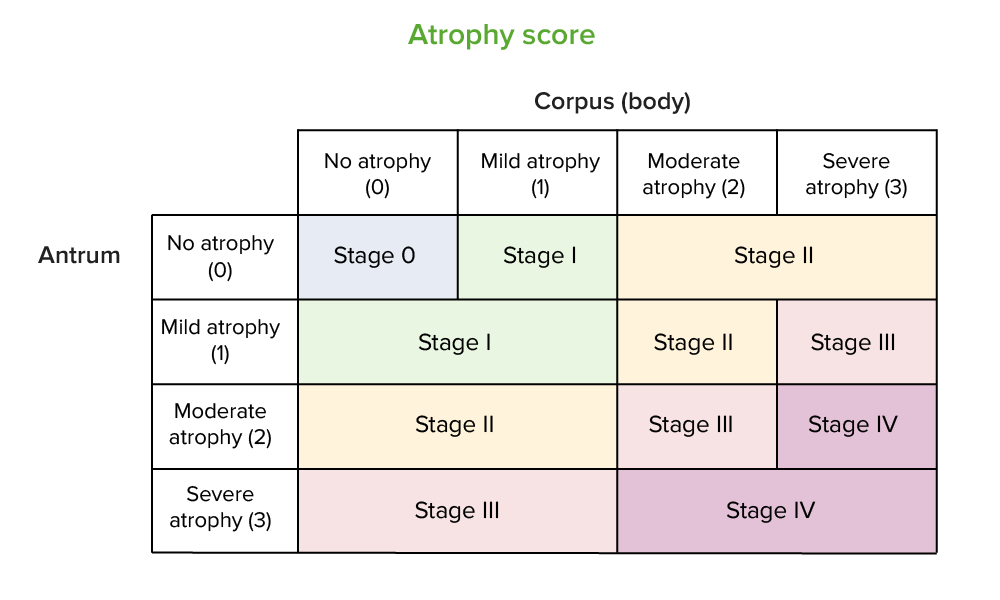
Operative link for gastritisGastritisGastritis refers to inflammation of the gastric mucosa. Gastritis may occur suddenly (acute gastritis) or slowly over time (chronic gastritis). Gastritis may be asymptomatic or with symptoms, including burning abdominal pain (which either worsens or improves with eating), dyspepsia, nausea, and vomiting. Gastritis assessment (OLGA):[10]
Helps stage chronic atrophic gastritisAtrophic gastritisGastritis with atrophy of the gastric mucosa, the gastric parietal cells, and the mucosal glands leading to achlorhydria. Atrophic gastritis usually progresses from chronic gastritis.Gastritis and predict cancer risk
Based on severity of atrophyAtrophyDecrease in the size of a cell, tissue, organ, or multiple organs, associated with a variety of pathological conditions such as abnormal cellular changes, ischemia, malnutrition, or hormonal changes.Cellular Adaptation in both the antrum and the corpus/body of the stomachStomachThe stomach is a muscular sac in the upper left portion of the abdomen that plays a critical role in digestion. The stomach develops from the foregut and connects the esophagus with the duodenum. Structurally, the stomach is C-shaped and forms a greater and lesser curvature and is divided grossly into regions: the cardia, fundus, body, and pylorus. Stomach: Anatomy
Operative link for gastritis assessment (OLGA) staging system
Image by Lecturio.
Epidemiology[1,2,11,13,16]
H. pyloriH. pyloriA spiral bacterium active as a human gastric pathogen. It is a gram-negative, urease-positive, curved or slightly spiral organism initially isolated in 1982 from patients with lesions of gastritis or peptic ulcers in Western Australia. Helicobacter pylori was originally classified in the genus campylobacter, but RNA sequencing, cellular fatty acid profiles, growth patterns, and other taxonomic characteristics indicate that the micro-organism should be included in the genus Helicobacter. It has been officially transferred to Helicobacter gen.Helicobacter infection: affects more than half the worldwide population, or 4.4 billion people
PrevalencePrevalenceThe total number of cases of a given disease in a specified population at a designated time. It is differentiated from incidence, which refers to the number of new cases in the population at a given time.Measures of Disease Frequency ranges from 19% in Switzerland to 88% in Nigeria
PrevalencePrevalenceThe total number of cases of a given disease in a specified population at a designated time. It is differentiated from incidence, which refers to the number of new cases in the population at a given time.Measures of Disease Frequency in the US is higher in certain groups:
Hispanics (60%)
African Americans (54%)
Adults over age 60 (50%)
H. pyloriH. pyloriA spiral bacterium active as a human gastric pathogen. It is a gram-negative, urease-positive, curved or slightly spiral organism initially isolated in 1982 from patients with lesions of gastritis or peptic ulcers in Western Australia. Helicobacter pylori was originally classified in the genus campylobacter, but RNA sequencing, cellular fatty acid profiles, growth patterns, and other taxonomic characteristics indicate that the micro-organism should be included in the genus Helicobacter. It has been officially transferred to Helicobacter gen.Helicobacter infection is associated with:
60% of gastric ulcers
80% of duodenal ulcers
Environmental metaplastic atrophic gastritisAtrophic gastritisGastritis with atrophy of the gastric mucosa, the gastric parietal cells, and the mucosal glands leading to achlorhydria. Atrophic gastritis usually progresses from chronic gastritis.Gastritis (EMAG) in 85% of cases
Most individuals with H. pyloriH. pyloriA spiral bacterium active as a human gastric pathogen. It is a gram-negative, urease-positive, curved or slightly spiral organism initially isolated in 1982 from patients with lesions of gastritis or peptic ulcers in Western Australia. Helicobacter pylori was originally classified in the genus campylobacter, but RNA sequencing, cellular fatty acid profiles, growth patterns, and other taxonomic characteristics indicate that the micro-organism should be included in the genus Helicobacter. It has been officially transferred to Helicobacter gen.Helicobacter never develop disease.
H. pyloriH. pyloriA spiral bacterium active as a human gastric pathogen. It is a gram-negative, urease-positive, curved or slightly spiral organism initially isolated in 1982 from patients with lesions of gastritis or peptic ulcers in Western Australia. Helicobacter pylori was originally classified in the genus campylobacter, but RNA sequencing, cellular fatty acid profiles, growth patterns, and other taxonomic characteristics indicate that the micro-organism should be included in the genus Helicobacter. It has been officially transferred to Helicobacter gen.HelicobacterprevalencePrevalenceThe total number of cases of a given disease in a specified population at a designated time. It is differentiated from incidence, which refers to the number of new cases in the population at a given time.Measures of Disease Frequency in the pediatric population is a major determinant ofH. pyloriH. pyloriA spiral bacterium active as a human gastric pathogen. It is a gram-negative, urease-positive, curved or slightly spiral organism initially isolated in 1982 from patients with lesions of gastritis or peptic ulcers in Western Australia. Helicobacter pylori was originally classified in the genus campylobacter, but RNA sequencing, cellular fatty acid profiles, growth patterns, and other taxonomic characteristics indicate that the micro-organism should be included in the genus Helicobacter. It has been officially transferred to Helicobacter gen.HelicobactergastritisGastritisGastritis refers to inflammation of the gastric mucosa. Gastritis may occur suddenly (acute gastritis) or slowly over time (chronic gastritis). Gastritis may be asymptomatic or with symptoms, including burning abdominal pain (which either worsens or improves with eating), dyspepsia, nausea, and vomiting. Gastritis.
Autoimmune metaplastic atrophic gastritisAtrophic gastritisGastritis with atrophy of the gastric mucosa, the gastric parietal cells, and the mucosal glands leading to achlorhydria. Atrophic gastritis usually progresses from chronic gastritis.Gastritis (AMAG, i.e., the pathologic process underlying pernicious anemiaPernicious anemiaA megaloblastic anemia occurring in children but more commonly in later life, characterized by histamine-fast achlorhydria, in which the laboratory and clinical manifestations are based on malabsorption of vitamin B12 due to a failure of the gastric mucosa to secrete adequate and potent intrinsic factor.Megaloblastic Anemia):
PrevalencePrevalenceThe total number of cases of a given disease in a specified population at a designated time. It is differentiated from incidence, which refers to the number of new cases in the population at a given time.Measures of Disease Frequency: 2% in Western countries
Risk increases with age
Women > men
Pathophysiology
H. pyloriH. pyloriA spiral bacterium active as a human gastric pathogen. It is a gram-negative, urease-positive, curved or slightly spiral organism initially isolated in 1982 from patients with lesions of gastritis or peptic ulcers in Western Australia. Helicobacter pylori was originally classified in the genus campylobacter, but RNA sequencing, cellular fatty acid profiles, growth patterns, and other taxonomic characteristics indicate that the micro-organism should be included in the genus Helicobacter. It has been officially transferred to Helicobacter gen.Helicobacter–associated gastritisGastritisGastritis refers to inflammation of the gastric mucosa. Gastritis may occur suddenly (acute gastritis) or slowly over time (chronic gastritis). Gastritis may be asymptomatic or with symptoms, including burning abdominal pain (which either worsens or improves with eating), dyspepsia, nausea, and vomiting. Gastritis[8,10,13]
Can be acute or chronic
H. pyloriH. pyloriA spiral bacterium active as a human gastric pathogen. It is a gram-negative, urease-positive, curved or slightly spiral organism initially isolated in 1982 from patients with lesions of gastritis or peptic ulcers in Western Australia. Helicobacter pylori was originally classified in the genus campylobacter, but RNA sequencing, cellular fatty acid profiles, growth patterns, and other taxonomic characteristics indicate that the micro-organism should be included in the genus Helicobacter. It has been officially transferred to Helicobacter gen.Helicobacterresides in the gastric mucus adjacent to epithelial cells and invades the lamina propriaLamina propriaWhipple’s Disease.
Acute (early stage):
Antral-predominant inflammationInflammationInflammation is a complex set of responses to infection and injury involving leukocytes as the principal cellular mediators in the body’s defense against pathogenic organisms. Inflammation is also seen as a response to tissue injury in the process of wound healing. The 5 cardinal signs of inflammation are pain, heat, redness, swelling, and loss of function. Inflammation or gastritisGastritisGastritis refers to inflammation of the gastric mucosa. Gastritis may occur suddenly (acute gastritis) or slowly over time (chronic gastritis). Gastritis may be asymptomatic or with symptoms, including burning abdominal pain (which either worsens or improves with eating), dyspepsia, nausea, and vomiting. Gastritis
↑GastrinGastrinA family of gastrointestinal peptide hormones that excite the secretion of gastric juice. They may also occur in the central nervous system where they are presumed to be neurotransmitters.Gastrointestinal Secretions + ↓ somatostatinSomatostatinA 14-amino acid peptide named for its ability to inhibit pituitary growth hormone release, also called somatotropin release-inhibiting factor. It is expressed in the central and peripheral nervous systems, the gut, and other organs. SRIF can also inhibit the release of thyroid-stimulating hormone; prolactin; insulin; and glucagon besides acting as a neurotransmitter and neuromodulator. In a number of species including humans, there is an additional form of somatostatin, srif-28 with a 14-amino acid extension at the n-terminal.Gastrointestinal Secretions = ↑ acid production
Increased acid exacerbates mucosal injury and inflammationInflammationInflammation is a complex set of responses to infection and injury involving leukocytes as the principal cellular mediators in the body’s defense against pathogenic organisms. Inflammation is also seen as a response to tissue injury in the process of wound healing. The 5 cardinal signs of inflammation are pain, heat, redness, swelling, and loss of function. Inflammation.
Due to antral location, duodenal ulcerDuodenal ulcerA peptic ulcer located in the duodenum.Peptic Ulcer Disease is a possible complication.
Progression from early-stage chronic gastritisGastritisGastritis refers to inflammation of the gastric mucosa. Gastritis may occur suddenly (acute gastritis) or slowly over time (chronic gastritis). Gastritis may be asymptomatic or with symptoms, including burning abdominal pain (which either worsens or improves with eating), dyspepsia, nausea, and vomiting. Gastritis to atrophic (later-stage) gastritisGastritisGastritis refers to inflammation of the gastric mucosa. Gastritis may occur suddenly (acute gastritis) or slowly over time (chronic gastritis). Gastritis may be asymptomatic or with symptoms, including burning abdominal pain (which either worsens or improves with eating), dyspepsia, nausea, and vomiting. Gastritis is a slow process as gastric glandsGastric glandsStomach: Anatomy disappear.
Chronic (late stage):
Environmental metaplastic atrophic gastritisAtrophic gastritisGastritis with atrophy of the gastric mucosa, the gastric parietal cells, and the mucosal glands leading to achlorhydria. Atrophic gastritis usually progresses from chronic gastritis.Gastritis(EMAG)
Ongoing inflammationInflammationInflammation is a complex set of responses to infection and injury involving leukocytes as the principal cellular mediators in the body’s defense against pathogenic organisms. Inflammation is also seen as a response to tissue injury in the process of wound healing. The 5 cardinal signs of inflammation are pain, heat, redness, swelling, and loss of function. Inflammation of gastric mucosaGastric mucosaLining of the stomach, consisting of an inner epithelium, a middle lamina propria, and an outer muscularis mucosae. The surface cells produce mucus that protects the stomach from attack by digestive acid and enzymes. When the epithelium invaginates into the lamina propria at various region of the stomach (cardia; gastric fundus; and pylorus), different tubular gastric glands are formed. These glands consist of cells that secrete mucus, enzymes, hydrochloric acid, or hormones.Stomach: Anatomy results in loss of G cells (gastrin-producing) and parietal cellsParietal cellsRounded or pyramidal cells of the gastric glands. They secrete hydrochloric acid and produce gastric intrinsic factor, a glycoprotein that binds vitamin B12.Stomach: Anatomy (acid-producing).
This change allows proximal migration ofH. pyloriH. pyloriA spiral bacterium active as a human gastric pathogen. It is a gram-negative, urease-positive, curved or slightly spiral organism initially isolated in 1982 from patients with lesions of gastritis or peptic ulcers in Western Australia. Helicobacter pylori was originally classified in the genus campylobacter, but RNA sequencing, cellular fatty acid profiles, growth patterns, and other taxonomic characteristics indicate that the micro-organism should be included in the genus Helicobacter. It has been officially transferred to Helicobacter gen.Helicobacter→ spread from the antrum to the gastric body
Effects:
Atrophic glands → intestinal metaplasiaMetaplasiaA condition in which there is a change of one adult cell type to another similar adult cell type.Cellular Adaptation of gastric mucosaGastric mucosaLining of the stomach, consisting of an inner epithelium, a middle lamina propria, and an outer muscularis mucosae. The surface cells produce mucus that protects the stomach from attack by digestive acid and enzymes. When the epithelium invaginates into the lamina propria at various region of the stomach (cardia; gastric fundus; and pylorus), different tubular gastric glands are formed. These glands consist of cells that secrete mucus, enzymes, hydrochloric acid, or hormones.Stomach: Anatomy (increased risk of gastric cancerGastric cancerGastric cancer is the 3rd-most common cause of cancer-related deaths worldwide. The majority of cases are from adenocarcinoma. The modifiable risk factors include Helicobacter pylori infection, smoking, and nitrate-rich diets. Gastric Cancer)
Loss of parietal cellsParietal cellsRounded or pyramidal cells of the gastric glands. They secrete hydrochloric acid and produce gastric intrinsic factor, a glycoprotein that binds vitamin B12.Stomach: Anatomy → ↓ intrinsic factorIntrinsic factorA glycoprotein secreted by the cells of the gastric glands that is required for the absorption of vitamin B 12 (cyanocobalamin). Deficiency of intrinsic factor leads to vitamin B12 deficiency and anemia, pernicious.Gastritis → B12 deficiency
Cancer risk:
H. pyloriH. pyloriA spiral bacterium active as a human gastric pathogen. It is a gram-negative, urease-positive, curved or slightly spiral organism initially isolated in 1982 from patients with lesions of gastritis or peptic ulcers in Western Australia. Helicobacter pylori was originally classified in the genus campylobacter, but RNA sequencing, cellular fatty acid profiles, growth patterns, and other taxonomic characteristics indicate that the micro-organism should be included in the genus Helicobacter. It has been officially transferred to Helicobacter gen.Helicobacterplays a pivotal role in the development of gastric adenocarcinomaGastric adenocarcinomaGastric Cancer over decades (10- to 15- fold increased risk).
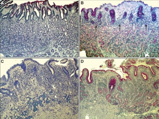
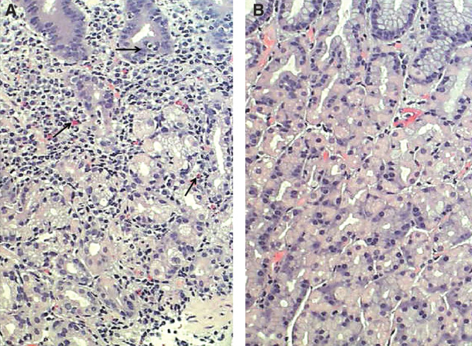
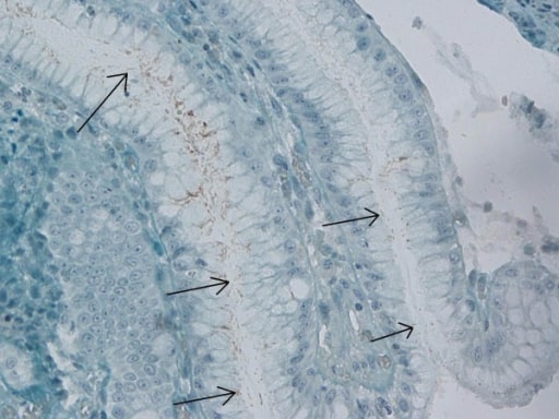
Chronic H. pylori gastritis (EMAG in C and D): A: Normal mucosa of the corpus B: Non-atrophic gastritis: mild mononuclear inflammation seen in upper layer of the mucosa (superficial gastritis as indicated by arrows). Gland layer is still intact. C: Moderate atrophic gastritis: intense chronic mononuclear inflammation occurs in the lower layers, accompanied by atrophy of the oxyntic glands (indicating stomach with hypochlorhydria). Acid secretion is impaired due to loss of parietal cells (in the oxyntic glands). D: Severe atrophic gastritis, showing mild inflammation but there is loss of oxyntic glands.
Image: “Corpus mucosa” by Patolab Oy, Espoo, Finland and Tartu State University, Tartu, Estonia. License: CC BY 4.0
Autoimmune metaplastic atrophic gastritisAtrophic gastritisGastritis with atrophy of the gastric mucosa, the gastric parietal cells, and the mucosal glands leading to achlorhydria. Atrophic gastritis usually progresses from chronic gastritis.Gastritis (AMAG)[9,11,13]
Chronic corpus-predominant inflammationInflammationInflammation is a complex set of responses to infection and injury involving leukocytes as the principal cellular mediators in the body’s defense against pathogenic organisms. Inflammation is also seen as a response to tissue injury in the process of wound healing. The 5 cardinal signs of inflammation are pain, heat, redness, swelling, and loss of function. Inflammation or gastritisGastritisGastritis refers to inflammation of the gastric mucosa. Gastritis may occur suddenly (acute gastritis) or slowly over time (chronic gastritis). Gastritis may be asymptomatic or with symptoms, including burning abdominal pain (which either worsens or improves with eating), dyspepsia, nausea, and vomiting. Gastritis
Spares the antrum, the lower portion of the stomachStomachThe stomach is a muscular sac in the upper left portion of the abdomen that plays a critical role in digestion. The stomach develops from the foregut and connects the esophagus with the duodenum. Structurally, the stomach is C-shaped and forms a greater and lesser curvature and is divided grossly into regions: the cardia, fundus, body, and pylorus. Stomach: Anatomy
Associated with chronic T cell–mediated autoimmune disease:
T cellsT cellsLymphocytes responsible for cell-mediated immunity. Two types have been identified – cytotoxic (t-lymphocytes, cytotoxic) and helper T-lymphocytes (t-lymphocytes, helper-inducer). They are formed when lymphocytes circulate through the thymus gland and differentiate to thymocytes. When exposed to an antigen, they divide rapidly and produce large numbers of new T cells sensitized to that antigen.T cells: Types and Functions destroy oxyntic mucosa.
AntibodiesAntibodiesImmunoglobulins (Igs), also known as antibodies, are glycoprotein molecules produced by plasma cells that act in immune responses by recognizing and binding particular antigens. The various Ig classes are IgG (the most abundant), IgM, IgE, IgD, and IgA, which differ in their biologic features, structure, target specificity, and distribution.Immunoglobulins: Types and Functions against parietal cellsParietal cellsRounded or pyramidal cells of the gastric glands. They secrete hydrochloric acid and produce gastric intrinsic factor, a glycoprotein that binds vitamin B12.Stomach: Anatomy and intrinsic factorIntrinsic factorA glycoprotein secreted by the cells of the gastric glands that is required for the absorption of vitamin B 12 (cyanocobalamin). Deficiency of intrinsic factor leads to vitamin B12 deficiency and anemia, pernicious.Gastritis
Effects:
Loss of oxyntic glands, replaced by atrophic mucosa → intestinal metaplasiaMetaplasiaA condition in which there is a change of one adult cell type to another similar adult cell type.Cellular Adaptation
Pathologic changes lead to reduced acid production → hypochlorhydria/achlorhydriaAchlorhydriaA lack of hydrochloric acid in gastric juice despite stimulation of gastric secretion.Gastritis → ↑ gastrinGastrinA family of gastrointestinal peptide hormones that excite the secretion of gastric juice. They may also occur in the central nervous system where they are presumed to be neurotransmitters.Gastrointestinal Secretions (loss of negative feedbackNegative feedbackHypothalamic and Pituitary Hormones)
Loss of parietal cellsParietal cellsRounded or pyramidal cells of the gastric glands. They secrete hydrochloric acid and produce gastric intrinsic factor, a glycoprotein that binds vitamin B12.Stomach: Anatomy and intrinsic factorIntrinsic factorA glycoprotein secreted by the cells of the gastric glands that is required for the absorption of vitamin B 12 (cyanocobalamin). Deficiency of intrinsic factor leads to vitamin B12 deficiency and anemia, pernicious.Gastritis due to antibodiesAntibodiesImmunoglobulins (Igs), also known as antibodies, are glycoprotein molecules produced by plasma cells that act in immune responses by recognizing and binding particular antigens. The various Ig classes are IgG (the most abundant), IgM, IgE, IgD, and IgA, which differ in their biologic features, structure, target specificity, and distribution.Immunoglobulins: Types and Functions → vitamin B12malabsorptionMalabsorptionGeneral term for a group of malnutrition syndromes caused by failure of normal intestinal absorption of nutrients.Malabsorption and Maldigestion and pernicious anemiaPernicious anemiaA megaloblastic anemia occurring in children but more commonly in later life, characterized by histamine-fast achlorhydria, in which the laboratory and clinical manifestations are based on malabsorption of vitamin B12 due to a failure of the gastric mucosa to secrete adequate and potent intrinsic factor.Megaloblastic Anemia
Cancer risk:
Gastric neuroendocrine/carcinoid tumorsCarcinoid tumorsCarcinoid tumors are small, well-differentiated, slow-growing neuroendocrine tumors (NET). Carcinoid syndrome describes the signs and symptoms associated with unregulated vasoactive hormone production by neuroendocrine tumors. Carcinoid tumors are most commonly found in the GI and bronchopulmonary tracts. Carcinoid Tumors and Syndrome: Elevated gastrinGastrinA family of gastrointestinal peptide hormones that excite the secretion of gastric juice. They may also occur in the central nervous system where they are presumed to be neurotransmitters.Gastrointestinal Secretions also stimulates enterochromaffin cells → hyperplasiaHyperplasiaAn increase in the number of cells in a tissue or organ without tumor formation. It differs from hypertrophy, which is an increase in bulk without an increase in the number of cells.Cellular Adaptation → dysplasia
Damage to the epithelial lining with no (or minimal) associated inflammationInflammationInflammation is a complex set of responses to infection and injury involving leukocytes as the principal cellular mediators in the body’s defense against pathogenic organisms. Inflammation is also seen as a response to tissue injury in the process of wound healing. The 5 cardinal signs of inflammation are pain, heat, redness, swelling, and loss of function. Inflammation
Associated with mucosal injury from destruction of the normal protective barrier (mucins, bicarbonateBicarbonateInorganic salts that contain the -HCO3 radical. They are an important factor in determining the ph of the blood and the concentration of bicarbonate ions is regulated by the kidney. Levels in the blood are an index of the alkali reserve or buffering capacity.Electrolytes, and epitheliumEpitheliumThe epithelium is a complex of specialized cellular organizations arranged into sheets and lining cavities and covering the surfaces of the body. The cells exhibit polarity, having an apical and a basal pole. Structures important for the epithelial integrity and function involve the basement membrane, the semipermeable sheet on which the cells rest, and interdigitations, as well as cellular junctions. Surface Epithelium: Histology), which may result in:
Bleeding (hemorrhagic gastropathygastropathyDamage to the epithelial lining with no or minimal associated inflammation and is technically a separate entity from gastritisGastritis)
Common causes:
Reactive (chemical) gastropathygastropathyDamage to the epithelial lining with no or minimal associated inflammation and is technically a separate entity from gastritisGastritis
Medications: NSAIDsNSAIDSPrimary vs Secondary Headaches, ironIronA metallic element with atomic symbol fe, atomic number 26, and atomic weight 55. 85. It is an essential constituent of hemoglobins; cytochromes; and iron-binding proteins. It plays a role in cellular redox reactions and in the transport of oxygen.Trace Elements salts, others
Alcohol
BileBileAn emulsifying agent produced in the liver and secreted into the duodenum. Its composition includes bile acids and salts; cholesterol; and electrolytes. It aids digestion of fats in the duodenum.Gallbladder and Biliary Tract: Anatomy reflux
Ischemic gastropathygastropathyDamage to the epithelial lining with no or minimal associated inflammation and is technically a separate entity from gastritisGastritis:
Associated with critical illness (e.g., shockShockShock is a life-threatening condition associated with impaired circulation that results in tissue hypoxia. The different types of shock are based on the underlying cause: distributive (↑ cardiac output (CO), ↓ systemic vascular resistance (SVR)), cardiogenic (↓ CO, ↑ SVR), hypovolemic (↓ CO, ↑ SVR), obstructive (↓ CO), and mixed. Types of Shock) and injury (e.g., trauma)
Represents end-organ failure of the stomachStomachThe stomach is a muscular sac in the upper left portion of the abdomen that plays a critical role in digestion. The stomach develops from the foregut and connects the esophagus with the duodenum. Structurally, the stomach is C-shaped and forms a greater and lesser curvature and is divided grossly into regions: the cardia, fundus, body, and pylorus. Stomach: Anatomy in critical illness
Vascular gastropathygastropathyDamage to the epithelial lining with no or minimal associated inflammation and is technically a separate entity from gastritisGastritis:
Portal hypertensive (congestive) gastropathygastropathyDamage to the epithelial lining with no or minimal associated inflammation and is technically a separate entity from gastritisGastritis
Gastric antral vascular ectasiaGastric Antral Vascular EctasiaA distinct vascular lesion in the pyloric antrum that is characterized by tortuous dilated blood vessels (ectasia) radiating outward from the pylorus. The vessel pattern resembles the stripes on the surface of a watermelon. This lesion causes both acute and chronic gastrointestinal hemorrhage.Gastrointestinal Bleeding (GAVE)
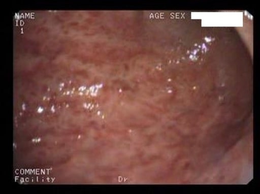
Acute erosive gastritis changes (endoscopic view) in a patient who has had prolonged COX-2 NSAID therapy
Image: “Acute erosive gastritis” by Department of Medical Sciences II, Medical Rehabilitation, University of Medicine and Pharmacy of Craiova, Romania. License: CC BY 2.0
ScreeningScreeningPreoperative Care of asymptomatic individuals for H. pyloriH. pyloriA spiral bacterium active as a human gastric pathogen. It is a gram-negative, urease-positive, curved or slightly spiral organism initially isolated in 1982 from patients with lesions of gastritis or peptic ulcers in Western Australia. Helicobacter pylori was originally classified in the genus campylobacter, but RNA sequencing, cellular fatty acid profiles, growth patterns, and other taxonomic characteristics indicate that the micro-organism should be included in the genus Helicobacter. It has been officially transferred to Helicobacter gen.Helicobacteris not recommended. Tests are done in symptomatic patientsPatientsIndividuals participating in the health care system for the purpose of receiving therapeutic, diagnostic, or preventive procedures.Clinician–Patient Relationship to evaluate for gastritisGastritisGastritis refers to inflammation of the gastric mucosa. Gastritis may occur suddenly (acute gastritis) or slowly over time (chronic gastritis). Gastritis may be asymptomatic or with symptoms, including burning abdominal pain (which either worsens or improves with eating), dyspepsia, nausea, and vomiting. Gastritis and peptic ulcerPeptic ulcerPeptic ulcer disease (PUD) refers to the full-thickness ulcerations of duodenal or gastric mucosa. The ulcerations form when exposure to acid and digestive enzymes overcomes mucosal defense mechanisms. The most common etiologies include Helicobacter pylori (H. pylori) infection and prolonged use of non-steroidal anti-inflammatory drugs (NSAIDs). Peptic Ulcer Disease disease.
Clinical presentation[8,10]
May be asymptomatic and present with anemiaAnemiaAnemia is a condition in which individuals have low Hb levels, which can arise from various causes. Anemia is accompanied by a reduced number of RBCs and may manifest with fatigue, shortness of breath, pallor, and weakness. Subtypes are classified by the size of RBCs, chronicity, and etiology. Anemia: Overview and Types on lab tests
Symptoms of acute gastritisGastritisGastritis refers to inflammation of the gastric mucosa. Gastritis may occur suddenly (acute gastritis) or slowly over time (chronic gastritis). Gastritis may be asymptomatic or with symptoms, including burning abdominal pain (which either worsens or improves with eating), dyspepsia, nausea, and vomiting. Gastritis:
NauseaNauseaAn unpleasant sensation in the stomach usually accompanied by the urge to vomit. Common causes are early pregnancy, sea and motion sickness, emotional stress, intense pain, food poisoning, and various enteroviruses.Antiemetics, sometimes vomitingVomitingThe forcible expulsion of the contents of the stomach through the mouth.Hypokalemia
H. pyloriH. pyloriA spiral bacterium active as a human gastric pathogen. It is a gram-negative, urease-positive, curved or slightly spiral organism initially isolated in 1982 from patients with lesions of gastritis or peptic ulcers in Western Australia. Helicobacter pylori was originally classified in the genus campylobacter, but RNA sequencing, cellular fatty acid profiles, growth patterns, and other taxonomic characteristics indicate that the micro-organism should be included in the genus Helicobacter. It has been officially transferred to Helicobacter gen.HelicobactergastritisGastritisGastritis refers to inflammation of the gastric mucosa. Gastritis may occur suddenly (acute gastritis) or slowly over time (chronic gastritis). Gastritis may be asymptomatic or with symptoms, including burning abdominal pain (which either worsens or improves with eating), dyspepsia, nausea, and vomiting. Gastritis:
Acute: asymptomatic or mild, self-limited dyspepsiaDyspepsiaImpaired digestion, especially after eating.Lactose Intolerance
Symptoms of peptic ulcerPeptic ulcerPeptic ulcer disease (PUD) refers to the full-thickness ulcerations of duodenal or gastric mucosa. The ulcerations form when exposure to acid and digestive enzymes overcomes mucosal defense mechanisms. The most common etiologies include Helicobacter pylori (H. pylori) infection and prolonged use of non-steroidal anti-inflammatory drugs (NSAIDs). Peptic Ulcer Disease disease (similar symptoms of acute gastritisGastritisGastritis refers to inflammation of the gastric mucosa. Gastritis may occur suddenly (acute gastritis) or slowly over time (chronic gastritis). Gastritis may be asymptomatic or with symptoms, including burning abdominal pain (which either worsens or improves with eating), dyspepsia, nausea, and vomiting. Gastritis)
IronIronA metallic element with atomic symbol fe, atomic number 26, and atomic weight 55. 85. It is an essential constituent of hemoglobins; cytochromes; and iron-binding proteins. It plays a role in cellular redox reactions and in the transport of oxygen.Trace Elements deficiency or B12-deficiency anemiaAnemiaAnemia is a condition in which individuals have low Hb levels, which can arise from various causes. Anemia is accompanied by a reduced number of RBCs and may manifest with fatigue, shortness of breath, pallor, and weakness. Subtypes are classified by the size of RBCs, chronicity, and etiology. Anemia: Overview and Types
AMAG:
May be asymptomatic, but often with dyspepsiaDyspepsiaImpaired digestion, especially after eating.Lactose Intolerance
B12 deficiency:
FatigueFatigueThe state of weariness following a period of exertion, mental or physical, characterized by a decreased capacity for work and reduced efficiency to respond to stimuli.Fibromyalgia
Glossitis
Mild cognitive impairment and other neurologic manifestations
IronIronA metallic element with atomic symbol fe, atomic number 26, and atomic weight 55. 85. It is an essential constituent of hemoglobins; cytochromes; and iron-binding proteins. It plays a role in cellular redox reactions and in the transport of oxygen.Trace Elements deficiency anemiaAnemiaAnemia is a condition in which individuals have low Hb levels, which can arise from various causes. Anemia is accompanied by a reduced number of RBCs and may manifest with fatigue, shortness of breath, pallor, and weakness. Subtypes are classified by the size of RBCs, chronicity, and etiology. Anemia: Overview and Types: occurs earlier than B12 deficiency (pernicious anemiaPernicious anemiaA megaloblastic anemia occurring in children but more commonly in later life, characterized by histamine-fast achlorhydria, in which the laboratory and clinical manifestations are based on malabsorption of vitamin B12 due to a failure of the gastric mucosa to secrete adequate and potent intrinsic factor.Megaloblastic Anemia) because of decreased acid secretionSecretionCoagulation Studies
Increased association with other autoimmune diseasesAutoimmune diseasesDisorders that are characterized by the production of antibodies that react with host tissues or immune effector cells that are autoreactive to endogenous peptides.Selective IgA Deficiency (e.g., autoimmune thyroidThyroidThe thyroid gland is one of the largest endocrine glands in the human body. The thyroid gland is a highly vascular, brownish-red gland located in the visceral compartment of the anterior region of the neck.Thyroid Gland: Anatomy disease, type 1Type 1Spinal Muscular AtrophydiabetesDiabetesDiabetes mellitus (DM) is a metabolic disease characterized by hyperglycemia and dysfunction of the regulation of glucose metabolism by insulin. Type 1 DM is diagnosed mostly in children and young adults as the result of autoimmune destruction of β cells in the pancreas and the resulting lack of insulin. Type 2 DM has a significant association with obesity and is characterized by insulin resistance.Diabetes Mellitus mellitus, Addison’s diseaseAddison’s DiseaseAdrenal insufficiency (AI) is the inadequate production of adrenocortical hormones: glucocorticoids, mineralocorticoids, and adrenal androgens. Primary AI, also called Addison’s disease, is caused by autoimmune disease, infections, and malignancy, among others.Adrenal Insufficiency and Addison Disease)
Other: hematemesisHematemesisVomiting of blood that is either fresh bright red, or older ‘coffee-ground’ in character. It generally indicates bleeding of the upper gastrointestinal tract.Mallory-Weiss Syndrome (Mallory-Weiss Tear) (hemorrhagic gastropathygastropathyDamage to the epithelial lining with no or minimal associated inflammation and is technically a separate entity from gastritisGastritis)
Diagnosis
Indications for H. pyloriH. pyloriA spiral bacterium active as a human gastric pathogen. It is a gram-negative, urease-positive, curved or slightly spiral organism initially isolated in 1982 from patients with lesions of gastritis or peptic ulcers in Western Australia. Helicobacter pylori was originally classified in the genus campylobacter, but RNA sequencing, cellular fatty acid profiles, growth patterns, and other taxonomic characteristics indicate that the micro-organism should be included in the genus Helicobacter. It has been officially transferred to Helicobacter gen.Helicobacter testing:[5]
PatientsPatientsIndividuals participating in the health care system for the purpose of receiving therapeutic, diagnostic, or preventive procedures.Clinician–Patient Relationship ≥ 60 years of age or younger patientsPatientsIndividuals participating in the health care system for the purpose of receiving therapeutic, diagnostic, or preventive procedures.Clinician–Patient Relationship with alarm symptoms of dyspepsiaDyspepsiaImpaired digestion, especially after eating.Lactose Intolerance:
VomitingVomitingThe forcible expulsion of the contents of the stomach through the mouth.Hypokalemia
Bleeding or anemiaAnemiaAnemia is a condition in which individuals have low Hb levels, which can arise from various causes. Anemia is accompanied by a reduced number of RBCs and may manifest with fatigue, shortness of breath, pallor, and weakness. Subtypes are classified by the size of RBCs, chronicity, and etiology. Anemia: Overview and Types
DysphagiaDysphagiaDysphagia is the subjective sensation of difficulty swallowing. Symptoms can range from a complete inability to swallow, to the sensation of solids or liquids becoming “stuck.” Dysphagia is classified as either oropharyngeal or esophageal, with esophageal dysphagia having 2 sub-types: functional and mechanical. Dysphagia
PatientsPatientsIndividuals participating in the health care system for the purpose of receiving therapeutic, diagnostic, or preventive procedures.Clinician–Patient Relationship starting chronic treatment with NSAIDsNSAIDSPrimary vs Secondary Headaches or who are taking long-term low-dose aspirinAspirinThe prototypical analgesic used in the treatment of mild to moderate pain. It has anti-inflammatory and antipyretic properties and acts as an inhibitor of cyclooxygenase which results in the inhibition of the biosynthesis of prostaglandins. Aspirin also inhibits platelet aggregation and is used in the prevention of arterial and venous thrombosis.Nonsteroidal Antiinflammatory Drugs (NSAIDs)
Unexplained ironIronA metallic element with atomic symbol fe, atomic number 26, and atomic weight 55. 85. It is an essential constituent of hemoglobins; cytochromes; and iron-binding proteins. It plays a role in cellular redox reactions and in the transport of oxygen.Trace Elements deficiency anemiaAnemiaAnemia is a condition in which individuals have low Hb levels, which can arise from various causes. Anemia is accompanied by a reduced number of RBCs and may manifest with fatigue, shortness of breath, pallor, and weakness. Subtypes are classified by the size of RBCs, chronicity, and etiology. Anemia: Overview and Types
Current peptic ulcerPeptic ulcerPeptic ulcer disease (PUD) refers to the full-thickness ulcerations of duodenal or gastric mucosa. The ulcerations form when exposure to acid and digestive enzymes overcomes mucosal defense mechanisms. The most common etiologies include Helicobacter pylori (H. pylori) infection and prolonged use of non-steroidal anti-inflammatory drugs (NSAIDs). Peptic Ulcer Disease disease (PUDPUDPeptic ulcer disease (PUD) refers to the full-thickness ulcerations of duodenal or gastric mucosa. The ulcerations form when exposure to acid and digestive enzymes overcomes mucosal defense mechanisms. The most common etiologies include Helicobacter pylori (H. pylori) infection and prolonged use of non-steroidal anti-inflammatory drugs (NSAIDs). Peptic Ulcer Disease) or a past history of PUDPUDPeptic ulcer disease (PUD) refers to the full-thickness ulcerations of duodenal or gastric mucosa. The ulcerations form when exposure to acid and digestive enzymes overcomes mucosal defense mechanisms. The most common etiologies include Helicobacter pylori (H. pylori) infection and prolonged use of non-steroidal anti-inflammatory drugs (NSAIDs). Peptic Ulcer Disease (without documented cure)
The development of upper GI symptoms in patientsPatientsIndividuals participating in the health care system for the purpose of receiving therapeutic, diagnostic, or preventive procedures.Clinician–Patient Relationship with pernicious anemiaPernicious anemiaA megaloblastic anemia occurring in children but more commonly in later life, characterized by histamine-fast achlorhydria, in which the laboratory and clinical manifestations are based on malabsorption of vitamin B12 due to a failure of the gastric mucosa to secrete adequate and potent intrinsic factor.Megaloblastic Anemia
Low-grade gastric mucosaGastric mucosaLining of the stomach, consisting of an inner epithelium, a middle lamina propria, and an outer muscularis mucosae. The surface cells produce mucus that protects the stomach from attack by digestive acid and enzymes. When the epithelium invaginates into the lamina propria at various region of the stomach (cardia; gastric fundus; and pylorus), different tubular gastric glands are formed. These glands consist of cells that secrete mucus, enzymes, hydrochloric acid, or hormones.Stomach: Anatomy–associated lymphoid tissue (MALTMALTColon, Cecum, and Appendix: Anatomy) lymphomaLymphomaA general term for various neoplastic diseases of the lymphoid tissue.Imaging of the Mediastinum
History of endoscopic resection of early gastric cancerGastric cancerGastric cancer is the 3rd-most common cause of cancer-related deaths worldwide. The majority of cases are from adenocarcinoma. The modifiable risk factors include Helicobacter pylori infection, smoking, and nitrate-rich diets. Gastric Cancer
Adults with idiopathicIdiopathicDermatomyositis thrombocytopenic purpura (ITPITPImmune thrombocytopenic purpura (ITP), formerly known as idiopathic thrombocytopenic purpura, is a condition that develops secondary to immune-mediated destruction of platelets, resulting in thrombocytopenia (platelet count < 100,000/mm³). Immune thrombocytopenic purpura can be either primary or secondary due to drugs or underlying disease. Immune Thrombocytopenic Purpura)
Notes:
For patientsPatientsIndividuals participating in the health care system for the purpose of receiving therapeutic, diagnostic, or preventive procedures.Clinician–Patient Relationship < age 60 with uninvestigated dyspepsiaDyspepsiaImpaired digestion, especially after eating.Lactose Intolerance without alarming features, noninvasive testing is acceptable.
Typical symptoms of GERDGERDGastroesophageal reflux disease (GERD) occurs when the stomach acid frequently flows back into the esophagus. This backwash (acid reflux) can irritate the lining of the esophagus, causing symptoms such as retrosternal burning pain (heartburn). Gastroesophageal Reflux Disease (GERD) without a history of peptic ulcerPeptic ulcerPeptic ulcer disease (PUD) refers to the full-thickness ulcerations of duodenal or gastric mucosa. The ulcerations form when exposure to acid and digestive enzymes overcomes mucosal defense mechanisms. The most common etiologies include Helicobacter pylori (H. pylori) infection and prolonged use of non-steroidal anti-inflammatory drugs (NSAIDs). Peptic Ulcer Disease disease (PUDPUDPeptic ulcer disease (PUD) refers to the full-thickness ulcerations of duodenal or gastric mucosa. The ulcerations form when exposure to acid and digestive enzymes overcomes mucosal defense mechanisms. The most common etiologies include Helicobacter pylori (H. pylori) infection and prolonged use of non-steroidal anti-inflammatory drugs (NSAIDs). Peptic Ulcer Disease) or dyspepsiaDyspepsiaImpaired digestion, especially after eating.Lactose Intolerance do not require H. pyloriH. pyloriA spiral bacterium active as a human gastric pathogen. It is a gram-negative, urease-positive, curved or slightly spiral organism initially isolated in 1982 from patients with lesions of gastritis or peptic ulcers in Western Australia. Helicobacter pylori was originally classified in the genus campylobacter, but RNA sequencing, cellular fatty acid profiles, growth patterns, and other taxonomic characteristics indicate that the micro-organism should be included in the genus Helicobacter. It has been officially transferred to Helicobacter gen.Helicobactertesting.
Noninvasive laboratory studies:
Noninvasive H. pyloriH. pyloriA spiral bacterium active as a human gastric pathogen. It is a gram-negative, urease-positive, curved or slightly spiral organism initially isolated in 1982 from patients with lesions of gastritis or peptic ulcers in Western Australia. Helicobacter pylori was originally classified in the genus campylobacter, but RNA sequencing, cellular fatty acid profiles, growth patterns, and other taxonomic characteristics indicate that the micro-organism should be included in the genus Helicobacter. It has been officially transferred to Helicobacter gen.Helicobacter testing:
Stool antigenAntigenSubstances that are recognized by the immune system and induce an immune reaction.Vaccination assay (preferred for initial diagnosis and to confirm eradication)
Rapid and inexpensive
Reduced accuracy if patient is taking PPIs, antibiotics, bismuth, or sucralfate
Capsules with radioactively labeled ureaUreaA compound formed in the liver from ammonia produced by the deamination of amino acids. It is the principal end product of protein catabolism and constitutes about one half of the total urinary solids.Urea Cycle (13C or 14C) are swallowed; the ureaUreaA compound formed in the liver from ammonia produced by the deamination of amino acids. It is the principal end product of protein catabolism and constitutes about one half of the total urinary solids.Urea Cycle is absorbed.
UreaseUreaseAn enzyme that catalyzes the conversion of urea and water to carbon dioxide and ammonia.Nocardia/Nocardiosis produced by H. pyloriH. pyloriA spiral bacterium active as a human gastric pathogen. It is a gram-negative, urease-positive, curved or slightly spiral organism initially isolated in 1982 from patients with lesions of gastritis or peptic ulcers in Western Australia. Helicobacter pylori was originally classified in the genus campylobacter, but RNA sequencing, cellular fatty acid profiles, growth patterns, and other taxonomic characteristics indicate that the micro-organism should be included in the genus Helicobacter. It has been officially transferred to Helicobacter gen.Helicobacter splits labeled ureaUreaA compound formed in the liver from ammonia produced by the deamination of amino acids. It is the principal end product of protein catabolism and constitutes about one half of the total urinary solids.Urea Cycle → produces 13C- or 14C-labeled CO2, which can be detected in exhaled breaths 20‒30 minutes later by massMassThree-dimensional lesion that occupies a space within the breastImaging of the Breast spectrometry or scintillation
If H. pyloriH. pyloriA spiral bacterium active as a human gastric pathogen. It is a gram-negative, urease-positive, curved or slightly spiral organism initially isolated in 1982 from patients with lesions of gastritis or peptic ulcers in Western Australia. Helicobacter pylori was originally classified in the genus campylobacter, but RNA sequencing, cellular fatty acid profiles, growth patterns, and other taxonomic characteristics indicate that the micro-organism should be included in the genus Helicobacter. It has been officially transferred to Helicobacter gen.Helicobacter is not present → unhydrolyzed labeled ureaUreaA compound formed in the liver from ammonia produced by the deamination of amino acids. It is the principal end product of protein catabolism and constitutes about one half of the total urinary solids.Urea Cycle is excreted by the kidneysKidneysThe kidneys are a pair of bean-shaped organs located retroperitoneally against the posterior wall of the abdomen on either side of the spine. As part of the urinary tract, the kidneys are responsible for blood filtration and excretion of water-soluble waste in the urine.Kidneys: Anatomy, and there is no labeled CO₂ in breath
Note: PatientsPatientsIndividuals participating in the health care system for the purpose of receiving therapeutic, diagnostic, or preventive procedures.Clinician–Patient Relationship should discontinue gastritisGastritisGastritis refers to inflammation of the gastric mucosa. Gastritis may occur suddenly (acute gastritis) or slowly over time (chronic gastritis). Gastritis may be asymptomatic or with symptoms, including burning abdominal pain (which either worsens or improves with eating), dyspepsia, nausea, and vomiting. Gastritis medications (antibiotics, PPIs, bismuth, sucralfate) prior the test to avoid false negativeFalse negativeAn FN test result indicates a person does not have the disease when, in fact, they do.Epidemiological Values of Diagnostic Tests results.
Sensitivity and specificitySensitivity and SpecificityBinary classification measures to assess test results. Sensitivity or recall rate is the proportion of true positives. Specificity is the probability of correctly determining the absence of a condition.Epidemiological Values of Diagnostic Tests: both > 95%
Serologic tests are not recommended.
AMAG:[13]
CBC: anemiaAnemiaAnemia is a condition in which individuals have low Hb levels, which can arise from various causes. Anemia is accompanied by a reduced number of RBCs and may manifest with fatigue, shortness of breath, pallor, and weakness. Subtypes are classified by the size of RBCs, chronicity, and etiology. Anemia: Overview and Types (↓ ironIronA metallic element with atomic symbol fe, atomic number 26, and atomic weight 55. 85. It is an essential constituent of hemoglobins; cytochromes; and iron-binding proteins. It plays a role in cellular redox reactions and in the transport of oxygen.Trace Elements, ↓ B12 level)
SerologySerologyThe study of serum, especially of antigen-antibody reactions in vitro.Yellow Fever Virus:
Positive for antibodiesAntibodiesImmunoglobulins (Igs), also known as antibodies, are glycoprotein molecules produced by plasma cells that act in immune responses by recognizing and binding particular antigens. The various Ig classes are IgG (the most abundant), IgM, IgE, IgD, and IgA, which differ in their biologic features, structure, target specificity, and distribution.Immunoglobulins: Types and Functions to parietal cellsParietal cellsRounded or pyramidal cells of the gastric glands. They secrete hydrochloric acid and produce gastric intrinsic factor, a glycoprotein that binds vitamin B12.Stomach: Anatomy and intrinsic factorIntrinsic factorA glycoprotein secreted by the cells of the gastric glands that is required for the absorption of vitamin B 12 (cyanocobalamin). Deficiency of intrinsic factor leads to vitamin B12 deficiency and anemia, pernicious.Gastritis
Elevated fasting serum gastrinGastrinA family of gastrointestinal peptide hormones that excite the secretion of gastric juice. They may also occur in the central nervous system where they are presumed to be neurotransmitters.Gastrointestinal Secretions level
Markedly low serum pepsinogen I levels or low pepsinogen I/II ratio
ScreeningScreeningPreoperative Care for autoimmune thyroidThyroidThe thyroid gland is one of the largest endocrine glands in the human body. The thyroid gland is a highly vascular, brownish-red gland located in the visceral compartment of the anterior region of the neck.Thyroid Gland: Anatomy disease (due to high risk of concurrent autoimmune thyroiditisAutoimmune thyroiditisInflammatory disease of the thyroid gland due to autoimmune responses leading to lymphocytic infiltration of the gland. It is characterized by the presence of circulating thyroid antigen-specific T-cells and thyroid autoantibodies. The clinical signs can range from hypothyroidism to thyrotoxicosis depending on the type of autoimmune thyroiditis.Thyroiditis (Hashimoto’s): Thyroid function testsThyroid Function TestsBlood tests used to evaluate the functioning of the thyroid gland.Ion Channel Myopathy (TSH, anti-TPO antibodiesAntibodiesImmunoglobulins (Igs), also known as antibodies, are glycoprotein molecules produced by plasma cells that act in immune responses by recognizing and binding particular antigens. The various Ig classes are IgG (the most abundant), IgM, IgE, IgD, and IgA, which differ in their biologic features, structure, target specificity, and distribution.Immunoglobulins: Types and Functions)
Consider screeningScreeningPreoperative Care for other autoimmune disorders: type 1Type 1Spinal Muscular AtrophydiabetesDiabetesDiabetes mellitus (DM) is a metabolic disease characterized by hyperglycemia and dysfunction of the regulation of glucose metabolism by insulin. Type 1 DM is diagnosed mostly in children and young adults as the result of autoimmune destruction of β cells in the pancreas and the resulting lack of insulin. Type 2 DM has a significant association with obesity and is characterized by insulin resistance.Diabetes Mellitus, Addison’s diseaseAddison’s DiseaseAdrenal insufficiency (AI) is the inadequate production of adrenocortical hormones: glucocorticoids, mineralocorticoids, and adrenal androgens. Primary AI, also called Addison’s disease, is caused by autoimmune disease, infections, and malignancy, among others.Adrenal Insufficiency and Addison Disease, vitiligoVitiligoVitiligo is the most common depigmenting disorder and is caused by the destruction of melanocytes. Patients present with hypo- or depigmented macules or patches which often occur on the face, hands, knees, and/or genitalia. Vitiligo
EMAG:[9]
GastrinGastrinA family of gastrointestinal peptide hormones that excite the secretion of gastric juice. They may also occur in the central nervous system where they are presumed to be neurotransmitters.Gastrointestinal Secretions level is not as elevated as with AMAG.
Pernicious anemiaPernicious anemiaA megaloblastic anemia occurring in children but more commonly in later life, characterized by histamine-fast achlorhydria, in which the laboratory and clinical manifestations are based on malabsorption of vitamin B12 due to a failure of the gastric mucosa to secrete adequate and potent intrinsic factor.Megaloblastic Anemia is absent.
No autoantibodiesAutoantibodiesAntibodies that react with self-antigens (autoantigens) of the organism that produced them.Blotting Techniques against parietal cellsParietal cellsRounded or pyramidal cells of the gastric glands. They secrete hydrochloric acid and produce gastric intrinsic factor, a glycoprotein that binds vitamin B12.Stomach: Anatomy and intrinsic factorIntrinsic factorA glycoprotein secreted by the cells of the gastric glands that is required for the absorption of vitamin B 12 (cyanocobalamin). Deficiency of intrinsic factor leads to vitamin B12 deficiency and anemia, pernicious.Gastritis
Esophagogastroduodenoscopy (EGD):[4,13]
Findings, often nonspecific, may include
May not have noticeable pathologic changes
Findings, often nonspecific, may include:
Mucosal erythemaErythemaRedness of the skin produced by congestion of the capillaries. This condition may result from a variety of disease processes.Chalazion/friable mucosa
Classic appearance of atrophic gastritisAtrophic gastritisGastritis with atrophy of the gastric mucosa, the gastric parietal cells, and the mucosal glands leading to achlorhydria. Atrophic gastritis usually progresses from chronic gastritis.Gastritis:[4]
Pale mucosa
Increased visibility of vasculature
Flattening of rugal folds (advanced atrophic gastritisAtrophic gastritisGastritis with atrophy of the gastric mucosa, the gastric parietal cells, and the mucosal glands leading to achlorhydria. Atrophic gastritis usually progresses from chronic gastritis.Gastritis)
Light blue crests and white opaque fields (intestinal metaplasiaMetaplasiaA condition in which there is a change of one adult cell type to another similar adult cell type.Cellular Adaptation)
BiopsyBiopsyRemoval and pathologic examination of specimens from the living body.Ewing Sarcoma:[4,13,14]
Required for diagnosis of atrophic gastritisAtrophic gastritisGastritis with atrophy of the gastric mucosa, the gastric parietal cells, and the mucosal glands leading to achlorhydria. Atrophic gastritis usually progresses from chronic gastritis.Gastritis (AMAG, EMAG)[13]
Multiple biopsies recommended (corpus and antrum)
All abnormal-looking areas are biopsied.
Adjacent normal tissue also included
Testing for H. pyloriH. pyloriA spiral bacterium active as a human gastric pathogen. It is a gram-negative, urease-positive, curved or slightly spiral organism initially isolated in 1982 from patients with lesions of gastritis or peptic ulcers in Western Australia. Helicobacter pylori was originally classified in the genus campylobacter, but RNA sequencing, cellular fatty acid profiles, growth patterns, and other taxonomic characteristics indicate that the micro-organism should be included in the genus Helicobacter. It has been officially transferred to Helicobacter gen.Helicobacterin biopsyBiopsyRemoval and pathologic examination of specimens from the living body.Ewing Sarcoma specimen:[4,14]
Point-of-care PCRPCRPolymerase chain reaction (PCR) is a technique that amplifies DNA fragments exponentially for analysis. The process is highly specific, allowing for the targeting of specific genomic sequences, even with minuscule sample amounts. The PCR cycles multiple times through 3 phases: denaturation of the template DNA, annealing of a specific primer to the individual DNA strands, and synthesis/elongation of new DNA molecules.Polymerase Chain Reaction (PCR) test kits (can also detect mutations that cause clarithromycinClarithromycinA semisynthetic macrolide antibiotic derived from erythromycin that is active against a variety of microorganisms. It can inhibit protein synthesis in bacteria by reversibly binding to the 50s ribosomal subunits. This inhibits the translocation of aminoacyl transfer-RNA and prevents peptide chain elongation.Macrolides and KetolidesresistanceResistancePhysiologically, the opposition to flow of air caused by the forces of friction. As a part of pulmonary function testing, it is the ratio of driving pressure to the rate of air flow.Ventilation: Mechanics of Breathing)
Rapid ureaseUreaseAn enzyme that catalyzes the conversion of urea and water to carbon dioxide and ammonia.Nocardia/Nocardiosis test (RUT):
UreaseUreaseAn enzyme that catalyzes the conversion of urea and water to carbon dioxide and ammonia.Nocardia/Nocardiosis produced by H. pyloriH. pyloriA spiral bacterium active as a human gastric pathogen. It is a gram-negative, urease-positive, curved or slightly spiral organism initially isolated in 1982 from patients with lesions of gastritis or peptic ulcers in Western Australia. Helicobacter pylori was originally classified in the genus campylobacter, but RNA sequencing, cellular fatty acid profiles, growth patterns, and other taxonomic characteristics indicate that the micro-organism should be included in the genus Helicobacter. It has been officially transferred to Helicobacter gen.Helicobacterliberates ammoniaAmmoniaA colorless alkaline gas. It is formed in the body during decomposition of organic materials during a large number of metabolically important reactions. Note that the aqueous form of ammonia is referred to as ammonium hydroxide.Acid-Base Balance from ureaUreaA compound formed in the liver from ammonia produced by the deamination of amino acids. It is the principal end product of protein catabolism and constitutes about one half of the total urinary solids.Urea Cycle → alkaline pHpHThe quantitative measurement of the acidity or basicity of a solution.Acid-Base Balance changes the color of the reagent
If suspicious clinical history but negative RUT → histology
Bacterial culture of biopsyBiopsyRemoval and pathologic examination of specimens from the living body.Ewing Sarcoma specimens: of limited use since H. pyloriH. pyloriA spiral bacterium active as a human gastric pathogen. It is a gram-negative, urease-positive, curved or slightly spiral organism initially isolated in 1982 from patients with lesions of gastritis or peptic ulcers in Western Australia. Helicobacter pylori was originally classified in the genus campylobacter, but RNA sequencing, cellular fatty acid profiles, growth patterns, and other taxonomic characteristics indicate that the micro-organism should be included in the genus Helicobacter. It has been officially transferred to Helicobacter gen.Helicobacteris fastidiousFastidiousBordetella
Acute H. pyloriH. pyloriA spiral bacterium active as a human gastric pathogen. It is a gram-negative, urease-positive, curved or slightly spiral organism initially isolated in 1982 from patients with lesions of gastritis or peptic ulcers in Western Australia. Helicobacter pylori was originally classified in the genus campylobacter, but RNA sequencing, cellular fatty acid profiles, growth patterns, and other taxonomic characteristics indicate that the micro-organism should be included in the genus Helicobacter. It has been officially transferred to Helicobacter gen.HelicobactergastritisGastritisGastritis refers to inflammation of the gastric mucosa. Gastritis may occur suddenly (acute gastritis) or slowly over time (chronic gastritis). Gastritis may be asymptomatic or with symptoms, including burning abdominal pain (which either worsens or improves with eating), dyspepsia, nausea, and vomiting. Gastritis findings:
Presence of H. pyloriH. pyloriA spiral bacterium active as a human gastric pathogen. It is a gram-negative, urease-positive, curved or slightly spiral organism initially isolated in 1982 from patients with lesions of gastritis or peptic ulcers in Western Australia. Helicobacter pylori was originally classified in the genus campylobacter, but RNA sequencing, cellular fatty acid profiles, growth patterns, and other taxonomic characteristics indicate that the micro-organism should be included in the genus Helicobacter. It has been officially transferred to Helicobacter gen.Helicobacteron histology: curved, flagellated, gram-negative rods
Mucosal neutrophilsNeutrophilsGranular leukocytes having a nucleus with three to five lobes connected by slender threads of chromatin, and cytoplasm containing fine inconspicuous granules and stainable by neutral dyes.Innate Immunity: Phagocytes and Antigen Presentation
Chronic H. pyloriH. pyloriA spiral bacterium active as a human gastric pathogen. It is a gram-negative, urease-positive, curved or slightly spiral organism initially isolated in 1982 from patients with lesions of gastritis or peptic ulcers in Western Australia. Helicobacter pylori was originally classified in the genus campylobacter, but RNA sequencing, cellular fatty acid profiles, growth patterns, and other taxonomic characteristics indicate that the micro-organism should be included in the genus Helicobacter. It has been officially transferred to Helicobacter gen.HelicobactergastritisGastritisGastritis refers to inflammation of the gastric mucosa. Gastritis may occur suddenly (acute gastritis) or slowly over time (chronic gastritis). Gastritis may be asymptomatic or with symptoms, including burning abdominal pain (which either worsens or improves with eating), dyspepsia, nausea, and vomiting. Gastritis findings:
LymphocytesLymphocytesLymphocytes are heterogeneous WBCs involved in immune response. Lymphocytes develop from the bone marrow, starting from hematopoietic stem cells (HSCs) and progressing to common lymphoid progenitors (CLPs). B and T lymphocytes and natural killer (NK) cells arise from the lineage.Lymphocytes: Histology, plasmaPlasmaThe residual portion of blood that is left after removal of blood cells by centrifugation without prior blood coagulation.Transfusion Products cells, eosinophilsEosinophilsGranular leukocytes with a nucleus that usually has two lobes connected by a slender thread of chromatin, and cytoplasm containing coarse, round granules that are uniform in size and stainable by eosin.Innate Immunity: Phagocytes and Antigen Presentation
AtrophyAtrophyDecrease in the size of a cell, tissue, organ, or multiple organs, associated with a variety of pathological conditions such as abnormal cellular changes, ischemia, malnutrition, or hormonal changes.Cellular Adaptation (if present) typically starts in the antrum and expands proximally.[13]
Early AMAG findings:
Deep inflammationInflammationInflammation is a complex set of responses to infection and injury involving leukocytes as the principal cellular mediators in the body’s defense against pathogenic organisms. Inflammation is also seen as a response to tissue injury in the process of wound healing. The 5 cardinal signs of inflammation are pain, heat, redness, swelling, and loss of function. Inflammation, eosinophilsEosinophilsGranular leukocytes with a nucleus that usually has two lobes connected by a slender thread of chromatin, and cytoplasm containing coarse, round granules that are uniform in size and stainable by eosin.Innate Immunity: Phagocytes and Antigen Presentation
Destruction of oxyntic glands
Intestinal or pseudopyloric metaplasiaMetaplasiaA condition in which there is a change of one adult cell type to another similar adult cell type.Cellular Adaptation
AtrophyAtrophyDecrease in the size of a cell, tissue, organ, or multiple organs, associated with a variety of pathological conditions such as abnormal cellular changes, ischemia, malnutrition, or hormonal changes.Cellular Adaptation is typically confined to the corpus and fundusFundusThe superior portion of the body of the stomach above the level of the cardiac notch.Stomach: Anatomy (antral sparing).[13]
Advanced AMAG findings:
Metaplastic glands replace the oxyntic glands.
Megaloblastic epithelial cells
Chronic gastritis. A: mucosal inflammation with many mononuclear cells (arrows); B: normal mucosa.
Image: “Corpus mucosa” by Informa Healthcare. License: CC BY 4.0
Immunohistochemical stain for H. pylori in the gastric antrum
Image: “Immunohistochemical H. pylori” by Department of Gastroenterology, Bakirkoy Dr Sadi Konuk Training and Research Hospital, Istanbul, Turkey. License: CC BY 2.0
IronIronA metallic element with atomic symbol fe, atomic number 26, and atomic weight 55. 85. It is an essential constituent of hemoglobins; cytochromes; and iron-binding proteins. It plays a role in cellular redox reactions and in the transport of oxygen.Trace Elements deficiency treatment
Avoid direct gastric irritants
Treatment for H. pylori is guided by penicillinPenicillinRheumatic FeverallergyAllergyAn abnormal adaptive immune response that may or may not involve antigen-specific IgEType I Hypersensitivity Reaction or risk factors for macrolide resistanceResistancePhysiologically, the opposition to flow of air caused by the forces of friction. As a part of pulmonary function testing, it is the ratio of driving pressure to the rate of air flow.Ventilation: Mechanics of Breathing.
H. pyloriH. pyloriA spiral bacterium active as a human gastric pathogen. It is a gram-negative, urease-positive, curved or slightly spiral organism initially isolated in 1982 from patients with lesions of gastritis or peptic ulcers in Western Australia. Helicobacter pylori was originally classified in the genus campylobacter, but RNA sequencing, cellular fatty acid profiles, growth patterns, and other taxonomic characteristics indicate that the micro-organism should be included in the genus Helicobacter. It has been officially transferred to Helicobacter gen.Helicobacter–associated gastritisGastritisGastritis refers to inflammation of the gastric mucosa. Gastritis may occur suddenly (acute gastritis) or slowly over time (chronic gastritis). Gastritis may be asymptomatic or with symptoms, including burning abdominal pain (which either worsens or improves with eating), dyspepsia, nausea, and vomiting. Gastritis management[4,5]
Treatment aimed to eradicate H. pyloriH. pyloriA spiral bacterium active as a human gastric pathogen. It is a gram-negative, urease-positive, curved or slightly spiral organism initially isolated in 1982 from patients with lesions of gastritis or peptic ulcers in Western Australia. Helicobacter pylori was originally classified in the genus campylobacter, but RNA sequencing, cellular fatty acid profiles, growth patterns, and other taxonomic characteristics indicate that the micro-organism should be included in the genus Helicobacter. It has been officially transferred to Helicobacter gen.Helicobacter
Preferred option (treatment-naive): 14-day optimized bismuth quadruple therapy (BQT) ~ PPI (standard BID), bismuth, metronidazoleMetronidazoleA nitroimidazole used to treat amebiasis; vaginitis; trichomonas infections; giardiasis; anaerobic bacteria; and treponemal infections.Pyogenic Liver Abscess, and tetracyclineTetracyclineA naphthacene antibiotic that inhibits amino Acyl tRNA binding during protein synthesis.Drug-Induced Liver Injury
PPIs
OmeprazoleOmeprazoleA 4-methoxy-3, 5-dimethylpyridyl, 5-methoxybenzimidazole derivative of timoprazole that is used in the therapy of stomach ulcers and zollinger-ellison syndrome. The drug inhibits an h(+)-k(+)-exchanging ATPase which is found in gastric parietal cells.Gastric Acid Drugs 20 mg twice a day
LansoprazoleLansoprazoleA 2, 2, 2-trifluoroethoxypyridyl derivative of timoprazole that is used in the therapy of stomach ulcers and zollinger-ellison syndrome. The drug inhibits h(+)-k(+)-exchanging ATPase which is found in gastric parietal cells. Lansoprazole is a racemic mixture of (r)- and (s)-isomers.Gastric Acid Drugs 30 mg twice a day
PantoprazolePantoprazole2-pyridinylmethylsulfinylbenzimidazole proton pump inhibitor that is used in the treatment of gastroesophageal reflux and peptic ulcer.Gastric Acid Drugs 40‒80 mg twice a day
RabeprazoleRabeprazoleA 4-(3-methoxypropoxy)-3-methylpyridinyl derivative of timoprazole that is used in the therapy of stomach ulcers and zollinger-ellison syndrome. The drug inhibits h(+)-k(+)-exchanging ATPase which is found in gastric parietal cells.Gastric Acid Drugs 20‒40 mg twice a day
Bismuth subcitrate 120 to 300 mg 4 times a day (not available in the U.S.)
TetracyclineTetracyclineA naphthacene antibiotic that inhibits amino Acyl tRNA binding during protein synthesis.Drug-Induced Liver Injury 500 mg 4 times a day
MetronidazoleMetronidazoleA nitroimidazole used to treat amebiasis; vaginitis; trichomonas infections; giardiasis; anaerobic bacteria; and treponemal infections.Pyogenic Liver Abscess 500 mg 3 or 4 times a day
Another regimen uses fixed dose combination capsules plus PPI (Pylera taken 4 x a day for 10 days).
Alternatives (treatment-naive)
RifabutinRifabutinA broad-spectrum antibiotic that is being used as prophylaxis against disseminated Mycobacterium avium complex infection in HIV-positive patients.Diseases of the Uvea triple therapy: fixed dose combination capsules (4 capsules= omeprazoleOmeprazoleA 4-methoxy-3, 5-dimethylpyridyl, 5-methoxybenzimidazole derivative of timoprazole that is used in the therapy of stomach ulcers and zollinger-ellison syndrome. The drug inhibits an h(+)-k(+)-exchanging ATPase which is found in gastric parietal cells.Gastric Acid Drugs 40 mg, rifabutinRifabutinA broad-spectrum antibiotic that is being used as prophylaxis against disseminated Mycobacterium avium complex infection in HIV-positive patients.Diseases of the Uvea 50 mg , amoxicillinAmoxicillinA broad-spectrum semisynthetic antibiotic similar to ampicillin except that its resistance to gastric acid permits higher serum levels with oral administration.Penicillins 1 g) taken 3 times a day for 14 days
Vonaprazan-amoxicillin dual therapy for 14 days
Vonaprazan 20 mg twice a day
AmoxicillinAmoxicillinA broad-spectrum semisynthetic antibiotic similar to ampicillin except that its resistance to gastric acid permits higher serum levels with oral administration.Penicillins 1 g 3x a day
Clarithromycin-based PPI triple therapy (for 14 days) should only be used in cases of confirmed clarithromycinClarithromycinA semisynthetic macrolide antibiotic derived from erythromycin that is active against a variety of microorganisms. It can inhibit protein synthesis in bacteria by reversibly binding to the 50s ribosomal subunits. This inhibits the translocation of aminoacyl transfer-RNA and prevents peptide chain elongation.Macrolides and Ketolides susceptibility.
Vonaprazan triple therapy
Vonaprazan 20 mg twice a day
AmoxicillinAmoxicillinA broad-spectrum semisynthetic antibiotic similar to ampicillin except that its resistance to gastric acid permits higher serum levels with oral administration.Penicillins 1 g twice a day
ClarithromycinClarithromycinA semisynthetic macrolide antibiotic derived from erythromycin that is active against a variety of microorganisms. It can inhibit protein synthesis in bacteria by reversibly binding to the 50s ribosomal subunits. This inhibits the translocation of aminoacyl transfer-RNA and prevents peptide chain elongation.Macrolides and Ketolides 500 mg twice a day
Confirmation of eradication[4,5]
Hold PPI for 1–2 weeks prior to testing.
Test all patientsPatientsIndividuals participating in the health care system for the purpose of receiving therapeutic, diagnostic, or preventive procedures.Clinician–Patient Relationship ≥ 4 weeks after completion of antibiotic therapy with any of these:
Stool antigenAntigenSubstances that are recognized by the immune system and induce an immune reaction.Vaccination test
EGD (biopsyBiopsyRemoval and pathologic examination of specimens from the living body.Ewing Sarcoma)
Approximately 20% of H. pyloriH. pyloriA spiral bacterium active as a human gastric pathogen. It is a gram-negative, urease-positive, curved or slightly spiral organism initially isolated in 1982 from patients with lesions of gastritis or peptic ulcers in Western Australia. Helicobacter pylori was originally classified in the genus campylobacter, but RNA sequencing, cellular fatty acid profiles, growth patterns, and other taxonomic characteristics indicate that the micro-organism should be included in the genus Helicobacter. It has been officially transferred to Helicobacter gen.Helicobacter cases fail to be eradicated by the initial attempt.
If infection persists, try to use different antibiotics, and/or test for antibiotic susceptibility by culture or molecular (PCRPCRPolymerase chain reaction (PCR) is a technique that amplifies DNA fragments exponentially for analysis. The process is highly specific, allowing for the targeting of specific genomic sequences, even with minuscule sample amounts. The PCR cycles multiple times through 3 phases: denaturation of the template DNA, annealing of a specific primer to the individual DNA strands, and synthesis/elongation of new DNA molecules.Polymerase Chain Reaction (PCR)) methods (less sensitive).
LevofloxacinLevofloxacinThe l-isomer of ofloxacin.Fluoroquinolones or clarithromycinClarithromycinA semisynthetic macrolide antibiotic derived from erythromycin that is active against a variety of microorganisms. It can inhibit protein synthesis in bacteria by reversibly binding to the 50s ribosomal subunits. This inhibits the translocation of aminoacyl transfer-RNA and prevents peptide chain elongation.Macrolides and Ketolides should not be used in salvage therapy unless there is confirmed H. pyloriH. pyloriA spiral bacterium active as a human gastric pathogen. It is a gram-negative, urease-positive, curved or slightly spiral organism initially isolated in 1982 from patients with lesions of gastritis or peptic ulcers in Western Australia. Helicobacter pylori was originally classified in the genus campylobacter, but RNA sequencing, cellular fatty acid profiles, growth patterns, and other taxonomic characteristics indicate that the micro-organism should be included in the genus Helicobacter. It has been officially transferred to Helicobacter gen.Helicobacter susceptibility to these antibiotics.
Salvage therapy:
Optimized bismuth quadruple therapy (BQT) or Fixed dose combination + PPI (Pylera) if this regimen was not previously used or was not previously completed
RifabutinRifabutinA broad-spectrum antibiotic that is being used as prophylaxis against disseminated Mycobacterium avium complex infection in HIV-positive patients.Diseases of the Uvea triple therapy if optimized BQT was previously used
High-dose dual therapy:
AmoxicillinAmoxicillinA broad-spectrum semisynthetic antibiotic similar to ampicillin except that its resistance to gastric acid permits higher serum levels with oral administration.Penicillins 1g 3x a day
Vonaprazan 20 mg or PPI 2 to 3x a day
Requires antibiotic susceptibility testing:
Vonaprazan triple therapy
LevofloxacinLevofloxacinThe l-isomer of ofloxacin.Fluoroquinolones triple therapy (PPI twice a day, amoxicillinAmoxicillinA broad-spectrum semisynthetic antibiotic similar to ampicillin except that its resistance to gastric acid permits higher serum levels with oral administration.Penicillins 1 g twice a day, levofloxacinLevofloxacinThe l-isomer of ofloxacin.Fluoroquinolones 500 mg a day)
Note: Despite eradication, normal gastric mucosaGastric mucosaLining of the stomach, consisting of an inner epithelium, a middle lamina propria, and an outer muscularis mucosae. The surface cells produce mucus that protects the stomach from attack by digestive acid and enzymes. When the epithelium invaginates into the lamina propria at various region of the stomach (cardia; gastric fundus; and pylorus), different tubular gastric glands are formed. These glands consist of cells that secrete mucus, enzymes, hydrochloric acid, or hormones.Stomach: Anatomy may not recover from significant atrophyAtrophyDecrease in the size of a cell, tissue, organ, or multiple organs, associated with a variety of pathological conditions such as abnormal cellular changes, ischemia, malnutrition, or hormonal changes.Cellular Adaptation → cancer risk remains elevated (especially for OLGA stages III and IV)
AMAG management[4,13]
No specific treatment other than H. pyloriH. pyloriA spiral bacterium active as a human gastric pathogen. It is a gram-negative, urease-positive, curved or slightly spiral organism initially isolated in 1982 from patients with lesions of gastritis or peptic ulcers in Western Australia. Helicobacter pylori was originally classified in the genus campylobacter, but RNA sequencing, cellular fatty acid profiles, growth patterns, and other taxonomic characteristics indicate that the micro-organism should be included in the genus Helicobacter. It has been officially transferred to Helicobacter gen.Helicobactereradication
SurveillanceSurveillanceDevelopmental Milestones and Normal GrowthendoscopyEndoscopyProcedures of applying endoscopes for disease diagnosis and treatment. Endoscopy involves passing an optical instrument through a small incision in the skin i.e., percutaneous; or through a natural orifice and along natural body pathways such as the digestive tract; and/or through an incision in the wall of a tubular structure or organ, i.e. Transluminal, to examine or perform surgery on the interior parts of the body.Gastroesophageal Reflux Disease (GERD) every 3–5 years to look for signs of cancer is controversial and not agreed on, but recommendation is found in the 2019 European Society for Gastrointestinal EndoscopyEndoscopyProcedures of applying endoscopes for disease diagnosis and treatment. Endoscopy involves passing an optical instrument through a small incision in the skin i.e., percutaneous; or through a natural orifice and along natural body pathways such as the digestive tract; and/or through an incision in the wall of a tubular structure or organ, i.e. Transluminal, to examine or perform surgery on the interior parts of the body.Gastroesophageal Reflux Disease (GERD) (ESGE) guidelines.[7]
Evaluate for:
IronIronA metallic element with atomic symbol fe, atomic number 26, and atomic weight 55. 85. It is an essential constituent of hemoglobins; cytochromes; and iron-binding proteins. It plays a role in cellular redox reactions and in the transport of oxygen.Trace Elements and vitamin B-12 deficiencies
Other autoimmune diseasesAutoimmune diseasesDisorders that are characterized by the production of antibodies that react with host tissues or immune effector cells that are autoreactive to endogenous peptides.Selective IgA Deficiency (e.g., autoimmune thyroidThyroidThe thyroid gland is one of the largest endocrine glands in the human body. The thyroid gland is a highly vascular, brownish-red gland located in the visceral compartment of the anterior region of the neck.Thyroid Gland: Anatomy disease, type 1Type 1Spinal Muscular AtrophydiabetesDiabetesDiabetes mellitus (DM) is a metabolic disease characterized by hyperglycemia and dysfunction of the regulation of glucose metabolism by insulin. Type 1 DM is diagnosed mostly in children and young adults as the result of autoimmune destruction of β cells in the pancreas and the resulting lack of insulin. Type 2 DM has a significant association with obesity and is characterized by insulin resistance.Diabetes Mellitus mellitus, Addison disease)
Complications[4]
Peptic ulcerPeptic ulcerPeptic ulcer disease (PUD) refers to the full-thickness ulcerations of duodenal or gastric mucosa. The ulcerations form when exposure to acid and digestive enzymes overcomes mucosal defense mechanisms. The most common etiologies include Helicobacter pylori (H. pylori) infection and prolonged use of non-steroidal anti-inflammatory drugs (NSAIDs). Peptic Ulcer Disease disease: requires continued acid suppressionSuppressionDefense Mechanisms for 4 to 8 weeks
Intestinal-type gastric cancerGastric cancerGastric cancer is the 3rd-most common cause of cancer-related deaths worldwide. The majority of cases are from adenocarcinoma. The modifiable risk factors include Helicobacter pylori infection, smoking, and nitrate-rich diets. Gastric Cancer:
Associated with AMAG and EMAG
Progression of mucosal atrophyAtrophyDecrease in the size of a cell, tissue, organ, or multiple organs, associated with a variety of pathological conditions such as abnormal cellular changes, ischemia, malnutrition, or hormonal changes.Cellular Adaptation to intestinal metaplasiaMetaplasiaA condition in which there is a change of one adult cell type to another similar adult cell type.Cellular Adaptation
MetaplasiaMetaplasiaA condition in which there is a change of one adult cell type to another similar adult cell type.Cellular Adaptation progresses to dysplasia and eventually to invasive adenocarcinoma.
MALTomaMALTomaExtranodal marginal zone lymphoma (EMZL) of mucosa-associated lymphoid tissue (also called MALToma, MALT lymphoma, and pseudolymphoma) is a group of non-Hodgkin’s lymphomas that have historically been grouped together because they appear to arise from postgerminal center marginal zone B cells and share a similar immunophenotype. MALT Lymphoma (mucosa-associated lymphoid tumorTumorInflammation):
B-cell lymphomaLymphomaA general term for various neoplastic diseases of the lymphoid tissue.Imaging of the Mediastinum of the stomachStomachThe stomach is a muscular sac in the upper left portion of the abdomen that plays a critical role in digestion. The stomach develops from the foregut and connects the esophagus with the duodenum. Structurally, the stomach is C-shaped and forms a greater and lesser curvature and is divided grossly into regions: the cardia, fundus, body, and pylorus. Stomach: Anatomy
Clear association with H. pyloriH. pyloriA spiral bacterium active as a human gastric pathogen. It is a gram-negative, urease-positive, curved or slightly spiral organism initially isolated in 1982 from patients with lesions of gastritis or peptic ulcers in Western Australia. Helicobacter pylori was originally classified in the genus campylobacter, but RNA sequencing, cellular fatty acid profiles, growth patterns, and other taxonomic characteristics indicate that the micro-organism should be included in the genus Helicobacter. It has been officially transferred to Helicobacter gen.Helicobacterinfection
Some cases are cured with H. pyloriH. pyloriA spiral bacterium active as a human gastric pathogen. It is a gram-negative, urease-positive, curved or slightly spiral organism initially isolated in 1982 from patients with lesions of gastritis or peptic ulcers in Western Australia. Helicobacter pylori was originally classified in the genus campylobacter, but RNA sequencing, cellular fatty acid profiles, growth patterns, and other taxonomic characteristics indicate that the micro-organism should be included in the genus Helicobacter. It has been officially transferred to Helicobacter gen.Helicobactereradication
Differential Diagnosis
Ménétrier’s disease (MD): protein-losing gastropathygastropathyDamage to the epithelial lining with no or minimal associated inflammation and is technically a separate entity from gastritisGastritis characterized by giant hyperplasticHyperplasticColon Polyps mucosal folds of the stomachStomachThe stomach is a muscular sac in the upper left portion of the abdomen that plays a critical role in digestion. The stomach develops from the foregut and connects the esophagus with the duodenum. Structurally, the stomach is C-shaped and forms a greater and lesser curvature and is divided grossly into regions: the cardia, fundus, body, and pylorus. Stomach: Anatomy body and fundusFundusThe superior portion of the body of the stomach above the level of the cardiac notch.Stomach: Anatomy.MDpresents with abdominal painAbdominal PainAcute Abdomen, nauseaNauseaAn unpleasant sensation in the stomach usually accompanied by the urge to vomit. Common causes are early pregnancy, sea and motion sickness, emotional stress, intense pain, food poisoning, and various enteroviruses.Antiemetics, and vomitingVomitingThe forcible expulsion of the contents of the stomach through the mouth.Hypokalemia, but associated weight lossWeight lossDecrease in existing body weight.Bariatric Surgery and diarrheaDiarrheaDiarrhea is defined as ≥ 3 watery or loose stools in a 24-hour period. There are a multitude of etiologies, which can be classified based on the underlying mechanism of disease. The duration of symptoms (acute or chronic) and characteristics of the stools (e.g., watery, bloody, steatorrheic, mucoid) can help guide further diagnostic evaluation. Diarrhea can help differentiate MD from gastritisGastritisGastritis refers to inflammation of the gastric mucosa. Gastritis may occur suddenly (acute gastritis) or slowly over time (chronic gastritis). Gastritis may be asymptomatic or with symptoms, including burning abdominal pain (which either worsens or improves with eating), dyspepsia, nausea, and vomiting. Gastritis. Further evaluation via EGD and biopsyBiopsyRemoval and pathologic examination of specimens from the living body.Ewing Sarcoma can help confirm MD.
Peptic ulcerPeptic ulcerPeptic ulcer disease (PUD) refers to the full-thickness ulcerations of duodenal or gastric mucosa. The ulcerations form when exposure to acid and digestive enzymes overcomes mucosal defense mechanisms. The most common etiologies include Helicobacter pylori (H. pylori) infection and prolonged use of non-steroidal anti-inflammatory drugs (NSAIDs). Peptic Ulcer Disease disease (PUDPUDPeptic ulcer disease (PUD) refers to the full-thickness ulcerations of duodenal or gastric mucosa. The ulcerations form when exposure to acid and digestive enzymes overcomes mucosal defense mechanisms. The most common etiologies include Helicobacter pylori (H. pylori) infection and prolonged use of non-steroidal anti-inflammatory drugs (NSAIDs). Peptic Ulcer Disease): full-thickness ulcer involving the gastric or duodenal wall. Peptic ulcerPeptic ulcerPeptic ulcer disease (PUD) refers to the full-thickness ulcerations of duodenal or gastric mucosa. The ulcerations form when exposure to acid and digestive enzymes overcomes mucosal defense mechanisms. The most common etiologies include Helicobacter pylori (H. pylori) infection and prolonged use of non-steroidal anti-inflammatory drugs (NSAIDs). Peptic Ulcer Disease disease presents with abdominal painAbdominal PainAcute Abdomen, nauseaNauseaAn unpleasant sensation in the stomach usually accompanied by the urge to vomit. Common causes are early pregnancy, sea and motion sickness, emotional stress, intense pain, food poisoning, and various enteroviruses.Antiemetics/vomitingVomitingThe forcible expulsion of the contents of the stomach through the mouth.Hypokalemia, and, sometimes, upper gastrointestinal bleed. PUDPUDPeptic ulcer disease (PUD) refers to the full-thickness ulcerations of duodenal or gastric mucosa. The ulcerations form when exposure to acid and digestive enzymes overcomes mucosal defense mechanisms. The most common etiologies include Helicobacter pylori (H. pylori) infection and prolonged use of non-steroidal anti-inflammatory drugs (NSAIDs). Peptic Ulcer Disease is also associated with H. pyloriH. pyloriA spiral bacterium active as a human gastric pathogen. It is a gram-negative, urease-positive, curved or slightly spiral organism initially isolated in 1982 from patients with lesions of gastritis or peptic ulcers in Western Australia. Helicobacter pylori was originally classified in the genus campylobacter, but RNA sequencing, cellular fatty acid profiles, growth patterns, and other taxonomic characteristics indicate that the micro-organism should be included in the genus Helicobacter. It has been officially transferred to Helicobacter gen.Helicobacter. Diagnosis is made by endoscopyEndoscopyProcedures of applying endoscopes for disease diagnosis and treatment. Endoscopy involves passing an optical instrument through a small incision in the skin i.e., percutaneous; or through a natural orifice and along natural body pathways such as the digestive tract; and/or through an incision in the wall of a tubular structure or organ, i.e. Transluminal, to examine or perform surgery on the interior parts of the body.Gastroesophageal Reflux Disease (GERD).
CholelithiasisCholelithiasisCholelithiasis (gallstones) is the presence of stones in the gallbladder. Most gallstones are cholesterol stones, while the rest are composed of bilirubin (pigment stones) and other mixed components. Patients are commonly asymptomatic but may present with biliary colic (intermittent pain in the right upper quadrant). Cholelithiasis/cholecystitisCholecystitisCholecystitis is the inflammation of the gallbladder (GB) usually caused by the obstruction of the cystic duct (acute cholecystitis). Mechanical irritation by gallstones can also produce chronic GB inflammation. Cholecystitis is one of the most common complications of cholelithiasis but inflammation without gallstones can occur in a minority of patients. Cholecystitis: gallstonesGallstonesCholelithiasis (gallstones) is the presence of stones in the gallbladder. Most gallstones are cholesterol stones, while the rest are composed of bilirubin (pigment stones) and other mixed components. Patients are commonly asymptomatic but may present with biliary colic (intermittent pain in the right upper quadrant).Cholelithiasis obstructing cysticCysticFibrocystic Change duct (cholelithiasisCholelithiasisCholelithiasis (gallstones) is the presence of stones in the gallbladder. Most gallstones are cholesterol stones, while the rest are composed of bilirubin (pigment stones) and other mixed components. Patients are commonly asymptomatic but may present with biliary colic (intermittent pain in the right upper quadrant). Cholelithiasis) that can be associated with inflammationInflammationInflammation is a complex set of responses to infection and injury involving leukocytes as the principal cellular mediators in the body’s defense against pathogenic organisms. Inflammation is also seen as a response to tissue injury in the process of wound healing. The 5 cardinal signs of inflammation are pain, heat, redness, swelling, and loss of function. Inflammation of the gallbladderGallbladderThe gallbladder is a pear-shaped sac, located directly beneath the liver, that sits on top of the superior part of the duodenum. The primary functions of the gallbladder include concentrating and storing up to 50 mL of bile. Gallbladder and Biliary Tract: Anatomy (cholecystitisCholecystitisCholecystitis is the inflammation of the gallbladder (GB) usually caused by the obstruction of the cystic duct (acute cholecystitis). Mechanical irritation by gallstones can also produce chronic GB inflammation. Cholecystitis is one of the most common complications of cholelithiasis but inflammation without gallstones can occur in a minority of patients. Cholecystitis). CholecystitisCholecystitisCholecystitis is the inflammation of the gallbladder (GB) usually caused by the obstruction of the cystic duct (acute cholecystitis). Mechanical irritation by gallstones can also produce chronic GB inflammation. Cholecystitis is one of the most common complications of cholelithiasis but inflammation without gallstones can occur in a minority of patients. Cholecystitis commonly presents with right upper quadrantRight upper quadrantAnterior Abdominal Wall: Anatomy and epigastric painEpigastric painMallory-Weiss Syndrome (Mallory-Weiss Tear), nauseaNauseaAn unpleasant sensation in the stomach usually accompanied by the urge to vomit. Common causes are early pregnancy, sea and motion sickness, emotional stress, intense pain, food poisoning, and various enteroviruses.Antiemetics, and vomitingVomitingThe forcible expulsion of the contents of the stomach through the mouth.Hypokalemia. Unlike gastritisGastritisGastritis refers to inflammation of the gastric mucosa. Gastritis may occur suddenly (acute gastritis) or slowly over time (chronic gastritis). Gastritis may be asymptomatic or with symptoms, including burning abdominal pain (which either worsens or improves with eating), dyspepsia, nausea, and vomiting. Gastritis, the condition is frequently associated with right upper quadrantRight upper quadrantAnterior Abdominal Wall: AnatomypainPainAn unpleasant sensation induced by noxious stimuli which are detected by nerve endings of nociceptive neurons.Pain: Types and Pathways and tenderness.
Viral gastroenteritisGastroenteritisGastroenteritis is inflammation of the stomach and intestines, commonly caused by infections from bacteria, viruses, or parasites. Transmission may be foodborne, fecal-oral, or through animal contact. Common clinical features include abdominal pain, diarrhea, vomiting, fever, and dehydration.Gastroenteritis: acute self-limited illness associated with diffuse abdominal painAbdominal PainAcute Abdomen, vomitingVomitingThe forcible expulsion of the contents of the stomach through the mouth.Hypokalemia, and diarrheaDiarrheaDiarrhea is defined as ≥ 3 watery or loose stools in a 24-hour period. There are a multitude of etiologies, which can be classified based on the underlying mechanism of disease. The duration of symptoms (acute or chronic) and characteristics of the stools (e.g., watery, bloody, steatorrheic, mucoid) can help guide further diagnostic evaluation. Diarrhea. Watery diarrheaWatery diarrheaRotavirus can help differentiate this condition from acute gastritisGastritisGastritis refers to inflammation of the gastric mucosa. Gastritis may occur suddenly (acute gastritis) or slowly over time (chronic gastritis). Gastritis may be asymptomatic or with symptoms, including burning abdominal pain (which either worsens or improves with eating), dyspepsia, nausea, and vomiting. Gastritis.
PancreatitisPancreatitisInflammation of the pancreas. Pancreatitis is classified as acute unless there are computed tomographic or endoscopic retrograde cholangiopancreatographic findings of chronic pancreatitis. The two most common forms of acute pancreatitis are alcoholic pancreatitis and gallstone pancreatitis.Acute Pancreatitis: inflammationInflammationInflammation is a complex set of responses to infection and injury involving leukocytes as the principal cellular mediators in the body’s defense against pathogenic organisms. Inflammation is also seen as a response to tissue injury in the process of wound healing. The 5 cardinal signs of inflammation are pain, heat, redness, swelling, and loss of function. Inflammation of the pancreasPancreasThe pancreas lies mostly posterior to the stomach and extends across the posterior abdominal wall from the duodenum on the right to the spleen on the left. This organ has both exocrine and endocrine tissue. Pancreas: Anatomy; can be acute or chronic. PancreatitisPancreatitisInflammation of the pancreas. Pancreatitis is classified as acute unless there are computed tomographic or endoscopic retrograde cholangiopancreatographic findings of chronic pancreatitis. The two most common forms of acute pancreatitis are alcoholic pancreatitis and gallstone pancreatitis.Acute Pancreatitis usually presents with epigastric painEpigastric painMallory-Weiss Syndrome (Mallory-Weiss Tear) radiating to the back, nauseaNauseaAn unpleasant sensation in the stomach usually accompanied by the urge to vomit. Common causes are early pregnancy, sea and motion sickness, emotional stress, intense pain, food poisoning, and various enteroviruses.Antiemetics, vomitingVomitingThe forcible expulsion of the contents of the stomach through the mouth.Hypokalemia, and bloatingBloatingConstipation. Bloodwork will show elevated amylaseAmylaseA group of amylolytic enzymes that cleave starch, glycogen, and related alpha-1, 4-glucans.Digestion and Absorption and lipaseLipaseAn enzyme of the hydrolase class that catalyzes the reaction of triacylglycerol and water to yield diacylglycerol and a fatty acid anion. It is produced by glands on the tongue and by the pancreas and initiates the digestion of dietary fats.Malabsorption and Maldigestion.
Billing and Coding
Diagnosis Codes:
These codes are used to classify gastritisGastritisGastritis refers to inflammation of the gastric mucosa. Gastritis may occur suddenly (acute gastritis) or slowly over time (chronic gastritis). Gastritis may be asymptomatic or with symptoms, including burning abdominal pain (which either worsens or improves with eating), dyspepsia, nausea, and vomiting. Gastritis, or inflammationInflammationInflammation is a complex set of responses to infection and injury involving leukocytes as the principal cellular mediators in the body’s defense against pathogenic organisms. Inflammation is also seen as a response to tissue injury in the process of wound healing. The 5 cardinal signs of inflammation are pain, heat, redness, swelling, and loss of function. Inflammation of the stomachStomachThe stomach is a muscular sac in the upper left portion of the abdomen that plays a critical role in digestion. The stomach develops from the foregut and connects the esophagus with the duodenum. Structurally, the stomach is C-shaped and forms a greater and lesser curvature and is divided grossly into regions: the cardia, fundus, body, and pylorus. Stomach: Anatomy lining. The codes specify the acuity (acute vs. chronic) and can indicate the presence of bleeding.
Coding System
Code
Description
ICD-10-CM
K29.70
GastritisGastritisGastritis refers to inflammation of the gastric mucosa. Gastritis may occur suddenly (acute gastritis) or slowly over time (chronic gastritis). Gastritis may be asymptomatic or with symptoms, including burning abdominal pain (which either worsens or improves with eating), dyspepsia, nausea, and vomiting. Gastritis, unspecified, without bleeding
ICD-10-CM
K29.01
Acute gastritisGastritisGastritis refers to inflammation of the gastric mucosa. Gastritis may occur suddenly (acute gastritis) or slowly over time (chronic gastritis). Gastritis may be asymptomatic or with symptoms, including burning abdominal pain (which either worsens or improves with eating), dyspepsia, nausea, and vomiting. Gastritis with bleeding
ICD-10-CM
K29.50
Chronic gastritisGastritisGastritis refers to inflammation of the gastric mucosa. Gastritis may occur suddenly (acute gastritis) or slowly over time (chronic gastritis). Gastritis may be asymptomatic or with symptoms, including burning abdominal pain (which either worsens or improves with eating), dyspepsia, nausea, and vomiting. Gastritis, unspecified, without bleeding
Evaluation & Workup:
These codes are for an upper endoscopyEndoscopyProcedures of applying endoscopes for disease diagnosis and treatment. Endoscopy involves passing an optical instrument through a small incision in the skin i.e., percutaneous; or through a natural orifice and along natural body pathways such as the digestive tract; and/or through an incision in the wall of a tubular structure or organ, i.e. Transluminal, to examine or perform surgery on the interior parts of the body.Gastroesophageal Reflux Disease (GERD) (EGD) with biopsyBiopsyRemoval and pathologic examination of specimens from the living body.Ewing Sarcoma to directly visualize the stomachStomachThe stomach is a muscular sac in the upper left portion of the abdomen that plays a critical role in digestion. The stomach develops from the foregut and connects the esophagus with the duodenum. Structurally, the stomach is C-shaped and forms a greater and lesser curvature and is divided grossly into regions: the cardia, fundus, body, and pylorus. Stomach: Anatomy lining and obtain tissue samples, and for testing for H. pyloriH. pyloriA spiral bacterium active as a human gastric pathogen. It is a gram-negative, urease-positive, curved or slightly spiral organism initially isolated in 1982 from patients with lesions of gastritis or peptic ulcers in Western Australia. Helicobacter pylori was originally classified in the genus campylobacter, but RNA sequencing, cellular fatty acid profiles, growth patterns, and other taxonomic characteristics indicate that the micro-organism should be included in the genus Helicobacter. It has been officially transferred to Helicobacter gen.Helicobacter, a common cause of chronic gastritisGastritisGastritis refers to inflammation of the gastric mucosa. Gastritis may occur suddenly (acute gastritis) or slowly over time (chronic gastritis). Gastritis may be asymptomatic or with symptoms, including burning abdominal pain (which either worsens or improves with eating), dyspepsia, nausea, and vomiting. Gastritis.
Coding System
Code
Description
CPT
43239
Upper gastrointestinal endoscopyEndoscopyProcedures of applying endoscopes for disease diagnosis and treatment. Endoscopy involves passing an optical instrument through a small incision in the skin i.e., percutaneous; or through a natural orifice and along natural body pathways such as the digestive tract; and/or through an incision in the wall of a tubular structure or organ, i.e. Transluminal, to examine or perform surgery on the interior parts of the body.Gastroesophageal Reflux Disease (GERD)…; with biopsyBiopsyRemoval and pathologic examination of specimens from the living body.Ewing Sarcoma, single or multiple
CPT
87076
Culture, bacterial; anaerobic isolate, additional methods required for definitive identificationIdentificationDefense Mechanisms, each isolate (Rapid UreaseUreaseAn enzyme that catalyzes the conversion of urea and water to carbon dioxide and ammonia.Nocardia/Nocardiosis Test for H. pyloriH. pyloriA spiral bacterium active as a human gastric pathogen. It is a gram-negative, urease-positive, curved or slightly spiral organism initially isolated in 1982 from patients with lesions of gastritis or peptic ulcers in Western Australia. Helicobacter pylori was originally classified in the genus campylobacter, but RNA sequencing, cellular fatty acid profiles, growth patterns, and other taxonomic characteristics indicate that the micro-organism should be included in the genus Helicobacter. It has been officially transferred to Helicobacter gen.Helicobacter)
Medications:
These codes are for medications to treat gastritisGastritisGastritis refers to inflammation of the gastric mucosa. Gastritis may occur suddenly (acute gastritis) or slowly over time (chronic gastritis). Gastritis may be asymptomatic or with symptoms, including burning abdominal pain (which either worsens or improves with eating), dyspepsia, nausea, and vomiting. Gastritis, primarily proton pumpPumpACES and RUSH: Resuscitation Ultrasound Protocols inhibitors (PPIs) to reduce stomachStomachThe stomach is a muscular sac in the upper left portion of the abdomen that plays a critical role in digestion. The stomach develops from the foregut and connects the esophagus with the duodenum. Structurally, the stomach is C-shaped and forms a greater and lesser curvature and is divided grossly into regions: the cardia, fundus, body, and pylorus. Stomach: Anatomy acid, and a multidrug regimen to eradicate H. pyloriH. pyloriA spiral bacterium active as a human gastric pathogen. It is a gram-negative, urease-positive, curved or slightly spiral organism initially isolated in 1982 from patients with lesions of gastritis or peptic ulcers in Western Australia. Helicobacter pylori was originally classified in the genus campylobacter, but RNA sequencing, cellular fatty acid profiles, growth patterns, and other taxonomic characteristics indicate that the micro-organism should be included in the genus Helicobacter. It has been officially transferred to Helicobacter gen.Helicobacter if it is present.
Coding System
Code
Description
RxNorm
7646
OmeprazoleOmeprazoleA 4-methoxy-3, 5-dimethylpyridyl, 5-methoxybenzimidazole derivative of timoprazole that is used in the therapy of stomach ulcers and zollinger-ellison syndrome. The drug inhibits an h(+)-k(+)-exchanging ATPase which is found in gastric parietal cells.Gastric Acid Drugs (ingredient)
RxNorm
2582
ClarithromycinClarithromycinA semisynthetic macrolide antibiotic derived from erythromycin that is active against a variety of microorganisms. It can inhibit protein synthesis in bacteria by reversibly binding to the 50s ribosomal subunits. This inhibits the translocation of aminoacyl transfer-RNA and prevents peptide chain elongation.Macrolides and Ketolides (ingredient)
RxNorm
834
AmoxicillinAmoxicillinA broad-spectrum semisynthetic antibiotic similar to ampicillin except that its resistance to gastric acid permits higher serum levels with oral administration.Penicillins (ingredient)
Complications:
These codes document the potential long-term consequences of chronic gastritisGastritisGastritis refers to inflammation of the gastric mucosa. Gastritis may occur suddenly (acute gastritis) or slowly over time (chronic gastritis). Gastritis may be asymptomatic or with symptoms, including burning abdominal pain (which either worsens or improves with eating), dyspepsia, nausea, and vomiting. Gastritis, including the development of peptic ulcers and an increased risk of gastric cancerGastric cancerGastric cancer is the 3rd-most common cause of cancer-related deaths worldwide. The majority of cases are from adenocarcinoma. The modifiable risk factors include Helicobacter pylori infection, smoking, and nitrate-rich diets. Gastric Cancer.
Coding System
Code
Description
ICD-10-CM
K25.9
Gastric ulcerGastric ulcerUlceration of the gastric mucosa due to contact with gastric juice. It is often associated with Helicobacter pylori infection or consumption of nonsteroidal anti-inflammatory drugs (NSAIDS).Peptic Ulcer Disease, unspecified as acute or chronic, without hemorrhage or perforationPerforationA pathological hole in an organ, blood vessel or other soft part of the body, occurring in the absence of external force.Esophagitis
ICD-10-CM
C16.9
Malignant neoplasm of stomachStomachThe stomach is a muscular sac in the upper left portion of the abdomen that plays a critical role in digestion. The stomach develops from the foregut and connects the esophagus with the duodenum. Structurally, the stomach is C-shaped and forms a greater and lesser curvature and is divided grossly into regions: the cardia, fundus, body, and pylorus. Stomach: Anatomy, unspecified
References
Kuipers, E. J. (2019). Acid peptic disease. In Crow, M. K., et al. (Eds.), Goldman-Cecil Medicine. (26th ed., vol. 1, pp. 871–881).
Hooi, J. K. Y., Lai, W. Y., et al. (2017). Global prevalence of Helicobacter pylori infection: systematic review and meta-analysis. Gastroenterology, 153:420–429. doi: 10.1053/j.gastro.2017.04.022
Siddique, O., Ovalle, A., et al. (2018). Helicobacter pylori infection: an update for the internist in the age of increasing global antibiotic resistance. American Journal of Medicine, 131, 473–479. doi: 10.1016/j.amjmed.2017.12.024
Katelaris, P., Hunt, R., Bazzoli, F., Cohen, H., Fock, K. M., Gemilyan, M., Malfertheiner, P., Mégraud, F., Piscoya, A., Quach, D., Vakil, N., Vaz Coelho, L. G., LeMair, A., & Melberg, J. (2023). Helicobacter pylori World Gastroenterology Organization Global Guideline. Journal of clinical gastroenterology, 57(2), 111–126. https://doi.org/10.1097/MCG.0000000000001719
Chey, W. D., Howden, C. W., Moss, S. F., Morgan, D. R., Greer, K. B., Grover, S., & Shah, S. C. (2024). ACG Clinical Guideline: Treatment of Helicobacter pylori Infection. The American journal of gastroenterology, 119(9), 1730–1753. https://doi.org/10.14309/ajg.0000000000002968
National Institute for Health and Care Excellence (NICE). (2019). Gastro-oesophageal reflux disease and dyspepsia in adults: investigation and management. Clinical guideline [CG184]. https://www.nice.org.uk/guidance/cg184
Pimentel-Nunes, P., Libânio, D., et al. (2019). Management of epithelial precancerous conditions and lesions in the stomach (MAPS II): European Society of Gastrointestinal Endoscopy (ESGE), European Helicobacter and Microbiota Study Group (EHMSG), European Society of Pathology (ESP), and Sociedade Portuguesa de Endoscopia Digestiva (SPED) guideline update 2019. Endoscopy, 51(4), 365–388. doi: 10.1055/a-0859-1883
Sipponen, P., Price, A. B. (2011). The Sydney System for classification of gastritis 20 years ago. Journal of Gastroenterology and Hepatology, 26(Suppl 1), 31–34. https://doi.org/10.1111/j.1440-1746.2010.06536.x
Shah, S. C., Piazuelo, M. B., Kuipers, E. J., Li, D. (2021). AGA clinical practice update on the diagnosis and management of atrophic gastritis: expert review. Gastroenterology, 161(4), 1325.e7–1332.e7. https://doi.org/10.1053/j.gastro.2021.06.078
Uotani, T., Graham, D. Y.(2015). Diagnosis of Helicobacter pylori using the rapid urease test. Annals of Translational Medicine, 3(1), 9. doi: 10.3978/j.issn.2305-5839.2014.12.04. PMID: 25705641; PMCID: PMC4293486.
Rugge, Massimo, et al. (2018). OLGA Gastritis Staging for the prediction of gastric cancer risk: A long-term follow-up study of 7436 patients. The American Journal of Gastroenterology, 113(11):1621–28. https://doi.org/10.1038/s41395-018-0353-8.
Feldman, M., Friedman, L.S., Brandt, L.J. Sleisenger and Fordtran’s Gastrointestinal and Liver Disease (Kindle Location 68453). Elsevier Health Sciences. Kindle Edition.
Create your free account or log in to continue reading!