ProteinsProteinsLinear polypeptides that are synthesized on ribosomes and may be further modified, crosslinked, cleaved, or assembled into complex proteins with several subunits. The specific sequence of amino acids determines the shape the polypeptide will take, during protein folding, and the function of the protein.Energy Homeostasis have an extensive range of functions in the body. Structural proteinsStructural proteinsProteins and Peptides help maintain the physical integrity of cells and allow movement of substances within cells. Catalytic proteinsCatalytic proteinsProteins and Peptides are enzymesEnzymesEnzymes are complex protein biocatalysts that accelerate chemical reactions without being consumed by them. Due to the body's constant metabolic needs, the absence of enzymes would make life unsustainable, as reactions would occur too slowly without these molecules. Basics of Enzymes, which are critical in almost all biologic functions (e.g., metabolism, coagulation, digestionDigestionDigestion refers to the process of the mechanical and chemical breakdown of food into smaller particles, which can then be absorbed and utilized by the body.Digestion and Absorption). CommunicationCommunicationThe exchange or transmission of ideas, attitudes, or beliefs between individuals or groups.Decision-making Capacity and Legal Competence, signaling, and regulatory proteinsRegulatory proteinsProteins and Peptides are critical in coordinating responses throughout the organism, and include receptorsReceptorsReceptors are proteins located either on the surface of or within a cell that can bind to signaling molecules known as ligands (e.g., hormones) and cause some type of response within the cell.Receptors, hormonesHormonesHormones are messenger molecules that are synthesized in one part of the body and move through the bloodstream to exert specific regulatory effects on another part of the body. Hormones play critical roles in coordinating cellular activities throughout the body in response to the constant changes in both the internal and external environments. Hormones: Overview and Types, neurotransmitters, intracellular signaling moleculesSignaling moleculesSecond Messengers (such as kinasesKinasesMacrolides and Ketolides and G-proteins), and transcription factorsTranscription FactorsEndogenous substances, usually proteins, which are effective in the initiation, stimulation, or termination of the genetic transcription process.Stages of Transcription. Additionally, proteinsProteinsLinear polypeptides that are synthesized on ribosomes and may be further modified, crosslinked, cleaved, or assembled into complex proteins with several subunits. The specific sequence of amino acids determines the shape the polypeptide will take, during protein folding, and the function of the protein.Energy Homeostasis are involved in transportation of substances through the bloodstream, as well as across cell membranes. ProteinsProteinsLinear polypeptides that are synthesized on ribosomes and may be further modified, crosslinked, cleaved, or assembled into complex proteins with several subunits. The specific sequence of amino acids determines the shape the polypeptide will take, during protein folding, and the function of the protein.Energy Homeostasis also play a critical role in the immune systemImmune systemThe body's defense mechanism against foreign organisms or substances and deviant native cells. It includes the humoral immune response and the cell-mediated response and consists of a complex of interrelated cellular, molecular, and genetic components.Primary Lymphatic Organs.
ProteinsProteinsLinear polypeptides that are synthesized on ribosomes and may be further modified, crosslinked, cleaved, or assembled into complex proteins with several subunits. The specific sequence of amino acids determines the shape the polypeptide will take, during protein folding, and the function of the protein.Energy Homeostasis are 1 of the 3 major macronutrients used in the body. ProteinsProteinsLinear polypeptides that are synthesized on ribosomes and may be further modified, crosslinked, cleaved, or assembled into complex proteins with several subunits. The specific sequence of amino acids determines the shape the polypeptide will take, during protein folding, and the function of the protein.Energy Homeostasis are made up of amino acidsAmino acidsOrganic compounds that generally contain an amino (-NH2) and a carboxyl (-COOH) group. Twenty alpha-amino acids are the subunits which are polymerized to form proteins.Basics of Amino Acids (AAs) and have an extensive range of functions in the body, including:
Structural functions:
Maintaining shape and physical integrity
E.g., collagenCollagenA polypeptide substance comprising about one third of the total protein in mammalian organisms. It is the main constituent of skin; connective tissue; and the organic substance of bones (bone and bones) and teeth (tooth).Connective Tissue: Histology, keratinKeratinA class of fibrous proteins or scleroproteins that represents the principal constituent of epidermis; hair; nails; horny tissues, and the organic matrix of tooth enamel. Two major conformational groups have been characterized, alpha-keratin, whose peptide backbone forms a coiled-coil alpha helical structure consisting of type I keratin and a type II keratin, and beta-keratin, whose backbone forms a zigzag or pleated sheet structure. Alpha-keratins have been classified into at least 20 subtypes. In addition multiple isoforms of subtypes have been found which may be due to gene duplication.Seborrheic Keratosis, elastin
Movement:
Moving substances within cells (e.g., kinesinKinesinA microtubule-associated mechanical adenosine triphosphatase, that uses the energy of ATP hydrolysis to move organelles along microtubules toward the plus end of the microtubule. The protein is found in squid axoplasm, optic lobes, and in bovine brain. Bovine kinesin is a heterotetramer composed of two heavy (120 kda) and two light (62 kda) chains.The Cell: Cytosol and Cytoskeleton moving along microtubulesMicrotubulesSlender, cylindrical filaments found in the cytoskeleton of plant and animal cells. They are composed of the protein tubulin and are influenced by tubulin modulators.The Cell: Cytosol and Cytoskeleton)
Muscle contraction (e.g., myosinMyosinA diverse superfamily of proteins that function as translocating proteins. They share the common characteristics of being able to bind actins and hydrolyze mgATP. Myosins generally consist of heavy chains which are involved in locomotion, and light chains which are involved in regulation. Within the structure of myosin heavy chain are three domains: the head, the neck and the tail. The head region of the heavy chain contains the actin binding domain and mgATPase domain which provides energy for locomotion. The neck region is involved in binding the light-chains. The tail region provides the anchoring point that maintains the position of the heavy chain. The superfamily of myosins is organized into structural classes based upon the type and arrangement of the subunits they contain.Skeletal Muscle Contraction moving along actinActinFilamentous proteins that are the main constituent of the thin filaments of muscle fibers. The filaments (known also as filamentous or f-actin) can be dissociated into their globular subunits; each subunit is composed of a single polypeptide 375 amino acids long. This is known as globular or g-actin. In conjunction with myosins, actin is responsible for the contraction and relaxation of muscle.Skeletal Muscle Contraction filaments)
Catalysis (i.e., enzymesEnzymesEnzymes are complex protein biocatalysts that accelerate chemical reactions without being consumed by them. Due to the body’s constant metabolic needs, the absence of enzymes would make life unsustainable, as reactions would occur too slowly without these molecules. Basics of Enzymes); some examples include:
Digestive enzymesEnzymesEnzymes are complex protein biocatalysts that accelerate chemical reactions without being consumed by them. Due to the body’s constant metabolic needs, the absence of enzymes would make life unsustainable, as reactions would occur too slowly without these molecules. Basics of Enzymes
EnzymesEnzymesEnzymes are complex protein biocatalysts that accelerate chemical reactions without being consumed by them. Due to the body’s constant metabolic needs, the absence of enzymes would make life unsustainable, as reactions would occur too slowly without these molecules. Basics of Enzymes catalyzing metabolic and catabolic processes (e.g., Krebs cycleKrebs cycleThe citric acid cycle, also known as the tricarboxylic acid (TCA) cycle or the krebs cycle, is a cyclic set of reactions that occurs in the mitochondrial matrix. The TCA cycle is the continuation of any metabolic pathway that produces pyruvate, which is converted into its main substrate, acetyl-CoA.Citric Acid Cycle)
Clotting cascade
Regulatory and signaling proteinsProteinsLinear polypeptides that are synthesized on ribosomes and may be further modified, crosslinked, cleaved, or assembled into complex proteins with several subunits. The specific sequence of amino acids determines the shape the polypeptide will take, during protein folding, and the function of the protein.Energy Homeostasis, including:
ReceptorsReceptorsReceptors are proteins located either on the surface of or within a cell that can bind to signaling molecules known as ligands (e.g., hormones) and cause some type of response within the cell.Receptors
HormonesHormonesHormones are messenger molecules that are synthesized in one part of the body and move through the bloodstream to exert specific regulatory effects on another part of the body. Hormones play critical roles in coordinating cellular activities throughout the body in response to the constant changes in both the internal and external environments. Hormones: Overview and Types
Transcription factorsTranscription FactorsEndogenous substances, usually proteins, which are effective in the initiation, stimulation, or termination of the genetic transcription process.Stages of Transcription
Transport and storage molecules (e.g., albuminAlbuminSerum albumin from humans. It is an essential carrier of both endogenous substances, such as fatty acids and bilirubin, and of xenobiotics in the blood.Liver Function Tests, ferritinFerritinIron-containing proteins that are widely distributed in animals, plants, and microorganisms. Their major function is to store iron in a nontoxic bioavailable form. Each ferritin molecule consists of ferric iron in a hollow protein shell (apoferritins) made of 24 subunits of various sequences depending on the species and tissue types.Hereditary Hemochromatosis, apolipoproteins, membrane channelsChannelsThe Cell: Cell Membrane)
Immunologic functions: antibodiesAntibodiesImmunoglobulins (Igs), also known as antibodies, are glycoprotein molecules produced by plasma cells that act in immune responses by recognizing and binding particular antigens. The various Ig classes are IgG (the most abundant), IgM, IgE, IgD, and IgA, which differ in their biologic features, structure, target specificity, and distribution.Immunoglobulins: Types and Functions
Often only primary and secondary structure (lack more complex tertiary and quaternary structure)
Often have frequently repeating AAs
Often have a lot of glycineGlycineA non-essential amino acid. It is found primarily in gelatin and silk fibroin and used therapeutically as a nutrient. It is also a fast inhibitory neurotransmitter.Synthesis of Nonessential Amino Acids
CollagenCollagenA polypeptide substance comprising about one third of the total protein in mammalian organisms. It is the main constituent of skin; connective tissue; and the organic substance of bones (bone and bones) and teeth (tooth).Connective Tissue: Histology: cartilageCartilageCartilage is a type of connective tissue derived from embryonic mesenchyme that is responsible for structural support, resilience, and the smoothness of physical actions. Perichondrium (connective tissue membrane surrounding cartilage) compensates for the absence of vasculature in cartilage by providing nutrition and support. Cartilage: Histology, connective tissueConnective tissueConnective tissues originate from embryonic mesenchyme and are present throughout the body except inside the brain and spinal cord. The main function of connective tissues is to provide structural support to organs. Connective tissues consist of cells and an extracellular matrix.Connective Tissue: Histology
Keratins: hair, nails
Elastin and fibrillin: connective tissueConnective tissueConnective tissues originate from embryonic mesenchyme and are present throughout the body except inside the brain and spinal cord. The main function of connective tissues is to provide structural support to organs. Connective tissues consist of cells and an extracellular matrix.Connective Tissue: Histology
ActinActinFilamentous proteins that are the main constituent of the thin filaments of muscle fibers. The filaments (known also as filamentous or f-actin) can be dissociated into their globular subunits; each subunit is composed of a single polypeptide 375 amino acids long. This is known as globular or g-actin. In conjunction with myosins, actin is responsible for the contraction and relaxation of muscle.Skeletal Muscle Contraction: many structural and contractile functions
Muscle contraction
Forms part of the cytoskeletonCytoskeletonThe network of filaments, tubules, and interconnecting filamentous bridges which give shape, structure, and organization to the cytoplasm.The Cell: Cytosol and Cytoskeleton
Movement of vesiclesVesiclesFemale Genitourinary Examination and organellesOrganellesA cell is a complex unit that performs several complex functions. An organelle is a specialized subunit within a cell that fulfills a specific role or function. Organelles are enclosed within their own lipid bilayers or are unbound by membranes. The Cell: Organelles within the cell
TubulinTubulinA microtubule subunit protein found in large quantities in mammalian brain. It has also been isolated from sperm flagellum; cilia; and other sources. Structurally, the protein is a dimer with a molecular weight of approximately 120, 000 and a sedimentation coefficient of 5. 8s. It binds to colchicine; vincristine; and vinblastine.Flucytosine, Griseofulvin, and Terbinafine: forms microtubulesMicrotubulesSlender, cylindrical filaments found in the cytoskeleton of plant and animal cells. They are composed of the protein tubulin and are influenced by tubulin modulators.The Cell: Cytosol and Cytoskeleton
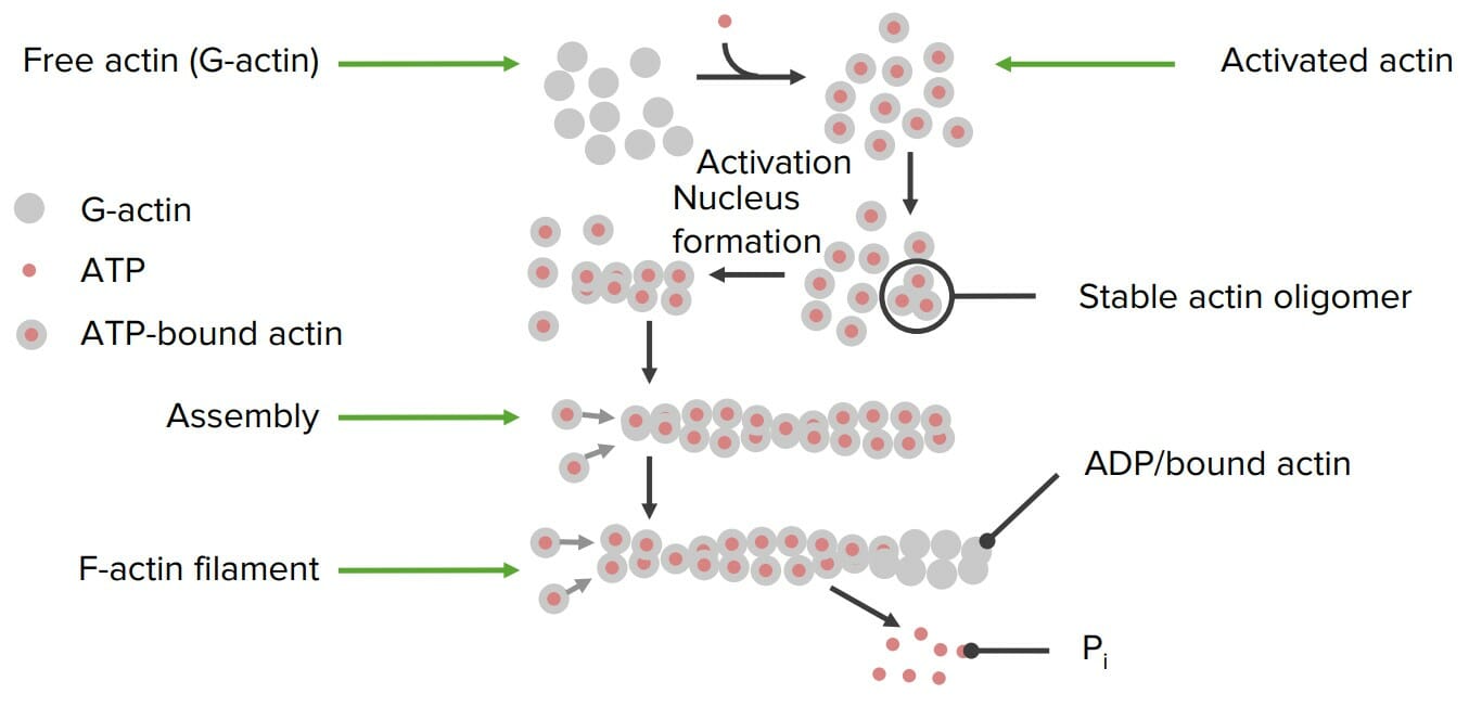
Formation of actin filaments from individual actin proteins: Free actin is activated with ATP. An activation nucleus forms and polymerization begins. Once the filament is assembled, an individual phosphate (Pi) is released from the individual actin proteins, which “deactivates” it, resulting in a stable final filament. An ADP remains bound to each actin.
KinesinKinesinA microtubule-associated mechanical adenosine triphosphatase, that uses the energy of ATP hydrolysis to move organelles along microtubules toward the plus end of the microtubule. The protein is found in squid axoplasm, optic lobes, and in bovine brain. Bovine kinesin is a heterotetramer composed of two heavy (120 kda) and two light (62 kda) chains.The Cell: Cytosol and Cytoskeleton and dyneinDyneinA family of multisubunit cytoskeletal motor proteins that use the energy of ATP hydrolysis, generated by a ring of aaa ATPases in the dynein heavy chain, to power a variety of cellular functions. Dyneins fall into two major classes based upon structural and functional criteria.The Cell: Cytosol and Cytoskeleton:
Use ATP energy to grab cargo and walk along microtubulesMicrotubulesSlender, cylindrical filaments found in the cytoskeleton of plant and animal cells. They are composed of the protein tubulin and are influenced by tubulin modulators.The Cell: Cytosol and Cytoskeleton
Move substances within the cell
KinesinKinesinA microtubule-associated mechanical adenosine triphosphatase, that uses the energy of ATP hydrolysis to move organelles along microtubules toward the plus end of the microtubule. The protein is found in squid axoplasm, optic lobes, and in bovine brain. Bovine kinesin is a heterotetramer composed of two heavy (120 kda) and two light (62 kda) chains.The Cell: Cytosol and Cytoskeleton walks one way down the microtubulesMicrotubulesSlender, cylindrical filaments found in the cytoskeleton of plant and animal cells. They are composed of the protein tubulin and are influenced by tubulin modulators.The Cell: Cytosol and Cytoskeleton; dyneinDyneinA family of multisubunit cytoskeletal motor proteins that use the energy of ATP hydrolysis, generated by a ring of aaa ATPases in the dynein heavy chain, to power a variety of cellular functions. Dyneins fall into two major classes based upon structural and functional criteria.The Cell: Cytosol and Cytoskeleton walks the other way
MyosinMyosinA diverse superfamily of proteins that function as translocating proteins. They share the common characteristics of being able to bind actins and hydrolyze mgATP. Myosins generally consist of heavy chains which are involved in locomotion, and light chains which are involved in regulation. Within the structure of myosin heavy chain are three domains: the head, the neck and the tail. The head region of the heavy chain contains the actin binding domain and mgATPase domain which provides energy for locomotion. The neck region is involved in binding the light-chains. The tail region provides the anchoring point that maintains the position of the heavy chain. The superfamily of myosins is organized into structural classes based upon the type and arrangement of the subunits they contain.Skeletal Muscle Contraction:
Uses ATP energy to walk down actinActinFilamentous proteins that are the main constituent of the thin filaments of muscle fibers. The filaments (known also as filamentous or f-actin) can be dissociated into their globular subunits; each subunit is composed of a single polypeptide 375 amino acids long. This is known as globular or g-actin. In conjunction with myosins, actin is responsible for the contraction and relaxation of muscle.Skeletal Muscle Contraction filaments via a process known as cross-bridge cyclingCross-bridge cyclingSkeletal Muscle Contraction
Causes muscular contraction
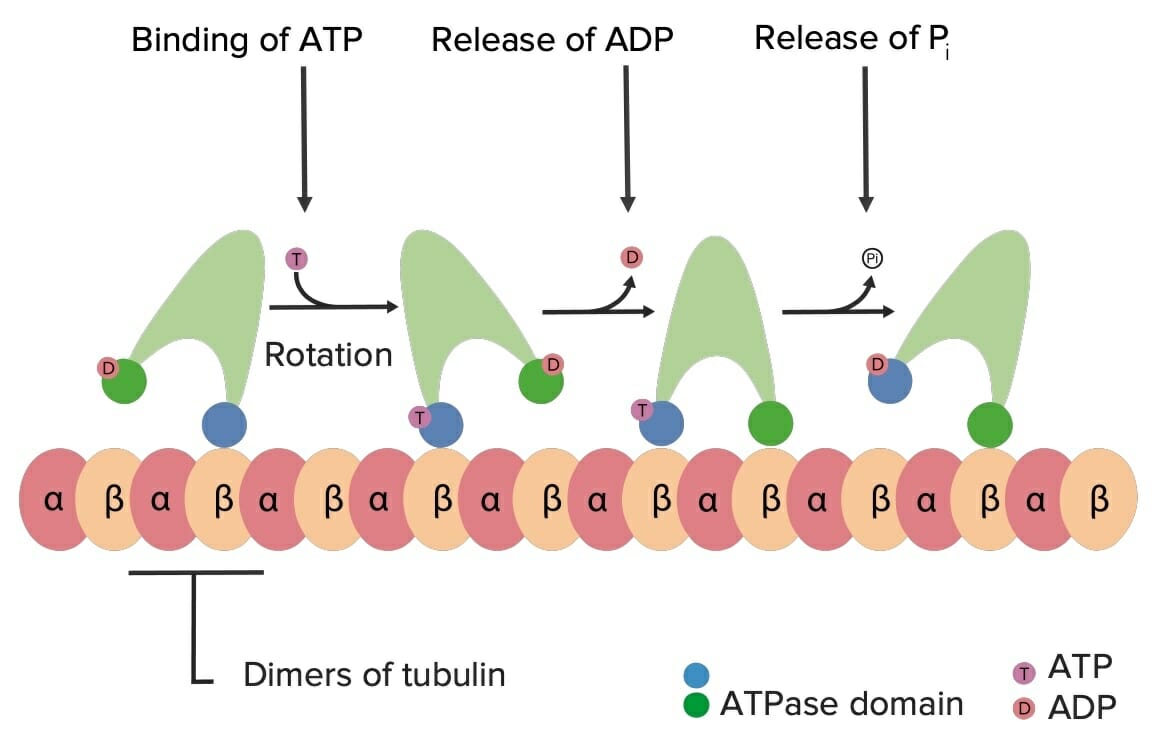
Example of how kinesin walks down microtubules using ATP energy: When ATP binds to kinesin, it results in a conformational change in the molecule, which causes it to “swivel” on the tubulin. This leads to a “walking” motion of the kinesin (and its cargo) down the microtubule “highways” within the cell. Pi: phosphate
These proteinsProteinsLinear polypeptides that are synthesized on ribosomes and may be further modified, crosslinked, cleaved, or assembled into complex proteins with several subunits. The specific sequence of amino acids determines the shape the polypeptide will take, during protein folding, and the function of the protein.Energy Homeostasis are responsible for:
Transmitting messages between and within cells
Responding to the environment
Coordinating responses between cells and organ systems
Membrane-bound receptorsReceptorsReceptors are proteins located either on the surface of or within a cell that can bind to signaling molecules known as ligands (e.g., hormones) and cause some type of response within the cell.Receptors:
Integrated into the cell membraneCell MembraneA cell membrane (also known as the plasma membrane or plasmalemma) is a biological membrane that separates the cell contents from the outside environment. A cell membrane is composed of a phospholipid bilayer and proteins that function to protect cellular DNA and mediate the exchange of ions and molecules. The Cell: Cell Membrane
Many are known as 7TM: they have 7 transmembrane coiled domains
Respond to an outside signal (e.g., hormone) → induces a conformational change → initiate a 2nd messenger within the cell
Intracellular receptorsReceptorsReceptors are proteins located either on the surface of or within a cell that can bind to signaling molecules known as ligands (e.g., hormones) and cause some type of response within the cell.Receptors:
ProteinsProteinsLinear polypeptides that are synthesized on ribosomes and may be further modified, crosslinked, cleaved, or assembled into complex proteins with several subunits. The specific sequence of amino acids determines the shape the polypeptide will take, during protein folding, and the function of the protein.Energy Homeostasis within a cell that bindBINDHyperbilirubinemia of the Newborn and respond to an outside signal (e.g., hormone)
Common with the cholesterol-derived lipophilic hormonesHormonesHormones are messenger molecules that are synthesized in one part of the body and move through the bloodstream to exert specific regulatory effects on another part of the body. Hormones play critical roles in coordinating cellular activities throughout the body in response to the constant changes in both the internal and external environments. Hormones: Overview and Types (e.g., estrogenEstrogenCompounds that interact with estrogen receptors in target tissues to bring about the effects similar to those of estradiol. Estrogens stimulate the female reproductive organs, and the development of secondary female sex characteristics. Estrogenic chemicals include natural, synthetic, steroidal, or non-steroidal compounds.Ovaries: AnatomyreceptorReceptorReceptors are proteins located either on the surface of or within a cell that can bind to signaling molecules known as ligands (e.g., hormones) and cause some type of response within the cell.Receptors)
Peptide hormonesHormonesHormones are messenger molecules that are synthesized in one part of the body and move through the bloodstream to exert specific regulatory effects on another part of the body. Hormones play critical roles in coordinating cellular activities throughout the body in response to the constant changes in both the internal and external environments. Hormones: Overview and Types:
Travel within the blood from one location to its target cells
Can bindBINDHyperbilirubinemia of the NewbornreceptorsReceptorsReceptors are proteins located either on the surface of or within a cell that can bind to signaling molecules known as ligands (e.g., hormones) and cause some type of response within the cell.Receptors on the membrane or within the cell
E.g., insulinInsulinInsulin is a peptide hormone that is produced by the beta cells of the pancreas. Insulin plays a role in metabolic functions such as glucose uptake, glycolysis, glycogenesis, lipogenesis, and protein synthesis. Exogenous insulin may be needed for individuals with diabetes mellitus, in whom there is a deficiency in endogenous insulin or increased insulin resistance. Insulin, oxytocin, many others
Neurotransmitters:
AAAAAmyloidosis derivatives that communicate nerve signals
E.g., norepinephrineNorepinephrinePrecursor of epinephrine that is secreted by the adrenal medulla and is a widespread central and autonomic neurotransmitter. Norepinephrine is the principal transmitter of most postganglionic sympathetic fibers, and of the diffuse projection system in the brain that arises from the locus ceruleus.Receptors and Neurotransmitters of the CNS, dopamineDopamineOne of the catecholamine neurotransmitters in the brain. It is derived from tyrosine and is the precursor to norepinephrine and epinephrine. Dopamine is a major transmitter in the extrapyramidal system of the brain, and important in regulating movement.Receptors and Neurotransmitters of the CNS
Signal transductionTransductionThe transfer of bacterial DNA by phages from an infected bacterium to another bacterium. This also refers to the transfer of genes into eukaryotic cells by viruses. This naturally occurring process is routinely employed as a gene transfer technique.BacteriologyproteinsProteinsLinear polypeptides that are synthesized on ribosomes and may be further modified, crosslinked, cleaved, or assembled into complex proteins with several subunits. The specific sequence of amino acids determines the shape the polypeptide will take, during protein folding, and the function of the protein.Energy Homeostasis:
ProteinsProteinsLinear polypeptides that are synthesized on ribosomes and may be further modified, crosslinked, cleaved, or assembled into complex proteins with several subunits. The specific sequence of amino acids determines the shape the polypeptide will take, during protein folding, and the function of the protein.Energy Homeostasis within a cell that pass the signal along
Primary mechanisms:
Creating or releasing secondary messengers (e.g., cAMPcAMPAn adenine nucleotide containing one phosphate group which is esterified to both the 3′- and 5′-positions of the sugar moiety. It is a second messenger and a key intracellular regulator, functioning as a mediator of activity for a number of hormones, including epinephrine, glucagon, and acth.Phosphodiesterase Inhibitors, IP3 (inositol trisphosphateInositol trisphosphateIntracellular messenger formed by the action of phospholipase C on phosphatidylinositol 4, 5-bisphosphate, which is one of the phospholipids that make up the cell membrane. Inositol 1, 4, 5-trisphosphate is released into the cytoplasm where it releases calcium ions from internal stores within the cell’s endoplasmic reticulum. These calcium ions stimulate the activity of B kinase or calmodulin.Second Messengers), CaCACondylomata acuminata are a clinical manifestation of genital HPV infection. Condylomata acuminata are described as raised, pearly, flesh-colored, papular, cauliflower-like lesions seen in the anogenital region that may cause itching, pain, or bleeding.Condylomata Acuminata (Genital Warts)2+)
PhosphorylationPhosphorylationThe introduction of a phosphoryl group into a compound through the formation of an ester bond between the compound and a phosphorus moiety.Post-translational Protein Processing cascades: A molecule phosphorylates the next molecule, activating that molecule so that it can go on to phosphorylate another molecule, and so on.
Examples:
G-proteins: frequently coupled to membrane receptorsReceptorsReceptors are proteins located either on the surface of or within a cell that can bind to signaling molecules known as ligands (e.g., hormones) and cause some type of response within the cell.Receptors
Transcription factorsTranscription FactorsEndogenous substances, usually proteins, which are effective in the initiation, stimulation, or termination of the genetic transcription process.Stages of Transcription (TFs):
Control which genesGenesA category of nucleic acid sequences that function as units of heredity and which code for the basic instructions for the development, reproduction, and maintenance of organisms.DNA Types and Structure are transcribed (i.e., control geneGeneA category of nucleic acid sequences that function as units of heredity and which code for the basic instructions for the development, reproduction, and maintenance of organisms.Basic Terms of Genetics expression)
Some of these proteinsProteinsLinear polypeptides that are synthesized on ribosomes and may be further modified, crosslinked, cleaved, or assembled into complex proteins with several subunits. The specific sequence of amino acids determines the shape the polypeptide will take, during protein folding, and the function of the protein.Energy Homeostasis are capable of binding to specific DNADNAA deoxyribonucleotide polymer that is the primary genetic material of all cells. Eukaryotic and prokaryotic organisms normally contain DNA in a double-stranded state, yet several important biological processes transiently involve single-stranded regions. DNA, which consists of a polysugar-phosphate backbone possessing projections of purines (adenine and guanine) and pyrimidines (thymine and cytosine), forms a double helix that is held together by hydrogen bonds between these purines and pyrimidines (adenine to thymine and guanine to cytosine).DNA Types and Structure sequences (known as enhancer or repressor sequences).
Transcription factorsTranscription FactorsEndogenous substances, usually proteins, which are effective in the initiation, stimulation, or termination of the genetic transcription process.Stages of Transcription may either:
Be required for transcriptionTranscriptionTranscription of genetic information is the first step in gene expression. Transcription is the process by which DNA is used as a template to make mRNA. This process is divided into 3 stages: initiation, elongation, and termination. Stages of Transcription to occur
Prevent transcriptionTranscriptionTranscription of genetic information is the first step in gene expression. Transcription is the process by which DNA is used as a template to make mRNA. This process is divided into 3 stages: initiation, elongation, and termination. Stages of Transcription from occurring
An incoming signal often affects TFs:
Allows the cell to respond by, for example, increasing or decreasing a particular protein product
That protein product may, in turn, be another TF, which controls the expression of a different geneGeneA category of nucleic acid sequences that function as units of heredity and which code for the basic instructions for the development, reproduction, and maintenance of organisms.Basic Terms of Genetics.
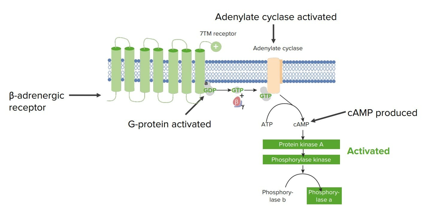
Example of how proteins are involved in cell signaling in a hepatocyte: β-adrenergic receptors are membrane-bound 7TM receptors (receptor with 7 transmembrane domains) that is bound to a G-protein on the cytosolic side, and they respond to circulating catecholamines (e.g., epinephrine, a monoamine derived from amino acids). Epinephrine induces a conformational change in the receptor, which activates the attached G-protein. The G-protein binds GTP and releases 2 of its subunits (β and 𝝲). The remaining GTP-bound α unit then activates another membrane-bound protein called adenylate cyclase. The adenylate cyclase converts ATP to cAMP, which is a common intracellular 2nd messenger. Here, cAMP activates protein kinase A (PKA), which phosphorylates phosphorylase kinase, activating it. The phosphorylase kinase then phosphorylates glycogen phosphorylase B, creating glycogen phosphorylase A, which is able to break down glycogen to produce molecules of glucose. In this example, the signaling molecule (epinephrine) triggered the formation of an intracellular 2nd messenger and then a phosphorylation cascade, resulting in release of glucose from the hepatocyte.
Image by Lecturio.
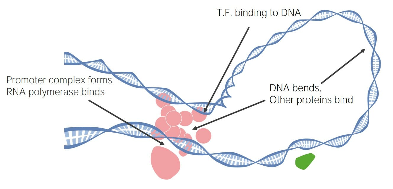
Transcription factors (TFs) can bind DNA, creating large transcription complexes that either promote or inhibit transcription, ultimately regulating gene expression.
EnzymesEnzymesEnzymes are complex protein biocatalysts that accelerate chemical reactions without being consumed by them. Due to the body’s constant metabolic needs, the absence of enzymes would make life unsustainable, as reactions would occur too slowly without these molecules. Basics of Enzymes
A class of proteinsProteinsLinear polypeptides that are synthesized on ribosomes and may be further modified, crosslinked, cleaved, or assembled into complex proteins with several subunits. The specific sequence of amino acids determines the shape the polypeptide will take, during protein folding, and the function of the protein.Energy Homeostasis capable of catalyzing reactions
Binding sites are highly specific to a particular antigenAntigenSubstances that are recognized by the immune system and induce an immune reaction.Vaccination
Can speed up the rate of a reaction by more than a quadrillion times
Involved in a wide variety of biologic functions, including:
Metabolism
Cellular respirationRespirationThe act of breathing with the lungs, consisting of inhalation, or the taking into the lungs of the ambient air, and of exhalation, or the expelling of the modified air which contains more carbon dioxide than the air taken in.Nose Anatomy (External & Internal)
Growth and development
DigestionDigestionDigestion refers to the process of the mechanical and chemical breakdown of food into smaller particles, which can then be absorbed and utilized by the body.Digestion and Absorption
Coagulation
Models for understanding enzyme function
There are 2 primary models that help explain how enzymesEnzymesEnzymes are complex protein biocatalysts that accelerate chemical reactions without being consumed by them. Due to the body’s constant metabolic needs, the absence of enzymes would make life unsustainable, as reactions would occur too slowly without these molecules. Basics of Enzymes work:
Fischer lock and key model (older model):
Postulates that the substrateSubstrateA substance upon which the enzyme acts.Basics of Enzymes fits into the enzyme’s binding site like a key fitting into a lock
Describes the specificity of antigenAntigenSubstances that are recognized by the immune system and induce an immune reaction.Vaccination/substrateSubstrateA substance upon which the enzyme acts.Basics of Enzymes to enzyme, but not how the actual catalyzation works
Koshland induced fit model (newer model):
Postulates that the enzyme’s binding site is close to fitting, but not a perfect fit for, the substrateSubstrateA substance upon which the enzyme acts.Basics of Enzymes
This means that when the substrateSubstrateA substance upon which the enzyme acts.Basics of Enzymes binds, there is a slight conformational shift in the enzyme → tension is stored as potential energy
This tension/energy then acts on the substrateSubstrateA substance upon which the enzyme acts.Basics of Enzymes, causing the reaction to occur.
Image showing the 2 theories of enzyme–substrate interaction
Another important function of proteinsProteinsLinear polypeptides that are synthesized on ribosomes and may be further modified, crosslinked, cleaved, or assembled into complex proteins with several subunits. The specific sequence of amino acids determines the shape the polypeptide will take, during protein folding, and the function of the protein.Energy Homeostasis is to transport and/or store biomolecules, including substances such as oxygen, vitamins and mineralsMineralsElectrolytes, hormonesHormonesHormones are messenger molecules that are synthesized in one part of the body and move through the bloodstream to exert specific regulatory effects on another part of the body. Hormones play critical roles in coordinating cellular activities throughout the body in response to the constant changes in both the internal and external environments. Hormones: Overview and Types, and more.
Circulating proteinsProteinsLinear polypeptides that are synthesized on ribosomes and may be further modified, crosslinked, cleaved, or assembled into complex proteins with several subunits. The specific sequence of amino acids determines the shape the polypeptide will take, during protein folding, and the function of the protein.Energy Homeostasis
Circulating proteinsProteinsLinear polypeptides that are synthesized on ribosomes and may be further modified, crosslinked, cleaved, or assembled into complex proteins with several subunits. The specific sequence of amino acids determines the shape the polypeptide will take, during protein folding, and the function of the protein.Energy Homeostasis carry substances through the blood and/or interstitial spaces; examples include:
Heme and myoglobinMyoglobinA conjugated protein which is the oxygen-transporting pigment of muscle. It is made up of one globin polypeptide chain and one heme group.Rhabdomyolysis:
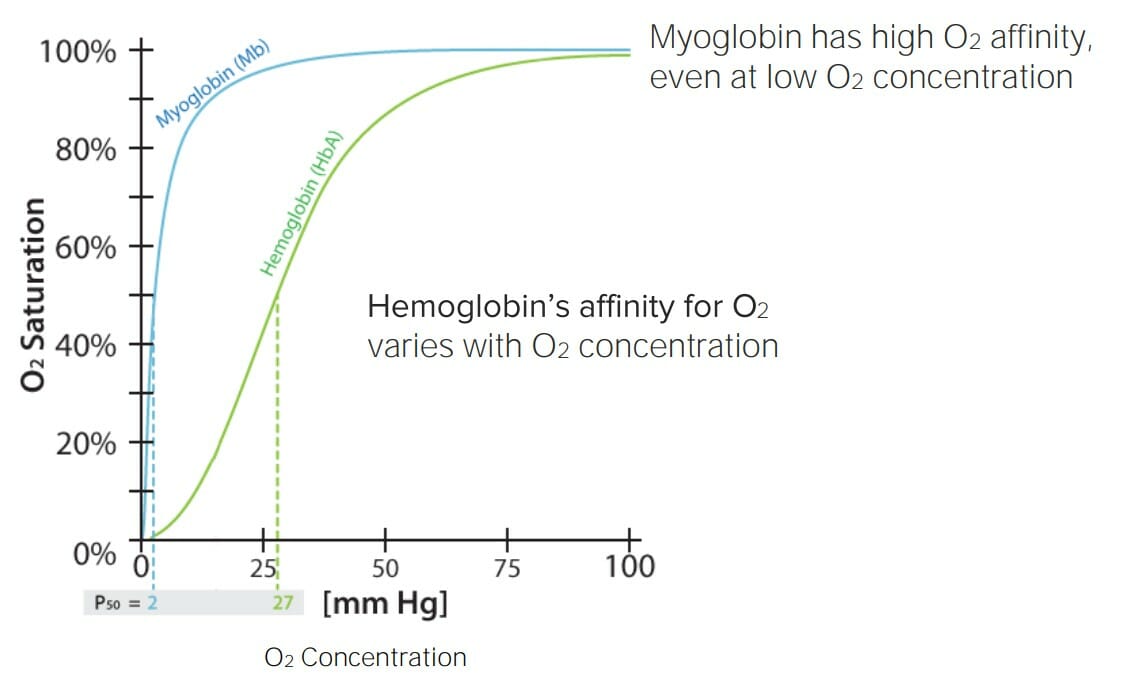
Heme’s properties make it an excellent O2 transport molecule:
Affinity for O2 varies depending on the surrounding O2 concentration
Readily binds O2 when O2 concentration is high → easily binds up O2 in the lungsLungsLungs are the main organs of the respiratory system. Lungs are paired viscera located in the thoracic cavity and are composed of spongy tissue. The primary function of the lungs is to oxygenate blood and eliminate CO2. Lungs: Anatomy during inhalation
Readily releases O2 when O2 concentrations are low → easily releases O2 in the tissues
MyoglobinMyoglobinA conjugated protein which is the oxygen-transporting pigment of muscle. It is made up of one globin polypeptide chain and one heme group.Rhabdomyolysis‘s properties make it an excellent O2 storage molecule:
Affinity for O2 is high, even at low O2 concentrations
Readily binds O2, regardless of the surrounding O2 concentration
Highly concentrated in muscles, where O2 is stored until needed during exercise
AlbuminAlbuminSerum albumin from humans. It is an essential carrier of both endogenous substances, such as fatty acids and bilirubin, and of xenobiotics in the blood.Liver Function Tests:
Binds to many other substances (e.g., hormonesHormonesHormones are messenger molecules that are synthesized in one part of the body and move through the bloodstream to exert specific regulatory effects on another part of the body. Hormones play critical roles in coordinating cellular activities throughout the body in response to the constant changes in both the internal and external environments. Hormones: Overview and Types, drugs) and transports them throughout the body
Also the primary modulator of plasmaPlasmaThe residual portion of blood that is left after removal of blood cells by centrifugation without prior blood coagulation.Transfusion Productsoncotic pressureOncotic PressureEdema
FerritinFerritinIron-containing proteins that are widely distributed in animals, plants, and microorganisms. Their major function is to store iron in a nontoxic bioavailable form. Each ferritin molecule consists of ferric iron in a hollow protein shell (apoferritins) made of 24 subunits of various sequences depending on the species and tissue types.Hereditary Hemochromatosis: an iron-binding protein that serves as the primary storage form of ironIronA metallic element with atomic symbol fe, atomic number 26, and atomic weight 55. 85. It is an essential constituent of hemoglobins; cytochromes; and iron-binding proteins. It plays a role in cellular redox reactions and in the transport of oxygen.Trace Elements in the body (an ironIronA metallic element with atomic symbol fe, atomic number 26, and atomic weight 55. 85. It is an essential constituent of hemoglobins; cytochromes; and iron-binding proteins. It plays a role in cellular redox reactions and in the transport of oxygen.Trace ElementsreservoirReservoirAnimate or inanimate sources which normally harbor disease-causing organisms and thus serve as potential sources of disease outbreaks. Reservoirs are distinguished from vectors (disease vectors) and carriers, which are agents of disease transmission rather than continuing sources of potential disease outbreaks. Humans may serve both as disease reservoirs and carriers.Escherichia coli)
Affinity of hemoglobin and myoglobin for oxygen (O2) depending on O2 saturation: Note how hemoglobin’s affinity varies based on the surrounding O2 saturation. This means that hemoglobin will readily bind O2 when O2 is plentiful (e.g., during inhalation in the lungs), but will readily release it when O2 saturation is low (e.g., in the tissues). This makes hemoglobin an excellent O2 transport molecule. On the other hand, myoglobin has a high affinity for O2 regardless of the surrounding O2 saturation, meaning it will readily bind O2 and will not release it until the surrounding O2 saturation is nearly 0. This makes myoglobin an excellent O2 storage molecule. P50: pressure at which 50% of the molecules (hemoglobin or myoglobin) are saturated with O2/p>
Image by Lecturio.
Membrane-bound proteinsProteinsLinear polypeptides that are synthesized on ribosomes and may be further modified, crosslinked, cleaved, or assembled into complex proteins with several subunits. The specific sequence of amino acids determines the shape the polypeptide will take, during protein folding, and the function of the protein.Energy Homeostasis
Membrane-bound proteinsProteinsLinear polypeptides that are synthesized on ribosomes and may be further modified, crosslinked, cleaved, or assembled into complex proteins with several subunits. The specific sequence of amino acids determines the shape the polypeptide will take, during protein folding, and the function of the protein.Energy Homeostasis move substances through the cell membraneCell MembraneA cell membrane (also known as the plasma membrane or plasmalemma) is a biological membrane that separates the cell contents from the outside environment. A cell membrane is composed of a phospholipid bilayer and proteins that function to protect cellular DNA and mediate the exchange of ions and molecules. The Cell: Cell Membrane. Examples include:
Allow a particular molecule to travel through the membrane
Examples:
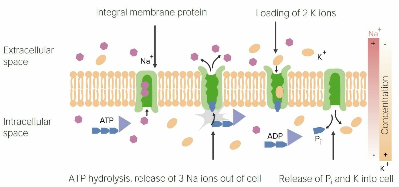
Epithelial sodiumSodiumA member of the alkali group of metals. It has the atomic symbol na, atomic number 11, and atomic weight 23.HyponatremiachannelsChannelsThe Cell: Cell Membrane (ENaCs): channelsChannelsThe Cell: Cell Membrane that reabsorb Na+ in the distal convoluted tubules in the kidneysKidneysThe kidneys are a pair of bean-shaped organs located retroperitoneally against the posterior wall of the abdomen on either side of the spine. As part of the urinary tract, the kidneys are responsible for blood filtration and excretion of water-soluble waste in the urine.Kidneys: Anatomy (site of action for amilorideAmilorideA pyrazine compound inhibiting sodium reabsorption through sodium channels in renal epithelial cells. This inhibition creates a negative potential in the luminal membranes of principal cells, located in the distal convoluted tubule and collecting duct. Negative potential reduces secretion of potassium and hydrogen ions. Amiloride is used in conjunction with diuretics to spare potassium loss.Liddle Syndrome, a K+-sparing diuretic)
Moves 3 Na+ out of the cell and 2 K+ into the cell (both ions are moving against their concentration gradients)
Requires ATP energy
Establishes an electrochemical gradientElectrochemical gradientThe Cell: Cell Membrane that is used in multiple processes (e.g., nerve transmission, nutrient absorptionAbsorptionAbsorption involves the uptake of nutrient molecules and their transfer from the lumen of the GI tract across the enterocytes and into the interstitial space, where they can be taken up in the venous or lymphatic circulation.Digestion and Absorption)
Image showing the functioning of the Na/K-ATPase transporter, which is a major function of proteins Pi: phosphate
Globular glycoproteinsGlycoproteinsConjugated protein-carbohydrate compounds including mucins, mucoid, and amyloid glycoproteins.Basics of Carbohydrates produced by the adaptive immune systemImmune systemThe body’s defense mechanism against foreign organisms or substances and deviant native cells. It includes the humoral immune response and the cell-mediated response and consists of a complex of interrelated cellular, molecular, and genetic components.Primary Lymphatic Organs
Neutralize toxins and pathogens by binding to and “covering” attachment sites
Activate the complement systemComplement systemSerum glycoproteins participating in the host defense mechanism of complement activation that creates the complement membrane attack complex. Included are glycoproteins in the various pathways of complement activation (classical complement pathway; alternative complement pathway; and lectin complement pathway).Innate Immunity: Barriers, Complement, and Cytokines
Opsonization (coating pathogens that enhance phagocytosisPhagocytosisThe engulfing and degradation of microorganisms; other cells that are dead, dying, or pathogenic; and foreign particles by phagocytic cells (phagocytes).Innate Immunity: Phagocytes and Antigen Presentation)
Present in 2 forms:
Soluble: secreted in the blood
Membrane-bound
Structure
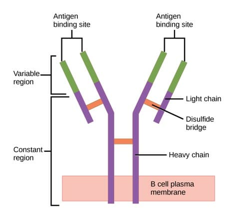
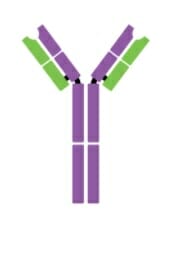
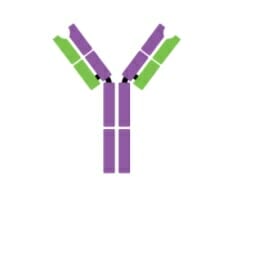
Consist of 4 polypeptide chains:
2 heavy chainsHeavy chainsThe largest of polypeptide chains comprising immunoglobulins. They contain 450 to 600 amino acid residues per chain, and have molecular weights of 51-72 kda.Immunoglobulins: Types and Functions: form a Y shape joined by disulfide bonds
2 light joins: bound to the heavy chainsHeavy chainsThe largest of polypeptide chains comprising immunoglobulins. They contain 450 to 600 amino acid residues per chain, and have molecular weights of 51-72 kda.Immunoglobulins: Types and Functions along the upper arms of the Y by disulfide bonds
2 primary regions:
VariableVariableVariables represent information about something that can change. The design of the measurement scales, or of the methods for obtaining information, will determine the data gathered and the characteristics of that data. As a result, a variable can be qualitative or quantitative, and may be further classified into subgroups.Types of Variables regions:
Located at the tips of the light and heavy chainsHeavy chainsThe largest of polypeptide chains comprising immunoglobulins. They contain 450 to 600 amino acid residues per chain, and have molecular weights of 51-72 kda.Immunoglobulins: Types and Functions
Responsible for identifying antigens
Can produce an incredibly wide variety of variableVariableVariables represent information about something that can change. The design of the measurement scales, or of the methods for obtaining information, will determine the data gathered and the characteristics of that data. As a result, a variable can be qualitative or quantitative, and may be further classified into subgroups.Types of Variables regions owing to DNADNAA deoxyribonucleotide polymer that is the primary genetic material of all cells. Eukaryotic and prokaryotic organisms normally contain DNA in a double-stranded state, yet several important biological processes transiently involve single-stranded regions. DNA, which consists of a polysugar-phosphate backbone possessing projections of purines (adenine and guanine) and pyrimidines (thymine and cytosine), forms a double helix that is held together by hydrogen bonds between these purines and pyrimidines (adenine to thymine and guanine to cytosine).DNA Types and Structure shuffling
Constant regions: regions that are relatively consistent in structure among antibodiesAntibodiesImmunoglobulins (Igs), also known as antibodies, are glycoprotein molecules produced by plasma cells that act in immune responses by recognizing and binding particular antigens. The various Ig classes are IgG (the most abundant), IgM, IgE, IgD, and IgA, which differ in their biologic features, structure, target specificity, and distribution.Immunoglobulins: Types and Functions within the same class
Structure of the antibody (regions): Antibody has a unique variable region (formed by heavy and light chains) capable of binding a different antigen and a constant region (formed by heavy chains).
Image: “Figure 42.22” by OpenStax. License: CC BY 4.0, cropped by Lecturio.
There are 5 different classes of immunoglobulinsImmunoglobulinsImmunoglobulins (Igs), also known as antibodies, are glycoprotein molecules produced by plasma cells that act in immune responses by recognizing and binding particular antigens. The various Ig classes are IgG (the most abundant), IgM, IgE, IgD, and IgA, which differ in their biologic features, structure, target specificity, and distribution.Immunoglobulins: Types and Functions:
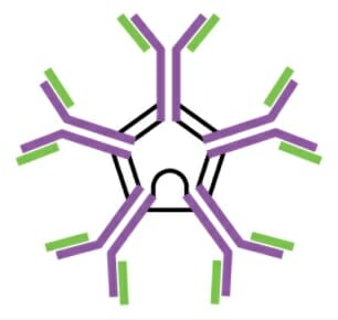
IgMIgMA class of immunoglobulin bearing mu chains (immunoglobulin mu-chains). Igm can fix complement. The name comes from its high molecular weight and originally being called a macroglobulin.Immunoglobulins: Types and Functions:
Facilitates activation of the B cellsB cellsLymphoid cells concerned with humoral immunity. They are short-lived cells resembling bursa-derived lymphocytes of birds in their production of immunoglobulin upon appropriate stimulation.B cells: Types and Functions by binding to helper T cellsT cellsLymphocytes responsible for cell-mediated immunity. Two types have been identified – cytotoxic (t-lymphocytes, cytotoxic) and helper T-lymphocytes (t-lymphocytes, helper-inducer). They are formed when lymphocytes circulate through the thymus gland and differentiate to thymocytes. When exposed to an antigen, they divide rapidly and produce large numbers of new T cells sensitized to that antigen.T cells: Types and Functions
Fixes complement, leading to lysis of microorganisms
Can agglutinate pathogens, thus facilitating pathogen eliminationEliminationThe initial damage and destruction of tumor cells by innate and adaptive immunity. Completion of the phase means no cancer growth. Cancer Immunotherapy
Monomer form serves as a B-cell receptorReceptorReceptors are proteins located either on the surface of or within a cell that can bind to signaling molecules known as ligands (e.g., hormones) and cause some type of response within the cell.Receptors (BCRBCRLymphocytes: Histology) in naive B cellsNaive B cellsB cells: Types and Functions.
IgGIgGThe major immunoglobulin isotype class in normal human serum. There are several isotype subclasses of igg, for example, igg1, igg2a, and igg2b.Hypersensitivity Pneumonitis:
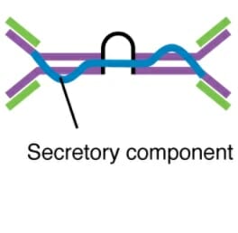
IgAIgARepresents 15-20% of the human serum immunoglobulins, mostly as the 4-chain polymer in humans or dimer in other mammals. Secretory iga is the main immunoglobulin in secretions.Immunoglobulins: Types and Functions:
Major IgIgX-linked Agammaglobulinemia in secretions: tears, salivaSalivaThe clear, viscous fluid secreted by the salivary glands and mucous glands of the mouth. It contains mucins, water, organic salts, and ptyalin.Salivary Glands: Anatomy, colostrumColostrumThe thin, yellow, serous fluid secreted by the mammary glands during pregnancy and immediately postpartum before lactation begins. It consists of immunologically active substances, white blood cells, water, protein, fat, and carbohydrates.Breastfeeding, mucus
Binding of allergen to IgEIgEAn immunoglobulin associated with mast cells. Overexpression has been associated with allergic hypersensitivity.Immunoglobulins: Types and Functions triggers release of inflammatory mediators from mast cellsMast cellsGranulated cells that are found in almost all tissues, most abundantly in the skin and the gastrointestinal tract. Like the basophils, mast cells contain large amounts of histamine and heparin. Unlike basophils, mast cells normally remain in the tissues and do not circulate in the blood. Mast cells, derived from the bone marrow stem cells, are regulated by the stem cell factor.Innate Immunity: Phagocytes and Antigen Presentation and basophilsBasophilsGranular leukocytes characterized by a relatively pale-staining, lobate nucleus and cytoplasm containing coarse dark-staining granules of variable size and stainable by basic dyes.Innate Immunity: Phagocytes and Antigen Presentation (allergic response)
Important in eliminationEliminationThe initial damage and destruction of tumor cells by innate and adaptive immunity. Completion of the phase means no cancer growth. Cancer Immunotherapy of parasites: eosinophilsEosinophilsGranular leukocytes with a nucleus that usually has two lobes connected by a slender thread of chromatin, and cytoplasm containing coarse, round granules that are uniform in size and stainable by eosin.Innate Immunity: Phagocytes and Antigen PresentationbindBINDHyperbilirubinemia of the Newborn to IgE-coated helminthsHelminthsCommonly known as parasitic worms, this group includes the acanthocephala; nematoda; and platyhelminths. Some authors consider certain species of leeches that can become temporarily parasitic as helminths.Anthelmintic Drugs → kills the parasite
IgMIgMA class of immunoglobulin bearing mu chains (immunoglobulin mu-chains). Igm can fix complement. The name comes from its high molecular weight and originally being called a macroglobulin.Immunoglobulins: Types and Functions pentameter
IgGIgGThe major immunoglobulin isotype class in normal human serum. There are several isotype subclasses of igg, for example, igg1, igg2a, and igg2b.Hypersensitivity Pneumonitis monometer
Secretory IgAIgARepresents 15-20% of the human serum immunoglobulins, mostly as the 4-chain polymer in humans or dimer in other mammals. Secretory iga is the main immunoglobulin in secretions.Immunoglobulins: Types and Functions dimer
IgEIgEAn immunoglobulin associated with mast cells. Overexpression has been associated with allergic hypersensitivity.Immunoglobulins: Types and Functions monomer
IgDIgDAn immunoglobulin which accounts for less than 1% of plasma immunoglobulin. It is found on the membrane of many circulating B lymphocytes.Immunoglobulins: Types and Functions monomer
Heavy chainsHeavy chainsThe largest of polypeptide chains comprising immunoglobulins. They contain 450 to 600 amino acid residues per chain, and have molecular weights of 51-72 kda.Immunoglobulins: Types and Functions
μ
γ
α
ε
δ
Number of antigen-binding sites
10
2
4
2
2
Molecular weight (daltons)
900,000
150,000
385,000
200,000
180,000
Percentage of total antibody in serum
6%
80%
13%
0.002%
1%
Crosses placentaPlacentaA highly vascularized mammalian fetal-maternal organ and major site of transport of oxygen, nutrients, and fetal waste products. It includes a fetal portion (chorionic villi) derived from trophoblasts and a maternal portion (decidua) derived from the uterine endometrium. The placenta produces an array of steroid, protein and peptide hormones (placental hormones).Placenta, Umbilical Cord, and Amniotic Cavity
No
Yes
No
No
No
Fixes complement
Yes
Yes
No
No
No
FcFcCrystallizable fragments composed of the carboxy-terminal halves of both immunoglobulin heavy chains linked to each other by disulfide bonds. Fc fragments contain the carboxy-terminal parts of the heavy chain constant regions that are responsible for the effector functions of an immunoglobulin (complement fixation, binding to the cell membrane via fc receptors, and placental transport). This fragment can be obtained by digestion of immunoglobulins with the proteolytic enzyme papain.Immunoglobulins: Types and Functions binds to
Phagocytes
Mast cellsMast cellsGranulated cells that are found in almost all tissues, most abundantly in the skin and the gastrointestinal tract. Like the basophils, mast cells contain large amounts of histamine and heparin. Unlike basophils, mast cells normally remain in the tissues and do not circulate in the blood. Mast cells, derived from the bone marrow stem cells, are regulated by the stem cell factor.Innate Immunity: Phagocytes and Antigen Presentation and basophilsBasophilsGranular leukocytes characterized by a relatively pale-staining, lobate nucleus and cytoplasm containing coarse dark-staining granules of variable size and stainable by basic dyes.Innate Immunity: Phagocytes and Antigen Presentation
Function
Main antibody of primary responses, best at fixing complement; the monomer form of IgMIgMA class of immunoglobulin bearing mu chains (immunoglobulin mu-chains). Igm can fix complement. The name comes from its high molecular weight and originally being called a macroglobulin.Immunoglobulins: Types and Functions serves as the B-cell receptorReceptorReceptors are proteins located either on the surface of or within a cell that can bind to signaling molecules known as ligands (e.g., hormones) and cause some type of response within the cell.Receptors
Main blood antibody of secondary responses, neutralizes toxins, opsonization
Secrets into mucus, tears, salivaSalivaThe clear, viscous fluid secreted by the salivary glands and mucous glands of the mouth. It contains mucins, water, organic salts, and ptyalin.Salivary Glands: Anatomy, colostrumColostrumThe thin, yellow, serous fluid secreted by the mammary glands during pregnancy and immediately postpartum before lactation begins. It consists of immunologically active substances, white blood cells, water, protein, fat, and carbohydrates.Breastfeeding
Antibody of allergic and antiparasitic activity
B-cell receptorReceptorReceptors are proteins located either on the surface of or within a cell that can bind to signaling molecules known as ligands (e.g., hormones) and cause some type of response within the cell.Receptors
A countless number of clinical disorders are caused by abnormalities or deficiencies of proteinsProteinsLinear polypeptides that are synthesized on ribosomes and may be further modified, crosslinked, cleaved, or assembled into complex proteins with several subunits. The specific sequence of amino acids determines the shape the polypeptide will take, during protein folding, and the function of the protein.Energy Homeostasis and/or abnormal protein metabolism. A few examples are listed below.
Enzyme abnormalities/deficiencies
HypercoagulableHypercoagulableHypercoagulable states (also referred to as thrombophilias) are a group of hematologic diseases defined by an increased risk of clot formation (i.e., thrombosis) due to either an increase in procoagulants, a decrease in anticoagulants, or a decrease in fibrinolysis. Hypercoagulable States or hypocoagulableHypocoagulableHypocoagulable conditions, also known as bleeding disorders or bleeding diathesis, are a diverse group of diseases that result in abnormal hemostasis. Physiologic hemostasis is dependent on the integrity of endothelial cells, subendothelial matrix, platelets, and coagulation factors. The hypocoagulable states result from abnormalities in one or more of these contributors, resulting in ineffective thrombosis and bleeding.Hypocoagulable Conditions states: Deficiencies or mutations of enzymesEnzymesEnzymes are complex protein biocatalysts that accelerate chemical reactions without being consumed by them. Due to the body’s constant metabolic needs, the absence of enzymes would make life unsustainable, as reactions would occur too slowly without these molecules. Basics of Enzymes involved in the coagulation cascadeCoagulation cascadeThe coagulation cascade is a series of reactions that ultimately generates a strong, cross-linked fibrin clot.Hemostasis can result in hypercoagulableHypercoagulableHypercoagulable states (also referred to as thrombophilias) are a group of hematologic diseases defined by an increased risk of clot formation (i.e., thrombosis) due to either an increase in procoagulants, a decrease in anticoagulants, or a decrease in fibrinolysis. Hypercoagulable States or hypocoagulableHypocoagulableHypocoagulable conditions, also known as bleeding disorders or bleeding diathesis, are a diverse group of diseases that result in abnormal hemostasis. Physiologic hemostasis is dependent on the integrity of endothelial cells, subendothelial matrix, platelets, and coagulation factors. The hypocoagulable states result from abnormalities in one or more of these contributors, resulting in ineffective thrombosis and bleeding.Hypocoagulable Conditions states.
Hemophilias: deficiencies of factor VIIIFactor VIIIFactor VIII of blood coagulation. Antihemophilic factor that is part of the factor viii/von Willebrand factor complex. Factor VIII is produced in the liver and acts in the intrinsic pathway of blood coagulation. It serves as a cofactor in factor X activation and this action is markedly enhanced by small amounts of thrombin.Hemostasis (hemophiliaHemophiliaThe hemophilias are a group of inherited, or sometimes acquired, disorders of secondary hemostasis due to deficiency of specific clotting factors. Hemophilia A is a deficiency of factor VIII, hemophilia B a deficiency of factor IX, and hemophilia C a deficiency of factor XI. Patients present with bleeding events that may be spontaneous or associated with minor or major trauma.Hemophilia A), factor IXFactor IXStorage-stable blood coagulation factor acting in the intrinsic pathway of blood coagulation. Its activated form, ixa, forms a complex with factor VIII and calcium on platelet factor 3 to activate factor X to Xa.Hemostasis (hemophiliaHemophiliaThe hemophilias are a group of inherited, or sometimes acquired, disorders of secondary hemostasis due to deficiency of specific clotting factors. Hemophilia A is a deficiency of factor VIII, hemophilia B a deficiency of factor IX, and hemophilia C a deficiency of factor XI. Patients present with bleeding events that may be spontaneous or associated with minor or major trauma.Hemophilia B), or factor XI (hemophiliaHemophiliaThe hemophilias are a group of inherited, or sometimes acquired, disorders of secondary hemostasis due to deficiency of specific clotting factors. Hemophilia A is a deficiency of factor VIII, hemophilia B a deficiency of factor IX, and hemophilia C a deficiency of factor XI. Patients present with bleeding events that may be spontaneous or associated with minor or major trauma.Hemophilia C), all of which are important enzymesEnzymesEnzymes are complex protein biocatalysts that accelerate chemical reactions without being consumed by them. Due to the body’s constant metabolic needs, the absence of enzymes would make life unsustainable, as reactions would occur too slowly without these molecules. Basics of Enzymes required to form clots. Hemophilias result in a hypocoagulableHypocoagulableHypocoagulable conditions, also known as bleeding disorders or bleeding diathesis, are a diverse group of diseases that result in abnormal hemostasis. Physiologic hemostasis is dependent on the integrity of endothelial cells, subendothelial matrix, platelets, and coagulation factors. The hypocoagulable states result from abnormalities in one or more of these contributors, resulting in ineffective thrombosis and bleeding.Hypocoagulable Conditions state and present with abnormal bleeding.
Phenylketonuria: metabolic disorder caused by mutations in the phenylalanine hydroxylasePhenylalanine hydroxylaseAn enzyme of the oxidoreductase class that catalyzes the formation of l-tyrosine, dihydrobiopterin, and water from l-phenylalanine, tetrahydrobiopterin, and oxygen.Synthesis of Nonessential Amino Acids (PAHPAHThe glycine amide of 4-aminobenzoic acid. Its sodium salt is used as a diagnostic aid to measure effective renal plasma flow (ERPF) and excretory capacity.Glomerular Filtration) geneGeneA category of nucleic acid sequences that function as units of heredity and which code for the basic instructions for the development, reproduction, and maintenance of organisms.Basic Terms of Genetics that encode the enzyme PAHPAHThe glycine amide of 4-aminobenzoic acid. Its sodium salt is used as a diagnostic aid to measure effective renal plasma flow (ERPF) and excretory capacity.Glomerular Filtration, which converts phenylalaninePhenylalanineAn essential aromatic amino acid that is a precursor of melanin; dopamine; noradrenalin (norepinephrine), and thyroxine.Synthesis of Nonessential Amino Acids to tyrosineTyrosineA non-essential amino acid. In animals it is synthesized from phenylalanine. It is also the precursor of epinephrine; thyroid hormones; and melanin.Synthesis of Nonessential Amino Acids. A disruption of this conversion leads to an accumulation of phenylalaninePhenylalanineAn essential aromatic amino acid that is a precursor of melanin; dopamine; noradrenalin (norepinephrine), and thyroxine.Synthesis of Nonessential Amino Acids, which causes damage to white matterWhite MatterThe region of central nervous system that appears lighter in color than the other type, gray matter. It mainly consists of myelinated nerve fibers and contains few neuronal cell bodies or dendrites.Brown-Séquard Syndrome tracts and myelin through unknown mechanisms, leading to neurologic deficitsNeurologic DeficitsHigh-Risk Headaches. In most cases, tyrosineTyrosineA non-essential amino acid. In animals it is synthesized from phenylalanine. It is also the precursor of epinephrine; thyroid hormones; and melanin.Synthesis of Nonessential Amino Acids levels are normal or slightly low.
Lysosomal storage diseases (LSDs): genetic mutationsGenetic MutationsCarcinogenesis of lysosomal enzymesEnzymesEnzymes are complex protein biocatalysts that accelerate chemical reactions without being consumed by them. Due to the body’s constant metabolic needs, the absence of enzymes would make life unsustainable, as reactions would occur too slowly without these molecules. Basics of Enzymes leading to dysfunctional metabolism and accumulation of glycosaminoglycans, glycoproteinsGlycoproteinsConjugated protein-carbohydrate compounds including mucins, mucoid, and amyloid glycoproteins.Basics of Carbohydrates, or glycolipidsGlycolipidsLipid attached to carbohydrate, outward-facing.The Cell: Cell Membrane. Examples of LSDs include Gaucher diseaseGaucher diseaseGaucher Disease (GD) is an autosomal recessive lysosomal storage disorder caused by a deficiency of glucocerebrosidase enzyme activity, resulting in accumulation of glucocerebroside in cells and certain organs. The disease is categorized into 3 types with variable clinical presentation. Gaucher Disease, Tay-Sachs diseaseTay-Sachs diseaseTay-Sachs disease is an autosomal recessive lysosomal storage disorder caused by genetic mutations in the hexosaminidase A (HEXA) gene, leading to progressive neurodegeneration. Classic symptoms in infants include rapid degeneration of cognitive and neuromuscular abilities, progressive blindness, and a macular cherry-red spot on physical examination.Tay-Sachs Disease, and mucopolysaccharidosesMucopolysaccharidosesThe mucopolysaccharidoses, a subset of the lysosomal storage diseases, are a group of inherited disorders characterized by absent or defective enzymes needed to break down carbohydrate chains called glycosaminoglycans (GAGs). These disorders lead to the accumulation of GAGs within cells.Mucopolysaccharidoses.
Glycogen storage diseasesGlycogen Storage DiseasesA group of inherited metabolic disorders involving the enzymes responsible for the synthesis and degradation of glycogen. In some patients, prominent liver involvement is presented. In others, more generalized storage of glycogen occurs, sometimes with prominent cardiac involvement.Benign Liver Tumors (GSDs): disorders characterized by abnormal glycogen breakdown due to genetic defects of one of the key enzymesEnzymesEnzymes are complex protein biocatalysts that accelerate chemical reactions without being consumed by them. Due to the body’s constant metabolic needs, the absence of enzymes would make life unsustainable, as reactions would occur too slowly without these molecules. Basics of Enzymes involved in the process. Deficiency of these enzymesEnzymesEnzymes are complex protein biocatalysts that accelerate chemical reactions without being consumed by them. Due to the body’s constant metabolic needs, the absence of enzymes would make life unsustainable, as reactions would occur too slowly without these molecules. Basics of Enzymes can cause hypoglycemiaHypoglycemiaHypoglycemia is an emergency condition defined as a serum glucose level ≤ 70 mg/dL (≤ 3.9 mmol/L) in diabetic patients. In nondiabetic patients, there is no specific or defined limit for normal serum glucose levels, and hypoglycemia is defined mainly by its clinical features. Hypoglycemia and/or abnormal glycogen deposition in tissues. The most common GSDs include von Gierke, Pompe, Cori, and McArdle diseases.
ScurvyScurvyAn acquired blood vessel disorder caused by severe deficiency of vitamin C (ascorbic acid) in the diet leading to defective collagen formation in small blood vessels. Scurvy is characterized by bleeding in any tissue, weakness, anemia, spongy gums, and a brawny induration of the muscles of the calves and legs.Water-soluble Vitamins and their Deficiencies: a dietary deficiency of vitamin CVitamin CA six carbon compound related to glucose. It is found naturally in citrus fruits and many vegetables. Ascorbic acid is an essential nutrient in human diets, and necessary to maintain connective tissue and bone. Its biologically active form, vitamin C, functions as a reducing agent and coenzyme in several metabolic pathways. Vitamin C is considered an antioxidant.Water-soluble Vitamins and their Deficiencies resulting in abnormal collagenCollagenA polypeptide substance comprising about one third of the total protein in mammalian organisms. It is the main constituent of skin; connective tissue; and the organic substance of bones (bone and bones) and teeth (tooth).Connective Tissue: Histology. Vitamin CVitamin CA six carbon compound related to glucose. It is found naturally in citrus fruits and many vegetables. Ascorbic acid is an essential nutrient in human diets, and necessary to maintain connective tissue and bone. Its biologically active form, vitamin C, functions as a reducing agent and coenzyme in several metabolic pathways. Vitamin C is considered an antioxidant.Water-soluble Vitamins and their Deficiencies is required for the hydroxylation of prolineProlineA non-essential amino acid that is synthesized from glutamic acid. It is an essential component of collagen and is important for proper functioning of joints and tendons.Synthesis of Nonessential Amino Acids in collagenCollagenA polypeptide substance comprising about one third of the total protein in mammalian organisms. It is the main constituent of skin; connective tissue; and the organic substance of bones (bone and bones) and teeth (tooth).Connective Tissue: Histology fibers. The hydroxyproline allows the formation of many hydrogen bonds, linking collagenCollagenA polypeptide substance comprising about one third of the total protein in mammalian organisms. It is the main constituent of skin; connective tissue; and the organic substance of bones (bone and bones) and teeth (tooth).Connective Tissue: Histology fibers together, which is very important for collagenCollagenA polypeptide substance comprising about one third of the total protein in mammalian organisms. It is the main constituent of skin; connective tissue; and the organic substance of bones (bone and bones) and teeth (tooth).Connective Tissue: Histology strength.
Duchenne’s muscular dystrophyMuscular DystrophyBecker Muscular Dystrophy (DMDDMDDuchenne muscular dystrophy (DMD) is an X-linked recessive genetic disorder that is caused by a mutation in the dmd gene. The mutation leads to the production of abnormal dystrophin, resulting in muscle-fiber destruction and replacement with fatty or fibrous tissue.Duchenne Muscular Dystrophy): an X-linked recessiveX-Linked RecessiveDuchenne Muscular Dystrophy genetic disorder resulting in abnormal dystrophinDystrophinA muscle protein localized in surface membranes which is the product of the Duchenne/Becker muscular dystrophy gene. Individuals with Duchenne muscular dystrophy usually lack dystrophin completely while those with Becker muscular dystrophy have dystrophin of an altered size. It shares features with other cytoskeletal proteins such as spectrin and alpha-actinin but the precise function of dystrophin is not clear. One possible role might be to preserve the integrity and alignment of the plasma membrane to the myofibrils during muscle contraction and relaxation.Blotting Techniques. DystrophinDystrophinA muscle protein localized in surface membranes which is the product of the Duchenne/Becker muscular dystrophy gene. Individuals with Duchenne muscular dystrophy usually lack dystrophin completely while those with Becker muscular dystrophy have dystrophin of an altered size. It shares features with other cytoskeletal proteins such as spectrin and alpha-actinin but the precise function of dystrophin is not clear. One possible role might be to preserve the integrity and alignment of the plasma membrane to the myofibrils during muscle contraction and relaxation.Blotting Techniques is a structural glycoprotein linking the cytoskeletonCytoskeletonThe network of filaments, tubules, and interconnecting filamentous bridges which give shape, structure, and organization to the cytoplasm.The Cell: Cytosol and Cytoskeleton and the extracellular matrixExtracellular matrixA meshwork-like substance found within the extracellular space and in association with the basement membrane of the cell surface. It promotes cellular proliferation and provides a supporting structure to which cells or cell lysates in culture dishes adhere.Hypertrophic and Keloid Scars of muscle (required for normal muscle function). Unable to regenerate normally, the muscle tissue is replaced with fibrousFibrousFibrocystic Change and fatty tissue.
Sickle cell anemiaSickle cell anemiaA disease characterized by chronic hemolytic anemia, episodic painful crises, and pathologic involvement of many organs. It is the clinical expression of homozygosity for hemoglobin S.Sickle Cell Disease: group of genetic disorders in which an abnormal hemoglobin protein (hemoglobin SHemoglobin SAn abnormal hemoglobin resulting from the substitution of valine for glutamic acid at position 6 of the beta chain of the globin moiety. The heterozygous state results in sickle cell trait, the homozygous in sickle cell anemia.Sickle Cell Disease) transforms RBCsRBCsErythrocytes, or red blood cells (RBCs), are the most abundant cells in the blood. While erythrocytes in the fetus are initially produced in the yolk sac then the liver, the bone marrow eventually becomes the main site of production.Erythrocytes: Histology into a sickle-shaped cell. This transformationTransformationChange brought about to an organism’s genetic composition by unidirectional transfer (transfection; transduction, genetic; conjugation, genetic, etc.) and incorporation of foreign DNA into prokaryotic or eukaryotic cells by recombination of part or all of that DNA into the cell’s genome.Bacteriology results in chronic anemiaAnemiaAnemia is a condition in which individuals have low Hb levels, which can arise from various causes. Anemia is accompanied by a reduced number of RBCs and may manifest with fatigue, shortness of breath, pallor, and weakness. Subtypes are classified by the size of RBCs, chronicity, and etiology. Anemia: Overview and Types, vaso-occlusive episodes, painPainAn unpleasant sensation induced by noxious stimuli which are detected by nerve endings of nociceptive neurons.Pain: Types and Pathways, and organ damage.
CysticCysticFibrocystic ChangefibrosisFibrosisAny pathological condition where fibrous connective tissue invades any organ, usually as a consequence of inflammation or other injury.Bronchiolitis Obliterans:autosomal recessiveAutosomal recessiveAutosomal inheritance, both dominant and recessive, refers to the transmission of genes from the 22 autosomal chromosomes. Autosomal recessive diseases are only expressed when 2 copies of the recessive allele are inherited.Autosomal Recessive and Autosomal Dominant Inheritance disorder caused by mutations in the geneGeneA category of nucleic acid sequences that function as units of heredity and which code for the basic instructions for the development, reproduction, and maintenance of organisms.Basic Terms of GeneticsCFTR. The mutations lead to dysfunction of chlorideChlorideInorganic compounds derived from hydrochloric acid that contain the Cl- ion.ElectrolyteschannelsChannelsThe Cell: Cell Membrane, which results in hyperviscous mucus and the accumulation of secretions.
Abnormal signaling and receptorReceptorReceptors are proteins located either on the surface of or within a cell that can bind to signaling molecules known as ligands (e.g., hormones) and cause some type of response within the cell.ReceptorsproteinsProteinsLinear polypeptides that are synthesized on ribosomes and may be further modified, crosslinked, cleaved, or assembled into complex proteins with several subunits. The specific sequence of amino acids determines the shape the polypeptide will take, during protein folding, and the function of the protein.Energy Homeostasis
Myasthenia gravisMyasthenia GravisMyasthenia gravis (MG) is an autoimmune neuromuscular disorder characterized by weakness and fatigability of skeletal muscles caused by dysfunction/destruction of acetylcholine receptors at the neuromuscular junction. MG presents with fatigue, ptosis, diplopia, dysphagia, respiratory difficulties, and progressive weakness in the limbs, leading to difficulty in movement. Myasthenia Gravis:autoimmune neuromuscular disorder characterized by weakness and fatigability of skeletal musclesSkeletal musclesA subtype of striated muscle, attached by tendons to the skeleton. Skeletal muscles are innervated and their movement can be consciously controlled. They are also called voluntary muscles.Muscle Tissue: Histology caused by dysfunction/destruction of acetylcholineAcetylcholineA neurotransmitter found at neuromuscular junctions, autonomic ganglia, parasympathetic effector junctions, a subset of sympathetic effector junctions, and at many sites in the central nervous system.Receptors and Neurotransmitters of the CNSreceptorsReceptorsReceptors are proteins located either on the surface of or within a cell that can bind to signaling molecules known as ligands (e.g., hormones) and cause some type of response within the cell.Receptors at the neuromuscular junctionNeuromuscular junctionThe synapse between a neuron and a muscle.Skeletal Muscle Contraction. Myasthenia presents with fatigueFatigueThe state of weariness following a period of exertion, mental or physical, characterized by a decreased capacity for work and reduced efficiency to respond to stimuli.Fibromyalgia, ptosisPtosisCranial Nerve Palsies, diplopiaDiplopiaA visual symptom in which a single object is perceived by the visual cortex as two objects rather than one. Disorders associated with this condition include refractive errors; strabismus; oculomotor nerve diseases; trochlear nerve diseases; abducens nerve diseases; and diseases of the brain stem and occipital lobe.Myasthenia Gravis, dysphagiaDysphagiaDysphagia is the subjective sensation of difficulty swallowing. Symptoms can range from a complete inability to swallow, to the sensation of solids or liquids becoming “stuck.” Dysphagia is classified as either oropharyngeal or esophageal, with esophageal dysphagia having 2 sub-types: functional and mechanical. Dysphagia, respiratory difficulties, and progressive weakness in the limbs, leading to difficulty in movement.
Graves’ disease:autoimmune disorderAutoimmune DisorderSeptic Arthritis characterized by the presence of circulating antibodiesAntibodiesImmunoglobulins (Igs), also known as antibodies, are glycoprotein molecules produced by plasma cells that act in immune responses by recognizing and binding particular antigens. The various Ig classes are IgG (the most abundant), IgM, IgE, IgD, and IgA, which differ in their biologic features, structure, target specificity, and distribution.Immunoglobulins: Types and Functions against the thyroid-stimulating hormoneThyroid-stimulating hormoneA glycoprotein hormone secreted by the adenohypophysis. Thyrotropin stimulates thyroid gland by increasing the iodide transport, synthesis and release of thyroid hormones (thyroxine and triiodothyronine).Thyroid Hormones (TSH) receptorsReceptorsReceptors are proteins located either on the surface of or within a cell that can bind to signaling molecules known as ligands (e.g., hormones) and cause some type of response within the cell.Receptors, causing the thyroidThyroidThe thyroid gland is one of the largest endocrine glands in the human body. The thyroid gland is a highly vascular, brownish-red gland located in the visceral compartment of the anterior region of the neck.Thyroid Gland: Anatomy gland to hyperfunction.
Type 2 diabetesDiabetesDiabetes mellitus (DM) is a metabolic disease characterized by hyperglycemia and dysfunction of the regulation of glucose metabolism by insulin. Type 1 DM is diagnosed mostly in children and young adults as the result of autoimmune destruction of β cells in the pancreas and the resulting lack of insulin. Type 2 DM has a significant association with obesity and is characterized by insulin resistance.Diabetes Mellitus mellitus:due primarily to peripheral insulin resistanceInsulin resistanceDiminished effectiveness of insulin in lowering blood sugar levels: requiring the use of 200 units or more of insulin per day to prevent hyperglycemia or ketosis.Diabetes Mellitus. InsulinInsulinInsulin is a peptide hormone that is produced by the beta cells of the pancreas. Insulin plays a role in metabolic functions such as glucose uptake, glycolysis, glycogenesis, lipogenesis, and protein synthesis. Exogenous insulin may be needed for individuals with diabetes mellitus, in whom there is a deficiency in endogenous insulin or increased insulin resistance. Insulin itself is a peptide hormone that is responsible for maintaining normal blood glucoseGlucoseA primary source of energy for living organisms. It is naturally occurring and is found in fruits and other parts of plants in its free state. It is used therapeutically in fluid and nutrient replacement.Lactose Intolerance levels. Chronically elevated blood glucoseGlucoseA primary source of energy for living organisms. It is naturally occurring and is found in fruits and other parts of plants in its free state. It is used therapeutically in fluid and nutrient replacement.Lactose Intolerance results in chronically elevated insulinInsulinInsulin is a peptide hormone that is produced by the beta cells of the pancreas. Insulin plays a role in metabolic functions such as glucose uptake, glycolysis, glycogenesis, lipogenesis, and protein synthesis. Exogenous insulin may be needed for individuals with diabetes mellitus, in whom there is a deficiency in endogenous insulin or increased insulin resistance. InsulinsecretionSecretionCoagulation Studies, which in turn results in down-regulationDown-RegulationA negative regulatory effect on physiological processes at the molecular, cellular, or systemic level. At the molecular level, the major regulatory sites include membrane receptors, genes (gene expression regulation), mRNAs, and proteins.Pharmacokinetics and Pharmacodynamics and a decreased sensitivity of the insulinInsulinInsulin is a peptide hormone that is produced by the beta cells of the pancreas. Insulin plays a role in metabolic functions such as glucose uptake, glycolysis, glycogenesis, lipogenesis, and protein synthesis. Exogenous insulin may be needed for individuals with diabetes mellitus, in whom there is a deficiency in endogenous insulin or increased insulin resistance. InsulinreceptorReceptorReceptors are proteins located either on the surface of or within a cell that can bind to signaling molecules known as ligands (e.g., hormones) and cause some type of response within the cell.ReceptorsproteinsProteinsLinear polypeptides that are synthesized on ribosomes and may be further modified, crosslinked, cleaved, or assembled into complex proteins with several subunits. The specific sequence of amino acids determines the shape the polypeptide will take, during protein folding, and the function of the protein.Energy Homeostasis.
Complete androgen insensitivity syndromeComplete Androgen Insensitivity SyndromeAndrogen Insensitivity Syndrome:X-linked recessiveX-Linked RecessiveDuchenne Muscular Dystrophy condition in which a genetic mutationMutationGenetic mutations are errors in DNA that can cause protein misfolding and dysfunction. There are various types of mutations, including chromosomal, point, frameshift, and expansion mutations. Types of Mutations affects the function of androgen receptorsReceptorsReceptors are proteins located either on the surface of or within a cell that can bind to signaling molecules known as ligands (e.g., hormones) and cause some type of response within the cell.Receptors, leading to testosteroneTestosteroneA potent androgenic steroid and major product secreted by the leydig cells of the testis. Its production is stimulated by luteinizing hormone from the pituitary gland. In turn, testosterone exerts feedback control of the pituitary LH and FSH secretion. Depending on the tissues, testosterone can be further converted to dihydrotestosterone or estradiol.Androgens and AntiandrogensresistanceResistancePhysiologically, the opposition to flow of air caused by the forces of friction. As a part of pulmonary function testing, it is the ratio of driving pressure to the rate of air flow.Ventilation: Mechanics of Breathing. Individuals will have a 46,XY karyotypeKaryotypeThe full set of chromosomes presented as a systematized array of metaphase chromosomes from a photomicrograph of a single cell nucleus arranged in pairs in descending order of size and according to the position of the centromere.Congenital Malformations of the Female Reproductive System and undescended testesTestesGonadal Hormones, with external female genitalia and breast development (due to peripheral conversion of the excess testosteroneTestosteroneA potent androgenic steroid and major product secreted by the leydig cells of the testis. Its production is stimulated by luteinizing hormone from the pituitary gland. In turn, testosterone exerts feedback control of the pituitary LH and FSH secretion. Depending on the tissues, testosterone can be further converted to dihydrotestosterone or estradiol.Androgens and Antiandrogens to estrogenEstrogenCompounds that interact with estrogen receptors in target tissues to bring about the effects similar to those of estradiol. Estrogens stimulate the female reproductive organs, and the development of secondary female sex characteristics. Estrogenic chemicals include natural, synthetic, steroidal, or non-steroidal compounds.Ovaries: Anatomy).
Autoimmune disorders
Systemic lupus erythematosusSystemic lupus erythematosusSystemic lupus erythematosus (SLE) is a chronic autoimmune, inflammatory condition that causes immune-complex deposition in organs, resulting in systemic manifestations. Women, particularly those of African American descent, are more commonly affected.Systemic Lupus Erythematosus (SLESLESystemic lupus erythematosus (SLE) is a chronic autoimmune, inflammatory condition that causes immune-complex deposition in organs, resulting in systemic manifestations. Women, particularly those of African American descent, are more commonly affected.Systemic Lupus Erythematosus): chronic, autoimmune, inflammatory condition that causes immune-complex deposition in organs, resulting in systemic manifestations. Notable clinical features include a malar rashMalar RashSystemic Lupus Erythematosus, nondestructive arthritisArthritisAcute or chronic inflammation of joints.Osteoarthritis, lupus nephritisLupus nephritisGlomerulonephritis associated with autoimmune disease systemic lupus erythematosus. Lupus nephritis is histologically classified into 6 classes: class I – normal glomeruli, class II – pure mesangial alterations, class III – focal segmental glomerulonephritis, class IV – diffuse glomerulonephritis, class V – diffuse membranous glomerulonephritis, and class VI – advanced sclerosing glomerulonephritis (the world health organization classification 1982).Diffuse Proliferative Glomerulonephritis, serositisSerositisInflammation of a serous membrane.Systemic Lupus Erythematosus, cytopeniasCytopeniasIPEX Syndrome, thromboembolic disease, seizuresSeizuresA seizure is abnormal electrical activity of the neurons in the cerebral cortex that can manifest in numerous ways depending on the region of the brain affected. Seizures consist of a sudden imbalance that occurs between the excitatory and inhibitory signals in cortical neurons, creating a net excitation. The 2 major classes of seizures are focal and generalized. Seizures, and/or psychosis.
Rheumatoid arthritisArthritisAcute or chronic inflammation of joints.Osteoarthritis (RA):symmetric, inflammatory polyarthritisPolyarthritisRheumatoid Arthritis. Rheumatoid arthritisArthritisAcute or chronic inflammation of joints.Osteoarthritis typically presents in middle-aged women with joint swellingSwellingInflammation, painPainAn unpleasant sensation induced by noxious stimuli which are detected by nerve endings of nociceptive neurons.Pain: Types and Pathways, and morning stiffness. The pathophysiology is incompletely understood, but in many individuals, there is an increased expression of the enzyme converting arginineArginineAn essential amino acid that is physiologically active in the l-form.Urea Cycle to citrullineCitrullineUrea Cycle; antibodiesAntibodiesImmunoglobulins (Igs), also known as antibodies, are glycoprotein molecules produced by plasma cells that act in immune responses by recognizing and binding particular antigens. The various Ig classes are IgG (the most abundant), IgM, IgE, IgD, and IgA, which differ in their biologic features, structure, target specificity, and distribution.Immunoglobulins: Types and FunctionsbindBINDHyperbilirubinemia of the Newborn to these citrullinated proteinsProteinsLinear polypeptides that are synthesized on ribosomes and may be further modified, crosslinked, cleaved, or assembled into complex proteins with several subunits. The specific sequence of amino acids determines the shape the polypeptide will take, during protein folding, and the function of the protein.Energy Homeostasis, resulting in activation of the complement systemComplement systemSerum glycoproteins participating in the host defense mechanism of complement activation that creates the complement membrane attack complex. Included are glycoproteins in the various pathways of complement activation (classical complement pathway; alternative complement pathway; and lectin complement pathway).Innate Immunity: Barriers, Complement, and Cytokines.
IgAIgARepresents 15-20% of the human serum immunoglobulins, mostly as the 4-chain polymer in humans or dimer in other mammals. Secretory iga is the main immunoglobulin in secretions.Immunoglobulins: Types and Functions nephropathy (Berger disease): renal disease characterized by IgAIgARepresents 15-20% of the human serum immunoglobulins, mostly as the 4-chain polymer in humans or dimer in other mammals. Secretory iga is the main immunoglobulin in secretions.Immunoglobulins: Types and Functions deposition in the mesangiumMesangiumThe thin membranous structure supporting the adjoining glomerular capillaries. It is composed of glomerular mesangial cells and their extracellular matrix.IgA Nephropathy. Berger disease is the most common cause of primary glomerulonephritis in most developed countries. Common presenting features are gross hematuriaHematuriaPresence of blood in the urine.Renal Cell Carcinoma or asymptomatic, microscopic hematuriaHematuriaPresence of blood in the urine.Renal Cell Carcinoma on urinalysisUrinalysisExamination of urine by chemical, physical, or microscopic means. Routine urinalysis usually includes performing chemical screening tests, determining specific gravity, observing any unusual color or odor, screening for bacteriuria, and examining the sediment microscopically.Urinary Tract Infections (UTIs) in Children with a preceding upper respiratory or GI infectionGI infectionMicrosporidia/Microsporidiosis.
References
Ajomiwe, N., Mba, O. I., Joshua, C. C., & Nwonumara, G. N. (2024). Protein nutrition: Understanding structure, digestibility, and bioavailability for optimal health. Foods, 13(11), 1771. https://doi.org/10.3390/foods13111771
Boadu, F., & Cheng, J. (2024). Improving protein function prediction by learning and integrating representations of protein sequences and function labels. Bioinformatics Advances, 4(1), vbae120. https://doi.org/10.1093/bioadv/vbae120
Lin, B., Zhang, W., Zhang, Y., Zhao, Z., Wang, Y., Zhu, C., & Pan, R. (2024). A comprehensive review and comparison of existing computational methods for protein function prediction. Briefings in Bioinformatics, 25(4), bbae289. https://doi.org/10.1093/bib/bbae289