Hereditary hemochromatosis (HH) is an autosomal recessive disorder most often associated with HFE gene mutations. Patients have increased iron intestinal absorption and iron deposition in several organs, such as the liver, heart, skin, and pancreas. The clinical presentation is usually with nonspecific symptoms and signs such as fatigue and joint pain in patients over 40, abnormal liver function tests, and hypogonadism in men. Diagnosis is made by laboratory testing showing high transferrin saturation or ferritin indicative of iron overload. Other testing includes genetic testing for HFE variants and MRI to quantify liver or heart iron overload. If untreated, HH can lead to cirrhosis, type 2 diabetes, and bronze-colored skin. Other findings depend on the organ(s) involved. Genetic screening is recommended for family members. Management requires phlebotomy (or iron chelation therapy in some cases) to prevent disease progression. The prognosisdepends on the severity of iron overload and organ injury at the time of diagnosis and the extent of iron removal. The presence of hepatic fibrosis is a poor prognostic factor.
Hereditary hemochromatosisHemochromatosisA disorder of iron metabolism characterized by a triad of hemosiderosis; liver cirrhosis; and diabetes mellitus. It is caused by massive iron deposits in parenchymal cells that may develop after a prolonged increase of iron absorption.Hereditary Hemochromatosis (HHHHHereditary hemochromatosis (HH) is an autosomal recessive disorder most often associated with hfe gene mutations. Patients have increased iron intestinal absorption and iron deposition in several organs, such as the liver, heart, skin, and pancreas. The clinical presentation includes the triad of cirrhosis, diabetes, and skin bronzing.Hereditary Hemochromatosis) is an inherited disorder characterized by iron overloadIron overloadAn excessive accumulation of iron in the body due to a greater than normal absorption of iron from the gastrointestinal tract or from parenteral injection. This may arise from idiopathic hemochromatosis, excessive iron intake, chronic alcoholism, certain types of refractory anemia, or transfusional hemosiderosis.Hereditary Hemochromatosis that results in tissue injury and fibrosisFibrosisAny pathological condition where fibrous connective tissue invades any organ, usually as a consequence of inflammation or other injury.Bronchiolitis Obliterans.
Etiology[1,7–9,11]
There are 4 types of hereditary hemochromatosisHemochromatosisA disorder of iron metabolism characterized by a triad of hemosiderosis; liver cirrhosis; and diabetes mellitus. It is caused by massive iron deposits in parenchymal cells that may develop after a prolonged increase of iron absorption.Hereditary Hemochromatosis; classification depends on the mutated geneGeneA category of nucleic acid sequences that function as units of heredity and which code for the basic instructions for the development, reproduction, and maintenance of organisms.Basic Terms of Genetics.
HFE geneGeneA category of nucleic acid sequences that function as units of heredity and which code for the basic instructions for the development, reproduction, and maintenance of organisms.Basic Terms of Genetics:
Specific for HHHHHereditary hemochromatosis (HH) is an autosomal recessive disorder most often associated with hfe gene mutations. Patients have increased iron intestinal absorption and iron deposition in several organs, such as the liver, heart, skin, and pancreas. The clinical presentation includes the triad of cirrhosis, diabetes, and skin bronzing.Hereditary Hemochromatosis (type 1Type 1Spinal Muscular Atrophy, most common)
Within the human leukocyte antigenAntigenSubstances that are recognized by the immune system and induce an immune reaction.Vaccination (HLA) region on chromosomeChromosomeIn a prokaryotic cell or in the nucleus of a eukaryotic cell, a structure consisting of or containing DNA which carries the genetic information essential to the cell.Basic Terms of Genetics 6
Common missense mutations in the HFEgeneGeneA category of nucleic acid sequences that function as units of heredity and which code for the basic instructions for the development, reproduction, and maintenance of organisms.Basic Terms of Genetics:
C282Y (cysteine-to-tyrosine substitution at amino acidAmino acidAmino acids (AAs) are composed of a central carbon atom attached to a carboxyl group, an amino group, a hydrogen atom, and a side chain (R group). Basics of Amino Acids position 282)
H63D (histidine-to-aspartic acid substitution at amino acidAmino acidAmino acids (AAs) are composed of a central carbon atom attached to a carboxyl group, an amino group, a hydrogen atom, and a side chain (R group). Basics of Amino Acids position 63)
Common HHHHHereditary hemochromatosis (HH) is an autosomal recessive disorder most often associated with hfe gene mutations. Patients have increased iron intestinal absorption and iron deposition in several organs, such as the liver, heart, skin, and pancreas. The clinical presentation includes the triad of cirrhosis, diabetes, and skin bronzing.Hereditary Hemochromatosis genotypes:
Homozygosity for C282Y (C282Y/C282Y)
Compound heterozygosity (C282Y/H63D)
Less common: Homozygosity for H63D (H63D/H63D) has generally lower likelihood of clinically significant iron overloadIron overloadAn excessive accumulation of iron in the body due to a greater than normal absorption of iron from the gastrointestinal tract or from parenteral injection. This may arise from idiopathic hemochromatosis, excessive iron intake, chronic alcoholism, certain types of refractory anemia, or transfusional hemosiderosis.Hereditary Hemochromatosis
Low and variableVariableVariables represent information about something that can change. The design of the measurement scales, or of the methods for obtaining information, will determine the data gathered and the characteristics of that data. As a result, a variable can be qualitative or quantitative, and may be further classified into subgroups.Types of Variables clinical penetrancePenetranceThe percent frequency with which a dominant or homozygous recessive gene or gene combination manifests itself in the phenotype of the carriers.Familial Juvenile Polyposis:
Not all people who are homozygous for the HFE mutationMutationGenetic mutations are errors in DNA that can cause protein misfolding and dysfunction. There are various types of mutations, including chromosomal, point, frameshift, and expansion mutations. Types of Mutations develop hemochromatosisHemochromatosisA disorder of iron metabolism characterized by a triad of hemosiderosis; liver cirrhosis; and diabetes mellitus. It is caused by massive iron deposits in parenchymal cells that may develop after a prolonged increase of iron absorption.Hereditary Hemochromatosis.
50% of women and 25% of men have normal serum ferritinFerritinIron-containing proteins that are widely distributed in animals, plants, and microorganisms. Their major function is to store iron in a nontoxic bioavailable form. Each ferritin molecule consists of ferric iron in a hollow protein shell (apoferritins) made of 24 subunits of various sequences depending on the species and tissue types.Hereditary Hemochromatosis levels and iron overloadIron overloadAn excessive accumulation of iron in the body due to a greater than normal absorption of iron from the gastrointestinal tract or from parenteral injection. This may arise from idiopathic hemochromatosis, excessive iron intake, chronic alcoholism, certain types of refractory anemia, or transfusional hemosiderosis.Hereditary Hemochromatosis–related disease develops in a minority.
Non-HFE geneGeneA category of nucleic acid sequences that function as units of heredity and which code for the basic instructions for the development, reproduction, and maintenance of organisms.Basic Terms of Genetics:
Mutations in other genesGenesA category of nucleic acid sequences that function as units of heredity and which code for the basic instructions for the development, reproduction, and maintenance of organisms.DNA Types and Structure involved in ironIronA metallic element with atomic symbol fe, atomic number 26, and atomic weight 55. 85. It is an essential constituent of hemoglobins; cytochromes; and iron-binding proteins. It plays a role in cellular redox reactions and in the transport of oxygen.Trace Elements metabolism:
HAMP (hepcidinHepcidinForms of hepcidin, a cationic amphipathic peptide synthesized in the liver as a prepropeptide which is first processed into prohepcidin and then into the biologically active hepcidin forms, including in human the 20-, 22-, and 25-amino acid residue peptide forms. Hepcidin acts as a homeostatic regulators of iron metabolism and also possesses antimicrobial activity.Hereditary Hemochromatosis antimicrobial peptide)geneGeneA category of nucleic acid sequences that function as units of heredity and which code for the basic instructions for the development, reproduction, and maintenance of organisms.Basic Terms of Genetics or HJV (juvenile hemochromatosisJuvenile HemochromatosisHereditary Hemochromatosis) genesGenesA category of nucleic acid sequences that function as units of heredity and which code for the basic instructions for the development, reproduction, and maintenance of organisms.DNA Types and Structure → type 2 HHHHHereditary hemochromatosis (HH) is an autosomal recessive disorder most often associated with hfe gene mutations. Patients have increased iron intestinal absorption and iron deposition in several organs, such as the liver, heart, skin, and pancreas. The clinical presentation includes the triad of cirrhosis, diabetes, and skin bronzing.Hereditary Hemochromatosis
TFR2 (transferrinTransferrinAn iron-binding beta1-globulin that is synthesized in the liver and secreted into the blood. It plays a central role in the transport of iron throughout the circulation.Heme MetabolismreceptorReceptorReceptors are proteins located either on the surface of or within a cell that can bind to signaling molecules known as ligands (e.g., hormones) and cause some type of response within the cell.Receptors 2) geneGeneA category of nucleic acid sequences that function as units of heredity and which code for the basic instructions for the development, reproduction, and maintenance of organisms.Basic Terms of Genetics → type 3Type 3Spinal Muscular AtrophyHHHHHereditary hemochromatosis (HH) is an autosomal recessive disorder most often associated with hfe gene mutations. Patients have increased iron intestinal absorption and iron deposition in several organs, such as the liver, heart, skin, and pancreas. The clinical presentation includes the triad of cirrhosis, diabetes, and skin bronzing.Hereditary Hemochromatosis
SLC40A1 (solute carrierCarrierVaccination family 40 member 1) geneGeneA category of nucleic acid sequences that function as units of heredity and which code for the basic instructions for the development, reproduction, and maintenance of organisms.Basic Terms of Genetics → type 4Type 4Spinal Muscular AtrophyHHHHHereditary hemochromatosis (HH) is an autosomal recessive disorder most often associated with hfe gene mutations. Patients have increased iron intestinal absorption and iron deposition in several organs, such as the liver, heart, skin, and pancreas. The clinical presentation includes the triad of cirrhosis, diabetes, and skin bronzing.Hereditary Hemochromatosis (ferroportinFerroportinHelps export iron from the intestinal cell.Heme Metabolism disease)
Other rare genetic disorders
Effects of geneGeneA category of nucleic acid sequences that function as units of heredity and which code for the basic instructions for the development, reproduction, and maintenance of organisms.Basic Terms of Genetics mutations:
↑ Iron absorptionIron absorptionDigestion and Absorption in the intestine: up to 2–4 mg of dietary ironIronA metallic element with atomic symbol fe, atomic number 26, and atomic weight 55. 85. It is an essential constituent of hemoglobins; cytochromes; and iron-binding proteins. It plays a role in cellular redox reactions and in the transport of oxygen.Trace Elements/day (normal: 1–2 mg/day)
↑ IronIronA metallic element with atomic symbol fe, atomic number 26, and atomic weight 55. 85. It is an essential constituent of hemoglobins; cytochromes; and iron-binding proteins. It plays a role in cellular redox reactions and in the transport of oxygen.Trace Elements deposition in different organs (e.g., liverLiverThe liver is the largest gland in the human body. The liver is found in the superior right quadrant of the abdomen and weighs approximately 1.5 kilograms. Its main functions are detoxification, metabolism, nutrient storage (e.g., iron and vitamins), synthesis of coagulation factors, formation of bile, filtration, and storage of blood. Liver: Anatomy)
Table: Types of hereditary hemochromatosisHemochromatosisA disorder of iron metabolism characterized by a triad of hemosiderosis; liver cirrhosis; and diabetes mellitus. It is caused by massive iron deposits in parenchymal cells that may develop after a prolonged increase of iron absorption.Hereditary Hemochromatosis[1-3]
Types
MutationMutationGenetic mutations are errors in DNA that can cause protein misfolding and dysfunction. There are various types of mutations, including chromosomal, point, frameshift, and expansion mutations. Types of Mutations
Description
1
HFE
Classic form of hemochromatosisHemochromatosisA disorder of iron metabolism characterized by a triad of hemosiderosis; liver cirrhosis; and diabetes mellitus. It is caused by massive iron deposits in parenchymal cells that may develop after a prolonged increase of iron absorption.Hereditary Hemochromatosis
Autosomal recessiveAutosomal recessiveAutosomal inheritance, both dominant and recessive, refers to the transmission of genes from the 22 autosomal chromosomes. Autosomal recessive diseases are only expressed when 2 copies of the recessive allele are inherited.Autosomal Recessive and Autosomal Dominant Inheritance
2
2a: HJV (encodes hemojuvelinHemojuvelinHereditary Hemochromatosis)
2b: HAMP (encodes hepcidinHepcidinForms of hepcidin, a cationic amphipathic peptide synthesized in the liver as a prepropeptide which is first processed into prohepcidin and then into the biologically active hepcidin forms, including in human the 20-, 22-, and 25-amino acid residue peptide forms. Hepcidin acts as a homeostatic regulators of iron metabolism and also possesses antimicrobial activity.Hereditary Hemochromatosis)
Younger age at onset
Autosomal recessiveAutosomal recessiveAutosomal inheritance, both dominant and recessive, refers to the transmission of genes from the 22 autosomal chromosomes. Autosomal recessive diseases are only expressed when 2 copies of the recessive allele are inherited.Autosomal Recessive and Autosomal Dominant Inheritance
3
TFR2
Intermediate severity
Autosomal recessiveAutosomal recessiveAutosomal inheritance, both dominant and recessive, refers to the transmission of genes from the 22 autosomal chromosomes. Autosomal recessive diseases are only expressed when 2 copies of the recessive allele are inherited.Autosomal Recessive and Autosomal Dominant Inheritance
4
SLC40A1
Autosomal dominantAutosomal dominantAutosomal inheritance, both dominant and recessive, refers to the transmission of genes from the 22 autosomal chromosomes. Autosomal dominant diseases are expressed when only 1 copy of the dominant allele is inherited. Autosomal Recessive and Autosomal Dominant Inheritance
Iron overloadIron overloadAn excessive accumulation of iron in the body due to a greater than normal absorption of iron from the gastrointestinal tract or from parenteral injection. This may arise from idiopathic hemochromatosis, excessive iron intake, chronic alcoholism, certain types of refractory anemia, or transfusional hemosiderosis.Hereditary Hemochromatosis[1,6,11]
It is important to note that causes of iron overloadIron overloadAn excessive accumulation of iron in the body due to a greater than normal absorption of iron from the gastrointestinal tract or from parenteral injection. This may arise from idiopathic hemochromatosis, excessive iron intake, chronic alcoholism, certain types of refractory anemia, or transfusional hemosiderosis.Hereditary Hemochromatosis may overlap or occur at the same time.
Secondary iron overloadIron overloadAn excessive accumulation of iron in the body due to a greater than normal absorption of iron from the gastrointestinal tract or from parenteral injection. This may arise from idiopathic hemochromatosis, excessive iron intake, chronic alcoholism, certain types of refractory anemia, or transfusional hemosiderosis.Hereditary Hemochromatosis disorders (acquired):
Common causes:
Increased ironIronA metallic element with atomic symbol fe, atomic number 26, and atomic weight 55. 85. It is an essential constituent of hemoglobins; cytochromes; and iron-binding proteins. It plays a role in cellular redox reactions and in the transport of oxygen.Trace Elements intake (transfusions)
Ineffective erythropoiesisErythropoiesisThe production of red blood cells (erythrocytes). In humans, erythrocytes are produced by the yolk sac in the first trimester; by the liver in the second trimester; by the bone marrow in the third trimester and after birth. In normal individuals, the erythrocyte count in the peripheral blood remains relatively constant implying a balance between the rate of erythrocyte production and rate of destruction.Erythrocytes: Histology (thalassemiaThalassemiaThalassemia is a hereditary cause of microcytic hypochromic anemia and results from a deficiency in either the α or β globin chains, resulting in hemoglobinopathy. The presentation of thalassemia depends on the number of defective chains present and can range from being asymptomatic to rendering the more severely affected patients to be transfusion dependent. Thalassemia)
Loss of hepatocyte massMassThree-dimensional lesion that occupies a space within the breastImaging of the Breast resulting in reduced hepcidinHepcidinForms of hepcidin, a cationic amphipathic peptide synthesized in the liver as a prepropeptide which is first processed into prohepcidin and then into the biologically active hepcidin forms, including in human the 20-, 22-, and 25-amino acid residue peptide forms. Hepcidin acts as a homeostatic regulators of iron metabolism and also possesses antimicrobial activity.Hereditary Hemochromatosis (chronic liverLiverThe liver is the largest gland in the human body. The liver is found in the superior right quadrant of the abdomen and weighs approximately 1.5 kilograms. Its main functions are detoxification, metabolism, nutrient storage (e.g., iron and vitamins), synthesis of coagulation factors, formation of bile, filtration, and storage of blood. Liver: Anatomy disease)
IronIronA metallic element with atomic symbol fe, atomic number 26, and atomic weight 55. 85. It is an essential constituent of hemoglobins; cytochromes; and iron-binding proteins. It plays a role in cellular redox reactions and in the transport of oxygen.Trace Elements deposits affecting different organs exhibit similar clinical and pathologic features as HHHHHereditary hemochromatosis (HH) is an autosomal recessive disorder most often associated with hfe gene mutations. Patients have increased iron intestinal absorption and iron deposition in several organs, such as the liver, heart, skin, and pancreas. The clinical presentation includes the triad of cirrhosis, diabetes, and skin bronzing.Hereditary Hemochromatosis.
Epidemiology
A common genetic disorder among those of Northern European ancestry[1,2,6,7]
1 in 200–500 people
Highest in people of Celtic or Nordic origin
C282Y variant:
Most common finding in patientsPatientsIndividuals participating in the health care system for the purpose of receiving therapeutic, diagnostic, or preventive procedures.Clinician–Patient Relationship with HHHHHereditary hemochromatosis (HH) is an autosomal recessive disorder most often associated with hfe gene mutations. Patients have increased iron intestinal absorption and iron deposition in several organs, such as the liver, heart, skin, and pancreas. The clinical presentation includes the triad of cirrhosis, diabetes, and skin bronzing.Hereditary Hemochromatosis
85% of patientsPatientsIndividuals participating in the health care system for the purpose of receiving therapeutic, diagnostic, or preventive procedures.Clinician–Patient Relationship with HHHHHereditary hemochromatosis (HH) is an autosomal recessive disorder most often associated with hfe gene mutations. Patients have increased iron intestinal absorption and iron deposition in several organs, such as the liver, heart, skin, and pancreas. The clinical presentation includes the triad of cirrhosis, diabetes, and skin bronzing.Hereditary Hemochromatosis are homozygous for the C282Y mutationMutationGenetic mutations are errors in DNA that can cause protein misfolding and dysfunction. There are various types of mutations, including chromosomal, point, frameshift, and expansion mutations. Types of Mutations.
MortalityMortalityAll deaths reported in a given population.Measures of Health Status: 1.7 cases associated with hemochromatosisHemochromatosisA disorder of iron metabolism characterized by a triad of hemosiderosis; liver cirrhosis; and diabetes mellitus. It is caused by massive iron deposits in parenchymal cells that may develop after a prolonged increase of iron absorption.Hereditary Hemochromatosis per 10,000 deaths
Pathophysiology
The mechanism of iron overloadIron overloadAn excessive accumulation of iron in the body due to a greater than normal absorption of iron from the gastrointestinal tract or from parenteral injection. This may arise from idiopathic hemochromatosis, excessive iron intake, chronic alcoholism, certain types of refractory anemia, or transfusional hemosiderosis.Hereditary Hemochromatosis in all 4 types of hemochromatosisHemochromatosisA disorder of iron metabolism characterized by a triad of hemosiderosis; liver cirrhosis; and diabetes mellitus. It is caused by massive iron deposits in parenchymal cells that may develop after a prolonged increase of iron absorption.Hereditary Hemochromatosis is increased iron absorptionIron absorptionDigestion and Absorption by the GI tract; it is regulated by hepcidinHepcidinForms of hepcidin, a cationic amphipathic peptide synthesized in the liver as a prepropeptide which is first processed into prohepcidin and then into the biologically active hepcidin forms, including in human the 20-, 22-, and 25-amino acid residue peptide forms. Hepcidin acts as a homeostatic regulators of iron metabolism and also possesses antimicrobial activity.Hereditary Hemochromatosis and results in tissue ironIronA metallic element with atomic symbol fe, atomic number 26, and atomic weight 55. 85. It is an essential constituent of hemoglobins; cytochromes; and iron-binding proteins. It plays a role in cellular redox reactions and in the transport of oxygen.Trace Elements deposition.The shared pathway is inappropriately low hepcidinHepcidinForms of hepcidin, a cationic amphipathic peptide synthesized in the liver as a prepropeptide which is first processed into prohepcidin and then into the biologically active hepcidin forms, including in human the 20-, 22-, and 25-amino acid residue peptide forms. Hepcidin acts as a homeostatic regulators of iron metabolism and also possesses antimicrobial activity.Hereditary Hemochromatosis signaling and/or impaired ferroportinFerroportinHelps export iron from the intestinal cell.Heme Metabolism regulation.[11–14]
IronIronA metallic element with atomic symbol fe, atomic number 26, and atomic weight 55. 85. It is an essential constituent of hemoglobins; cytochromes; and iron-binding proteins. It plays a role in cellular redox reactions and in the transport of oxygen.Trace ElementshomeostasisHomeostasisThe processes whereby the internal environment of an organism tends to remain balanced and stable.Cell Injury and Death
Dietary ironIronA metallic element with atomic symbol fe, atomic number 26, and atomic weight 55. 85. It is an essential constituent of hemoglobins; cytochromes; and iron-binding proteins. It plays a role in cellular redox reactions and in the transport of oxygen.Trace Elementsundergoes intestinal uptake in the duodenumDuodenumThe shortest and widest portion of the small intestine adjacent to the pylorus of the stomach. It is named for having the length equal to about the width of 12 fingers.Small Intestine: Anatomy.
IronIronA metallic element with atomic symbol fe, atomic number 26, and atomic weight 55. 85. It is an essential constituent of hemoglobins; cytochromes; and iron-binding proteins. It plays a role in cellular redox reactions and in the transport of oxygen.Trace Elements moves from the duodenumDuodenumThe shortest and widest portion of the small intestine adjacent to the pylorus of the stomach. It is named for having the length equal to about the width of 12 fingers.Small Intestine: Anatomy via the ironIronA metallic element with atomic symbol fe, atomic number 26, and atomic weight 55. 85. It is an essential constituent of hemoglobins; cytochromes; and iron-binding proteins. It plays a role in cellular redox reactions and in the transport of oxygen.Trace Elements exporter, ferroportinFerroportinHelps export iron from the intestinal cell.Heme Metabolism (FpnFpnHereditary Hemochromatosis), and enters the circulationCirculationThe movement of the blood as it is pumped through the cardiovascular system.ABCDE Assessment via transferrinTransferrinAn iron-binding beta1-globulin that is synthesized in the liver and secreted into the blood. It plays a central role in the transport of iron throughout the circulation.Heme Metabolism (Tf).
TransferrinTransferrinAn iron-binding beta1-globulin that is synthesized in the liver and secreted into the blood. It plays a central role in the transport of iron throughout the circulation.Heme Metabolism supplies ironIronA metallic element with atomic symbol fe, atomic number 26, and atomic weight 55. 85. It is an essential constituent of hemoglobins; cytochromes; and iron-binding proteins. It plays a role in cellular redox reactions and in the transport of oxygen.Trace Elements to the bone marrowBone marrowThe soft tissue filling the cavities of bones. Bone marrow exists in two types, yellow and red. Yellow marrow is found in the large cavities of large bones and consists mostly of fat cells and a few primitive blood cells. Red marrow is a hematopoietic tissue and is the site of production of erythrocytes and granular leukocytes. Bone marrow is made up of a framework of connective tissue containing branching fibers with the frame being filled with marrow cells.Bone Marrow: Composition and Hematopoiesis for hemoglobin synthesisSynthesisPolymerase Chain Reaction (PCR).
As the red blood cellsRed blood cellsErythrocytes, or red blood cells (RBCs), are the most abundant cells in the blood. While erythrocytes in the fetus are initially produced in the yolk sac then the liver, the bone marrow eventually becomes the main site of production.Erythrocytes: Histology (RBCsRBCsErythrocytes, or red blood cells (RBCs), are the most abundant cells in the blood. While erythrocytes in the fetus are initially produced in the yolk sac then the liver, the bone marrow eventually becomes the main site of production.Erythrocytes: Histology) age, they are phagocytosed by macrophagesMacrophagesThe relatively long-lived phagocytic cell of mammalian tissues that are derived from blood monocytes. Main types are peritoneal macrophages; alveolar macrophages; histiocytes; kupffer cells of the liver; and osteoclasts. They may further differentiate within chronic inflammatory lesions to epithelioid cells or may fuse to form foreign body giant cells or langhans giant cells.Innate Immunity: Phagocytes and Antigen Presentation (in the spleenSpleenThe spleen is the largest lymphoid organ in the body, located in the LUQ of the abdomen, superior to the left kidney and posterior to the stomach at the level of the 9th-11th ribs just below the diaphragm. The spleen is highly vascular and acts as an important blood filter, cleansing the blood of pathogens and damaged erythrocytes. Spleen: Anatomy), releasing ironIronA metallic element with atomic symbol fe, atomic number 26, and atomic weight 55. 85. It is an essential constituent of hemoglobins; cytochromes; and iron-binding proteins. It plays a role in cellular redox reactions and in the transport of oxygen.Trace Elements again into the circulationCirculationThe movement of the blood as it is pumped through the cardiovascular system.ABCDE Assessment through FpnFpnHereditary Hemochromatosis.
IronIronA metallic element with atomic symbol fe, atomic number 26, and atomic weight 55. 85. It is an essential constituent of hemoglobins; cytochromes; and iron-binding proteins. It plays a role in cellular redox reactions and in the transport of oxygen.Trace Elements that is not used for biochemical processes is stored in ferritinFerritinIron-containing proteins that are widely distributed in animals, plants, and microorganisms. Their major function is to store iron in a nontoxic bioavailable form. Each ferritin molecule consists of ferric iron in a hollow protein shell (apoferritins) made of 24 subunits of various sequences depending on the species and tissue types.Hereditary Hemochromatosis.
IronIronA metallic element with atomic symbol fe, atomic number 26, and atomic weight 55. 85. It is an essential constituent of hemoglobins; cytochromes; and iron-binding proteins. It plays a role in cellular redox reactions and in the transport of oxygen.Trace Elements regulation via hepcidinHepcidinForms of hepcidin, a cationic amphipathic peptide synthesized in the liver as a prepropeptide which is first processed into prohepcidin and then into the biologically active hepcidin forms, including in human the 20-, 22-, and 25-amino acid residue peptide forms. Hepcidin acts as a homeostatic regulators of iron metabolism and also possesses antimicrobial activity.Hereditary Hemochromatosis[5]
Certain conditions require a decrease or increase in iron absorptionIron absorptionDigestion and Absorption and circulating ironIronA metallic element with atomic symbol fe, atomic number 26, and atomic weight 55. 85. It is an essential constituent of hemoglobins; cytochromes; and iron-binding proteins. It plays a role in cellular redox reactions and in the transport of oxygen.Trace Elements, a pathway regulated by hepcidinHepcidinForms of hepcidin, a cationic amphipathic peptide synthesized in the liver as a prepropeptide which is first processed into prohepcidin and then into the biologically active hepcidin forms, including in human the 20-, 22-, and 25-amino acid residue peptide forms. Hepcidin acts as a homeostatic regulators of iron metabolism and also possesses antimicrobial activity.Hereditary Hemochromatosis:
Liver-derived peptide regulating the plasmaPlasmaThe residual portion of blood that is left after removal of blood cells by centrifugation without prior blood coagulation.Transfusion ProductsironIronA metallic element with atomic symbol fe, atomic number 26, and atomic weight 55. 85. It is an essential constituent of hemoglobins; cytochromes; and iron-binding proteins. It plays a role in cellular redox reactions and in the transport of oxygen.Trace Elements concentration
Inhibits intestinal ironIronA metallic element with atomic symbol fe, atomic number 26, and atomic weight 55. 85. It is an essential constituent of hemoglobins; cytochromes; and iron-binding proteins. It plays a role in cellular redox reactions and in the transport of oxygen.Trace Elements uptake
Inhibits the release of ironIronA metallic element with atomic symbol fe, atomic number 26, and atomic weight 55. 85. It is an essential constituent of hemoglobins; cytochromes; and iron-binding proteins. It plays a role in cellular redox reactions and in the transport of oxygen.Trace Elements from macrophagesMacrophagesThe relatively long-lived phagocytic cell of mammalian tissues that are derived from blood monocytes. Main types are peritoneal macrophages; alveolar macrophages; histiocytes; kupffer cells of the liver; and osteoclasts. They may further differentiate within chronic inflammatory lesions to epithelioid cells or may fuse to form foreign body giant cells or langhans giant cells.Innate Immunity: Phagocytes and Antigen Presentation with old RBCsRBCsErythrocytes, or red blood cells (RBCs), are the most abundant cells in the blood. While erythrocytes in the fetus are initially produced in the yolk sac then the liver, the bone marrow eventually becomes the main site of production.Erythrocytes: Histology
IronIronA metallic element with atomic symbol fe, atomic number 26, and atomic weight 55. 85. It is an essential constituent of hemoglobins; cytochromes; and iron-binding proteins. It plays a role in cellular redox reactions and in the transport of oxygen.Trace Elements sensing is mediated by different proteinsProteinsLinear polypeptides that are synthesized on ribosomes and may be further modified, crosslinked, cleaved, or assembled into complex proteins with several subunits. The specific sequence of amino acids determines the shape the polypeptide will take, during protein folding, and the function of the protein.Energy Homeostasis:
HFE
TransferrinTransferrinAn iron-binding beta1-globulin that is synthesized in the liver and secreted into the blood. It plays a central role in the transport of iron throughout the circulation.Heme Metabolism (Tf) receptorReceptorReceptors are proteins located either on the surface of or within a cell that can bind to signaling molecules known as ligands (e.g., hormones) and cause some type of response within the cell.Receptors 2 (Tfr2)
↑ IronIronA metallic element with atomic symbol fe, atomic number 26, and atomic weight 55. 85. It is an essential constituent of hemoglobins; cytochromes; and iron-binding proteins. It plays a role in cellular redox reactions and in the transport of oxygen.Trace Elements: ↑ hepcidinHepcidinForms of hepcidin, a cationic amphipathic peptide synthesized in the liver as a prepropeptide which is first processed into prohepcidin and then into the biologically active hepcidin forms, including in human the 20-, 22-, and 25-amino acid residue peptide forms. Hepcidin acts as a homeostatic regulators of iron metabolism and also possesses antimicrobial activity.Hereditary Hemochromatosis to reduce ironIronA metallic element with atomic symbol fe, atomic number 26, and atomic weight 55. 85. It is an essential constituent of hemoglobins; cytochromes; and iron-binding proteins. It plays a role in cellular redox reactions and in the transport of oxygen.Trace Elements
↑ InflammationInflammationInflammation is a complex set of responses to infection and injury involving leukocytes as the principal cellular mediators in the body’s defense against pathogenic organisms. Inflammation is also seen as a response to tissue injury in the process of wound healing. The 5 cardinal signs of inflammation are pain, heat, redness, swelling, and loss of function. Inflammation: ↑ hepcidinHepcidinForms of hepcidin, a cationic amphipathic peptide synthesized in the liver as a prepropeptide which is first processed into prohepcidin and then into the biologically active hepcidin forms, including in human the 20-, 22-, and 25-amino acid residue peptide forms. Hepcidin acts as a homeostatic regulators of iron metabolism and also possesses antimicrobial activity.Hereditary Hemochromatosis to limitLimitA value (e.g., pressure or time) that should not be exceeded and which is specified by the operator to protect the lungInvasive Mechanical VentilationironIronA metallic element with atomic symbol fe, atomic number 26, and atomic weight 55. 85. It is an essential constituent of hemoglobins; cytochromes; and iron-binding proteins. It plays a role in cellular redox reactions and in the transport of oxygen.Trace Elements availability to microorganisms
↑ ErythropoietinErythropoietinGlycoprotein hormone, secreted chiefly by the kidney in the adult and the liver in the fetus, that acts on erythroid stem cells of the bone marrow to stimulate proliferation and differentiation.Erythrocytes: Histology: ↓ hepcidinHepcidinForms of hepcidin, a cationic amphipathic peptide synthesized in the liver as a prepropeptide which is first processed into prohepcidin and then into the biologically active hepcidin forms, including in human the 20-, 22-, and 25-amino acid residue peptide forms. Hepcidin acts as a homeostatic regulators of iron metabolism and also possesses antimicrobial activity.Hereditary Hemochromatosis to increase ironIronA metallic element with atomic symbol fe, atomic number 26, and atomic weight 55. 85. It is an essential constituent of hemoglobins; cytochromes; and iron-binding proteins. It plays a role in cellular redox reactions and in the transport of oxygen.Trace Elements for hematopoiesisHematopoiesisThe development and formation of various types of blood cells. Hematopoiesis can take place in the bone marrow (medullary) or outside the bone marrow (extramedullary hematopoiesis).Bone Marrow: Composition and Hematopoiesis
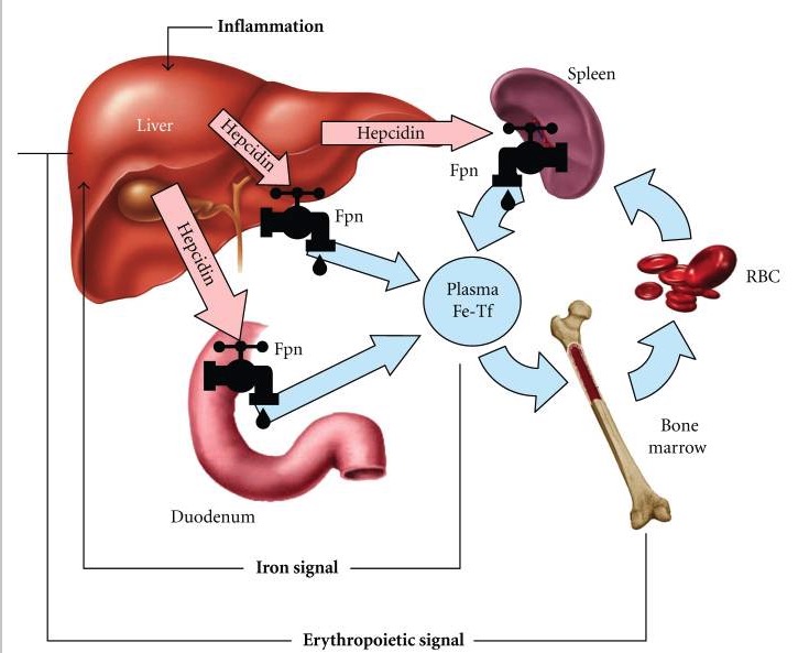
Hepcidin-ferroportin (Fpn) interaction determines the flow of iron into plasma: 1. Dietary iron undergoes intestinal uptake in the duodenum. 2.
Iron moves from the duodenum via the iron exporter, ferroportin (Fpn), and enters the circulation via transferrin (Tf). 3.
Tf supplies iron to the bone marrow for hemoglobin synthesis. 4.
As the red blood cells (RBC) age, they are phagocytosed by macrophages (mainly in the spleen), releasing iron again into the circulation through ferroportin. 5.
When there is increased iron or inflammation, the liver-derived hepcidin increases; this reduces duodenal absorption of iron and the release of iron from the degradation of old RBCs. 6.
When there is a need for more RBC formation, erythropoietin inhibits hepcidin to allow more iron absorption (from the intestine) and release from the macrophages with RBCs.
Image: “Hepcidin-ferroportin interaction” by David Geffen School of Medicine at UCLA, CHS 37-131, 10833 LeConte Avenue, Los Angeles, CA 90095-1690, USA. License: CC BY 3.0
Mutations in the hepcidinHepcidinForms of hepcidin, a cationic amphipathic peptide synthesized in the liver as a prepropeptide which is first processed into prohepcidin and then into the biologically active hepcidin forms, including in human the 20-, 22-, and 25-amino acid residue peptide forms. Hepcidin acts as a homeostatic regulators of iron metabolism and also possesses antimicrobial activity.Hereditary Hemochromatosis pathway[5,11]
HHHHHereditary hemochromatosis (HH) is an autosomal recessive disorder most often associated with hfe gene mutations. Patients have increased iron intestinal absorption and iron deposition in several organs, such as the liver, heart, skin, and pancreas. The clinical presentation includes the triad of cirrhosis, diabetes, and skin bronzing.Hereditary Hemochromatosis:
GeneGeneA category of nucleic acid sequences that function as units of heredity and which code for the basic instructions for the development, reproduction, and maintenance of organisms.Basic Terms of Genetics mutations involving the HFE protein and non-HFEproteinsProteinsLinear polypeptides that are synthesized on ribosomes and may be further modified, crosslinked, cleaved, or assembled into complex proteins with several subunits. The specific sequence of amino acids determines the shape the polypeptide will take, during protein folding, and the function of the protein.Energy Homeostasis (Tfr2, hemojuvelinHemojuvelinHereditary Hemochromatosis, FpnFpnHereditary Hemochromatosis, and hepcidinHepcidinForms of hepcidin, a cationic amphipathic peptide synthesized in the liver as a prepropeptide which is first processed into prohepcidin and then into the biologically active hepcidin forms, including in human the 20-, 22-, and 25-amino acid residue peptide forms. Hepcidin acts as a homeostatic regulators of iron metabolism and also possesses antimicrobial activity.Hereditary Hemochromatosis) → decreased hepcidinHepcidinForms of hepcidin, a cationic amphipathic peptide synthesized in the liver as a prepropeptide which is first processed into prohepcidin and then into the biologically active hepcidin forms, including in human the 20-, 22-, and 25-amino acid residue peptide forms. Hepcidin acts as a homeostatic regulators of iron metabolism and also possesses antimicrobial activity.Hereditary Hemochromatosis release and increased iron absorptionIron absorptionDigestion and Absorption
Increased iron absorptionIron absorptionDigestion and Absorption → saturation of Tf and progressive increase of ferritinFerritinIron-containing proteins that are widely distributed in animals, plants, and microorganisms. Their major function is to store iron in a nontoxic bioavailable form. Each ferritin molecule consists of ferric iron in a hollow protein shell (apoferritins) made of 24 subunits of various sequences depending on the species and tissue types.Hereditary Hemochromatosis → iron overloadIron overloadAn excessive accumulation of iron in the body due to a greater than normal absorption of iron from the gastrointestinal tract or from parenteral injection. This may arise from idiopathic hemochromatosis, excessive iron intake, chronic alcoholism, certain types of refractory anemia, or transfusional hemosiderosis.Hereditary Hemochromatosis → ironIronA metallic element with atomic symbol fe, atomic number 26, and atomic weight 55. 85. It is an essential constituent of hemoglobins; cytochromes; and iron-binding proteins. It plays a role in cellular redox reactions and in the transport of oxygen.Trace Elements deposition
↑ ironIronA metallic element with atomic symbol fe, atomic number 26, and atomic weight 55. 85. It is an essential constituent of hemoglobins; cytochromes; and iron-binding proteins. It plays a role in cellular redox reactions and in the transport of oxygen.Trace Elements results in excess oxidative stressOxidative stressA disturbance in the prooxidant-antioxidant balance in favor of the former, leading to potential damage. Indicators of oxidative stress include damaged DNA bases, protein oxidation products, and lipid peroxidation products.Cell Injury and Death → inflammationInflammationInflammation is a complex set of responses to infection and injury involving leukocytes as the principal cellular mediators in the body’s defense against pathogenic organisms. Inflammation is also seen as a response to tissue injury in the process of wound healing. The 5 cardinal signs of inflammation are pain, heat, redness, swelling, and loss of function. Inflammation and cell injuryCell injuryThe cell undergoes a variety of changes in response to injury, which may or may not lead to cell death. Injurious stimuli trigger the process of cellular adaptation, whereby cells respond to withstand the harmful changes in their environment. Overwhelmed adaptive mechanisms lead to cell injury. Mild stimuli produce reversible injury. If the stimulus is severe or persistent, injury becomes irreversible. Cell Injury and Death → organ damage
Men usually present in their 40s or later; women have slower ironIronA metallic element with atomic symbol fe, atomic number 26, and atomic weight 55. 85. It is an essential constituent of hemoglobins; cytochromes; and iron-binding proteins. It plays a role in cellular redox reactions and in the transport of oxygen.Trace Elements accumulation (presumably due to menstrual blood loss) and present after menopauseMenopauseMenopause is a physiologic process in women characterized by the permanent cessation of menstruation that occurs after the loss of ovarian activity. Menopause can only be diagnosed retrospectively, after 12 months without menstrual bleeding. Menopause. With patientsPatientsIndividuals participating in the health care system for the purpose of receiving therapeutic, diagnostic, or preventive procedures.Clinician–Patient Relationship being diagnosed earlier since the beginning of the 21st century, end-organ damage secondary to iron overloadIron overloadAn excessive accumulation of iron in the body due to a greater than normal absorption of iron from the gastrointestinal tract or from parenteral injection. This may arise from idiopathic hemochromatosis, excessive iron intake, chronic alcoholism, certain types of refractory anemia, or transfusional hemosiderosis.Hereditary Hemochromatosis is not often seen in clinical practice.[9,12]
General findings[6,8,11]
Manifestations depend on the degree of ironIronA metallic element with atomic symbol fe, atomic number 26, and atomic weight 55. 85. It is an essential constituent of hemoglobins; cytochromes; and iron-binding proteins. It plays a role in cellular redox reactions and in the transport of oxygen.Trace Elements accumulation.
Often asymptomatic; diagnosed incidentally on blood tests or on family screeningScreeningPreoperative Care
There is no “typical presentation;” patientsPatientsIndividuals participating in the health care system for the purpose of receiving therapeutic, diagnostic, or preventive procedures.Clinician–Patient Relationship often report fatigueFatigueThe state of weariness following a period of exertion, mental or physical, characterized by a decreased capacity for work and reduced efficiency to respond to stimuli.Fibromyalgia, joint painPainAn unpleasant sensation induced by noxious stimuli which are detected by nerve endings of nociceptive neurons.Pain: Types and Pathways, and erectile dysfunctionErectile DysfunctionErectile dysfunction (ED) is defined as the inability to achieve or maintain a penile erection, resulting in difficulty to perform penetrative sexual intercourse. Local penile factors and systemic diseases, including diabetes, cardiac disease, and neurological disorders, can cause ED. Erectile Dysfunction in men.
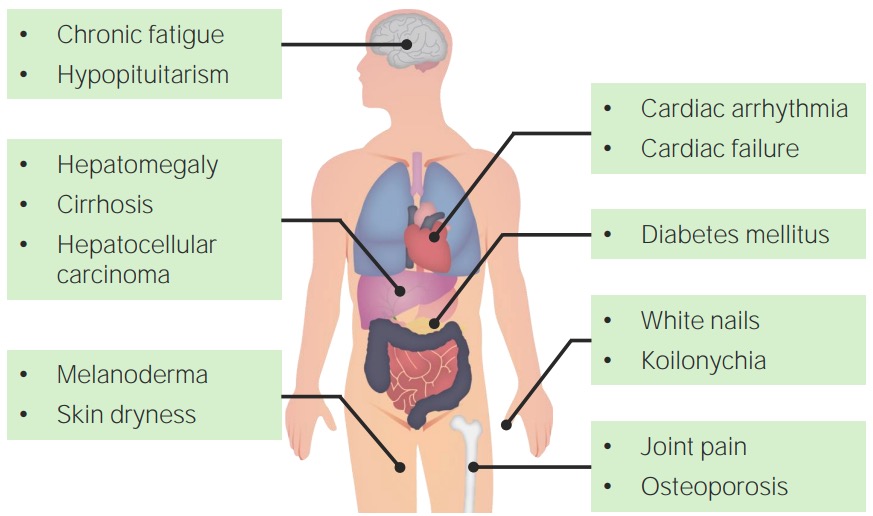
Clinical features have evolved substantially from the “usual presentation” a few decades ago—classic triad (found late in the disease):
CirrhosisCirrhosisCirrhosis is a late stage of hepatic parenchymal necrosis and scarring (fibrosis) most commonly due to hepatitis C infection and alcoholic liver disease. Patients may present with jaundice, ascites, and hepatosplenomegaly. Cirrhosis can also cause complications such as hepatic encephalopathy, portal hypertension, portal vein thrombosis, and hepatorenal syndrome. Cirrhosis
DiabetesDiabetesDiabetes mellitus (DM) is a metabolic disease characterized by hyperglycemia and dysfunction of the regulation of glucose metabolism by insulin. Type 1 DM is diagnosed mostly in children and young adults as the result of autoimmune destruction of β cells in the pancreas and the resulting lack of insulin. Type 2 DM has a significant association with obesity and is characterized by insulin resistance.Diabetes Mellitus (“bronze diabetesBronze DiabetesHereditary Hemochromatosis” due to the associated discoloration)
SkinSkinThe skin, also referred to as the integumentary system, is the largest organ of the body. The skin is primarily composed of the epidermis (outer layer) and dermis (deep layer). The epidermis is primarily composed of keratinocytes that undergo rapid turnover, while the dermis contains dense layers of connective tissue.Skin: Structure and Functions bronzing/increased pigmentation
Specific findings and complications[6,11,18]
LiverLiverThe liver is the largest gland in the human body. The liver is found in the superior right quadrant of the abdomen and weighs approximately 1.5 kilograms. Its main functions are detoxification, metabolism, nutrient storage (e.g., iron and vitamins), synthesis of coagulation factors, formation of bile, filtration, and storage of blood. Liver: Anatomy:
Abnormal liver function testsLiver function testsLiver function tests, also known as hepatic function panels, are one of the most commonly performed screening blood tests. Such tests are also used to detect, evaluate, and monitor acute and chronic liver diseases.Liver Function Tests
Chronic liverLiverThe liver is the largest gland in the human body. The liver is found in the superior right quadrant of the abdomen and weighs approximately 1.5 kilograms. Its main functions are detoxification, metabolism, nutrient storage (e.g., iron and vitamins), synthesis of coagulation factors, formation of bile, filtration, and storage of blood. Liver: Anatomy disease/cirrhosisCirrhosisCirrhosis is a late stage of hepatic parenchymal necrosis and scarring (fibrosis) most commonly due to hepatitis C infection and alcoholic liver disease. Patients may present with jaundice, ascites, and hepatosplenomegaly. Cirrhosis can also cause complications such as hepatic encephalopathy, portal hypertension, portal vein thrombosis, and hepatorenal syndrome. Cirrhosis: palmar erythemaPalmar ErythemaCirrhosis, spiderSpiderArthropods of the class arachnida, order araneae. Except for mites and ticks, spiders constitute the largest order of arachnids, with approximately 37, 000 species having been described. The majority of spiders are harmless, although some species can be regarded as moderately harmful since their bites can lead to quite severe local symptoms.Spider Bites angiomas, splenomegalySplenomegalySplenomegaly is pathologic enlargement of the spleen that is attributable to numerous causes, including infections, hemoglobinopathies, infiltrative processes, and outflow obstruction of the portal vein. Splenomegaly
Hepatic failureHepatic failureSevere inability of the liver to perform its normal metabolic functions, as evidenced by severe jaundice and abnormal serum levels of ammonia; bilirubin; alkaline phosphatase; aspartate aminotransferase; lactate dehydrogenases; and albumin/globulin ratio.Autoimmune Hepatitis: encephalopathyEncephalopathyHyper-IgM Syndrome, jaundiceJaundiceJaundice is the abnormal yellowing of the skin and/or sclera caused by the accumulation of bilirubin. Hyperbilirubinemia is caused by either an increase in bilirubin production or a decrease in the hepatic uptake, conjugation, or excretion of bilirubin. Jaundice, ascitesAscitesAscites is the pathologic accumulation of fluid within the peritoneal cavity that occurs due to an osmotic and/or hydrostatic pressure imbalance secondary to portal hypertension (cirrhosis, heart failure) or non-portal hypertension (hypoalbuminemia, malignancy, infection).Ascites
Hepatocellular carcinomaHepatocellular carcinomaHepatocellular carcinoma (HCC) typically arises in a chronically diseased or cirrhotic liver and is the most common primary liver cancer. Diagnosis may include ultrasound, CT, MRI, biopsy (if inconclusive imaging), and/or biomarkers. Hepatocellular Carcinoma (HCC) and Liver Metastases (HCCHCCHepatocellular carcinoma (HCC) typically arises in a chronically diseased or cirrhotic liver and is the most common primary liver cancer. Diagnosis may include ultrasound, CT, MRI, biopsy (if inconclusive imaging), and/or biomarkers. Hepatocellular Carcinoma (HCC) and Liver Metastases)
PancreasPancreasThe pancreas lies mostly posterior to the stomach and extends across the posterior abdominal wall from the duodenum on the right to the spleen on the left. This organ has both exocrine and endocrine tissue. Pancreas: Anatomy:
DiabetesDiabetesDiabetes mellitus (DM) is a metabolic disease characterized by hyperglycemia and dysfunction of the regulation of glucose metabolism by insulin. Type 1 DM is diagnosed mostly in children and young adults as the result of autoimmune destruction of β cells in the pancreas and the resulting lack of insulin. Type 2 DM has a significant association with obesity and is characterized by insulin resistance.Diabetes Mellitus: elevated glucoseGlucoseA primary source of energy for living organisms. It is naturally occurring and is found in fruits and other parts of plants in its free state. It is used therapeutically in fluid and nutrient replacement.Lactose Intolerance
SkinSkinThe skin, also referred to as the integumentary system, is the largest organ of the body. The skin is primarily composed of the epidermis (outer layer) and dermis (deep layer). The epidermis is primarily composed of keratinocytes that undergo rapid turnover, while the dermis contains dense layers of connective tissue.Skin: Structure and Functions:
Bronze/slate gray skinSkinThe skin, also referred to as the integumentary system, is the largest organ of the body. The skin is primarily composed of the epidermis (outer layer) and dermis (deep layer). The epidermis is primarily composed of keratinocytes that undergo rapid turnover, while the dermis contains dense layers of connective tissue.Skin: Structure and Functions coloration (melanodermaMelanodermaBronze/slate-grey skin coloration.Hereditary Hemochromatosis)
Discoloration prominent on face, neckNeckThe part of a human or animal body connecting the head to the rest of the body.Peritonsillar Abscess, forearmsForearmsPart of the upper extremity in humans and primates extending from the elbow to the wrist.Bowen Disease and Erythroplasia of Queyrat, and genital regions
KoilonychiaKoilonychiaIron Deficiency Anemia (spoon-shaped nails): a paradoxical finding; more frequently associated with ironIronA metallic element with atomic symbol fe, atomic number 26, and atomic weight 55. 85. It is an essential constituent of hemoglobins; cytochromes; and iron-binding proteins. It plays a role in cellular redox reactions and in the transport of oxygen.Trace Elements deficiency disorder
Heart:
Dilated cardiomyopathyDilated CardiomyopathyDilated cardiomyopathy (DCM) is the most common type of non-ischemic cardiomyopathy and a common cause of heart failure (HF). The cause may be idiopathic, familial, or secondary to a variety of underlying conditions. The disease is characterized by the enlargement of 1 or both ventricles and reduced systolic function. Dilated Cardiomyopathy, heart failureHeart FailureA heterogeneous condition in which the heart is unable to pump out sufficient blood to meet the metabolic need of the body. Heart failure can be caused by structural defects, functional abnormalities (ventricular dysfunction), or a sudden overload beyond its capacity. Chronic heart failure is more common than acute heart failure which results from sudden insult to cardiac function, such as myocardial infarction.Total Anomalous Pulmonary Venous Return (TAPVR)
OsteoporosisOsteoporosisOsteoporosis refers to a decrease in bone mass and density leading to an increased number of fractures. There are 2 forms of osteoporosis: primary, which is commonly postmenopausal or senile; and secondary, which is a manifestation of immobilization, underlying medical disorders, or long-term use of certain medications. Osteoporosis
PituitaryPituitaryA small, unpaired gland situated in the sella turcica. It is connected to the hypothalamus by a short stalk which is called the infundibulum.Hormones: Overview and Types gland and other endocrine glandsEndocrine glandsDuctless glands that secrete hormones directly into the blood circulation. These hormones influence the metabolism and other functions of cells in the body.Glandular Epithelium: Histology:
HypogonadismHypogonadismHypogonadism is a condition characterized by reduced or no sex hormone production by the testes or ovaries. Hypogonadism can result from primary (hypergonadotropic) or secondary (hypogonadotropic) failure. Symptoms include infertility, increased risk of osteoporosis, erectile dysfunction, decreased libido, and regression (or absence) of secondary sexual characteristics.Hypogonadism (most common pituitaryPituitaryA small, unpaired gland situated in the sella turcica. It is connected to the hypothalamus by a short stalk which is called the infundibulum.Hormones: Overview and Types dysfunction): infertilityInfertilityInfertility is the inability to conceive in the context of regular intercourse. The most common causes of infertility in women are related to ovulatory dysfunction or tubal obstruction, whereas, in men, abnormal sperm is a common cause. Infertility, loss of libidoLoss of libidoMale Sexual Dysfunction, amenorrheaAmenorrheaAbsence of menstruation.Congenital Malformations of the Female Reproductive System, testicular atrophyAtrophyDecrease in the size of a cell, tissue, organ, or multiple organs, associated with a variety of pathological conditions such as abnormal cellular changes, ischemia, malnutrition, or hormonal changes.Cellular Adaptation
Adrenal insufficiencyAdrenal InsufficiencyConditions in which the production of adrenal corticosteroids falls below the requirement of the body. Adrenal insufficiency can be caused by defects in the adrenal glands, the pituitary gland, or the hypothalamus.Adrenal Insufficiency and Addison Disease
HypothyroidismHypothyroidismHypothyroidism is a condition characterized by a deficiency of thyroid hormones. Iodine deficiency is the most common cause worldwide, but Hashimoto’s disease (autoimmune thyroiditis) is the leading cause in non-iodine-deficient regions. Hypothyroidism
HypoparathyroidismHypoparathyroidismHypoparathyroidism is defined as reduced parathyroid hormone (PTH) levels due to poor function of the parathyroid glands. The cause of hypoparathyroidism is most commonly iatrogenic following neck surgery, but it can also be associated with genetic or autoimmune disorders as well as infiltrative diseases causing destruction of the normal parathyroid tissue. Hypoparathyroidism
Decreased host defenses:
Impaired phagocytosisPhagocytosisThe engulfing and degradation of microorganisms; other cells that are dead, dying, or pathogenic; and foreign particles by phagocytic cells (phagocytes).Innate Immunity: Phagocytes and Antigen Presentation and reduced lymphocyte proliferation, with readily available ironIronA metallic element with atomic symbol fe, atomic number 26, and atomic weight 55. 85. It is an essential constituent of hemoglobins; cytochromes; and iron-binding proteins. It plays a role in cellular redox reactions and in the transport of oxygen.Trace Elements to microorganisms
↑ Risk of infection from siderophilic bacteriaBacteriaBacteria are prokaryotic single-celled microorganisms that are metabolically active and divide by binary fission. Some of these organisms play a significant role in the pathogenesis of diseases. Bacteriology (ListeriaListeriaListeria spp. are motile, flagellated, gram-positive, facultative intracellular bacilli. The major pathogenic species is Listeria monocytogenes. Listeria are part of the normal gastrointestinal flora of domestic mammals and poultry and are transmitted to humans through the ingestion of contaminated food, especially unpasteurized dairy products. Listeria Monocytogenes/Listeriosis monocytogenes, Yersinia enterocoliticaYersinia enterocoliticaA species of the genus yersinia, isolated from both man and animal. It is a frequent cause of bacterial gastroenteritis in children.Yersinia spp./Yersiniosis, andVibrio vulnificusVibrio vulnificusA species of halophilic bacteria in the genus vibrio, which lives in warm seawater. It can cause infections in those who eat raw contaminated seafood or have open wounds exposed to seawater.Vibrio)
HHHHHereditary hemochromatosis (HH) is an autosomal recessive disorder most often associated with hfe gene mutations. Patients have increased iron intestinal absorption and iron deposition in several organs, such as the liver, heart, skin, and pancreas. The clinical presentation includes the triad of cirrhosis, diabetes, and skin bronzing.Hereditary Hemochromatosis should be considered as a diagnosis in patientsPatientsIndividuals participating in the health care system for the purpose of receiving therapeutic, diagnostic, or preventive procedures.Clinician–Patient Relationship with elevated liver function testsLiver function testsLiver function tests, also known as hepatic function panels, are one of the most commonly performed screening blood tests. Such tests are also used to detect, evaluate, and monitor acute and chronic liver diseases.Liver Function Tests and abnormal ironIronA metallic element with atomic symbol fe, atomic number 26, and atomic weight 55. 85. It is an essential constituent of hemoglobins; cytochromes; and iron-binding proteins. It plays a role in cellular redox reactions and in the transport of oxygen.Trace Elements study results. Note that ferritinFerritinIron-containing proteins that are widely distributed in animals, plants, and microorganisms. Their major function is to store iron in a nontoxic bioavailable form. Each ferritin molecule consists of ferric iron in a hollow protein shell (apoferritins) made of 24 subunits of various sequences depending on the species and tissue types.Hereditary Hemochromatosis is an acute-phase reactant and can be falsely elevated in other diseases.
Laboratory tests[7,9,12,13,19]
Tf saturationTf saturationRatio of serum iron to total iron-binding capacity.Hereditary Hemochromatosis (TSATTSATRatio of serum iron to total iron-binding capacity.Hereditary Hemochromatosis): ratio of serum ironIronA metallic element with atomic symbol fe, atomic number 26, and atomic weight 55. 85. It is an essential constituent of hemoglobins; cytochromes; and iron-binding proteins. It plays a role in cellular redox reactions and in the transport of oxygen.Trace Elements to total iron-binding capacity (TIBC):
If > 45% → screen for hemochromatosisHemochromatosisA disorder of iron metabolism characterized by a triad of hemosiderosis; liver cirrhosis; and diabetes mellitus. It is caused by massive iron deposits in parenchymal cells that may develop after a prolonged increase of iron absorption.Hereditary Hemochromatosis
If > 60% (men) and > 50% (women) → highly specific for diagnosis
Serum ironIronA metallic element with atomic symbol fe, atomic number 26, and atomic weight 55. 85. It is an essential constituent of hemoglobins; cytochromes; and iron-binding proteins. It plays a role in cellular redox reactions and in the transport of oxygen.Trace Elements: > 150 µg/dL
TIBC: 200–300 µg/dL
Serum ferritinFerritinIron-containing proteins that are widely distributed in animals, plants, and microorganisms. Their major function is to store iron in a nontoxic bioavailable form. Each ferritin molecule consists of ferric iron in a hollow protein shell (apoferritins) made of 24 subunits of various sequences depending on the species and tissue types.Hereditary Hemochromatosis:
Less sensitive, because serum ferritinFerritinIron-containing proteins that are widely distributed in animals, plants, and microorganisms. Their major function is to store iron in a nontoxic bioavailable form. Each ferritin molecule consists of ferric iron in a hollow protein shell (apoferritins) made of 24 subunits of various sequences depending on the species and tissue types.Hereditary Hemochromatosis can be ↑ in other liverLiverThe liver is the largest gland in the human body. The liver is found in the superior right quadrant of the abdomen and weighs approximately 1.5 kilograms. Its main functions are detoxification, metabolism, nutrient storage (e.g., iron and vitamins), synthesis of coagulation factors, formation of bile, filtration, and storage of blood. Liver: Anatomy and inflammatory diseases (including HIVHIVAnti-HIV Drugs)
Indicative of disease when:
> 200 µg/L in premenopausal women
> 300 µg/L in men and in postmenopausal women
If > 1,000 µg/L → increased risk of organ damage
Abnormal liver function testsLiver function testsLiver function tests, also known as hepatic function panels, are one of the most commonly performed screening blood tests. Such tests are also used to detect, evaluate, and monitor acute and chronic liver diseases.Liver Function Tests: ↑ alanineAlanineA non-essential amino acid that occurs in high levels in its free state in plasma. It is produced from pyruvate by transamination. It is involved in sugar and acid metabolism, increases immunity, and provides energy for muscle tissue, brain, and the central nervous system.Synthesis of Nonessential Amino AcidstransaminaseTransaminaseA subclass of enzymes of the transferase class that catalyze the transfer of an amino group from a donor (generally an amino acid) to an acceptor (generally a 2-keto acid). Most of these enzymes are pyridoxyl phosphate proteins.Catabolism of Amino Acids (ALTALTAn enzyme that catalyzes the conversion of l-alanine and 2-oxoglutarate to pyruvate and l-glutamate.Liver Function Tests), ↑ aspartateAspartateOne of the non-essential amino acids commonly occurring in the l-form. It is found in animals and plants, especially in sugar cane and sugar beets. It may be a neurotransmitter.Synthesis of Nonessential Amino AcidstransaminaseTransaminaseA subclass of enzymes of the transferase class that catalyze the transfer of an amino group from a donor (generally an amino acid) to an acceptor (generally a 2-keto acid). Most of these enzymes are pyridoxyl phosphate proteins.Catabolism of Amino Acids (ASTASTEnzymes of the transferase class that catalyze the conversion of l-aspartate and 2-ketoglutarate to oxaloacetate and l-glutamate.Liver Function Tests)
Additional tests:
Hormone tests in cases consistent with pituitaryPituitaryA small, unpaired gland situated in the sella turcica. It is connected to the hypothalamus by a short stalk which is called the infundibulum.Hormones: Overview and Types disorders
Thyroid-stimulating hormoneThyroid-stimulating hormoneA glycoprotein hormone secreted by the adenohypophysis. Thyrotropin stimulates thyroid gland by increasing the iodide transport, synthesis and release of thyroid hormones (thyroxine and triiodothyronine).Thyroid Hormones (TSH)
Luteinizing hormone (LHLHA major gonadotropin secreted by the adenohypophysis. Luteinizing hormone regulates steroid production by the interstitial cells of the testis and the ovary. The preovulatory luteinizing hormone surge in females induces ovulation, and subsequent luteinization of the follicle. Luteinizing hormone consists of two noncovalently linked subunits, alpha and beta. Within a species, the alpha subunit is common in the three pituitary glycoprotein hormones (TSH, LH, and FSH), but the beta subunit is unique and confers its biological specificity.Menstrual Cycle) and follicle-stimulating hormone (FSHFSHA major gonadotropin secreted by the adenohypophysis. Follicle-stimulating hormone stimulates gametogenesis and the supporting cells such as the ovarian granulosa cells, the testicular sertoli cells, and leydig cells. Fsh consists of two noncovalently linked subunits, alpha and beta. Within a species, the alpha subunit is common in the three pituitary glycoprotein hormones (TSH, LH, and FSH), but the beta subunit is unique and confers its biological specificity.Menstrual Cycle) in patientsPatientsIndividuals participating in the health care system for the purpose of receiving therapeutic, diagnostic, or preventive procedures.Clinician–Patient Relationship with prematurePrematureChildbirth before 37 weeks of pregnancy (259 days from the first day of the mother’s last menstrual period, or 245 days after fertilization).Necrotizing EnterocolitisamenorrheaAmenorrheaAbsence of menstruation.Congenital Malformations of the Female Reproductive System or erectile dysfunctionErectile DysfunctionErectile dysfunction (ED) is defined as the inability to achieve or maintain a penile erection, resulting in difficulty to perform penetrative sexual intercourse. Local penile factors and systemic diseases, including diabetes, cardiac disease, and neurological disorders, can cause ED. Erectile Dysfunction
Hemoglobin A1c and metabolic panel in those with diabetesDiabetesDiabetes mellitus (DM) is a metabolic disease characterized by hyperglycemia and dysfunction of the regulation of glucose metabolism by insulin. Type 1 DM is diagnosed mostly in children and young adults as the result of autoimmune destruction of β cells in the pancreas and the resulting lack of insulin. Type 2 DM has a significant association with obesity and is characterized by insulin resistance.Diabetes Mellitus
Hepatitis BHepatitis BHepatitis B virus (HBV) is a partially double-stranded DNA virus, which belongs to the Orthohepadnavirus genus and the Hepadnaviridae family. Most individuals with acute HBV infection are asymptomatic or have mild, self-limiting symptoms. Chronic infection can be asymptomatic or create hepatic inflammation, leading to liver cirrhosis and hepatocellular carcinoma (HCC). Hepatitis B Virus and C testing if with hepatomegaly or elevated liver function testsLiver function testsLiver function tests, also known as hepatic function panels, are one of the most commonly performed screening blood tests. Such tests are also used to detect, evaluate, and monitor acute and chronic liver diseases.Liver Function Tests
Genetic testingGenetic TestingDetection of a mutation; genotype; karyotype; or specific alleles associated with genetic traits, heritable diseases, or predisposition to a disease, or that may lead to the disease in descendants. It includes prenatal genetic testing.Myotonic Dystrophies[11,19]
C282Y and H63D mutations in the HFEgeneGeneA category of nucleic acid sequences that function as units of heredity and which code for the basic instructions for the development, reproduction, and maintenance of organisms.Basic Terms of Genetics
Homozygous for C282Y or heterozygous for C282Y/H63D: indicative of HHHHHereditary hemochromatosis (HH) is an autosomal recessive disorder most often associated with hfe gene mutations. Patients have increased iron intestinal absorption and iron deposition in several organs, such as the liver, heart, skin, and pancreas. The clinical presentation includes the triad of cirrhosis, diabetes, and skin bronzing.Hereditary Hemochromatosis
Screen all 1st-degree relatives of patientsPatientsIndividuals participating in the health care system for the purpose of receiving therapeutic, diagnostic, or preventive procedures.Clinician–Patient Relationship diagnosed with HHHHHereditary hemochromatosis (HH) is an autosomal recessive disorder most often associated with hfe gene mutations. Patients have increased iron intestinal absorption and iron deposition in several organs, such as the liver, heart, skin, and pancreas. The clinical presentation includes the triad of cirrhosis, diabetes, and skin bronzing.Hereditary Hemochromatosis.
Do not order HFE genetic testingGenetic TestingDetection of a mutation; genotype; karyotype; or specific alleles associated with genetic traits, heritable diseases, or predisposition to a disease, or that may lead to the disease in descendants. It includes prenatal genetic testing.Myotonic Dystrophies for individuals without a family historyFamily HistoryAdult Health Maintenance of HHHHHereditary hemochromatosis (HH) is an autosomal recessive disorder most often associated with hfe gene mutations. Patients have increased iron intestinal absorption and iron deposition in several organs, such as the liver, heart, skin, and pancreas. The clinical presentation includes the triad of cirrhosis, diabetes, and skin bronzing.Hereditary Hemochromatosis or lab results showing iron overloadIron overloadAn excessive accumulation of iron in the body due to a greater than normal absorption of iron from the gastrointestinal tract or from parenteral injection. This may arise from idiopathic hemochromatosis, excessive iron intake, chronic alcoholism, certain types of refractory anemia, or transfusional hemosiderosis.Hereditary Hemochromatosis.[9]
Imaging
Magnetic resonance imaging (MRI) of the liverLiverThe liver is the largest gland in the human body. The liver is found in the superior right quadrant of the abdomen and weighs approximately 1.5 kilograms. Its main functions are detoxification, metabolism, nutrient storage (e.g., iron and vitamins), synthesis of coagulation factors, formation of bile, filtration, and storage of blood. Liver: Anatomy:[11,16]
Evaluates liverLiverThe liver is the largest gland in the human body. The liver is found in the superior right quadrant of the abdomen and weighs approximately 1.5 kilograms. Its main functions are detoxification, metabolism, nutrient storage (e.g., iron and vitamins), synthesis of coagulation factors, formation of bile, filtration, and storage of blood. Liver: Anatomy damage or tumorTumorInflammation development
Noninvasive way to estimate ironIronA metallic element with atomic symbol fe, atomic number 26, and atomic weight 55. 85. It is an essential constituent of hemoglobins; cytochromes; and iron-binding proteins. It plays a role in cellular redox reactions and in the transport of oxygen.Trace Elements stores
Assesses liverLiverThe liver is the largest gland in the human body. The liver is found in the superior right quadrant of the abdomen and weighs approximately 1.5 kilograms. Its main functions are detoxification, metabolism, nutrient storage (e.g., iron and vitamins), synthesis of coagulation factors, formation of bile, filtration, and storage of blood. Liver: AnatomyironIronA metallic element with atomic symbol fe, atomic number 26, and atomic weight 55. 85. It is an essential constituent of hemoglobins; cytochromes; and iron-binding proteins. It plays a role in cellular redox reactions and in the transport of oxygen.Trace Elements concentration (LIC), which is related to total body ironIronA metallic element with atomic symbol fe, atomic number 26, and atomic weight 55. 85. It is an essential constituent of hemoglobins; cytochromes; and iron-binding proteins. It plays a role in cellular redox reactions and in the transport of oxygen.Trace Elements store
LIC is expressed in milligrams of ironIronA metallic element with atomic symbol fe, atomic number 26, and atomic weight 55. 85. It is an essential constituent of hemoglobins; cytochromes; and iron-binding proteins. It plays a role in cellular redox reactions and in the transport of oxygen.Trace Elements per gram of tissue (mg/g) or micromolars of ironIronA metallic element with atomic symbol fe, atomic number 26, and atomic weight 55. 85. It is an essential constituent of hemoglobins; cytochromes; and iron-binding proteins. It plays a role in cellular redox reactions and in the transport of oxygen.Trace Elements per gram (μmol/g) of dry tissue.
Normal: < 1.8 mg/g (32 μmol/g)
Borderline: between 1.8 and 3.2 mg/g (57 μmol/g)
Mild iron overloadIron overloadAn excessive accumulation of iron in the body due to a greater than normal absorption of iron from the gastrointestinal tract or from parenteral injection. This may arise from idiopathic hemochromatosis, excessive iron intake, chronic alcoholism, certain types of refractory anemia, or transfusional hemosiderosis.Hereditary Hemochromatosis: 3.2–7.0 mg/g (57–125 μmol/g)
Moderate iron overloadIron overloadAn excessive accumulation of iron in the body due to a greater than normal absorption of iron from the gastrointestinal tract or from parenteral injection. This may arise from idiopathic hemochromatosis, excessive iron intake, chronic alcoholism, certain types of refractory anemia, or transfusional hemosiderosis.Hereditary Hemochromatosis: between 7.0 and 15.0 mg/g (125–269 μmol/g), which carries an increased risk of iron-induced complications
Severe iron overloadIron overloadAn excessive accumulation of iron in the body due to a greater than normal absorption of iron from the gastrointestinal tract or from parenteral injection. This may arise from idiopathic hemochromatosis, excessive iron intake, chronic alcoholism, certain types of refractory anemia, or transfusional hemosiderosis.Hereditary Hemochromatosis: > 15.0 mg/g (269 μmol/g), which is associated with increased risk of early death
Cardiac MRICardiac MRIImaging of the Heart and Great Vessels: performed for those with cardiac failureCardiac failureCongestive heart failure refers to the inability of the heart to supply the body with normal cardiac output to meet metabolic needs. Echocardiography can confirm the diagnosis and give information about the ejection fraction.Heart Failure
MRI of anterior pituitaryPituitaryA small, unpaired gland situated in the sella turcica. It is connected to the hypothalamus by a short stalk which is called the infundibulum.Hormones: Overview and Types: considered in patientsPatientsIndividuals participating in the health care system for the purpose of receiving therapeutic, diagnostic, or preventive procedures.Clinician–Patient Relationship with endocrine dysfunction
Radiography:
CardiomegalyCardiomegalyEnlargement of the heart, usually indicated by a cardiothoracic ratio above 0. 50. Heart enlargement may involve the right, the left, or both heart ventricles or heart atria. Cardiomegaly is a nonspecific symptom seen in patients with chronic systolic heart failure (heart failure) or several forms of cardiomyopathies.Ebstein’s Anomaly and pulmonary vascularity may be seen on chest-X-ray.
Obtain X-raysX-raysX-rays are high-energy particles of electromagnetic radiation used in the medical field for the generation of anatomical images. X-rays are projected through the body of a patient and onto a film, and this technique is called conventional or projectional radiography. X-rays of affected joints to evaluate for other arthropathies.
BiopsyBiopsyRemoval and pathologic examination of specimens from the living body.Ewing Sarcoma[2,11,13,19]
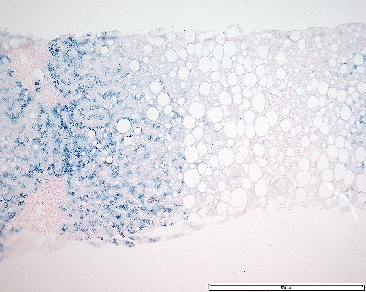
LiverLiverThe liver is the largest gland in the human body. The liver is found in the superior right quadrant of the abdomen and weighs approximately 1.5 kilograms. Its main functions are detoxification, metabolism, nutrient storage (e.g., iron and vitamins), synthesis of coagulation factors, formation of bile, filtration, and storage of blood. Liver: AnatomybiopsyBiopsyRemoval and pathologic examination of specimens from the living body.Ewing Sarcoma:
Performed when initial approach yields uncertain results (such as abnormal markers of iron overloadIron overloadAn excessive accumulation of iron in the body due to a greater than normal absorption of iron from the gastrointestinal tract or from parenteral injection. This may arise from idiopathic hemochromatosis, excessive iron intake, chronic alcoholism, certain types of refractory anemia, or transfusional hemosiderosis.Hereditary Hemochromatosis in those who are not C282Y homozygotes or compound heterozygotes) and/or stagingStagingMethods which attempt to express in replicable terms the extent of the neoplasm in the patient.Grading, Staging, and Metastasis of hepatic fibrosisHepatic fibrosisAutosomal Recessive Polycystic Kidney Disease (ARPKD) is needed
Not indicated just for the diagnosis of hepatic iron overloadIron overloadAn excessive accumulation of iron in the body due to a greater than normal absorption of iron from the gastrointestinal tract or from parenteral injection. This may arise from idiopathic hemochromatosis, excessive iron intake, chronic alcoholism, certain types of refractory anemia, or transfusional hemosiderosis.Hereditary Hemochromatosis
Indications:
Abnormal liver function testsLiver function testsLiver function tests, also known as hepatic function panels, are one of the most commonly performed screening blood tests. Such tests are also used to detect, evaluate, and monitor acute and chronic liver diseases.Liver Function Tests
FerritinFerritinIron-containing proteins that are widely distributed in animals, plants, and microorganisms. Their major function is to store iron in a nontoxic bioavailable form. Each ferritin molecule consists of ferric iron in a hollow protein shell (apoferritins) made of 24 subunits of various sequences depending on the species and tissue types.Hereditary Hemochromatosis > 1,000 µg/L
Other concurrent liverLiverThe liver is the largest gland in the human body. The liver is found in the superior right quadrant of the abdomen and weighs approximately 1.5 kilograms. Its main functions are detoxification, metabolism, nutrient storage (e.g., iron and vitamins), synthesis of coagulation factors, formation of bile, filtration, and storage of blood. Liver: Anatomy disease is suspected.
Helps determine the extent of liverLiverThe liver is the largest gland in the human body. The liver is found in the superior right quadrant of the abdomen and weighs approximately 1.5 kilograms. Its main functions are detoxification, metabolism, nutrient storage (e.g., iron and vitamins), synthesis of coagulation factors, formation of bile, filtration, and storage of blood. Liver: Anatomy damage and ironIronA metallic element with atomic symbol fe, atomic number 26, and atomic weight 55. 85. It is an essential constituent of hemoglobins; cytochromes; and iron-binding proteins. It plays a role in cellular redox reactions and in the transport of oxygen.Trace Elements concentration
PerforationPerforationA pathological hole in an organ, blood vessel or other soft part of the body, occurring in the absence of external force.Esophagitis
SkinSkinThe skin, also referred to as the integumentary system, is the largest organ of the body. The skin is primarily composed of the epidermis (outer layer) and dermis (deep layer). The epidermis is primarily composed of keratinocytes that undergo rapid turnover, while the dermis contains dense layers of connective tissue.Skin: Structure and Functions: may help support diagnosis; can sample any site except for legs (may be nonspecific owing to stasis)
Assess fibrosisFibrosisAny pathological condition where fibrous connective tissue invades any organ, usually as a consequence of inflammation or other injury.Bronchiolitis Obliterans
Presence of fibrosisFibrosisAny pathological condition where fibrous connective tissue invades any organ, usually as a consequence of inflammation or other injury.Bronchiolitis Obliterans indicates advanced liverLiverThe liver is the largest gland in the human body. The liver is found in the superior right quadrant of the abdomen and weighs approximately 1.5 kilograms. Its main functions are detoxification, metabolism, nutrient storage (e.g., iron and vitamins), synthesis of coagulation factors, formation of bile, filtration, and storage of blood. Liver: Anatomy disease and its detection affects management of patientsPatientsIndividuals participating in the health care system for the purpose of receiving therapeutic, diagnostic, or preventive procedures.Clinician–Patient Relationship.
Transient elastography:
Vibrating source is applied and shear waves created are measure by detector, estimating liverLiverThe liver is the largest gland in the human body. The liver is found in the superior right quadrant of the abdomen and weighs approximately 1.5 kilograms. Its main functions are detoxification, metabolism, nutrient storage (e.g., iron and vitamins), synthesis of coagulation factors, formation of bile, filtration, and storage of blood. Liver: Anatomy stiffness
Can be used to check for fibrosisFibrosisAny pathological condition where fibrous connective tissue invades any organ, usually as a consequence of inflammation or other injury.Bronchiolitis Obliterans in patientsPatientsIndividuals participating in the health care system for the purpose of receiving therapeutic, diagnostic, or preventive procedures.Clinician–Patient Relationship
Hepatocyte iron accumulation in a patient with hemochromatosis (stained with Perls Prussian blue)
Image: “Grade 3 hepatocyte iron accumulation” by Mathew, J. et al. License: CC BY 3.0.
Management and Prognosis
Management approach[11,13–15,19]
Objectives:
Remove excess ironIronA metallic element with atomic symbol fe, atomic number 26, and atomic weight 55. 85. It is an essential constituent of hemoglobins; cytochromes; and iron-binding proteins. It plays a role in cellular redox reactions and in the transport of oxygen.Trace Elements from the body → reduce ferritinFerritinIron-containing proteins that are widely distributed in animals, plants, and microorganisms. Their major function is to store iron in a nontoxic bioavailable form. Each ferritin molecule consists of ferric iron in a hollow protein shell (apoferritins) made of 24 subunits of various sequences depending on the species and tissue types.Hereditary Hemochromatosis level to < 1,000 µg/L (may take several years of therapy)
Reduce progression of disease and complications, including risk for HCCHCCHepatocellular carcinoma (HCC) typically arises in a chronically diseased or cirrhotic liver and is the most common primary liver cancer. Diagnosis may include ultrasound, CT, MRI, biopsy (if inconclusive imaging), and/or biomarkers. Hepatocellular Carcinoma (HCC) and Liver Metastases.
Hepatologist referral:
For liverLiverThe liver is the largest gland in the human body. The liver is found in the superior right quadrant of the abdomen and weighs approximately 1.5 kilograms. Its main functions are detoxification, metabolism, nutrient storage (e.g., iron and vitamins), synthesis of coagulation factors, formation of bile, filtration, and storage of blood. Liver: AnatomybiopsyBiopsyRemoval and pathologic examination of specimens from the living body.Ewing Sarcoma, if indicated
For initiation of therapy
Therapy in C282Y homozygotes:
In those with serum ferritinFerritinIron-containing proteins that are widely distributed in animals, plants, and microorganisms. Their major function is to store iron in a nontoxic bioavailable form. Each ferritin molecule consists of ferric iron in a hollow protein shell (apoferritins) made of 24 subunits of various sequences depending on the species and tissue types.Hereditary Hemochromatosis > 1,000 µg/L or elevated liver function testsLiver function testsLiver function tests, also known as hepatic function panels, are one of the most commonly performed screening blood tests. Such tests are also used to detect, evaluate, and monitor acute and chronic liver diseases.Liver Function Tests:
Perform liverLiverThe liver is the largest gland in the human body. The liver is found in the superior right quadrant of the abdomen and weighs approximately 1.5 kilograms. Its main functions are detoxification, metabolism, nutrient storage (e.g., iron and vitamins), synthesis of coagulation factors, formation of bile, filtration, and storage of blood. Liver: AnatomybiopsyBiopsyRemoval and pathologic examination of specimens from the living body.Ewing Sarcoma if indicated (for fibrosisFibrosisAny pathological condition where fibrous connective tissue invades any organ, usually as a consequence of inflammation or other injury.Bronchiolitis ObliteransstagingStagingMethods which attempt to express in replicable terms the extent of the neoplasm in the patient.Grading, Staging, and Metastasis and to rule out other liverLiverThe liver is the largest gland in the human body. The liver is found in the superior right quadrant of the abdomen and weighs approximately 1.5 kilograms. Its main functions are detoxification, metabolism, nutrient storage (e.g., iron and vitamins), synthesis of coagulation factors, formation of bile, filtration, and storage of blood. Liver: Anatomy disease).
Therapeutic phlebotomyPhlebotomyThe techniques used to draw blood from a vein for diagnostic purposes or for treatment of certain blood disorders such as erythrocytosis, hemochromatosis, polycythemia vera, and porphyria cutanea tarda.Hereditary Hemochromatosis
In those with elevated serum ferritinFerritinIron-containing proteins that are widely distributed in animals, plants, and microorganisms. Their major function is to store iron in a nontoxic bioavailable form. Each ferritin molecule consists of ferric iron in a hollow protein shell (apoferritins) made of 24 subunits of various sequences depending on the species and tissue types.Hereditary Hemochromatosis (>300 ng/mL in men and >200 ng/mL in women) but < 1,000 µg/L and normal elevated liver function testsLiver function testsLiver function tests, also known as hepatic function panels, are one of the most commonly performed screening blood tests. Such tests are also used to detect, evaluate, and monitor acute and chronic liver diseases.Liver Function Tests, proceed with therapeutic phlebotomyPhlebotomyThe techniques used to draw blood from a vein for diagnostic purposes or for treatment of certain blood disorders such as erythrocytosis, hemochromatosis, polycythemia vera, and porphyria cutanea tarda.Hereditary Hemochromatosis.[15]
While end-organ damage risk is low with ferritinFerritinIron-containing proteins that are widely distributed in animals, plants, and microorganisms. Their major function is to store iron in a nontoxic bioavailable form. Each ferritin molecule consists of ferric iron in a hollow protein shell (apoferritins) made of 24 subunits of various sequences depending on the species and tissue types.Hereditary Hemochromatosis < 1,000 µg/L, there are no consistent indicators of who will develop complications.
Initiating treatment (phlebotomyPhlebotomyThe techniques used to draw blood from a vein for diagnostic purposes or for treatment of certain blood disorders such as erythrocytosis, hemochromatosis, polycythemia vera, and porphyria cutanea tarda.Hereditary Hemochromatosis) is favored given that it is inexpensive, safe, and reduces mortalityMortalityAll deaths reported in a given population.Measures of Health Status from cardiovascular events.
In those with no ironIronA metallic element with atomic symbol fe, atomic number 26, and atomic weight 55. 85. It is an essential constituent of hemoglobins; cytochromes; and iron-binding proteins. It plays a role in cellular redox reactions and in the transport of oxygen.Trace Elements accumulation (normal transferrinTransferrinAn iron-binding beta1-globulin that is synthesized in the liver and secreted into the blood. It plays a central role in the transport of iron throughout the circulation.Heme Metabolism saturation and ferritinFerritinIron-containing proteins that are widely distributed in animals, plants, and microorganisms. Their major function is to store iron in a nontoxic bioavailable form. Each ferritin molecule consists of ferric iron in a hollow protein shell (apoferritins) made of 24 subunits of various sequences depending on the species and tissue types.Hereditary Hemochromatosis) and normal elevated liver function testsLiver function testsLiver function tests, also known as hepatic function panels, are one of the most commonly performed screening blood tests. Such tests are also used to detect, evaluate, and monitor acute and chronic liver diseases.Liver Function Tests:[11,15]
Low risk of developing relevant ironIronA metallic element with atomic symbol fe, atomic number 26, and atomic weight 55. 85. It is an essential constituent of hemoglobins; cytochromes; and iron-binding proteins. It plays a role in cellular redox reactions and in the transport of oxygen.Trace Elements accumulation
Observation and annual follow-up can be done.
Therapy in compound heterozygotes (C282Y/H63D):
Treatment initiation is individualized.
Risk of iron overloadIron overloadAn excessive accumulation of iron in the body due to a greater than normal absorption of iron from the gastrointestinal tract or from parenteral injection. This may arise from idiopathic hemochromatosis, excessive iron intake, chronic alcoholism, certain types of refractory anemia, or transfusional hemosiderosis.Hereditary Hemochromatosis is relatively low based on mutationMutationGenetic mutations are errors in DNA that can cause protein misfolding and dysfunction. There are various types of mutations, including chromosomal, point, frameshift, and expansion mutations. Types of Mutations but can occur in those with comorbiditiesComorbiditiesThe presence of co-existing or additional diseases with reference to an initial diagnosis or with reference to the index condition that is the subject of study. Comorbidity may affect the ability of affected individuals to function and also their survival; it may be used as a prognostic indicator for length of hospital stay, cost factors, and outcome or survival.St. Louis Encephalitis Virus (fatty liverLiverThe liver is the largest gland in the human body. The liver is found in the superior right quadrant of the abdomen and weighs approximately 1.5 kilograms. Its main functions are detoxification, metabolism, nutrient storage (e.g., iron and vitamins), synthesis of coagulation factors, formation of bile, filtration, and storage of blood. Liver: Anatomy, diabetesDiabetesDiabetes mellitus (DM) is a metabolic disease characterized by hyperglycemia and dysfunction of the regulation of glucose metabolism by insulin. Type 1 DM is diagnosed mostly in children and young adults as the result of autoimmune destruction of β cells in the pancreas and the resulting lack of insulin. Type 2 DM has a significant association with obesity and is characterized by insulin resistance.Diabetes Mellitus, alcoholicAlcoholicPersons who have a history of physical or psychological dependence on ethanol.Mallory-Weiss Syndrome (Mallory-Weiss Tear)liverLiverThe liver is the largest gland in the human body. The liver is found in the superior right quadrant of the abdomen and weighs approximately 1.5 kilograms. Its main functions are detoxification, metabolism, nutrient storage (e.g., iron and vitamins), synthesis of coagulation factors, formation of bile, filtration, and storage of blood. Liver: Anatomy disease).
Consider liverLiverThe liver is the largest gland in the human body. The liver is found in the superior right quadrant of the abdomen and weighs approximately 1.5 kilograms. Its main functions are detoxification, metabolism, nutrient storage (e.g., iron and vitamins), synthesis of coagulation factors, formation of bile, filtration, and storage of blood. Liver: AnatomybiopsyBiopsyRemoval and pathologic examination of specimens from the living body.Ewing Sarcoma to determine LIC, degree of hepatic fibrosisHepatic fibrosisAutosomal Recessive Polycystic Kidney Disease (ARPKD), and treatment benefit in these patientsPatientsIndividuals participating in the health care system for the purpose of receiving therapeutic, diagnostic, or preventive procedures.Clinician–Patient Relationship.
Those with elevated LIC and ferritinFerritinIron-containing proteins that are widely distributed in animals, plants, and microorganisms. Their major function is to store iron in a nontoxic bioavailable form. Each ferritin molecule consists of ferric iron in a hollow protein shell (apoferritins) made of 24 subunits of various sequences depending on the species and tissue types.Hereditary Hemochromatosis > 1,000 µg/L should have treatment initiated.
Therapeutic options[11,13,15,19]
PhlebotomyPhlebotomyThe techniques used to draw blood from a vein for diagnostic purposes or for treatment of certain blood disorders such as erythrocytosis, hemochromatosis, polycythemia vera, and porphyria cutanea tarda.Hereditary Hemochromatosis:
Treatment of choice
Performed weekly or twice weekly, removing 500 mL, and continued until the serum ferritinFerritinIron-containing proteins that are widely distributed in animals, plants, and microorganisms. Their major function is to store iron in a nontoxic bioavailable form. Each ferritin molecule consists of ferric iron in a hollow protein shell (apoferritins) made of 24 subunits of various sequences depending on the species and tissue types.Hereditary Hemochromatosis level is approximately 50–100 µg/L
Note that different expert groups recommend similar but not equal targets.
Check hemoglobin and hematocritHematocritThe volume of packed red blood cells in a blood specimen. The volume is measured by centrifugation in a tube with graduated markings, or with automated blood cell counters. It is an indicator of erythrocyte status in disease. For example, anemia shows a low value; polycythemia, a high value.Neonatal Polycythemia before each session.
Reduce frequency/volume if HbHbThe oxygen-carrying proteins of erythrocytes. They are found in all vertebrates and some invertebrates. The number of globin subunits in the hemoglobin quaternary structure differs between species. Structures range from monomeric to a variety of multimeric arrangements.Gas Exchange <12 g/dL and hold phlebotomyPhlebotomyThe techniques used to draw blood from a vein for diagnostic purposes or for treatment of certain blood disorders such as erythrocytosis, hemochromatosis, polycythemia vera, and porphyria cutanea tarda.Hereditary Hemochromatosis if HbHbThe oxygen-carrying proteins of erythrocytes. They are found in all vertebrates and some invertebrates. The number of globin subunits in the hemoglobin quaternary structure differs between species. Structures range from monomeric to a variety of multimeric arrangements.Gas Exchange <11 g/dL pending reassessment.
After ferritinFerritinIron-containing proteins that are widely distributed in animals, plants, and microorganisms. Their major function is to store iron in a nontoxic bioavailable form. Each ferritin molecule consists of ferric iron in a hollow protein shell (apoferritins) made of 24 subunits of various sequences depending on the species and tissue types.Hereditary Hemochromatosis drops to the goal level, it is maintained with phlebotomyPhlebotomyThe techniques used to draw blood from a vein for diagnostic purposes or for treatment of certain blood disorders such as erythrocytosis, hemochromatosis, polycythemia vera, and porphyria cutanea tarda.Hereditary Hemochromatosis (the frequency depends on serum iron studiesIron StudiesIron Deficiency Anemia and blood count).
Can improve or slow the natural disease progression
IronIronA metallic element with atomic symbol fe, atomic number 26, and atomic weight 55. 85. It is an essential constituent of hemoglobins; cytochromes; and iron-binding proteins. It plays a role in cellular redox reactions and in the transport of oxygen.Trace Elements chelating agents:
Treatment option for patientsPatientsIndividuals participating in the health care system for the purpose of receiving therapeutic, diagnostic, or preventive procedures.Clinician–Patient Relationship who are intolerant of or unresponsive to phlebotomyPhlebotomyThe techniques used to draw blood from a vein for diagnostic purposes or for treatment of certain blood disorders such as erythrocytosis, hemochromatosis, polycythemia vera, and porphyria cutanea tarda.Hereditary Hemochromatosis and/or have heart disease, anemiaAnemiaAnemia is a condition in which individuals have low Hb levels, which can arise from various causes. Anemia is accompanied by a reduced number of RBCs and may manifest with fatigue, shortness of breath, pallor, and weakness. Subtypes are classified by the size of RBCs, chronicity, and etiology. Anemia: Overview and Types, or difficult intravenous access
Options:
DeferoxamineDeferoxamineNatural product isolated from streptomyces pilosus. It forms iron complexes and is used as a chelating agent, particularly in the mesylate form.Hereditary Hemochromatosis: intravenous/subcutaneous
Has a relatively short half-lifeHalf-LifeThe time it takes for a substance (drug, radioactive nuclide, or other) to lose half of its pharmacologic, physiologic, or radiologic activity.Pharmacokinetics and Pharmacodynamics in the body, and tissue ironIronA metallic element with atomic symbol fe, atomic number 26, and atomic weight 55. 85. It is an essential constituent of hemoglobins; cytochromes; and iron-binding proteins. It plays a role in cellular redox reactions and in the transport of oxygen.Trace Elements stores release ironIronA metallic element with atomic symbol fe, atomic number 26, and atomic weight 55. 85. It is an essential constituent of hemoglobins; cytochromes; and iron-binding proteins. It plays a role in cellular redox reactions and in the transport of oxygen.Trace Elements continuously
IV infusion: 40–50 mg/kg/day infused over 8–24 hours (can be painful and time-consuming)
Potential adverse effects: ototoxicityOtotoxicityDamage to the ear or its function secondary to exposure to toxic substances such as drugs used in chemotherapy; immunotherapy; or radiation.Glycopeptides, visual loss (requiring periodic ophthalmologic and hearing exam)
Monitoring: elevated liver function testsLiver function testsLiver function tests, also known as hepatic function panels, are one of the most commonly performed screening blood tests. Such tests are also used to detect, evaluate, and monitor acute and chronic liver diseases.Liver Function Tests, serum chemistries/BUN/creatinine, and urine protein every 3 months
DeferiproneDeferiproneA pyridone derivative and iron chelator that is used in the treatment of iron overload in patients with thalassemia.Hereditary Hemochromatosis: oral chelator (↓ cardiac iron overloadIron overloadAn excessive accumulation of iron in the body due to a greater than normal absorption of iron from the gastrointestinal tract or from parenteral injection. This may arise from idiopathic hemochromatosis, excessive iron intake, chronic alcoholism, certain types of refractory anemia, or transfusional hemosiderosis.Hereditary Hemochromatosis)
Dosing: 75 mg/kg in 2 divided doses (using 1,000 mg twice-a-day tablet formulation only) or in 3 divided doses (using oral solution, 500 mg tablet, or 1,000 mg 3-times-a-day tablet formulation)
Maximum: 99 mg/kg/day
Potential adverse effects: neutropeniaNeutropeniaNeutrophils are an important component of the immune system and play a significant role in the eradication of infections. Low numbers of circulating neutrophils, referred to as neutropenia, predispose the body to recurrent infections or sepsis, though patients can also be asymptomatic. Neutropenia and agranulocytosisAgranulocytosisA decrease in the number of granulocytes; (basophils; eosinophils; and neutrophils).Lincosamides
DeferasiroxDeferasiroxA triazole and benzoate derivative that acts as a selective iron chelator. It is used in the management of chronic iron overload due to blood transfusion or non-transfusion dependent thalassemia.Hereditary Hemochromatosis: oral ironIronA metallic element with atomic symbol fe, atomic number 26, and atomic weight 55. 85. It is an essential constituent of hemoglobins; cytochromes; and iron-binding proteins. It plays a role in cellular redox reactions and in the transport of oxygen.Trace Elements chelator
Dosing: 20 mg/kg/once daily
Potential adverse effects: ↑ elevated liver function testsLiver function testsLiver function tests, also known as hepatic function panels, are one of the most commonly performed screening blood tests. Such tests are also used to detect, evaluate, and monitor acute and chronic liver diseases.Liver Function Tests, gastrointestinal upset, rashRashRocky Mountain Spotted Fever, and renal toxicityToxicityDosage Calculation
Erythrocytapheresis:[17]
Alternative to phlebotomyPhlebotomyThe techniques used to draw blood from a vein for diagnostic purposes or for treatment of certain blood disorders such as erythrocytosis, hemochromatosis, polycythemia vera, and porphyria cutanea tarda.Hereditary Hemochromatosis
Removes RBCsRBCsErythrocytes, or red blood cells (RBCs), are the most abundant cells in the blood. While erythrocytes in the fetus are initially produced in the yolk sac then the liver, the bone marrow eventually becomes the main site of production.Erythrocytes: Histology with the remaining components (e.g., plasmaPlasmaThe residual portion of blood that is left after removal of blood cells by centrifugation without prior blood coagulation.Transfusion ProductsproteinsProteinsLinear polypeptides that are synthesized on ribosomes and may be further modified, crosslinked, cleaved, or assembled into complex proteins with several subunits. The specific sequence of amino acids determines the shape the polypeptide will take, during protein folding, and the function of the protein.Energy Homeostasis, clotting factors, and plateletsPlateletsPlatelets are small cell fragments involved in hemostasis. Thrombopoiesis takes place primarily in the bone marrow through a series of cell differentiation and is influenced by several cytokines. Platelets are formed after fragmentation of the megakaryocyte cytoplasm. Platelets: Histology) returned to the patient
An option for those with hypoproteinemia or thrombocytopeniaThrombocytopeniaThrombocytopenia occurs when the platelet count is < 150,000 per microliter. The normal range for platelets is usually 150,000-450,000/µL of whole blood. Thrombocytopenia can be a result of decreased production, increased destruction, or splenic sequestration of platelets. Patients are often asymptomatic until platelet counts are < 50,000/µL. Thrombocytopenia
Up to 1,000 mL of RBCsRBCsErythrocytes, or red blood cells (RBCs), are the most abundant cells in the blood. While erythrocytes in the fetus are initially produced in the yolk sac then the liver, the bone marrow eventually becomes the main site of production.Erythrocytes: Histology can be removed per procedure.
More costly than phlebotomyPhlebotomyThe techniques used to draw blood from a vein for diagnostic purposes or for treatment of certain blood disorders such as erythrocytosis, hemochromatosis, polycythemia vera, and porphyria cutanea tarda.Hereditary Hemochromatosis
Additional management[11,15]
Dietary restrictions:
No ironIronA metallic element with atomic symbol fe, atomic number 26, and atomic weight 55. 85. It is an essential constituent of hemoglobins; cytochromes; and iron-binding proteins. It plays a role in cellular redox reactions and in the transport of oxygen.Trace Elements supplements
LimitLimitA value (e.g., pressure or time) that should not be exceeded and which is specified by the operator to protect the lungInvasive Mechanical Ventilationvitamin CVitamin CA six carbon compound related to glucose. It is found naturally in citrus fruits and many vegetables. Ascorbic acid is an essential nutrient in human diets, and necessary to maintain connective tissue and bone. Its biologically active form, vitamin C, functions as a reducing agent and coenzyme in several metabolic pathways. Vitamin C is considered an antioxidant.Water-soluble Vitamins and their Deficiencies supplements (↑ iron absorptionIron absorptionDigestion and Absorption).
No alcohol to limitLimitA value (e.g., pressure or time) that should not be exceeded and which is specified by the operator to protect the lungInvasive Mechanical VentilationliverLiverThe liver is the largest gland in the human body. The liver is found in the superior right quadrant of the abdomen and weighs approximately 1.5 kilograms. Its main functions are detoxification, metabolism, nutrient storage (e.g., iron and vitamins), synthesis of coagulation factors, formation of bile, filtration, and storage of blood. Liver: Anatomy injury
No raw seafood (Vibrio vulnificusVibrio vulnificusA species of halophilic bacteria in the genus vibrio, which lives in warm seawater. It can cause infections in those who eat raw contaminated seafood or have open wounds exposed to seawater.Vibrio contamination → sepsisSepsisSystemic inflammatory response syndrome with a proven or suspected infectious etiology. When sepsis is associated with organ dysfunction distant from the site of infection, it is called severe sepsis. When sepsis is accompanied by hypotension despite adequate fluid infusion, it is called septic shock.Sepsis and Septic Shock)
Hepatitis AHepatitis AHepatitis A is caused by the hepatitis A virus (HAV), a nonenveloped virus of the Picornaviridae family with single-stranded RNA. HAV causes an acute, highly contagious hepatitis with unspecific prodromal symptoms such as fever and malaise followed by jaundice and elevated liver transaminases. Hepatitis A Virus and B vaccinationVaccinationVaccination is the administration of a substance to induce the immune system to develop protection against a disease. Unlike passive immunization, which involves the administration of pre-performed antibodies, active immunization constitutes the administration of a vaccine to stimulate the body to produce its own antibodies.Vaccination is recommended for all patientsPatientsIndividuals participating in the health care system for the purpose of receiving therapeutic, diagnostic, or preventive procedures.Clinician–Patient Relationship with HHHHHereditary hemochromatosis (HH) is an autosomal recessive disorder most often associated with hfe gene mutations. Patients have increased iron intestinal absorption and iron deposition in several organs, such as the liver, heart, skin, and pancreas. The clinical presentation includes the triad of cirrhosis, diabetes, and skin bronzing.Hereditary Hemochromatosis.[12]
Complication-specific treatments:
Conventional therapy for diabetesDiabetesDiabetes mellitus (DM) is a metabolic disease characterized by hyperglycemia and dysfunction of the regulation of glucose metabolism by insulin. Type 1 DM is diagnosed mostly in children and young adults as the result of autoimmune destruction of β cells in the pancreas and the resulting lack of insulin. Type 2 DM has a significant association with obesity and is characterized by insulin resistance.Diabetes Mellitus, cirrhosisCirrhosisCirrhosis is a late stage of hepatic parenchymal necrosis and scarring (fibrosis) most commonly due to hepatitis C infection and alcoholic liver disease. Patients may present with jaundice, ascites, and hepatosplenomegaly. Cirrhosis can also cause complications such as hepatic encephalopathy, portal hypertension, portal vein thrombosis, and hepatorenal syndrome. Cirrhosis, and cardiac failureCardiac failureCongestive heart failure refers to the inability of the heart to supply the body with normal cardiac output to meet metabolic needs. Echocardiography can confirm the diagnosis and give information about the ejection fraction.Heart Failure/arrhythmia
Hormone replacement or gonadotropin therapy for pituitaryPituitaryA small, unpaired gland situated in the sella turcica. It is connected to the hypothalamus by a short stalk which is called the infundibulum.Hormones: Overview and Types effects
Insufficient data on occurrence of HCCHCCHepatocellular carcinoma (HCC) typically arises in a chronically diseased or cirrhotic liver and is the most common primary liver cancer. Diagnosis may include ultrasound, CT, MRI, biopsy (if inconclusive imaging), and/or biomarkers. Hepatocellular Carcinoma (HCC) and Liver Metastases in patientsPatientsIndividuals participating in the health care system for the purpose of receiving therapeutic, diagnostic, or preventive procedures.Clinician–Patient Relationship with HHHHHereditary hemochromatosis (HH) is an autosomal recessive disorder most often associated with hfe gene mutations. Patients have increased iron intestinal absorption and iron deposition in several organs, such as the liver, heart, skin, and pancreas. The clinical presentation includes the triad of cirrhosis, diabetes, and skin bronzing.Hereditary Hemochromatosis but without cirrhosisCirrhosisCirrhosis is a late stage of hepatic parenchymal necrosis and scarring (fibrosis) most commonly due to hepatitis C infection and alcoholic liver disease. Patients may present with jaundice, ascites, and hepatosplenomegaly. Cirrhosis can also cause complications such as hepatic encephalopathy, portal hypertension, portal vein thrombosis, and hepatorenal syndrome. Cirrhosis (low risk)
The American College of Gastroenterology (ACG) guidelines recommend against routine surveillanceSurveillanceDevelopmental Milestones and Normal Growth in patientsPatientsIndividuals participating in the health care system for the purpose of receiving therapeutic, diagnostic, or preventive procedures.Clinician–Patient Relationship with HHHHHereditary hemochromatosis (HH) is an autosomal recessive disorder most often associated with hfe gene mutations. Patients have increased iron intestinal absorption and iron deposition in several organs, such as the liver, heart, skin, and pancreas. The clinical presentation includes the triad of cirrhosis, diabetes, and skin bronzing.Hereditary Hemochromatosis and fibrosisFibrosisAny pathological condition where fibrous connective tissue invades any organ, usually as a consequence of inflammation or other injury.Bronchiolitis Obliterans of stage 3 or less. Tailor to fibrosisFibrosisAny pathological condition where fibrous connective tissue invades any organ, usually as a consequence of inflammation or other injury.Bronchiolitis Obliterans stage and additional risk factors (alcohol, diabetesDiabetesDiabetes mellitus (DM) is a metabolic disease characterized by hyperglycemia and dysfunction of the regulation of glucose metabolism by insulin. Type 1 DM is diagnosed mostly in children and young adults as the result of autoimmune destruction of β cells in the pancreas and the resulting lack of insulin. Type 2 DM has a significant association with obesity and is characterized by insulin resistance.Diabetes Mellitus, viral hepatitis)[11]
European Association for the study of the liverLiverThe liver is the largest gland in the human body. The liver is found in the superior right quadrant of the abdomen and weighs approximately 1.5 kilograms. Its main functions are detoxification, metabolism, nutrient storage (e.g., iron and vitamins), synthesis of coagulation factors, formation of bile, filtration, and storage of blood. Liver: Anatomy suggest ultrasound-based surveillanceSurveillanceDevelopmental Milestones and Normal Growth q6 months for advanced fibrosisFibrosisAny pathological condition where fibrous connective tissue invades any organ, usually as a consequence of inflammation or other injury.Bronchiolitis Obliterans (including F3/bridging fibrosisFibrosisAny pathological condition where fibrous connective tissue invades any organ, usually as a consequence of inflammation or other injury.Bronchiolitis Obliterans) and cirrhosisCirrhosisCirrhosis is a late stage of hepatic parenchymal necrosis and scarring (fibrosis) most commonly due to hepatitis C infection and alcoholic liver disease. Patients may present with jaundice, ascites, and hepatosplenomegaly. Cirrhosis can also cause complications such as hepatic encephalopathy, portal hypertension, portal vein thrombosis, and hepatorenal syndrome. Cirrhosis.
Surgical management for advanced complications:
Liver transplantationLiver transplantationThe transference of a part of or an entire liver from one human or animal to another.Hepatocellular Carcinoma (HCC) and Liver Metastases: for liver failureLiver failureSevere inability of the liver to perform its normal metabolic functions, as evidenced by severe jaundice and abnormal serum levels of ammonia; bilirubin; alkaline phosphatase; aspartate aminotransferase; lactate dehydrogenases; and albumin/globulin ratio.Autoimmune Hepatitis
ArthroplastyArthroplastySurgical reconstruction of a joint to relieve pain or restore motion.Osteoarthritis: for severe joint infiltration
PrognosisPrognosisA prediction of the probable outcome of a disease based on a individual’s condition and the usual course of the disease as seen in similar situations.Non-Hodgkin Lymphomas[10,11,15]
Effects of treatment/phlebotomyPhlebotomyThe techniques used to draw blood from a vein for diagnostic purposes or for treatment of certain blood disorders such as erythrocytosis, hemochromatosis, polycythemia vera, and porphyria cutanea tarda.Hereditary Hemochromatosis on complications:
Cardiac failureCardiac failureCongestive heart failure refers to the inability of the heart to supply the body with normal cardiac output to meet metabolic needs. Echocardiography can confirm the diagnosis and give information about the ejection fraction.Heart Failure may be reversed.
LiverLiverThe liver is the largest gland in the human body. The liver is found in the superior right quadrant of the abdomen and weighs approximately 1.5 kilograms. Its main functions are detoxification, metabolism, nutrient storage (e.g., iron and vitamins), synthesis of coagulation factors, formation of bile, filtration, and storage of blood. Liver: Anatomy function and skinSkinThe skin, also referred to as the integumentary system, is the largest organ of the body. The skin is primarily composed of the epidermis (outer layer) and dermis (deep layer). The epidermis is primarily composed of keratinocytes that undergo rapid turnover, while the dermis contains dense layers of connective tissue.Skin: Structure and Functions pigmentation improve.
40% of patientsPatientsIndividuals participating in the health care system for the purpose of receiving therapeutic, diagnostic, or preventive procedures.Clinician–Patient Relationship with diabetesDiabetesDiabetes mellitus (DM) is a metabolic disease characterized by hyperglycemia and dysfunction of the regulation of glucose metabolism by insulin. Type 1 DM is diagnosed mostly in children and young adults as the result of autoimmune destruction of β cells in the pancreas and the resulting lack of insulin. Type 2 DM has a significant association with obesity and is characterized by insulin resistance.Diabetes Mellitus reach better glucoseGlucoseA primary source of energy for living organisms. It is naturally occurring and is found in fruits and other parts of plants in its free state. It is used therapeutically in fluid and nutrient replacement.Lactose Intolerance control.
Minimal effect on arthropathyArthropathyOsteoarthritis and hypogonadismHypogonadismHypogonadism is a condition characterized by reduced or no sex hormone production by the testes or ovaries. Hypogonadism can result from primary (hypergonadotropic) or secondary (hypogonadotropic) failure. Symptoms include infertility, increased risk of osteoporosis, erectile dysfunction, decreased libido, and regression (or absence) of secondary sexual characteristics.Hypogonadism (unlikely to improve)
Once liverLiverThe liver is the largest gland in the human body. The liver is found in the superior right quadrant of the abdomen and weighs approximately 1.5 kilograms. Its main functions are detoxification, metabolism, nutrient storage (e.g., iron and vitamins), synthesis of coagulation factors, formation of bile, filtration, and storage of blood. Liver: AnatomycirrhosisCirrhosisCirrhosis is a late stage of hepatic parenchymal necrosis and scarring (fibrosis) most commonly due to hepatitis C infection and alcoholic liver disease. Patients may present with jaundice, ascites, and hepatosplenomegaly. Cirrhosis can also cause complications such as hepatic encephalopathy, portal hypertension, portal vein thrombosis, and hepatorenal syndrome. Cirrhosis sets in, it is irreversible; HCCHCCHepatocellular carcinoma (HCC) typically arises in a chronically diseased or cirrhotic liver and is the most common primary liver cancer. Diagnosis may include ultrasound, CT, MRI, biopsy (if inconclusive imaging), and/or biomarkers. Hepatocellular Carcinoma (HCC) and Liver Metastases is a late sequela.
Main causes of death:
Cardiac failureCardiac failureCongestive heart failure refers to the inability of the heart to supply the body with normal cardiac output to meet metabolic needs. Echocardiography can confirm the diagnosis and give information about the ejection fraction.Heart Failure
Hepatic failureHepatic failureSevere inability of the liver to perform its normal metabolic functions, as evidenced by severe jaundice and abnormal serum levels of ammonia; bilirubin; alkaline phosphatase; aspartate aminotransferase; lactate dehydrogenases; and albumin/globulin ratio.Autoimmune Hepatitis
Hepatocellular carcinomaHepatocellular carcinomaHepatocellular carcinoma (HCC) typically arises in a chronically diseased or cirrhotic liver and is the most common primary liver cancer. Diagnosis may include ultrasound, CT, MRI, biopsy (if inconclusive imaging), and/or biomarkers. Hepatocellular Carcinoma (HCC) and Liver Metastases
Differential Diagnosis
AlcoholicAlcoholicPersons who have a history of physical or psychological dependence on ethanol.Mallory-Weiss Syndrome (Mallory-Weiss Tear)liverLiverThe liver is the largest gland in the human body. The liver is found in the superior right quadrant of the abdomen and weighs approximately 1.5 kilograms. Its main functions are detoxification, metabolism, nutrient storage (e.g., iron and vitamins), synthesis of coagulation factors, formation of bile, filtration, and storage of blood. Liver: Anatomy disease: PatientsPatientsIndividuals participating in the health care system for the purpose of receiving therapeutic, diagnostic, or preventive procedures.Clinician–Patient Relationship with a history of chronic alcohol use disorderAlcohol use disorderAlcohol is one of the most commonly used addictive substances in the world. Alcohol use disorder (AUD) is defined as pathologic consumption of alcohol leading to impaired daily functioning. Acute alcohol intoxication presents with impairment in speech and motor functions and can be managed in most cases with supportive care. Alcohol Use Disorder who have developed chronic liverLiverThe liver is the largest gland in the human body. The liver is found in the superior right quadrant of the abdomen and weighs approximately 1.5 kilograms. Its main functions are detoxification, metabolism, nutrient storage (e.g., iron and vitamins), synthesis of coagulation factors, formation of bile, filtration, and storage of blood. Liver: Anatomy disease and cirrhosisCirrhosisCirrhosis is a late stage of hepatic parenchymal necrosis and scarring (fibrosis) most commonly due to hepatitis C infection and alcoholic liver disease. Patients may present with jaundice, ascites, and hepatosplenomegaly. Cirrhosis can also cause complications such as hepatic encephalopathy, portal hypertension, portal vein thrombosis, and hepatorenal syndrome. Cirrhosis may manifest with increased ironIronA metallic element with atomic symbol fe, atomic number 26, and atomic weight 55. 85. It is an essential constituent of hemoglobins; cytochromes; and iron-binding proteins. It plays a role in cellular redox reactions and in the transport of oxygen.Trace Elements and serum ferritinFerritinIron-containing proteins that are widely distributed in animals, plants, and microorganisms. Their major function is to store iron in a nontoxic bioavailable form. Each ferritin molecule consists of ferric iron in a hollow protein shell (apoferritins) made of 24 subunits of various sequences depending on the species and tissue types.Hereditary Hemochromatosis levels. Treatment is alcohol cessation.
Multiple transfusions: In patientsPatientsIndividuals participating in the health care system for the purpose of receiving therapeutic, diagnostic, or preventive procedures.Clinician–Patient Relationship who require a lot of blood transfusionsBlood transfusionsThe introduction of whole blood or blood component directly into the bloodstream.Transfusion Products (e.g., sickle cell diseaseSickle cell diseaseSickle cell disease (SCD) is a group of genetic disorders in which an abnormal Hb molecule (HbS) transforms RBCs into sickle-shaped cells, resulting in chronic anemia, vasoocclusive episodes, pain, and organ damage.Sickle Cell Disease, thalassemiaThalassemiaThalassemia is a hereditary cause of microcytic hypochromic anemia and results from a deficiency in either the α or β globin chains, resulting in hemoglobinopathy. The presentation of thalassemia depends on the number of defective chains present and can range from being asymptomatic to rendering the more severely affected patients to be transfusion dependent. Thalassemia, myelodysplastic syndrome), there may be an increased ironIronA metallic element with atomic symbol fe, atomic number 26, and atomic weight 55. 85. It is an essential constituent of hemoglobins; cytochromes; and iron-binding proteins. It plays a role in cellular redox reactions and in the transport of oxygen.Trace Elements level, comparable to patientsPatientsIndividuals participating in the health care system for the purpose of receiving therapeutic, diagnostic, or preventive procedures.Clinician–Patient Relationship with hemochromatosisHemochromatosisA disorder of iron metabolism characterized by a triad of hemosiderosis; liver cirrhosis; and diabetes mellitus. It is caused by massive iron deposits in parenchymal cells that may develop after a prolonged increase of iron absorption.Hereditary Hemochromatosis. Levels return to normal after transfusions are stopped.
Chronic anemiaAnemiaAnemia is a condition in which individuals have low Hb levels, which can arise from various causes. Anemia is accompanied by a reduced number of RBCs and may manifest with fatigue, shortness of breath, pallor, and weakness. Subtypes are classified by the size of RBCs, chronicity, and etiology. Anemia: Overview and Types: a condition of chronically low RBC counts and ineffective erythropoiesisErythropoiesisThe production of red blood cells (erythrocytes). In humans, erythrocytes are produced by the yolk sac in the first trimester; by the liver in the second trimester; by the bone marrow in the third trimester and after birth. In normal individuals, the erythrocyte count in the peripheral blood remains relatively constant implying a balance between the rate of erythrocyte production and rate of destruction.Erythrocytes: Histology. Due to the body adjusting to chronic anemiaAnemiaAnemia is a condition in which individuals have low Hb levels, which can arise from various causes. Anemia is accompanied by a reduced number of RBCs and may manifest with fatigue, shortness of breath, pallor, and weakness. Subtypes are classified by the size of RBCs, chronicity, and etiology. Anemia: Overview and Types, there is increased iron absorptionIron absorptionDigestion and Absorption.Treatment is ironIronA metallic element with atomic symbol fe, atomic number 26, and atomic weight 55. 85. It is an essential constituent of hemoglobins; cytochromes; and iron-binding proteins. It plays a role in cellular redox reactions and in the transport of oxygen.Trace Elements replacement in addition to treating the underlying cause.
Billing and Coding
Diagnosis Codes:
This code is used to diagnose hereditary hemochromatosisHemochromatosisA disorder of iron metabolism characterized by a triad of hemosiderosis; liver cirrhosis; and diabetes mellitus. It is caused by massive iron deposits in parenchymal cells that may develop after a prolonged increase of iron absorption.Hereditary Hemochromatosis, a genetic disorder that causes the body to absorb too much ironIronA metallic element with atomic symbol fe, atomic number 26, and atomic weight 55. 85. It is an essential constituent of hemoglobins; cytochromes; and iron-binding proteins. It plays a role in cellular redox reactions and in the transport of oxygen.Trace Elements from the diet, leading to iron overloadIron overloadAn excessive accumulation of iron in the body due to a greater than normal absorption of iron from the gastrointestinal tract or from parenteral injection. This may arise from idiopathic hemochromatosis, excessive iron intake, chronic alcoholism, certain types of refractory anemia, or transfusional hemosiderosis.Hereditary Hemochromatosis and organ damage.
Coding System
Code
Description
ICD-10-CM
E83.110
Hereditary hemochromatosisHemochromatosisA disorder of iron metabolism characterized by a triad of hemosiderosis; liver cirrhosis; and diabetes mellitus. It is caused by massive iron deposits in parenchymal cells that may develop after a prolonged increase of iron absorption.Hereditary Hemochromatosis
SNOMED CT
33590000
Hereditary hemochromatosisHemochromatosisA disorder of iron metabolism characterized by a triad of hemosiderosis; liver cirrhosis; and diabetes mellitus. It is caused by massive iron deposits in parenchymal cells that may develop after a prolonged increase of iron absorption.Hereditary Hemochromatosis (disorder)
Evaluation & Workup:
These codes are for the key laboratory tests used to diagnose hemochromatosisHemochromatosisA disorder of iron metabolism characterized by a triad of hemosiderosis; liver cirrhosis; and diabetes mellitus. It is caused by massive iron deposits in parenchymal cells that may develop after a prolonged increase of iron absorption.Hereditary Hemochromatosis, including iron studiesIron StudiesIron Deficiency Anemia (ferritinFerritinIron-containing proteins that are widely distributed in animals, plants, and microorganisms. Their major function is to store iron in a nontoxic bioavailable form. Each ferritin molecule consists of ferric iron in a hollow protein shell (apoferritins) made of 24 subunits of various sequences depending on the species and tissue types.Hereditary Hemochromatosis and transferrinTransferrinAn iron-binding beta1-globulin that is synthesized in the liver and secreted into the blood. It plays a central role in the transport of iron throughout the circulation.Heme Metabolism saturation) and HFE geneHFE geneHereditary Hemochromatosis testing to confirm the genetic mutationMutationGenetic mutations are errors in DNA that can cause protein misfolding and dysfunction. There are various types of mutations, including chromosomal, point, frameshift, and expansion mutations. Types of Mutations.
Coding System
Code
Description
CPT
82728
FerritinFerritinIron-containing proteins that are widely distributed in animals, plants, and microorganisms. Their major function is to store iron in a nontoxic bioavailable form. Each ferritin molecule consists of ferric iron in a hollow protein shell (apoferritins) made of 24 subunits of various sequences depending on the species and tissue types.Hereditary Hemochromatosis
CPT
84466
TransferrinTransferrinAn iron-binding beta1-globulin that is synthesized in the liver and secreted into the blood. It plays a central role in the transport of iron throughout the circulation.Heme Metabolism
CPT
81256
HFE (hemochromatosisHemochromatosisA disorder of iron metabolism characterized by a triad of hemosiderosis; liver cirrhosis; and diabetes mellitus. It is caused by massive iron deposits in parenchymal cells that may develop after a prolonged increase of iron absorption.Hereditary Hemochromatosis) geneGeneA category of nucleic acid sequences that function as units of heredity and which code for the basic instructions for the development, reproduction, and maintenance of organisms.Basic Terms of Genetics analysis
Procedures/Interventions:
This code is for therapeutic phlebotomyPhlebotomyThe techniques used to draw blood from a vein for diagnostic purposes or for treatment of certain blood disorders such as erythrocytosis, hemochromatosis, polycythemia vera, and porphyria cutanea tarda.Hereditary Hemochromatosis, the primary treatment for hemochromatosisHemochromatosisA disorder of iron metabolism characterized by a triad of hemosiderosis; liver cirrhosis; and diabetes mellitus. It is caused by massive iron deposits in parenchymal cells that may develop after a prolonged increase of iron absorption.Hereditary Hemochromatosis, which involves regularly removing blood from the body to reduce ironIronA metallic element with atomic symbol fe, atomic number 26, and atomic weight 55. 85. It is an essential constituent of hemoglobins; cytochromes; and iron-binding proteins. It plays a role in cellular redox reactions and in the transport of oxygen.Trace Elements stores to a safe level.
Coding System
Code
Description
CPT
99195
PhlebotomyPhlebotomyThe techniques used to draw blood from a vein for diagnostic purposes or for treatment of certain blood disorders such as erythrocytosis, hemochromatosis, polycythemia vera, and porphyria cutanea tarda.Hereditary Hemochromatosis, therapeutic (separate procedure)
Complications:
These codes are for the major long-term complications of iron overload, including cirrhosis of the liver, diabetes mellitus (“bronze diabetes”), and cardiomyopathyCardiomyopathyCardiomyopathy refers to a group of myocardial diseases associated with structural changes of the heart muscles (myocardium) and impaired systolic and/or diastolic function in the absence of other heart disorders (coronary artery disease, hypertension, valvular disease, and congenital heart disease). Cardiomyopathy: Overview and Types.
Coding System
Code
Description
ICD-10-CM
K74.60
Unspecified cirrhosisCirrhosisCirrhosis is a late stage of hepatic parenchymal necrosis and scarring (fibrosis) most commonly due to hepatitis C infection and alcoholic liver disease. Patients may present with jaundice, ascites, and hepatosplenomegaly. Cirrhosis can also cause complications such as hepatic encephalopathy, portal hypertension, portal vein thrombosis, and hepatorenal syndrome. Cirrhosis of liverLiverThe liver is the largest gland in the human body. The liver is found in the superior right quadrant of the abdomen and weighs approximately 1.5 kilograms. Its main functions are detoxification, metabolism, nutrient storage (e.g., iron and vitamins), synthesis of coagulation factors, formation of bile, filtration, and storage of blood. Liver: Anatomy
ICD-10-CM
E13.9
Other specified diabetesDiabetesDiabetes mellitus (DM) is a metabolic disease characterized by hyperglycemia and dysfunction of the regulation of glucose metabolism by insulin. Type 1 DM is diagnosed mostly in children and young adults as the result of autoimmune destruction of β cells in the pancreas and the resulting lack of insulin. Type 2 DM has a significant association with obesity and is characterized by insulin resistance.Diabetes Mellitus mellitus without complications
ICD-10-CM
I42.8
Other cardiomyopathiesCardiomyopathiesA group of diseases in which the dominant feature is the involvement of the cardiac muscle itself. Cardiomyopathies are classified according to their predominant pathophysiological features (dilated cardiomyopathy; hypertrophic cardiomyopathy; restrictive cardiomyopathy) or their etiological/pathological factors (cardiomyopathy, alcoholic; endocardial fibroelastosis).Cardiomyopathy: Overview and Types
Friedman, L. S. (2021). Hemochromatosis. In Papadakis, M. A., McPhee, S. J., Rabow, M. W. (Eds.), Current Medical Diagnosis & Treatment. McGraw-Hill.
Kelley, M., Joshi, N., Xie, Y., Borgaonkar, M. (2014). Iron overload is rare in patients homozygous for the H63D mutation. Canadian Journal of Gastroenterology and Hepatology, 28(4), 198–202. doi: 10.1155/2014/468521
Powell, L. W. (2018). Hemochromatosis. In Jameson, J., Fauci, A. S., Kasper, D. L., Hauser, S. L., Longo, D. L., Loscalzo, J. (Eds.), Harrison’s Principles of Internal Medicine (20th ed.). McGraw-Hill.
Kowdley, K. V. (2019). Iron overload (hemochromatosis). In Crow, M. K., et al. (Eds.), Goldman-Cecil Medicine (26th ed., vol. 2, pp. 1388–1392).
Friedman, L. S. (2023). Liver, biliary tract, & pancreas disorders: hemochromatosis. In Papadakis, M. A., et al. (Eds.), Current Medical Diagnosis & Treatment (62nd ed., pp. 709–711).
Fitzsimons, E. J., Cullis, J. O., et al. (2018). Diagnosis and therapy of genetic haemochromatosis (review and 2017 update). British Journal of Haematology, 181(3), 293–303. https://doi.org/10.1111/bjh.15164
Cullis, J. O., Fitzsimons, E. J., et al. (2018). Investigation and management of a raised serum ferritin. British Journal of Haematology, 181(3), 331–340. https://doi.org/10.1111/bjh.15166
Bacon, B. R., Adams, P. C., Kowdley, K. V., Powell, L. W., Tavill, A. S. (2011). Diagnosis and management of hemochromatosis: 2011 practice guideline by the American Association for the Study of Liver Diseases. Hepatology, 54(1), 328–343. https://doi.org/10.1002/hep.24330
Reeder, S. B., Yokoo, T., França, M., Hernando, D., Alberich-Bayarri, Á., Alústiza, J. M., Gandon, Y., Henninger, B., Hillenbrand, C., Jhaveri, K., Karçaaltıncaba, M., Kühn, J. P., Mojtahed, A., Serai, S. D., Ward, R., Wood, J. C., Yamamura, J., Martí-Bonmatí, L. (2023). Quantification of liver iron overload with MRI: review and guidelines from the ESGAR and SAR. Radiology, 307(1), e221856. https://doi.org/10.1148/radiol.221856
Rombout-Sestrienkova, E., Winkens, B., Essers, B. A., Nieman, F. H., Noord, P. A., Janssen, M. C., van Deursen, C. T., Boonen, A., Reuser-Kaasenbrood, E. P., Heeremans, J., van Kraaij, M., Masclee, A., Koek, G. H. (2016). Erythrocytapheresis versus phlebotomy in the maintenance treatment of HFE hemochromatosis patients: results from a randomized crossover trial. Transfusion, 56(1), 261–270. https://doi.org/10.1111/trf.13328
Tully, A. S., Trayes, K. P., & Studdiford, J. S. (2012). Evaluation of nail abnormalities. American family physician, 85(8), 779–787.
European Association for the Study of the Liver (2022). EASL Clinical Practice Guidelines on haemochromatosis. Journal of hepatology, 77(2), 479–502. https://doi.org/10.1016/j.jhep.2022.03.033
Create your free account or log in to continue reading!