Achieve Mastery of Medical Concepts

Study for medical school and boards with Lecturio

Familial Juvenile Polyposis

Familial juvenile polyposis, also known as juvenile polyposis syndrome, is an autosomal dominant Autosomal dominant Autosomal inheritance, both dominant and recessive, refers to the transmission of genes from the 22 autosomal chromosomes. Autosomal dominant diseases are expressed when only 1 copy of the dominant allele is inherited. Autosomal Recessive and Autosomal Dominant Inheritance condition characterized by the growth of hamartomatous polyps (juvenile type) in the gastrointestinal tract, mostly in the colon Colon The large intestines constitute the last portion of the digestive system. The large intestine consists of the cecum, appendix, colon (with ascending, transverse, descending, and sigmoid segments), rectum, and anal canal. The primary function of the colon is to remove water and compact the stool prior to expulsion from the body via the rectum and anal canal. Colon, Cecum, and Appendix: Anatomy. Polyposis syndromes are a group of inherited or acquired conditions characterized by the growth of polyps in the GI tract, and are associated with other extracolonic features. These syndromes are caused by mutations in specific genes Genes A category of nucleic acid sequences that function as units of heredity and which code for the basic instructions for the development, reproduction, and maintenance of organisms. DNA Types and Structure associated with tumor Tumor Inflammation suppression Suppression Defense Mechanisms or cell cycle Cell cycle The phases of the cell cycle include interphase (G1, S, and G2) and mitosis (prophase, metaphase, anaphase, and telophase). The cell's progression through these phases is punctuated by checkpoints regulated by cyclins, cyclin-dependent kinases, tumor suppressors, and their antagonists. Cell Cycle regulation. Familial juvenile polyposis is commonly associated with mutations in the SMAD4 ( chromosome Chromosome In a prokaryotic cell or in the nucleus of a eukaryotic cell, a structure consisting of or containing DNA which carries the genetic information essential to the cell. Basic Terms of Genetics 18q) and BMPR1A ( chromosome Chromosome In a prokaryotic cell or in the nucleus of a eukaryotic cell, a structure consisting of or containing DNA which carries the genetic information essential to the cell. Basic Terms of Genetics 10q) genes Genes A category of nucleic acid sequences that function as units of heredity and which code for the basic instructions for the development, reproduction, and maintenance of organisms. DNA Types and Structure. Diagnosis is made by visualizing 5 or more juvenile polyps on colonoscopy Colonoscopy Endoscopic examination, therapy or surgery of the luminal surface of the colon. Colorectal Cancer Screening, multiple juvenile polyps in other areas of the GI tract, or any number of juvenile polyps plus a positive family history Family History Adult Health Maintenance. Management is surgical in order to reduce the likelihood of GI bleeding and obstruction.

Last updated: Jan 16, 2024

Editorial responsibility: Stanley Oiseth, Lindsay Jones, Evelin Maza

Overview

Definition

Familial juvenile polyposis, or juvenile polyposis syndrome (JPS), is an autosomal dominant Autosomal dominant Autosomal inheritance, both dominant and recessive, refers to the transmission of genes from the 22 autosomal chromosomes. Autosomal dominant diseases are expressed when only 1 copy of the dominant allele is inherited. Autosomal Recessive and Autosomal Dominant Inheritance condition characterized by the growth of hamartomatous polyps (juvenile type) in the colon Colon The large intestines constitute the last portion of the digestive system. The large intestine consists of the cecum, appendix, colon (with ascending, transverse, descending, and sigmoid segments), rectum, and anal canal. The primary function of the colon is to remove water and compact the stool prior to expulsion from the body via the rectum and anal canal. Colon, Cecum, and Appendix: Anatomy.

Epidemiology

  • Juvenile polyp: most common polyp found in children:
  • JPS:
    • Incidence Incidence The number of new cases of a given disease during a given period in a specified population. It also is used for the rate at which new events occur in a defined population. It is differentiated from prevalence, which refers to all cases in the population at a given time. Measures of Disease Frequency: 1 in 100,000 individuals
    • Most develop polyps before the age of 20.
    • Approximately 75% of individuals have a family history Family History Adult Health Maintenance of the condition.
    • Increased risk of colorectal cancer Colorectal cancer Colorectal cancer (CRC) is the 2nd leading cause of cancer-related deaths in the United States. Colorectal cancer is a heterogeneous disease that arises from genetic and epigenetic abnormalities, with influence from environmental factors. Colorectal Cancer (CRC) and extracolonic cancer ( stomach Stomach The stomach is a muscular sac in the upper left portion of the abdomen that plays a critical role in digestion. The stomach develops from the foregut and connects the esophagus with the duodenum. Structurally, the stomach is C-shaped and forms a greater and lesser curvature and is divided grossly into regions: the cardia, fundus, body, and pylorus. Stomach: Anatomy, duodenum Duodenum The shortest and widest portion of the small intestine adjacent to the pylorus of the stomach. It is named for having the length equal to about the width of 12 fingers. Small Intestine: Anatomy, pancreas Pancreas The pancreas lies mostly posterior to the stomach and extends across the posterior abdominal wall from the duodenum on the right to the spleen on the left. This organ has both exocrine and endocrine tissue. Pancreas: Anatomy, and jejunum Jejunum The middle portion of the small intestine, between duodenum and ileum. It represents about 2/5 of the remaining portion of the small intestine below duodenum. Small Intestine: Anatomy)

Etiology

  • Most commonly associated with germline mutations in SMAD4 ( chromosome Chromosome In a prokaryotic cell or in the nucleus of a eukaryotic cell, a structure consisting of or containing DNA which carries the genetic information essential to the cell. Basic Terms of Genetics 18q) and BMPR1A ( chromosome Chromosome In a prokaryotic cell or in the nucleus of a eukaryotic cell, a structure consisting of or containing DNA which carries the genetic information essential to the cell. Basic Terms of Genetics 10q) genes Genes A category of nucleic acid sequences that function as units of heredity and which code for the basic instructions for the development, reproduction, and maintenance of organisms. DNA Types and Structure
  • An autosomal dominant Autosomal dominant Autosomal inheritance, both dominant and recessive, refers to the transmission of genes from the 22 autosomal chromosomes. Autosomal dominant diseases are expressed when only 1 copy of the dominant allele is inherited. Autosomal Recessive and Autosomal Dominant Inheritance condition with incomplete penetrance
  • Pathogenic mutation Mutation Genetic mutations are errors in DNA that can cause protein misfolding and dysfunction. There are various types of mutations, including chromosomal, point, frameshift, and expansion mutations. Types of Mutations of at least 1 gene Gene A category of nucleic acid sequences that function as units of heredity and which code for the basic instructions for the development, reproduction, and maintenance of organisms. Basic Terms of Genetics is found in 40%–50% of patients Patients Individuals participating in the health care system for the purpose of receiving therapeutic, diagnostic, or preventive procedures. Clinician–Patient Relationship.

Pathophysiology

  • SMAD4 and BMPR1A genes Genes A category of nucleic acid sequences that function as units of heredity and which code for the basic instructions for the development, reproduction, and maintenance of organisms. DNA Types and Structure exert a tumor Tumor Inflammation suppression Suppression Defense Mechanisms function. 
  • Mutations are most commonly germline-inherited defects, but can be de novo.
  • Mutations result in deficient tumor Tumor Inflammation growth factor beta 1 (TGF-β) signaling and poor control of cell proliferation → uncontrolled cell proliferation → hamartomas → cancer

Clinical Presentation

History

  • Typically presents with:
    • GI/rectal bleeding (most common presenting symptom)
    • Intussusception Intussusception Intussusception occurs when a part of the intestine (intussusceptum) telescopes into another part (intussuscipiens) of the intestine. The condition can cause obstruction and, if untreated, progress to bowel ischemia. Intussusception is most common in the pediatric population, but is occasionally encountered in adults. Intussusception
    • Obstruction
  • Patients Patients Individuals participating in the health care system for the purpose of receiving therapeutic, diagnostic, or preventive procedures. Clinician–Patient Relationship may also complain of protrusion of a mass Mass Three-dimensional lesion that occupies a space within the breast Imaging of the Breast through the anal sphincter ( rectal prolapse Rectal prolapse Rectal prolapse, also known as rectal procidentia, is the protrusion of rectal tissue through the anus. The tissue may include just the mucosa or the full thickness of the rectal wall. Common risk factors include chronic straining, constipation, bowel motility disorders, and weakening of the pelvic floor muscles. Rectal Prolapse).
  • Symptoms of anemia Anemia Anemia is a condition in which individuals have low Hb levels, which can arise from various causes. Anemia is accompanied by a reduced number of RBCs and may manifest with fatigue, shortness of breath, pallor, and weakness. Subtypes are classified by the size of RBCs, chronicity, and etiology. Anemia: Overview and Types:
    • Fatigue Fatigue The state of weariness following a period of exertion, mental or physical, characterized by a decreased capacity for work and reduced efficiency to respond to stimuli. Fibromyalgia
    • Generalized weakness
    • Loss of appetite
    • Cold hands or feet
    • Restless legs Restless legs A disorder characterized by aching or burning sensations in the lower and rarely the upper extremities that occur prior to sleep or may awaken the patient from sleep. Polyneuropathy
    • Palpitations Palpitations Ebstein’s Anomaly
    • Dyspnea Dyspnea Dyspnea is the subjective sensation of breathing discomfort. Dyspnea is a normal manifestation of heavy physical or psychological exertion, but also may be caused by underlying conditions (both pulmonary and extrapulmonary). Dyspnea

Findings

Other manifestations can include:

  • CNS:
  • Thorax:
    • Coarctation of aorta Aorta The main trunk of the systemic arteries. Mediastinum and Great Vessels: Anatomy
    • Atrial septal defects
    • Tetralogy of Fallot Tetralogy of Fallot Tetralogy of Fallot is the most common cyanotic congenital heart disease. The disease is the confluence of 4 pathologic cardiac features: overriding aorta, ventricular septal defect, right ventricular outflow obstruction, and right ventricular hypertrophy. Tetralogy of Fallot
  • Urogenital tract:
    • Undescended testes Testes Gonadal Hormones
    • Bifid uterus Uterus The uterus, cervix, and fallopian tubes are part of the internal female reproductive system. The uterus has a thick wall made of smooth muscle (the myometrium) and an inner mucosal layer (the endometrium). The most inferior portion of the uterus is the cervix, which connects the uterine cavity to the vagina. Uterus, Cervix, and Fallopian Tubes: Anatomy and vagina Vagina The vagina is the female genital canal, extending from the vulva externally to the cervix uteri internally. The structures have sexual, reproductive, and urinary functions and a rich blood supply, mainly arising from the internal iliac artery. Vagina, Vulva, and Pelvic Floor: Anatomy
    • Unilateral renal agenesis Agenesis Teratogenic Birth Defects
  • GI tract:
  • Vascular:
    • Hereditary hemorrhagic telangiectasias Telangiectasias Ataxia-telangiectasia (HHT): can present as epistaxis Epistaxis Bleeding from the nose. Granulomatosis with Polyangiitis and/or iron Iron A metallic element with atomic symbol fe, atomic number 26, and atomic weight 55. 85. It is an essential constituent of hemoglobins; cytochromes; and iron-binding proteins. It plays a role in cellular redox reactions and in the transport of oxygen. Trace Elements deficiency anemia Anemia Anemia is a condition in which individuals have low Hb levels, which can arise from various causes. Anemia is accompanied by a reduced number of RBCs and may manifest with fatigue, shortness of breath, pallor, and weakness. Subtypes are classified by the size of RBCs, chronicity, and etiology. Anemia: Overview and Types (from GI telangiectasia Telangiectasia Permanent dilation of preexisting blood vessels creating small focal red lesions, most commonly in the skin or mucous membranes. It is characterized by the prominence of skin blood vessels, such as vascular spiders. Chronic Venous Insufficiency)
    • Pulmonary, hepatic, and cerebral arteriovenous malformations Arteriovenous malformations Congenital vascular anomalies in the brain characterized by direct communication between an artery and a vein without passing through the capillaries. The locations and size of the shunts determine the symptoms including headaches; seizures; stroke; intracranial hemorrhages; mass effect; and vascular steal effect. Intracerebral Hemorrhage

Diagnosis

Criteria

Diagnosis is made by visualizing > 5 polyps on colonoscopy Colonoscopy Endoscopic examination, therapy or surgery of the luminal surface of the colon. Colorectal Cancer Screening, multiple juvenile polyps in other areas of the GI tract, or any number of polyps with a positive family history Family History Adult Health Maintenance.

Workup

  • CBC: anemia Anemia Anemia is a condition in which individuals have low Hb levels, which can arise from various causes. Anemia is accompanied by a reduced number of RBCs and may manifest with fatigue, shortness of breath, pallor, and weakness. Subtypes are classified by the size of RBCs, chronicity, and etiology. Anemia: Overview and Types
  • Barium enema Barium enema X-ray examination of lower gastrointestinal tract. Imaging is done while a barium compound (e.g., barium sulfate) fills the large intestine via the rectum as a contrast material. Diarrhea: helps detect polyps and structural changes in the lower GI tract
  • Colonoscopy Colonoscopy Endoscopic examination, therapy or surgery of the luminal surface of the colon. Colorectal Cancer Screening:
    • Polyps are typically round, smooth surfaced, red, and pedunculated.
    • Most patients Patients Individuals participating in the health care system for the purpose of receiving therapeutic, diagnostic, or preventive procedures. Clinician–Patient Relationship will have from 50–100 colorectal polyps.
    • Usually limited to the distal colon Colon The large intestines constitute the last portion of the digestive system. The large intestine consists of the cecum, appendix, colon (with ascending, transverse, descending, and sigmoid segments), rectum, and anal canal. The primary function of the colon is to remove water and compact the stool prior to expulsion from the body via the rectum and anal canal. Colon, Cecum, and Appendix: Anatomy and rectum Rectum The rectum and anal canal are the most terminal parts of the lower GI tract/large intestine that form a functional unit and control defecation. Fecal continence is maintained by several important anatomic structures including rectal folds, anal valves, the sling-like puborectalis muscle, and internal and external anal sphincters. Rectum and Anal Canal: Anatomy, although can be found throughout the GI tract
  • Histologic features of a juvenile polyp:
Multiple pedunculated and sessile, non-bleeding juvenile polyps

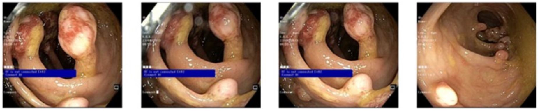
Multiple pedunculated and sessile, nonbleeding juvenile polyps

Image: “Multiple pedunculated and sessile, nonbleeding polyps” by Amna Ahmed and Badr Alsaleem. License: CC BY 4.0

Management

Observation

  • Long-term follow-up due to increased risk of CRC
  • Colonoscopy Colonoscopy Endoscopic examination, therapy or surgery of the luminal surface of the colon. Colorectal Cancer Screening: started at age 12–15 years
    • Can be started earlier if the patient becomes symptomatic
    • If negative: Repeat every 2–3 years.
    • If positive or polyps are removed: Repeat annually.
  • Upper endoscopy Endoscopy Procedures of applying endoscopes for disease diagnosis and treatment. Endoscopy involves passing an optical instrument through a small incision in the skin i.e., percutaneous; or through a natural orifice and along natural body pathways such as the digestive tract; and/or through an incision in the wall of a tubular structure or organ, i.e. Transluminal, to examine or perform surgery on the interior parts of the body. Gastroesophageal Reflux Disease (GERD): started at 12 years of age
    • Perform every 2–3 years.
    • If polyps are detected: Repeat annually.
  • Genetic testing Genetic Testing Detection of a mutation; genotype; karyotype; or specific alleles associated with genetic traits, heritable diseases, or predisposition to a disease, or that may lead to the disease in descendants. It includes prenatal genetic testing. Myotonic Dystrophies of SMAD4 or BMPR1A:
    • Recommended in individuals meeting the clinical criteria
    • Helps guide screening Screening Preoperative Care at-risk family members
  • Evaluate symptoms related to anemia Anemia Anemia is a condition in which individuals have low Hb levels, which can arise from various causes. Anemia is accompanied by a reduced number of RBCs and may manifest with fatigue, shortness of breath, pallor, and weakness. Subtypes are classified by the size of RBCs, chronicity, and etiology. Anemia: Overview and Types, telangiectasias Telangiectasias Ataxia-telangiectasia, and vascular malformations.

Surgical management

Complications

  • Anemia Anemia Anemia is a condition in which individuals have low Hb levels, which can arise from various causes. Anemia is accompanied by a reduced number of RBCs and may manifest with fatigue, shortness of breath, pallor, and weakness. Subtypes are classified by the size of RBCs, chronicity, and etiology. Anemia: Overview and Types
  • Bowel obstruction Bowel obstruction Any impairment, arrest, or reversal of the normal flow of intestinal contents toward the anal canal. Ascaris/Ascariasis

Prognosis Prognosis A prediction of the probable outcome of a disease based on a individual’s condition and the usual course of the disease as seen in similar situations. Non-Hodgkin Lymphomas

  • Lifetime risk of CRC: 39%–68% by age 60
  • Risk of gastric cancer Gastric cancer Gastric cancer is the 3rd-most common cause of cancer-related deaths worldwide. The majority of cases are from adenocarcinoma. The modifiable risk factors include Helicobacter pylori infection, smoking, and nitrate-rich diets. Gastric Cancer: up to 21%

Differential Diagnosis

  • Familial adenomatous polyps Adenomatous polyps Benign neoplasms derived from glandular epithelium. Colorectal Cancer ( FAP FAP Familial adenomatous polyposis (FAP) is an autosomal dominant inherited genetic disorder that presents with numerous adenomatous polyps in the colon. Familial adenomatous polyposis is the most common of the polyposis syndromes, which is a group of inherited or acquired conditions characterized by the growth of polyps in the GI tract, associated with other extracolonic features. Familial Adenomatous Polyposis): an autosomal dominant Autosomal dominant Autosomal inheritance, both dominant and recessive, refers to the transmission of genes from the 22 autosomal chromosomes. Autosomal dominant diseases are expressed when only 1 copy of the dominant allele is inherited. Autosomal Recessive and Autosomal Dominant Inheritance inherited genetic disorder that presents with numerous adenomatous polyps Adenomatous polyps Benign neoplasms derived from glandular epithelium. Colorectal Cancer in the colon Colon The large intestines constitute the last portion of the digestive system. The large intestine consists of the cecum, appendix, colon (with ascending, transverse, descending, and sigmoid segments), rectum, and anal canal. The primary function of the colon is to remove water and compact the stool prior to expulsion from the body via the rectum and anal canal. Colon, Cecum, and Appendix: Anatomy. Familial adenomatous polyps Adenomatous polyps Benign neoplasms derived from glandular epithelium. Colorectal Cancer is the most common of the polyposis syndromes. All patients Patients Individuals participating in the health care system for the purpose of receiving therapeutic, diagnostic, or preventive procedures. Clinician–Patient Relationship with FAP FAP Familial adenomatous polyposis (FAP) is an autosomal dominant inherited genetic disorder that presents with numerous adenomatous polyps in the colon. Familial adenomatous polyposis is the most common of the polyposis syndromes, which is a group of inherited or acquired conditions characterized by the growth of polyps in the GI tract, associated with other extracolonic features. Familial Adenomatous Polyposis will develop colon Colon The large intestines constitute the last portion of the digestive system. The large intestine consists of the cecum, appendix, colon (with ascending, transverse, descending, and sigmoid segments), rectum, and anal canal. The primary function of the colon is to remove water and compact the stool prior to expulsion from the body via the rectum and anal canal. Colon, Cecum, and Appendix: Anatomy cancer by age 35–40 years if left untreated. Management is with a surveillance Surveillance Developmental Milestones and Normal Growth program and colectomy.
  • Peutz-Jeghers syndrome Peutz-Jeghers Syndrome Peutz-Jeghers syndrome (PJS) is an autosomal-dominant inherited disorder characterized by GI polyps and mucocutaneous-pigmented macules. Peutz-Jeghers syndrome is 1 of the polyposis syndromes, a group of inherited or acquired conditions characterized by the growth of polyps in the GI tract and associated with other extracolonic features. Peutz-Jeghers Syndrome: an autosomal dominant Autosomal dominant Autosomal inheritance, both dominant and recessive, refers to the transmission of genes from the 22 autosomal chromosomes. Autosomal dominant diseases are expressed when only 1 copy of the dominant allele is inherited. Autosomal Recessive and Autosomal Dominant Inheritance inherited disorder characterized by GI polyps and mucocutaneous pigmented macules. Peutz-Jeghers syndrome Peutz-Jeghers Syndrome Peutz-Jeghers syndrome (PJS) is an autosomal-dominant inherited disorder characterized by GI polyps and mucocutaneous-pigmented macules. Peutz-Jeghers syndrome is 1 of the polyposis syndromes, a group of inherited or acquired conditions characterized by the growth of polyps in the GI tract and associated with other extracolonic features. Peutz-Jeghers Syndrome is caused by disruptions in the STK11 gene Gene A category of nucleic acid sequences that function as units of heredity and which code for the basic instructions for the development, reproduction, and maintenance of organisms. Basic Terms of Genetics and is associated with colonic (colorectal) and noncolonic (pancreatic, gastric, breast, uterine, cervical, lung, ovarian, and testicular) cancers. Management is with close surveillance Surveillance Developmental Milestones and Normal Growth and surgery.
  • Cowden disease: an autosomal dominant Autosomal dominant Autosomal inheritance, both dominant and recessive, refers to the transmission of genes from the 22 autosomal chromosomes. Autosomal dominant diseases are expressed when only 1 copy of the dominant allele is inherited. Autosomal Recessive and Autosomal Dominant Inheritance disorder caused by mutations in the tumor Tumor Inflammation suppressor gene Gene A category of nucleic acid sequences that function as units of heredity and which code for the basic instructions for the development, reproduction, and maintenance of organisms. Basic Terms of Genetics PTEN, and characterized by the development of multiple benign Benign Fibroadenoma hamartomas. The disease is a genodermatosis characterized by multiple benign Benign Fibroadenoma hamartomas in any location, mucocutaneous lesions, and macrocephaly Macrocephaly Cowden Syndrome. Management depends on the location of the hamartoma Hamartoma A focal malformation resembling a neoplasm, composed of an overgrowth of mature cells and tissues that normally occur in the affected area. Colorectal Cancer
  • Hereditary nonpolyposis colon Colon The large intestines constitute the last portion of the digestive system. The large intestine consists of the cecum, appendix, colon (with ascending, transverse, descending, and sigmoid segments), rectum, and anal canal. The primary function of the colon is to remove water and compact the stool prior to expulsion from the body via the rectum and anal canal. Colon, Cecum, and Appendix: Anatomy cancer: also known as Lynch syndrome Lynch syndrome Lynch syndrome, also called hereditary non-polyposis colorectal cancer (HNPCC), is the most common inherited colon cancer syndrome, and carries a significantly increased risk for endometrial cancer and other malignancies. Lynch syndrome has an autosomal dominant inheritance pattern involving pathogenic variants in one of the mismatch repair (MMR) genes or epithelial cell adhesion molecule (EpCAM). Lynch syndrome; an autosomal dominant Autosomal dominant Autosomal inheritance, both dominant and recessive, refers to the transmission of genes from the 22 autosomal chromosomes. Autosomal dominant diseases are expressed when only 1 copy of the dominant allele is inherited. Autosomal Recessive and Autosomal Dominant Inheritance disorder caused by mutations in the mismatch repair Mismatch repair A DNA repair pathway involved in correction of errors introduced during DNA replication when an incorrect base, which cannot form hydrogen bonds with the corresponding base in the parent strand, is incorporated into the daughter strand. Exonucleases recognize the base pair mismatch and cause a segment of polynucleotide chain to be excised from the daughter strand, thereby removing the mismatched base. Lynch syndrome ( MMR MMR A DNA repair pathway involved in correction of errors introduced during DNA replication when an incorrect base, which cannot form hydrogen bonds with the corresponding base in the parent strand, is incorporated into the daughter strand. Excinucleases recognize the base pair mismatch and cause a segment of polynucleotide chain to be excised from the daughter strand, thereby removing the mismatched base. Lynch syndrome) genes Genes A category of nucleic acid sequences that function as units of heredity and which code for the basic instructions for the development, reproduction, and maintenance of organisms. DNA Types and Structure, which ultimately lead to the development of colorectal cancer Colorectal cancer Colorectal cancer (CRC) is the 2nd leading cause of cancer-related deaths in the United States. Colorectal cancer is a heterogeneous disease that arises from genetic and epigenetic abnormalities, with influence from environmental factors. Colorectal Cancer in 1st-degree relatives. Management is with prophylactic colectomy and frequent endoscopy Endoscopy Procedures of applying endoscopes for disease diagnosis and treatment. Endoscopy involves passing an optical instrument through a small incision in the skin i.e., percutaneous; or through a natural orifice and along natural body pathways such as the digestive tract; and/or through an incision in the wall of a tubular structure or organ, i.e. Transluminal, to examine or perform surgery on the interior parts of the body. Gastroesophageal Reflux Disease (GERD) for surveillance Surveillance Developmental Milestones and Normal Growth
  • CRC: the 2nd leading cause of cancer-related deaths in the United States. Colorectal cancer Colorectal cancer Colorectal cancer (CRC) is the 2nd leading cause of cancer-related deaths in the United States. Colorectal cancer is a heterogeneous disease that arises from genetic and epigenetic abnormalities, with influence from environmental factors. Colorectal Cancer is a heterogeneous disease, arising from genetic and epigenetic abnormalities, with influence from environmental factors. Almost all cases of CRC are adenocarcinoma. Most cases are asymptomatic, so screening Screening Preoperative Care is important. Diagnosis is by colonoscopy Colonoscopy Endoscopic examination, therapy or surgery of the luminal surface of the colon. Colorectal Cancer Screening. Management is primarily surgical.

References

  1. Cohan, J.N., Varma, M.G. (2020). Large intestine. In Doherty, G.M.(Ed.), Current Diagnosis & Treatment: Surgery, 15e. McGraw-Hill. https://accessmedicine.mhmedical.com/content.aspx?bookid=2860&sectionid=241183142
  2. Shalkow, J. (2020). Colorectal Tumors in Adolescents and Young Adults. Emedicine. Retrieved June 20, 2021, from https://emedicine.medscape.com/article/993370-overview#a2
  3. Chung, D., Delgado, K. (2020). Juvenile polyposis syndrome. UpToDate. Retrieved June 21, 2021, from https://www.uptodate.com/contents/juvenile-polyposis-syndrome#H251243034
  4. Zbuk, K., Eng, C. (2007). Hamartomatous polyposis syndromes. Medscape. Retrieved February 26, 2022, from https://www.medscape.org/viewarticle/561747_1

Create your free account or log in to continue reading!

Sign up now and get free access to Lecturio with concept pages, medical videos, and questions for your medical education.

User Reviews

Details