Although fresh whole blood was the only product available in the early years of transfusion, the advent of whole-blood fractionation techniques has allowed for more efficient use of the various blood components. Fractionated transfusion products, prepared in blood transfusion centers, include RBCs, platelets, FFP, and cryoprecipitate. These products are transfused for different indications and each addresses different pathologies.
Blood transfusionsBlood transfusionsThe introduction of whole blood or blood component directly into the bloodstream.Transfusion Products are a very common medical procedure.[1,2,9]
21 million blood components are transfused each year in the United States.
Blood and its products are, at a minimum, screened for:
Hepatitis BHepatitis BHepatitis B virus (HBV) is a partially double-stranded DNA virus, which belongs to the Orthohepadnavirus genus and the Hepadnaviridae family. Most individuals with acute HBV infection are asymptomatic or have mild, self-limiting symptoms. Chronic infection can be asymptomatic or create hepatic inflammation, leading to liver cirrhosis and hepatocellular carcinoma (HCC). Hepatitis B Virus
Hepatitis CHepatitis CHepatitis C is an infection of the liver caused by the hepatitis C virus (HCV). The infection can be transmitted through infectious blood or body fluids and may be transmitted during childbirth or through IV drug use or sexual intercourse. Hepatitis C virus can cause both acute and chronic hepatitis, ranging from a mild to a serious, lifelong illness including liver cirrhosis and hepatocellular carcinoma (HCC).Hepatitis C Virus
Human T cell lymphotropic virusVirusViruses are infectious, obligate intracellular parasites composed of a nucleic acid core surrounded by a protein capsid. Viruses can be either naked (non-enveloped) or enveloped. The classification of viruses is complex and based on many factors, including type and structure of the nucleoid and capsid, the presence of an envelope, the replication cycle, and the host range. Virology (HTLV)
SyphilisSyphilisSyphilis is a bacterial infection caused by the spirochete Treponema pallidum pallidum (T. p. pallidum), which is usually spread through sexual contact. Syphilis has 4 clinical stages: primary, secondary, latent, and tertiary. Syphilis
Bacterial contaminants
Zika virusZika virusAn arbovirus in the flavivirus genus of the family flaviviridae. Originally isolated in the zika forest of uganda it has been introduced to Asia and the americas.Zika Virus Infection
Other: West Nile VirusWest Nile VirusWest Nile virus is an enveloped, positive-sense, single-stranded RNA virus of the genus Flavivirus. Birds are the primary hosts and the disease is most often transmitted by Culex mosquitoes. Most people infected with West Nile virus are asymptomatic. Some patients develop West Nile fever (a self-limited, febrile illness) and a very small proportion of patients develop West Nile neuroinvasive disease. West Nile Virus (seasonal/regional) and BabesiosisBabesiosisBabesiosis is an infection caused by a protozoa belonging to the genus, Babesia. The most common Babesia seen in the United States is B. microti, which is transmitted by the Ixodes tick. The protozoa thrive and replicate within host erythrocytes. Lysis of erythrocytes and the body’s immune response result in clinical symptoms. Babesia/Babesiosis (in specific geographic areas) are also often included.
PlateletsPlateletsPlatelets are small cell fragments involved in hemostasis. Thrombopoiesis takes place primarily in the bone marrow through a series of cell differentiation and is influenced by several cytokines. Platelets are formed after fragmentation of the megakaryocyte cytoplasm. Platelets: Histology
PlasmaPlasmaThe residual portion of blood that is left after removal of blood cells by centrifugation without prior blood coagulation.Transfusion Products
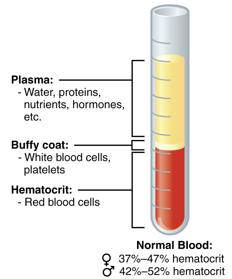
A centrifuged tube showing the components in whole blood (plasma, RBCs, platelets, and WBCs)
Image: “The cellular elements of blood include a vast number of erythrocytes and comparatively fewer leukocytes and platelets” by OpenStax College. License: CC BY 4.0
Units of PRBCs are created by removal of the majority of plasmaPlasmaThe residual portion of blood that is left after removal of blood cells by centrifugation without prior blood coagulation.Transfusion Products from a unit of whole blood:
Each unit contains:
200 mL of RBCsRBCsErythrocytes, or red blood cells (RBCs), are the most abundant cells in the blood. While erythrocytes in the fetus are initially produced in the yolk sac then the liver, the bone marrow eventually becomes the main site of production.Erythrocytes: Histology
70 mL of plasmaPlasmaThe residual portion of blood that is left after removal of blood cells by centrifugation without prior blood coagulation.Transfusion Products
HematocritHematocritThe volume of packed red blood cells in a blood specimen. The volume is measured by centrifugation in a tube with graduated markings, or with automated blood cell counters. It is an indicator of erythrocyte status in disease. For example, anemia shows a low value; polycythemia, a high value.Neonatal Polycythemia of 65%–80%
Volume of 250–300 mL
Can be stored up to 42 days
1 unit should:
↑ Hemoglobin by 1 g/dL
↑ HematocritHematocritThe volume of packed red blood cells in a blood specimen. The volume is measured by centrifugation in a tube with graduated markings, or with automated blood cell counters. It is an indicator of erythrocyte status in disease. For example, anemia shows a low value; polycythemia, a high value.Neonatal Polycythemia by 3%
Picture of the blood collection process: Collection bag hangs as blood is collected from a donor
Image: “Blood bag 2020” by 26RIJNA2020. License: CC0 1.0
PlateletsPlateletsPlatelets are small cell fragments involved in hemostasis. Thrombopoiesis takes place primarily in the bone marrow through a series of cell differentiation and is influenced by several cytokines. Platelets are formed after fragmentation of the megakaryocyte cytoplasm. Platelets: Histology[1,2,6,9]
PlateletsPlateletsPlatelets are small cell fragments involved in hemostasis. Thrombopoiesis takes place primarily in the bone marrow through a series of cell differentiation and is influenced by several cytokines. Platelets are formed after fragmentation of the megakaryocyte cytoplasm. Platelets: Histology (thrombocytes) are small, colorless fragments derived from megakaryocytes (precursor cells).
4–6 units are pooled to allow an adequate number of plateletsPlateletsPlatelets are small cell fragments involved in hemostasis. Thrombopoiesis takes place primarily in the bone marrow through a series of cell differentiation and is influenced by several cytokines. Platelets are formed after fragmentation of the megakaryocyte cytoplasm. Platelets: Histology per transfusion.
Advantages:
↓ Cost
Ease of collection
Disadvantages:
Each recipient is exposed to multiple donors.
↑ Risk of allergic reactionsAllergic ReactionsType I hypersensitivity reaction against plasma proteins in donor bloodTransfusion Reactions and infectionsInfectionsInvasion of the host organism by microorganisms or their toxins or by parasites that can cause pathological conditions or diseases.Chronic Granulomatous Disease
↑ Risk of HLA alloimmunization
Apheresis plateletsApheresis plateletsThe preparation of platelet concentrates with the return of red cells and platelet-poor plasma to the donor.Transfusion Products (single donor plateletsPlateletsPlatelets are small cell fragments involved in hemostasis. Thrombopoiesis takes place primarily in the bone marrow through a series of cell differentiation and is influenced by several cytokines. Platelets are formed after fragmentation of the megakaryocyte cytoplasm. Platelets: Histology):
PlateletsPlateletsPlatelets are small cell fragments involved in hemostasis. Thrombopoiesis takes place primarily in the bone marrow through a series of cell differentiation and is influenced by several cytokines. Platelets are formed after fragmentation of the megakaryocyte cytoplasm. Platelets: Histology are selectively removed from blood taken from the individual and then returned to the donor.
An apheresis platelet unit is equivalent to ≥ 6 units of plateletsPlateletsPlatelets are small cell fragments involved in hemostasis. Thrombopoiesis takes place primarily in the bone marrow through a series of cell differentiation and is influenced by several cytokines. Platelets are formed after fragmentation of the megakaryocyte cytoplasm. Platelets: Histology from whole blood.
PlasmaPlasmaThe residual portion of blood that is left after removal of blood cells by centrifugation without prior blood coagulation.Transfusion Products[1–3,9]
PlasmaPlasmaThe residual portion of blood that is left after removal of blood cells by centrifugation without prior blood coagulation.Transfusion Products is separated from RBCsRBCsErythrocytes, or red blood cells (RBCs), are the most abundant cells in the blood. While erythrocytes in the fetus are initially produced in the yolk sac then the liver, the bone marrow eventually becomes the main site of production.Erythrocytes: Histology and then fractionated.
Yellow liquid component of blood
Holds proteinsProteinsLinear polypeptides that are synthesized on ribosomes and may be further modified, crosslinked, cleaved, or assembled into complex proteins with several subunits. The specific sequence of amino acids determines the shape the polypeptide will take, during protein folding, and the function of the protein.Energy Homeostasis and other constituents of whole blood in suspension:
AlbuminAlbuminSerum albumin from humans. It is an essential carrier of both endogenous substances, such as fatty acids and bilirubin, and of xenobiotics in the blood.Liver Function Tests
Blood clotting factors
Immunoglobulin
Globulins
FibrinogenFibrinogenPlasma glycoprotein clotted by thrombin, composed of a dimer of three non-identical pairs of polypeptide chains (alpha, beta, gamma) held together by disulfide bonds. Fibrinogen clotting is a sol-gel change involving complex molecular arrangements: whereas fibrinogen is cleaved by thrombin to form polypeptides a and b, the proteolytic action of other enzymes yields different fibrinogen degradation products.Hemostasis
Contains all coagulation factorsCoagulation factorsEndogenous substances, usually proteins, that are involved in the blood coagulation process.Hemostasis and proteinsProteinsLinear polypeptides that are synthesized on ribosomes and may be further modified, crosslinked, cleaved, or assembled into complex proteins with several subunits. The specific sequence of amino acids determines the shape the polypeptide will take, during protein folding, and the function of the protein.Energy Homeostasis
PlasmaPlasmaThe residual portion of blood that is left after removal of blood cells by centrifugation without prior blood coagulation.Transfusion Products frozen < 24 hours after phlebotomyPhlebotomyThe techniques used to draw blood from a vein for diagnostic purposes or for treatment of certain blood disorders such as erythrocytosis, hemochromatosis, polycythemia vera, and porphyria cutanea tarda.Hereditary Hemochromatosis (PF24): contains ↓ levels of factor VIIIFactor VIIIFactor VIII of blood coagulation. Antihemophilic factor that is part of the factor viii/von Willebrand factor complex. Factor VIII is produced in the liver and acts in the intrinsic pathway of blood coagulation. It serves as a cofactor in factor X activation and this action is markedly enhanced by small amounts of thrombin.Hemostasis and protein C
↓ Levels of factor VFactor VHeat- and storage-labile plasma glycoprotein which accelerates the conversion of prothrombin to thrombin in blood coagulation. Factor V accomplishes this by forming a complex with factor Xa, phospholipid, and calcium (prothrombinase complex).Hemostasis and factor VIIIFactor VIIIFactor VIII of blood coagulation. Antihemophilic factor that is part of the factor viii/von Willebrand factor complex. Factor VIII is produced in the liver and acts in the intrinsic pathway of blood coagulation. It serves as a cofactor in factor X activation and this action is markedly enhanced by small amounts of thrombin.Hemostasis
Still contains all vitamin KVitamin KA lipid cofactor that is required for normal blood clotting. Several forms of vitamin K have been identified: vitamin K 1 (phytomenadione) derived from plants, vitamin K 2 (menaquinone) from bacteria, and synthetic naphthoquinone provitamins, vitamin K 3 (menadione). Vitamin k 3 provitamins, after being alkylated in vivo, exhibit the antifibrinolytic activity of vitamin k. Green leafy vegetables, liver, cheese, butter, and egg yolk are good sources of vitamin k.Fat-soluble Vitamins and their Deficiencies–dependent clotting factors
Derived from plasmaPlasmaThe residual portion of blood that is left after removal of blood cells by centrifugation without prior blood coagulation.Transfusion Products that is frozen ≤ 8 hours after collection (FFPFFPTransfusion Products)
The plasmaPlasmaThe residual portion of blood that is left after removal of blood cells by centrifugation without prior blood coagulation.Transfusion Products is thawed to between 1 and 6°C and subsequently centrifuged.
The fraction that precipitates out of solution is collected and refrozen to –18°C.
FibrinogenFibrinogenPlasma glycoprotein clotted by thrombin, composed of a dimer of three non-identical pairs of polypeptide chains (alpha, beta, gamma) held together by disulfide bonds. Fibrinogen clotting is a sol-gel change involving complex molecular arrangements: whereas fibrinogen is cleaved by thrombin to form polypeptides a and b, the proteolytic action of other enzymes yields different fibrinogen degradation products.Hemostasis: 150–300 mg of fibrinogenFibrinogenPlasma glycoprotein clotted by thrombin, composed of a dimer of three non-identical pairs of polypeptide chains (alpha, beta, gamma) held together by disulfide bonds. Fibrinogen clotting is a sol-gel change involving complex molecular arrangements: whereas fibrinogen is cleaved by thrombin to form polypeptides a and b, the proteolytic action of other enzymes yields different fibrinogen degradation products.Hemostasis; half-lifeHalf-LifeThe time it takes for a substance (drug, radioactive nuclide, or other) to lose half of its pharmacologic, physiologic, or radiologic activity.Pharmacokinetics and Pharmacodynamics: 100–150 hours
Factor VIIIFactor VIIIFactor VIII of blood coagulation. Antihemophilic factor that is part of the factor viii/von Willebrand factor complex. Factor VIII is produced in the liver and acts in the intrinsic pathway of blood coagulation. It serves as a cofactor in factor X activation and this action is markedly enhanced by small amounts of thrombin.Hemostasis: > 80 IU (range, 80–150); half-lifeHalf-LifeThe time it takes for a substance (drug, radioactive nuclide, or other) to lose half of its pharmacologic, physiologic, or radiologic activity.Pharmacokinetics and Pharmacodynamics: 12 hours
Factor XIII: 50–75 units; half-lifeHalf-LifeThe time it takes for a substance (drug, radioactive nuclide, or other) to lose half of its pharmacologic, physiologic, or radiologic activity.Pharmacokinetics and Pharmacodynamics: 150–300 hours
von Willebrand factorvon Willebrand factorA high-molecular-weight plasma protein, produced by endothelial cells and megakaryocytes, that is part of the factor VIII/von Willebrand factor complex. The von Willebrand factor has receptors for collagen, platelets, and ristocetin activity as well as the immunologically distinct antigenic determinants. It functions in adhesion of platelets to collagen and hemostatic plug formation. The prolonged bleeding time in von Willebrand diseases is due to the deficiency of this factor.Hemostasis: 100–150 units; half-lifeHalf-LifeThe time it takes for a substance (drug, radioactive nuclide, or other) to lose half of its pharmacologic, physiologic, or radiologic activity.Pharmacokinetics and Pharmacodynamics: 24 hours
FibronectinFibronectinGlycoproteins found on the surfaces of cells, particularly in fibrillar structures. The proteins are lost or reduced when these cells undergo viral or chemical transformation. They are highly susceptible to proteolysis and are substrates for activated blood coagulation factor VIII. The forms present in plasma are called cold-insoluble globulins.Connective Tissue: Histology: in variableVariableVariables represent information about something that can change. The design of the measurement scales, or of the methods for obtaining information, will determine the data gathered and the characteristics of that data. As a result, a variable can be qualitative or quantitative, and may be further classified into subgroups.Types of Variables concentrations
Breakdown of the blood components
Table: Breakdown of the blood components
Blood components
Subcomponents of the blood
Type
Production site
Main tasks
PlasmaPlasmaThe residual portion of blood that is left after removal of blood cells by centrifugation without prior blood coagulation.Transfusion Products 43%–63%
Water 92%
Liquid
Absorbed in GI tract or made during metabolism
Transport medium
PlasmaPlasmaThe residual portion of blood that is left after removal of blood cells by centrifugation without prior blood coagulation.Transfusion ProductsproteinsProteinsLinear polypeptides that are synthesized on ribosomes and may be further modified, crosslinked, cleaved, or assembled into complex proteins with several subunits. The specific sequence of amino acids determines the shape the polypeptide will take, during protein folding, and the function of the protein.Energy Homeostasis 7%
AlbuminAlbuminSerum albumin from humans. It is an essential carrier of both endogenous substances, such as fatty acids and bilirubin, and of xenobiotics in the blood.Liver Function Tests 54%–60%
LiverLiverThe liver is the largest gland in the human body. The liver is found in the superior right quadrant of the abdomen and weighs approximately 1.5 kilograms. Its main functions are detoxification, metabolism, nutrient storage (e.g., iron and vitamins), synthesis of coagulation factors, formation of bile, filtration, and storage of blood. Liver: Anatomy
Maintains osmotic concentration
Transports lipid molecules
Globulins 35%–38%
Alpha globulins: liverLiverThe liver is the largest gland in the human body. The liver is found in the superior right quadrant of the abdomen and weighs approximately 1.5 kilograms. Its main functions are detoxification, metabolism, nutrient storage (e.g., iron and vitamins), synthesis of coagulation factors, formation of bile, filtration, and storage of blood. Liver: Anatomy
Transport and maintain osmotic concentration
Beta globulins: liverLiverThe liver is the largest gland in the human body. The liver is found in the superior right quadrant of the abdomen and weighs approximately 1.5 kilograms. Its main functions are detoxification, metabolism, nutrient storage (e.g., iron and vitamins), synthesis of coagulation factors, formation of bile, filtration, and storage of blood. Liver: Anatomy
Transport and maintain osmotic concentration
Gamma globulins (immunoglobulinsImmunoglobulinsImmunoglobulins (Igs), also known as antibodies, are glycoprotein molecules produced by plasma cells that act in immune responses by recognizing and binding particular antigens. The various Ig classes are IgG (the most abundant), IgM, IgE, IgD, and IgA, which differ in their biologic features, structure, target specificity, and distribution.Immunoglobulins: Types and Functions): plasmaPlasmaThe residual portion of blood that is left after removal of blood cells by centrifugation without prior blood coagulation.Transfusion Products cells
Immune response
FibrinogenFibrinogenPlasma glycoprotein clotted by thrombin, composed of a dimer of three non-identical pairs of polypeptide chains (alpha, beta, gamma) held together by disulfide bonds. Fibrinogen clotting is a sol-gel change involving complex molecular arrangements: whereas fibrinogen is cleaved by thrombin to form polypeptides a and b, the proteolytic action of other enzymes yields different fibrinogen degradation products.Hemostasis 4%–7%
LiverLiverThe liver is the largest gland in the human body. The liver is found in the superior right quadrant of the abdomen and weighs approximately 1.5 kilograms. Its main functions are detoxification, metabolism, nutrient storage (e.g., iron and vitamins), synthesis of coagulation factors, formation of bile, filtration, and storage of blood. Liver: Anatomy
Blood clotting during hemostasisHemostasisHemostasis refers to the innate, stepwise body processes that occur following vessel injury, resulting in clot formation and cessation of bleeding. Hemostasis occurs in 2 phases, namely, primary and secondary. Primary hemostasis involves forming a plug that stops the bleeding temporarily. Secondary hemostasis involves the activation of the coagulation cascade.Hemostasis
HormonesHormonesHormones are messenger molecules that are synthesized in one part of the body and move through the bloodstream to exert specific regulatory effects on another part of the body. Hormones play critical roles in coordinating cellular activities throughout the body in response to the constant changes in both the internal and external environments. Hormones: Overview and Types
EnzymesEnzymesEnzymes are complex protein biocatalysts that accelerate chemical reactions without being consumed by them. Due to the body’s constant metabolic needs, the absence of enzymes would make life unsustainable, as reactions would occur too slowly without these molecules. Basics of Enzymes
Various locations
Regulate various body functions
Other dissolved substances 1%
Nutrients
Gases
Waste
Absorbed in GI tract
Replacement of cells in respiratory tract
Made in cells
Many different functions
Formed elements 37%–54%
ErythrocytesErythrocytesErythrocytes, or red blood cells (RBCs), are the most abundant cells in the blood. While erythrocytes in the fetus are initially produced in the yolk sac then the liver, the bone marrow eventually becomes the main site of production. Erythrocytes: Histology 99%
ErythrocytesErythrocytesErythrocytes, or red blood cells (RBCs), are the most abundant cells in the blood. While erythrocytes in the fetus are initially produced in the yolk sac then the liver, the bone marrow eventually becomes the main site of production. Erythrocytes: Histology
Red marrow
Transports gasses, O2, and some CO2
LeukocytesLeukocytesWhite blood cells. These include granular leukocytes (basophils; eosinophils; and neutrophils) as well as non-granular leukocytes (lymphocytes and monocytes).White Myeloid Cells: Histology < 1%
PlateletsPlateletsPlatelets are small cell fragments involved in hemostasis. Thrombopoiesis takes place primarily in the bone marrow through a series of cell differentiation and is influenced by several cytokines. Platelets are formed after fragmentation of the megakaryocyte cytoplasm. Platelets: Histology < 1%
Granular leukocytesLeukocytesWhite blood cells. These include granular leukocytes (basophils; eosinophils; and neutrophils) as well as non-granular leukocytes (lymphocytes and monocytes).White Myeloid Cells: Histology
NeutrophilsNeutrophilsGranular leukocytes having a nucleus with three to five lobes connected by slender threads of chromatin, and cytoplasm containing fine inconspicuous granules and stainable by neutral dyes.Innate Immunity: Phagocytes and Antigen Presentation
EosinophilsEosinophilsGranular leukocytes with a nucleus that usually has two lobes connected by a slender thread of chromatin, and cytoplasm containing coarse, round granules that are uniform in size and stainable by eosin.Innate Immunity: Phagocytes and Antigen Presentation
BasophilsBasophilsGranular leukocytes characterized by a relatively pale-staining, lobate nucleus and cytoplasm containing coarse dark-staining granules of variable size and stainable by basic dyes.Innate Immunity: Phagocytes and Antigen Presentation
Red marrow
Nonspecific immune response
Agranular leukocytesLeukocytesWhite blood cells. These include granular leukocytes (basophils; eosinophils; and neutrophils) as well as non-granular leukocytes (lymphocytes and monocytes).White Myeloid Cells: Histology
LymphocytesLymphocytesLymphocytes are heterogeneous WBCs involved in immune response. Lymphocytes develop from the bone marrow, starting from hematopoietic stem cells (HSCs) and progressing to common lymphoid progenitors (CLPs). B and T lymphocytes and natural killer (NK) cells arise from the lineage.Lymphocytes: Histology
MonocytesMonocytesLarge, phagocytic mononuclear leukocytes produced in the vertebrate bone marrow and released into the blood; contain a large, oval or somewhat indented nucleus surrounded by voluminous cytoplasm and numerous organelles.Innate Immunity: Phagocytes and Antigen Presentation
LymphocytesLymphocytesLymphocytes are heterogeneous WBCs involved in immune response. Lymphocytes develop from the bone marrow, starting from hematopoietic stem cells (HSCs) and progressing to common lymphoid progenitors (CLPs). B and T lymphocytes and natural killer (NK) cells arise from the lineage.Lymphocytes: Histology: bone marrowBone marrowThe soft tissue filling the cavities of bones. Bone marrow exists in two types, yellow and red. Yellow marrow is found in the large cavities of large bones and consists mostly of fat cells and a few primitive blood cells. Red marrow is a hematopoietic tissue and is the site of production of erythrocytes and granular leukocytes. Bone marrow is made up of a framework of connective tissue containing branching fibers with the frame being filled with marrow cells.Bone Marrow: Composition and Hematopoiesis and lymphoid tissue
LymphocytesLymphocytesLymphocytes are heterogeneous WBCs involved in immune response. Lymphocytes develop from the bone marrow, starting from hematopoietic stem cells (HSCs) and progressing to common lymphoid progenitors (CLPs). B and T lymphocytes and natural killer (NK) cells arise from the lineage.Lymphocytes: Histology: specific immune response
MonocytesMonocytesLarge, phagocytic mononuclear leukocytes produced in the vertebrate bone marrow and released into the blood; contain a large, oval or somewhat indented nucleus surrounded by voluminous cytoplasm and numerous organelles.Innate Immunity: Phagocytes and Antigen Presentation: red marrow
MonocytesMonocytesLarge, phagocytic mononuclear leukocytes produced in the vertebrate bone marrow and released into the blood; contain a large, oval or somewhat indented nucleus surrounded by voluminous cytoplasm and numerous organelles.Innate Immunity: Phagocytes and Antigen Presentation: nonspecific immune response
PlateletsPlateletsPlatelets are small cell fragments involved in hemostasis. Thrombopoiesis takes place primarily in the bone marrow through a series of cell differentiation and is influenced by several cytokines. Platelets are formed after fragmentation of the megakaryocyte cytoplasm. Platelets: Histology < 1%
—
Megakaryocytes: red marrow
HemostasisHemostasisHemostasis refers to the innate, stepwise body processes that occur following vessel injury, resulting in clot formation and cessation of bleeding. Hemostasis occurs in 2 phases, namely, primary and secondary. Primary hemostasis involves forming a plug that stops the bleeding temporarily. Secondary hemostasis involves the activation of the coagulation cascade.Hemostasis
Indications for Transfusion of Blood Products
Transfusion indications and thresholds may vary depending on practice location. The following information is based on US and UK literature and guidelines for adult patientsPatientsIndividuals participating in the health care system for the purpose of receiving therapeutic, diagnostic, or preventive procedures.Clinician–Patient Relationship.
PRBC transfusion
Indications and thresholds:[2,5,8,9,15,16,21,23,26–28,30]
The decision to transfuse should be made clinically, with laboratory data used to guide thresholds for transfusion and amounts to be transfused. When possible, patient education should be provided and consent obtained prior to transfusion of blood products.
Individuals with HbHbThe oxygen-carrying proteins of erythrocytes. They are found in all vertebrates and some invertebrates. The number of globin subunits in the hemoglobin quaternary structure differs between species. Structures range from monomeric to a variety of multimeric arrangements.Gas Exchange ≤ 7 g/dL who are symptomatic:
Dizzy
Weak
Short of breath
Chest painPainAn unpleasant sensation induced by noxious stimuli which are detected by nerve endings of nociceptive neurons.Pain: Types and Pathways
SyncopeSyncopeSyncope is a short-term loss of consciousness and loss of postural stability followed by spontaneous return of consciousness to the previous neurologic baseline without the need for resuscitation. The condition is caused by transient interruption of cerebral blood flow that may be benign or related to a underlying life-threatening condition. Syncope
HypotensionHypotensionHypotension is defined as low blood pressure, specifically < 90/60 mm Hg, and is most commonly a physiologic response. Hypotension may be mild, serious, or life threatening, depending on the cause. Hypotension
General hospitalized adults: < 7g/dL
Postoperative individuals or those undergoing surgery:
Hemodynamically stable individuals:
Cardiac surgeryCardiac surgeryCardiac surgery is the surgical management of cardiac abnormalities and of the great vessels of the thorax. In general terms, surgical intervention of the heart is performed to directly restore adequate pump function, correct inherent structural issues, and reestablish proper blood supply via the coronary circulation.Cardiac Surgery: HbHbThe oxygen-carrying proteins of erythrocytes. They are found in all vertebrates and some invertebrates. The number of globin subunits in the hemoglobin quaternary structure differs between species. Structures range from monomeric to a variety of multimeric arrangements.Gas Exchange ≤ 7.5‒8 g/dL
Orthopedic surgery: HbHbThe oxygen-carrying proteins of erythrocytes. They are found in all vertebrates and some invertebrates. The number of globin subunits in the hemoglobin quaternary structure differs between species. Structures range from monomeric to a variety of multimeric arrangements.Gas Exchange ≤ 8 g/dL
Underlying cardiovascular disease: HbHbThe oxygen-carrying proteins of erythrocytes. They are found in all vertebrates and some invertebrates. The number of globin subunits in the hemoglobin quaternary structure differs between species. Structures range from monomeric to a variety of multimeric arrangements.Gas Exchange ≤ 8 g/dL
Presence of symptoms of inadequate oxygen delivery:
Chest painPainAn unpleasant sensation induced by noxious stimuli which are detected by nerve endings of nociceptive neurons.Pain: Types and Pathways of cardiac origin
Orthostatic hypotensionOrthostatic hypotensionA significant drop in blood pressure after assuming a standing position. Orthostatic hypotension is a finding, and defined as a 20-mm hg decrease in systolic pressure or a 10-mm hg decrease in diastolic pressure 3 minutes after the person has risen from supine to standing. Symptoms generally include dizziness, blurred vision, and syncope.Hypotension
TachycardiaTachycardiaAbnormally rapid heartbeat, usually with a heart rate above 100 beats per minute for adults. Tachycardia accompanied by disturbance in the cardiac depolarization (cardiac arrhythmia) is called tachyarrhythmia.Sepsis in Children unresponsive to fluid resuscitationResuscitationThe restoration to life or consciousness of one apparently dead. .Neonatal Respiratory Distress Syndrome
Critically ill individuals:
HbHbThe oxygen-carrying proteins of erythrocytes. They are found in all vertebrates and some invertebrates. The number of globin subunits in the hemoglobin quaternary structure differs between species. Structures range from monomeric to a variety of multimeric arrangements.Gas Exchange ≤ 7 g/dL
Evidence of tissue hypoxiaHypoxiaSub-optimal oxygen levels in the ambient air of living organisms.Ischemic Cell Damage:
In individuals with acute coronary syndrome, HbHbThe oxygen-carrying proteins of erythrocytes. They are found in all vertebrates and some invertebrates. The number of globin subunits in the hemoglobin quaternary structure differs between species. Structures range from monomeric to a variety of multimeric arrangements.Gas Exchange should be maintained at > 8–9 g/dL.
In individuals with traumatic brain injuryTraumatic brain injuryA form of acquired brain injury which occurs when a sudden trauma causes damage to the brain.Le Fort Fractures, the target HbHbThe oxygen-carrying proteins of erythrocytes. They are found in all vertebrates and some invertebrates. The number of globin subunits in the hemoglobin quaternary structure differs between species. Structures range from monomeric to a variety of multimeric arrangements.Gas Exchange should be 7–9 g/dL.
These transition thresholds do not apply to individuals with active bleeding and:
Signs of ischemiaIschemiaA hypoperfusion of the blood through an organ or tissue caused by a pathologic constriction or obstruction of its blood vessels, or an absence of blood circulation.Ischemic Cell Damage or hypoxiaHypoxiaSub-optimal oxygen levels in the ambient air of living organisms.Ischemic Cell Damage related to anemiaAnemiaAnemia is a condition in which individuals have low Hb levels, which can arise from various causes. Anemia is accompanied by a reduced number of RBCs and may manifest with fatigue, shortness of breath, pallor, and weakness. Subtypes are classified by the size of RBCs, chronicity, and etiology. Anemia: Overview and Types
Children:
Critically Ill: < 7 g/dL for stable children in the ICUICUHospital units providing continuous surveillance and care to acutely ill patients.West Nile Virus (excluding those with cyanotic heart disease or hemoglobinopathiesHemoglobinopathiesA group of inherited disorders characterized by structural alterations within the hemoglobin molecule.Anemia: Overview and Types).
Congenital Heart Disease: Thresholds are based on the type of repair:
Biventricular repair: 7 g/dL
Single-ventricle palliation: 9 g/dL
Uncorrected heart disease: 7–9 g/dL
Indications for chronic anemiaAnemiaAnemia is a condition in which individuals have low Hb levels, which can arise from various causes. Anemia is accompanied by a reduced number of RBCs and may manifest with fatigue, shortness of breath, pallor, and weakness. Subtypes are classified by the size of RBCs, chronicity, and etiology. Anemia: Overview and Types:[9]
Symptomatic patientsPatientsIndividuals participating in the health care system for the purpose of receiving therapeutic, diagnostic, or preventive procedures.Clinician–Patient Relationship:
Treat to minimize symptoms
Transfuse ≤ 6 g/dL (usual triggerTriggerThe type of signal that initiates the inspiratory phase by the ventilatorInvasive Mechanical Ventilation to transfuse) but clinical context still matters.
Oncology patientsPatientsIndividuals participating in the health care system for the purpose of receiving therapeutic, diagnostic, or preventive procedures.Clinician–Patient Relationship:
< 7 g/dL
Base transfusion decision-making based on patient’s comorbiditiesComorbiditiesThe presence of co-existing or additional diseases with reference to an initial diagnosis or with reference to the index condition that is the subject of study. Comorbidity may affect the ability of affected individuals to function and also their survival; it may be used as a prognostic indicator for length of hospital stay, cost factors, and outcome or survival.St. Louis Encephalitis Virus
Sickle-cell disease (SCDSCDSickle cell disease (SCD) is a group of genetic disorders in which an abnormal Hb molecule (HbS) transforms RBCs into sickle-shaped cells, resulting in chronic anemia, vasoocclusive episodes, pain, and organ damage.Sickle Cell Disease):
Transfuse to 10 g/dL prior to induction of anesthesiaAnesthesiaA state characterized by loss of feeling or sensation. This depression of nerve function is usually the result of pharmacologic action and is induced to allow performance of surgery or other painful procedures.Anesthesiology: History and Basic Concepts.
Consult specialist for patientsPatientsIndividuals participating in the health care system for the purpose of receiving therapeutic, diagnostic, or preventive procedures.Clinician–Patient Relationship with HbHbThe oxygen-carrying proteins of erythrocytes. They are found in all vertebrates and some invertebrates. The number of globin subunits in the hemoglobin quaternary structure differs between species. Structures range from monomeric to a variety of multimeric arrangements.Gas Exchange > 8.5 g/dL on long-term hydroxyureaHydroxyureaAn antineoplastic agent that inhibits DNA synthesis through the inhibition of ribonucleoside diphosphate reductase.Antimetabolite Chemotherapy therapy or facing high-risk surgery.
PatientsPatientsIndividuals participating in the health care system for the purpose of receiving therapeutic, diagnostic, or preventive procedures.Clinician–Patient Relationship who may have higher HbHbThe oxygen-carrying proteins of erythrocytes. They are found in all vertebrates and some invertebrates. The number of globin subunits in the hemoglobin quaternary structure differs between species. Structures range from monomeric to a variety of multimeric arrangements.Gas Exchange S levels, are at risk for hyperviscosityHyperviscosityHypercoagulable States and are not on long-term treatment with hydroxyureaHydroxyureaAn antineoplastic agent that inhibits DNA synthesis through the inhibition of ribonucleoside diphosphate reductase.Antimetabolite Chemotherapy and/or transfusion therapy → avoid transfusion to hemoglobin > 10 g/dL
Aplastic crisis or acute splenic sequestrationSplenic sequestrationSevere Congenital Neutropenia with severe anemiaAnemiaAnemia is a condition in which individuals have low Hb levels, which can arise from various causes. Anemia is accompanied by a reduced number of RBCs and may manifest with fatigue, shortness of breath, pallor, and weakness. Subtypes are classified by the size of RBCs, chronicity, and etiology. Anemia: Overview and Types
Multisystem organ failure
Hepatic sequestration or intrahepatic cholestasis
Severe acute chest syndrome
Symptomatic anemiaAnemiaAnemia is a condition in which individuals have low Hb levels, which can arise from various causes. Anemia is accompanied by a reduced number of RBCs and may manifest with fatigue, shortness of breath, pallor, and weakness. Subtypes are classified by the size of RBCs, chronicity, and etiology. Anemia: Overview and Types
Acute stroke or TIATIATransient ischemic attack (TIA) is a temporary episode of neurologic dysfunction caused by ischemia without infarction that resolves completely when blood supply is restored. Transient ischemic attack is a neurologic emergency that warrants urgent medical attention. Transient Ischemic Attack (TIA) (prophylactic transfusion)
Wound healingWound healingWound healing is a physiological process involving tissue repair in response to injury. It involves a complex interaction of various cell types, cytokines, and inflammatory mediators. Wound healing stages include hemostasis, inflammation, granulation, and remodeling. Wound Healing
Patient’s sense of well-being
Compatibility:[9]
ABO: Must be compatible
Rh(D): Give Rh(D)-negative patientsPatientsIndividuals participating in the health care system for the purpose of receiving therapeutic, diagnostic, or preventive procedures.Clinician–Patient Relationship Rh(D)-negative PRBCs, when possible, to prevent alloimmunization.
Administration:[9,10,15,23,27,29]
Dose:
Typically 1 unit of PRBCs raises HbHbThe oxygen-carrying proteins of erythrocytes. They are found in all vertebrates and some invertebrates. The number of globin subunits in the hemoglobin quaternary structure differs between species. Structures range from monomeric to a variety of multimeric arrangements.Gas Exchange 1 g/dL (for an adult patient weighing 70‒80 kg)
Generally dosed as a single unit at a time to target a desired HbHbThe oxygen-carrying proteins of erythrocytes. They are found in all vertebrates and some invertebrates. The number of globin subunits in the hemoglobin quaternary structure differs between species. Structures range from monomeric to a variety of multimeric arrangements.Gas Exchange level (except in acute hemorrhage)
Order only the minimum number of units necessary to relieve symptoms or hit a safe target HbHbThe oxygen-carrying proteins of erythrocytes. They are found in all vertebrates and some invertebrates. The number of globin subunits in the hemoglobin quaternary structure differs between species. Structures range from monomeric to a variety of multimeric arrangements.Gas Exchange range.
Rate: In a nonactively bleeding patient, 1 unit over 90‒120 minutes (maximum: 4 hours)
Transfusion of plateletsPlateletsPlatelets are small cell fragments involved in hemostasis. Thrombopoiesis takes place primarily in the bone marrow through a series of cell differentiation and is influenced by several cytokines. Platelets are formed after fragmentation of the megakaryocyte cytoplasm. Platelets: Histology
Indications:[2,9,11,16,10,20,27]
PlateletsPlateletsPlatelets are small cell fragments involved in hemostasis. Thrombopoiesis takes place primarily in the bone marrow through a series of cell differentiation and is influenced by several cytokines. Platelets are formed after fragmentation of the megakaryocyte cytoplasm. Platelets: Histology may be used to treat a severe decrease in circulating plateletsPlateletsPlatelets are small cell fragments involved in hemostasis. Thrombopoiesis takes place primarily in the bone marrow through a series of cell differentiation and is influenced by several cytokines. Platelets are formed after fragmentation of the megakaryocyte cytoplasm. Platelets: Histology or functionally abnormal plateletsPlateletsPlatelets are small cell fragments involved in hemostasis. Thrombopoiesis takes place primarily in the bone marrow through a series of cell differentiation and is influenced by several cytokines. Platelets are formed after fragmentation of the megakaryocyte cytoplasm. Platelets: Histology.
Typical platelet transfusion thresholds:
< 100,000/μL: ocular surgery or neurosurgeryNeurosurgeryNeurosurgery is a specialized field focused on the surgical management of pathologies of the brain, spine, spinal cord, and peripheral nerves. General neurosurgery includes cases of trauma and emergencies. There are a number of specialized neurosurgical practices, including oncologic neurosurgery, spinal neurosurgery, and pediatric neurosurgery. Neurosurgery, no active bleeding
Active bleeding (thresholdThresholdMinimum voltage necessary to generate an action potential (an all-or-none response)Skeletal Muscle Contraction of 30,000/μL for nonsevere bleeding in UK)[16,20]
< 20,000/μL:
Preparation for central line insertion, lumbar punctureLumbar PunctureFebrile Infant, flexible bronchoscopyBronchoscopyEndoscopic examination, therapy or surgery of the bronchi.Laryngomalacia and Tracheomalacia, GI endoscopyEndoscopyProcedures of applying endoscopes for disease diagnosis and treatment. Endoscopy involves passing an optical instrument through a small incision in the skin i.e., percutaneous; or through a natural orifice and along natural body pathways such as the digestive tract; and/or through an incision in the wall of a tubular structure or organ, i.e. Transluminal, to examine or perform surgery on the interior parts of the body.Gastroesophageal Reflux Disease (GERD), or bone marrowBone marrowThe soft tissue filling the cavities of bones. Bone marrow exists in two types, yellow and red. Yellow marrow is found in the large cavities of large bones and consists mostly of fat cells and a few primitive blood cells. Red marrow is a hematopoietic tissue and is the site of production of erythrocytes and granular leukocytes. Bone marrow is made up of a framework of connective tissue containing branching fibers with the frame being filled with marrow cells.Bone Marrow: Composition and HematopoiesisbiopsyBiopsyRemoval and pathologic examination of specimens from the living body.Ewing Sarcoma
Unstable, nonbleeding patientsPatientsIndividuals participating in the health care system for the purpose of receiving therapeutic, diagnostic, or preventive procedures.Clinician–Patient Relationship
< 10,000/μL: prophylactically, even in asymptomatic individuals (reduces the risk of spontaneous bleeding)
For functionally abnormal plateletsPlateletsPlatelets are small cell fragments involved in hemostasis. Thrombopoiesis takes place primarily in the bone marrow through a series of cell differentiation and is influenced by several cytokines. Platelets are formed after fragmentation of the megakaryocyte cytoplasm. Platelets: Histology:
Life-threatening hemorrhage in patientsPatientsIndividuals participating in the health care system for the purpose of receiving therapeutic, diagnostic, or preventive procedures.Clinician–Patient Relationship on antiplatelet agentsAntiplatelet agentsAntiplatelet agents are medications that inhibit platelet aggregation, a critical step in the formation of the initial platelet plug. Abnormal, or inappropriate, platelet aggregation is a key step in the pathophysiology of arterial ischemic events. The primary categories of antiplatelet agents include aspirin, ADP inhibitors, phosphodiesterase/adenosine uptake inhibitors, and glycoprotein IIb/IIIa inhibitors. Antiplatelet Drugs (low-quality evidence)
Intrinsic causes of platelet dysfunction (e.g., Glanzmann thrombastheniaGlanzmann ThrombastheniaHypocoagulable Conditions, Bernard-Soulier syndrome) for which conservative management of bleeding has failed
Massive transfusion
ContraindicationsContraindicationsA condition or factor associated with a recipient that makes the use of a drug, procedure, or physical agent improper or inadvisable. Contraindications may be absolute (life threatening) or relative (higher risk of complications in which benefits may outweigh risks).Noninvasive Ventilation (relative) →plateletsPlateletsPlatelets are small cell fragments involved in hemostasis. Thrombopoiesis takes place primarily in the bone marrow through a series of cell differentiation and is influenced by several cytokines. Platelets are formed after fragmentation of the megakaryocyte cytoplasm. Platelets: Histology should not be transfused for the following, except for life-threatening hemorrhage:[2,16,20]
Autoimmune thrombocytopeniaThrombocytopeniaThrombocytopenia occurs when the platelet count is < 150,000 per microliter. The normal range for platelets is usually 150,000-450,000/µL of whole blood. Thrombocytopenia can be a result of decreased production, increased destruction, or splenic sequestration of platelets. Patients are often asymptomatic until platelet counts are < 50,000/µL. Thrombocytopenia
Thrombotic thrombocytopenic purpuraThrombotic thrombocytopenic purpuraThrombotic thrombocytopenic purpura (TTP) is a life-threatening condition due to either a congenital or an acquired deficiency of ADAMTS-13, a metalloproteinase that cleaves multimers of von Willebrand factor (VWF). The large multimers then aggregate excessive platelets resulting in microvascular thrombosis and an increase in consumption of platelets. Thrombotic Thrombocytopenic Purpura/hemolytic uremic syndromeHemolytic uremic syndromeA syndrome that is associated with microvascular diseases of the kidney, such as renal cortical necrosis. It is characterized by hemolytic anemia; thrombocytopenia; and acute renal failure.Hypocoagulable Conditions (TTPTTPThrombotic thrombocytopenic purpura (TTP) is a life-threatening condition due to either a congenital or an acquired deficiency of adamts-13, a metalloproteinase that cleaves multimers of von Willebrand factor (vWF). The large multimers then aggregate excessive platelets resulting in microvascular thrombosis and an increase in consumption of platelets.Thrombotic Thrombocytopenic Purpura/HUSHUSHemolytic uremic syndrome (HUS) is a clinical phenomenon most commonly seen in children that consists of a classic triad of microangiopathic hemolytic anemia, thrombocytopenia, and acute kidney injury. Hemolytic uremic syndrome is a major cause of acute kidney injury in children and is most commonly associated with a prodrome of diarrheal illness caused by shiga-like toxin-producing bacteria.Hemolytic Uremic Syndrome)
IdiopathicIdiopathicDermatomyositis thrombocytopenic purpura (ITPITPImmune thrombocytopenic purpura (ITP), formerly known as idiopathic thrombocytopenic purpura, is a condition that develops secondary to immune-mediated destruction of platelets, resulting in thrombocytopenia (platelet count < 100,000/mm³). Immune thrombocytopenic purpura can be either primary or secondary due to drugs or underlying disease. Immune Thrombocytopenic Purpura)
Extrinsic causes of platelet dysfunction (e.g., uremiaUremiaA clinical syndrome associated with the retention of renal waste products or uremic toxins in the blood. It is usually the result of renal insufficiency. Most uremic toxins are end products of protein or nitrogen catabolism, such as urea or creatinine. Severe uremia can lead to multiple organ dysfunctions with a constellation of symptoms.Acute Kidney Injury, hyperglobulinemia) in which other management options have failed
Compatibility:[9,20]
ABO:
Not required (always preferred, if possible)
Do ensure compatibility if plateletsPlateletsPlatelets are small cell fragments involved in hemostasis. Thrombopoiesis takes place primarily in the bone marrow through a series of cell differentiation and is influenced by several cytokines. Platelets are formed after fragmentation of the megakaryocyte cytoplasm. Platelets: Histology are suspended in donor plasmaPlasmaThe residual portion of blood that is left after removal of blood cells by centrifugation without prior blood coagulation.Transfusion Products.
Rh(D): Give Rh(D)-negative patientsPatientsIndividuals participating in the health care system for the purpose of receiving therapeutic, diagnostic, or preventive procedures.Clinician–Patient Relationship Rh(D)-negative plateletsPlateletsPlatelets are small cell fragments involved in hemostasis. Thrombopoiesis takes place primarily in the bone marrow through a series of cell differentiation and is influenced by several cytokines. Platelets are formed after fragmentation of the megakaryocyte cytoplasm. Platelets: Histology, when possible, to prevent alloimmunization.
Administration:[9–11,16,20]
Dose:
Transfuse to either:
HemostasisHemostasisHemostasis refers to the innate, stepwise body processes that occur following vessel injury, resulting in clot formation and cessation of bleeding. Hemostasis occurs in 2 phases, namely, primary and secondary. Primary hemostasis involves forming a plug that stops the bleeding temporarily. Secondary hemostasis involves the activation of the coagulation cascade.Hemostasis
Target platelet count
Platelet units typically refer to:
1 unit of apheresis plateletsApheresis plateletsThe preparation of platelet concentrates with the return of red cells and platelet-poor plasma to the donor.Transfusion Products
1 apheresis unit should raise platelet count 30,000-60,000/μL (contains about 3‒4 x 10¹¹plateletsPlateletsPlatelets are small cell fragments involved in hemostasis. Thrombopoiesis takes place primarily in the bone marrow through a series of cell differentiation and is influenced by several cytokines. Platelets are formed after fragmentation of the megakaryocyte cytoplasm. Platelets: Histology)
Rate: 30‒60 minutes/dose
Transfusion of plasmaPlasmaThe residual portion of blood that is left after removal of blood cells by centrifugation without prior blood coagulation.Transfusion Products products
Indications:[2,5,9,13,16,24]
INR > 1.5 and:
Inherited deficiency of coagulation factor II, V, X, or XI
Prophylactically in individuals on anticoagulant therapy before a procedure
Active or clinically significant bleeding
Emergent reversal of warfarinWarfarinAn anticoagulant that acts by inhibiting the synthesis of vitamin K-dependent coagulation factors. Warfarin is indicated for the prophylaxis and/or treatment of venous thrombosis and its extension, pulmonary embolism, and atrial fibrillation with embolization. It is also used as an adjunct in the prophylaxis of systemic embolism after myocardial infarction. Warfarin is also used as a rodenticide.Anticoagulants when 4-factor prothrombinProthrombinA plasma protein that is the inactive precursor of thrombin. It is converted to thrombin by a prothrombin activator complex consisting of factor Xa, factor V, phospholipid, and calcium ions.Hemostasis complex concentrate (PCC) is not available
Microvascular bleeding during massive transfusion (1:1:1 rule):[12]
1 unit of plasmaPlasmaThe residual portion of blood that is left after removal of blood cells by centrifugation without prior blood coagulation.Transfusion Products
1 unit of plateletsPlateletsPlatelets are small cell fragments involved in hemostasis. Thrombopoiesis takes place primarily in the bone marrow through a series of cell differentiation and is influenced by several cytokines. Platelets are formed after fragmentation of the megakaryocyte cytoplasm. Platelets: Histology
1 unit of RBCsRBCsErythrocytes, or red blood cells (RBCs), are the most abundant cells in the blood. While erythrocytes in the fetus are initially produced in the yolk sac then the liver, the bone marrow eventually becomes the main site of production.Erythrocytes: Histology
Hereditary angioedemaHereditary angioedemaForms of hereditary angioedema that occur due to mutations in the gene for complement C1 inhibitor protein. Type I hereditary angioedema is associated with reduced serum levels of complement C1 inhibitor protein. Type II hereditary angioedema is associated with the production of a non-functional complement C1 inhibitor protein.Hereditary Angioedema (C1 Esterase Inhibitor Deficiency): 2nd line option when C1 esterase inhibitorC1 esterase inhibitorAn endogenous 105-kda plasma glycoprotein produced primarily by the liver and monocytes. It inhibits a broad spectrum of proteases, including the complement C1r and the complement C1s proteases of the classical complement pathway, and the mannose-binding protein-associated serine proteases. C1-inh-deficient individuals suffer from hereditary angioedema types I and II.Hereditary Angioedema (C1 Esterase Inhibitor Deficiency) unavailable
Thrombotic microangiopathy: used as transfusion or as plasma exchangePlasma exchangeRemoval of plasma and replacement with various fluids, e.g., fresh frozen plasma, plasma protein fractions (ppf), albumin preparations, dextran solutions, saline. Used in treatment of autoimmune diseases, immune complex diseases, diseases of excess plasma factors, and other conditions.Thrombotic Thrombocytopenic Purpura
ContraindicationsContraindicationsA condition or factor associated with a recipient that makes the use of a drug, procedure, or physical agent improper or inadvisable. Contraindications may be absolute (life threatening) or relative (higher risk of complications in which benefits may outweigh risks).Noninvasive Ventilation:[2,9,16,19,24,25,27]
Not indicated solely to:
Increase blood volume
Increase albuminAlbuminSerum albumin from humans. It is an essential carrier of both endogenous substances, such as fatty acids and bilirubin, and of xenobiotics in the blood.Liver Function Tests levels
Coagulopathy that can be corrected by other means (e.g., warfarinWarfarinAn anticoagulant that acts by inhibiting the synthesis of vitamin K-dependent coagulation factors. Warfarin is indicated for the prophylaxis and/or treatment of venous thrombosis and its extension, pulmonary embolism, and atrial fibrillation with embolization. It is also used as an adjunct in the prophylaxis of systemic embolism after myocardial infarction. Warfarin is also used as a rodenticide.Anticoagulants adjustment, administration of vitamin KVitamin KA lipid cofactor that is required for normal blood clotting. Several forms of vitamin K have been identified: vitamin K 1 (phytomenadione) derived from plants, vitamin K 2 (menaquinone) from bacteria, and synthetic naphthoquinone provitamins, vitamin K 3 (menadione). Vitamin k 3 provitamins, after being alkylated in vivo, exhibit the antifibrinolytic activity of vitamin k. Green leafy vegetables, liver, cheese, butter, and egg yolk are good sources of vitamin k.Fat-soluble Vitamins and their Deficiencies)
Normalization of abnormal coagulation studiesCoagulation studiesCoagulation studies are a group of hematologic laboratory studies that reflect the function of blood vessels, platelets, and coagulation factors, which all interact with one another to achieve hemostasis. Coagulation studies are usually ordered to evaluate patients with bleeding or hypercoagulation disorders.Coagulation Studies in the absence of bleeding
Compatibility:[9]
ABO: must be compatible
Rh(D): not necessary
Administration:[9,10,14]
Dose:
Generally, 10‒20 mL/kg to achieve minimum hemostatic level for coagulation factorsCoagulation factorsEndogenous substances, usually proteins, that are involved in the blood coagulation process.Hemostasis
May require more, depending on the clinical situation
Should be guided by coagulation testing and/or clinical evidence of bleeding
Rate:
Healthy: 10‒20 mL/kg/hr
Volume overload or heart failureHeart FailureA heterogeneous condition in which the heart is unable to pump out sufficient blood to meet the metabolic need of the body. Heart failure can be caused by structural defects, functional abnormalities (ventricular dysfunction), or a sudden overload beyond its capacity. Chronic heart failure is more common than acute heart failure which results from sudden insult to cardiac function, such as myocardial infarction.Total Anomalous Pulmonary Venous Return (TAPVR) patient:
Decrease infusion rate
Should attempt to complete dose within 4 hours of thawing
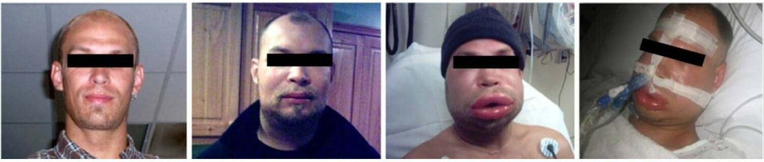
An individual experiencing acute attacks of hereditary angioedema
Image: “F1: HAE patient experiencing HAE attacks” by Bygum A. et al. License: CC BY 2.0
Indications (primarily a fibrinogenFibrinogenPlasma glycoprotein clotted by thrombin, composed of a dimer of three non-identical pairs of polypeptide chains (alpha, beta, gamma) held together by disulfide bonds. Fibrinogen clotting is a sol-gel change involving complex molecular arrangements: whereas fibrinogen is cleaved by thrombin to form polypeptides a and b, the proteolytic action of other enzymes yields different fibrinogen degradation products.Hemostasis replacement product):[2,5,9,16]
Hemorrhage after cardiac surgeryCardiac surgeryCardiac surgery is the surgical management of cardiac abnormalities and of the great vessels of the thorax. In general terms, surgical intervention of the heart is performed to directly restore adequate pump function, correct inherent structural issues, and reestablish proper blood supply via the coronary circulation.Cardiac Surgery
Massive hemorrhage or transfusion
Surgical bleeding
DICDICDisseminated intravascular coagulation (DIC) is a condition characterized by systemic bodywide activation of the coagulation cascade. This cascade results in both widespread microvascular thrombi contributing to multiple organ dysfunction and consumption of clotting factors and platelets, leading to hemorrhage. Disseminated Intravascular Coagulation with bleeding or severe hypofibrinogenemia despite FFPFFPTransfusion Products
FibrinogenFibrinogenPlasma glycoprotein clotted by thrombin, composed of a dimer of three non-identical pairs of polypeptide chains (alpha, beta, gamma) held together by disulfide bonds. Fibrinogen clotting is a sol-gel change involving complex molecular arrangements: whereas fibrinogen is cleaved by thrombin to form polypeptides a and b, the proteolytic action of other enzymes yields different fibrinogen degradation products.Hemostasis disorders associated with low or dysfunctional fibrinogenFibrinogenPlasma glycoprotein clotted by thrombin, composed of a dimer of three non-identical pairs of polypeptide chains (alpha, beta, gamma) held together by disulfide bonds. Fibrinogen clotting is a sol-gel change involving complex molecular arrangements: whereas fibrinogen is cleaved by thrombin to form polypeptides a and b, the proteolytic action of other enzymes yields different fibrinogen degradation products.Hemostasis (< 100 mg/dL or < 150 mg/dL with significant bleeding)
Uremic bleeding when other treatments (e.g., desmopressinDesmopressinHemophilia acetate) fail or are unavailable
Bleeding in individuals with liverLiverThe liver is the largest gland in the human body. The liver is found in the superior right quadrant of the abdomen and weighs approximately 1.5 kilograms. Its main functions are detoxification, metabolism, nutrient storage (e.g., iron and vitamins), synthesis of coagulation factors, formation of bile, filtration, and storage of blood. Liver: Anatomy disease
Outdated/ineffective uses:[9]
Bleeding patientsPatientsIndividuals participating in the health care system for the purpose of receiving therapeutic, diagnostic, or preventive procedures.Clinician–Patient Relationship with factor deficiencies other than VIII, XIII, fibrinogenFibrinogenPlasma glycoprotein clotted by thrombin, composed of a dimer of three non-identical pairs of polypeptide chains (alpha, beta, gamma) held together by disulfide bonds. Fibrinogen clotting is a sol-gel change involving complex molecular arrangements: whereas fibrinogen is cleaved by thrombin to form polypeptides a and b, the proteolytic action of other enzymes yields different fibrinogen degradation products.Hemostasis, or von Willebrand
PatientsPatientsIndividuals participating in the health care system for the purpose of receiving therapeutic, diagnostic, or preventive procedures.Clinician–Patient Relationship with thrombocytopenic bleeding
To reverse warfarinWarfarinAn anticoagulant that acts by inhibiting the synthesis of vitamin K-dependent coagulation factors. Warfarin is indicated for the prophylaxis and/or treatment of venous thrombosis and its extension, pulmonary embolism, and atrial fibrillation with embolization. It is also used as an adjunct in the prophylaxis of systemic embolism after myocardial infarction. Warfarin is also used as a rodenticide.Anticoagulants or other vitamin KVitamin KA lipid cofactor that is required for normal blood clotting. Several forms of vitamin K have been identified: vitamin K 1 (phytomenadione) derived from plants, vitamin K 2 (menaquinone) from bacteria, and synthetic naphthoquinone provitamins, vitamin K 3 (menadione). Vitamin k 3 provitamins, after being alkylated in vivo, exhibit the antifibrinolytic activity of vitamin k. Green leafy vegetables, liver, cheese, butter, and egg yolk are good sources of vitamin k.Fat-soluble Vitamins and their Deficiencies antagonist
Used for fibrinFibrinA protein derived from fibrinogen in the presence of thrombin, which forms part of the blood clot.Rapidly Progressive Glomerulonephritis sealants (surgical)
FibrinogenFibrinogenPlasma glycoprotein clotted by thrombin, composed of a dimer of three non-identical pairs of polypeptide chains (alpha, beta, gamma) held together by disulfide bonds. Fibrinogen clotting is a sol-gel change involving complex molecular arrangements: whereas fibrinogen is cleaved by thrombin to form polypeptides a and b, the proteolytic action of other enzymes yields different fibrinogen degradation products.Hemostasis level and goal
Estimated fibrinogenFibrinogenPlasma glycoprotein clotted by thrombin, composed of a dimer of three non-identical pairs of polypeptide chains (alpha, beta, gamma) held together by disulfide bonds. Fibrinogen clotting is a sol-gel change involving complex molecular arrangements: whereas fibrinogen is cleaved by thrombin to form polypeptides a and b, the proteolytic action of other enzymes yields different fibrinogen degradation products.Hemostasis content of 1 unit
Generally: 1‒2 bags of 5 pooled units (2 bags may increase fibrinogenFibrinogenPlasma glycoprotein clotted by thrombin, composed of a dimer of three non-identical pairs of polypeptide chains (alpha, beta, gamma) held together by disulfide bonds. Fibrinogen clotting is a sol-gel change involving complex molecular arrangements: whereas fibrinogen is cleaved by thrombin to form polypeptides a and b, the proteolytic action of other enzymes yields different fibrinogen degradation products.Hemostasis by around 50 to 100 mg/dL)
Rate: 10‒20 mL/kg/hr
Special Preparations
Leukocyte-reduced components[1,9,18]
Filtered to remove > 99% of WBC component (does not eliminate all WBCs but residual WBC content is < 5 × 10⁶ leukocytesLeukocytesWhite blood cells. These include granular leukocytes (basophils; eosinophils; and neutrophils) as well as non-granular leukocytes (lymphocytes and monocytes).White Myeloid Cells: Histology/unit)
↓ Risk of adverse patient reactions during transfusion:
Recurrent febrile nonhemolytic transfusion reactionsTransfusion reactionsTransfusion-related complications occur during or after a blood product is given. These complications can be classified as immunologic, non-immunologic and acute, and delayed. Non-immunologic reactions are caused by the transmission of disease in blood products, and immunologic reactions are antigen-antibody-mediated. Transfusion Reactions
HLA alloimmunization and HLA-mediated platelet refractoriness
Transmission of intracellular pathogensIntracellular pathogensIL-12 Receptor Deficiency (e.g., CMV, human T-lymphotropic virusVirusViruses are infectious, obligate intracellular parasites composed of a nucleic acid core surrounded by a protein capsid. Viruses can be either naked (non-enveloped) or enveloped. The classification of viruses is complex and based on many factors, including type and structure of the nucleoid and capsid, the presence of an envelope, the replication cycle, and the host range. Virology)
CytomegalovirusCytomegalovirusCMV is a ubiquitous double-stranded DNA virus belonging to the Herpesviridae family. CMV infections can be transmitted in bodily fluids, such as blood, saliva, urine, semen, and breast milk. The initial infection is usually asymptomatic in the immunocompetent host, or it can present with symptoms of mononucleosis. Cytomegalovirus (CMV) reduced-risk components[9]
Can be either:
Blood components from individuals who test negative for CMV antibodiesAntibodiesImmunoglobulins (Igs), also known as antibodies, are glycoprotein molecules produced by plasma cells that act in immune responses by recognizing and binding particular antigens. The various Ig classes are IgG (the most abundant), IgM, IgE, IgD, and IgA, which differ in their biologic features, structure, target specificity, and distribution.Immunoglobulins: Types and Functions
Leukoreduced blood components
Some risk remains
Considered for severely immunocompromisedimmunocompromisedA human or animal whose immunologic mechanism is deficient because of an immunodeficiency disorder or other disease or as the result of the administration of immunosuppressive drugs or radiation.GastroenteritispatientsPatientsIndividuals participating in the health care system for the purpose of receiving therapeutic, diagnostic, or preventive procedures.Clinician–Patient Relationship
Irradiated components[1,9,18]
Prepared by exposing the unit to 2500 cGy of radiationRadiationEmission or propagation of acoustic waves (sound), electromagnetic energy waves (such as light; radio waves; gamma rays; or x-rays), or a stream of subatomic particles (such as electrons; neutrons; protons; or alpha particles).Osteosarcoma
Inactivates donor T cellsT cellsLymphocytes responsible for cell-mediated immunity. Two types have been identified – cytotoxic (t-lymphocytes, cytotoxic) and helper T-lymphocytes (t-lymphocytes, helper-inducer). They are formed when lymphocytes circulate through the thymus gland and differentiate to thymocytes. When exposed to an antigen, they divide rapidly and produce large numbers of new T cells sensitized to that antigen.T cells: Types and Functions
↓ Risk of a graft-versus-host reaction in the recipient; required for those with risk factors:
Congenital cellular immunodeficiencyImmunodeficiencyChédiak-Higashi Syndrome (e.g., SCIDSCIDSevere combined immunodeficiency (SCID), also called “bubble boy disease,” is a rare genetic disorder in which the development of functional B and T cells is disturbed due to several genetic mutations that result in reduced or absent immune function.Severe Combined Immunodeficiency (SCID), DiGeorge syndromeDiGeorge syndromeDiGeorge syndrome (DGS) is a condition caused by a microdeletion at location q11.2 of chromosome 22 (thus also called 22q11.2 syndrome). There is a defective development of the third and fourth pharyngeal pouches, leading to thymic and parathyroid hypoplasia (causing T-cell immunodeficiency and hypocalcemia, respectively). DiGeorge Syndrome)
Intrauterine transfusion or infants who have received intrauterine transfusions
Infants and children suspected to have immune deficiency
Blood products that should be irradiated (regardless of the immune status of the recipient):
From a related donor
From an HLA-selected or crossmatched donor
Washed cellular components[1,9]
Washing with 0.9% NaCl depletes packed RBCsPacked RBCsTransfusion Products or plateletsPlateletsPlatelets are small cell fragments involved in hemostasis. Thrombopoiesis takes place primarily in the bone marrow through a series of cell differentiation and is influenced by several cytokines. Platelets are formed after fragmentation of the megakaryocyte cytoplasm. Platelets: Histology of most plasmaPlasmaThe residual portion of blood that is left after removal of blood cells by centrifugation without prior blood coagulation.Transfusion Products (> 95%)
Can result in loss of the desired components:
20% for RBCsRBCsErythrocytes, or red blood cells (RBCs), are the most abundant cells in the blood. While erythrocytes in the fetus are initially produced in the yolk sac then the liver, the bone marrow eventually becomes the main site of production.Erythrocytes: Histology
≥ 33% for plateletsPlateletsPlatelets are small cell fragments involved in hemostasis. Thrombopoiesis takes place primarily in the bone marrow through a series of cell differentiation and is influenced by several cytokines. Platelets are formed after fragmentation of the megakaryocyte cytoplasm. Platelets: Histology
Used for:
Anaphylactic and recurrent significant allergic transfusion reactionsTransfusion reactionsTransfusion-related complications occur during or after a blood product is given. These complications can be classified as immunologic, non-immunologic and acute, and delayed. Non-immunologic reactions are caused by the transmission of disease in blood products, and immunologic reactions are antigen-antibody-mediated. Transfusion Reactions (unresponsive to other prophylactic measures)
IgA deficiencyIgA deficiencyA dysgammaglobulinemia characterized by a deficiency of immunoglobulin a.Selective IgA Deficiency (particularly if history of anaphylactic reaction)
Avoidance of hyperkalemiaHyperkalemiaHyperkalemia is defined as a serum potassium (K+) concentration >5.2 mEq/L. Homeostatic mechanisms maintain the serum K+ concentration between 3.5 and 5.2 mEq/L, despite marked variation in dietary intake. Hyperkalemia can be due to a variety of causes, which include transcellular shifts, tissue breakdown, inadequate renal excretion, and drugs. Hyperkalemia in patientsPatientsIndividuals participating in the health care system for the purpose of receiving therapeutic, diagnostic, or preventive procedures.Clinician–Patient Relationship predisposed to arrhythmias (from rapid or large-volume transfusion)
Neonatal alloimmune thrombocytopeniaThrombocytopeniaThrombocytopenia occurs when the platelet count is < 150,000 per microliter. The normal range for platelets is usually 150,000-450,000/µL of whole blood. Thrombocytopenia can be a result of decreased production, increased destruction, or splenic sequestration of platelets. Patients are often asymptomatic until platelet counts are < 50,000/µL. Thrombocytopenia
Recurrent febrile nonhemolytic transfusion reactionsTransfusion reactionsTransfusion-related complications occur during or after a blood product is given. These complications can be classified as immunologic, non-immunologic and acute, and delayed. Non-immunologic reactions are caused by the transmission of disease in blood products, and immunologic reactions are antigen-antibody-mediated. Transfusion Reactions (unresponsive to leukocyte-reduced products and other prophylactic measures)
Volume reduction[9]
Reduced plasmaPlasmaThe residual portion of blood that is left after removal of blood cells by centrifugation without prior blood coagulation.Transfusion Products and storage medium with centrifugation
Blood products may be split if smaller aliquots are needed.
Often an option for neonatal and pediatric patientsPatientsIndividuals participating in the health care system for the purpose of receiving therapeutic, diagnostic, or preventive procedures.Clinician–Patient Relationship
Massive Blood Transfusion
Definitions
Major hemorrhage:[1,14,16]
Loss of > 1 blood volume within 24 hours (> 5 L in a 70-kg adult)
Mitigation of risks associated with massive transfusion
Protocol creation:[12,14]
Generally, institutionally based
Ideally developed by a multidisciplinary team, including:
Transfusion medicine/blood bank service
Emergency department
AnesthesiologyAnesthesiologyAnesthesiology is the field of medicine that focuses on interventions that bring a state of anesthesia upon an individual. General anesthesia is characterized by a reversible loss of consciousness along with analgesia, amnesia, and muscle relaxation. Anesthesiology: History and Basic Concepts department
Trauma service
Obstetrics
Medicine service
Should address:
Protocol logistics:
When to initiate
Mechanisms for continuation on transfer to other units/locations in the hospital
Blood product ratios (vary depending on protocol):
1:1 or 2:1 ratio of PRBCs:plasmaPlasmaThe residual portion of blood that is left after removal of blood cells by centrifugation without prior blood coagulation.Transfusion Products
1 adult therapeutic dose of plateletsPlateletsPlatelets are small cell fragments involved in hemostasis. Thrombopoiesis takes place primarily in the bone marrow through a series of cell differentiation and is influenced by several cytokines. Platelets are formed after fragmentation of the megakaryocyte cytoplasm. Platelets: Histology after 4‒6 units of PRBCs
CryoprecipitateCryoprecipitateTransfusion Products if fibrinogenFibrinogenPlasma glycoprotein clotted by thrombin, composed of a dimer of three non-identical pairs of polypeptide chains (alpha, beta, gamma) held together by disulfide bonds. Fibrinogen clotting is a sol-gel change involving complex molecular arrangements: whereas fibrinogen is cleaved by thrombin to form polypeptides a and b, the proteolytic action of other enzymes yields different fibrinogen degradation products.Hemostasis < 100‒150 mg/dL
Adjuncts:
Tranexamic acidTranexamic acidAntifibrinolytic hemostatic used in severe hemorrhage.Hemophilia or aminocaproic acid (antifibrinolytic agents)
PCC (if coagulopathy due to warfarinWarfarinAn anticoagulant that acts by inhibiting the synthesis of vitamin K-dependent coagulation factors. Warfarin is indicated for the prophylaxis and/or treatment of venous thrombosis and its extension, pulmonary embolism, and atrial fibrillation with embolization. It is also used as an adjunct in the prophylaxis of systemic embolism after myocardial infarction. Warfarin is also used as a rodenticide.Anticoagulants)
Prevent hypothermiaHypothermiaHypothermia can be defined as a drop in the core body temperature below 35°C (95°F) and is classified into mild, moderate, severe, and profound forms based on the degree of temperature decrease. Hypothermia:
Fluid warmers
Forced-air warming devices
Raise temperature of treatment room
Monitor labs frequently (at least hourly):
Coagulation studiesCoagulation studiesCoagulation studies are a group of hematologic laboratory studies that reflect the function of blood vessels, platelets, and coagulation factors, which all interact with one another to achieve hemostasis. Coagulation studies are usually ordered to evaluate patients with bleeding or hypercoagulation disorders.Coagulation Studies:
INR
aPTT
FibrinogenFibrinogenPlasma glycoprotein clotted by thrombin, composed of a dimer of three non-identical pairs of polypeptide chains (alpha, beta, gamma) held together by disulfide bonds. Fibrinogen clotting is a sol-gel change involving complex molecular arrangements: whereas fibrinogen is cleaved by thrombin to form polypeptides a and b, the proteolytic action of other enzymes yields different fibrinogen degradation products.Hemostasis
CBC:
HbHbThe oxygen-carrying proteins of erythrocytes. They are found in all vertebrates and some invertebrates. The number of globin subunits in the hemoglobin quaternary structure differs between species. Structures range from monomeric to a variety of multimeric arrangements.Gas Exchange and Hct
Treat electrolyte abnormalities (e.g., hypocalcemiaHypocalcemiaHypocalcemia, a serum calcium < 8.5 mg/dL, can result from various conditions. The causes may include hypoparathyroidism, drugs, disorders leading to vitamin D deficiency, and more. Calcium levels are regulated and affected by different elements such as dietary intake, parathyroid hormone (PTH), vitamin D, pH, and albumin. Presentation can range from an asymptomatic (mild deficiency) to a life-threatening condition (acute, significant deficiency). Hypocalcemia) and acidosisAcidosisA pathologic condition of acid accumulation or depletion of base in the body. The two main types are respiratory acidosis and metabolic acidosis, due to metabolic acid build up.Respiratory Acidosis
Billing and Coding
Indications for Transfusion:
These ICD-10 codesICD-10 CodesTransfusion Products (Clinical) represent the underlying medical reasons, such as acute anemiaAnemiaAnemia is a condition in which individuals have low Hb levels, which can arise from various causes. Anemia is accompanied by a reduced number of RBCs and may manifest with fatigue, shortness of breath, pallor, and weakness. Subtypes are classified by the size of RBCs, chronicity, and etiology. Anemia: Overview and Types or thrombocytopeniaThrombocytopeniaThrombocytopenia occurs when the platelet count is < 150,000 per microliter. The normal range for platelets is usually 150,000-450,000/µL of whole blood. Thrombocytopenia can be a result of decreased production, increased destruction, or splenic sequestration of platelets. Patients are often asymptomatic until platelet counts are < 50,000/µL. Thrombocytopenia, that establish the medical necessity of a blood transfusion for both clinical documentationDocumentationSystematic organization, storage, retrieval, and dissemination of specialized information, especially of a scientific or technical nature. It often involves authenticating or validating information.Advance Directives and billing.
Domain
Code
Description
ICD-10-CM
D62
Acute posthemorrhagic anemiaAnemiaAnemia is a condition in which individuals have low Hb levels, which can arise from various causes. Anemia is accompanied by a reduced number of RBCs and may manifest with fatigue, shortness of breath, pallor, and weakness. Subtypes are classified by the size of RBCs, chronicity, and etiology. Anemia: Overview and Types
ICD-10-CM
D64.9
AnemiaAnemiaAnemia is a condition in which individuals have low Hb levels, which can arise from various causes. Anemia is accompanied by a reduced number of RBCs and may manifest with fatigue, shortness of breath, pallor, and weakness. Subtypes are classified by the size of RBCs, chronicity, and etiology. Anemia: Overview and Types, unspecified
ICD-10-CM
D69.6
ThrombocytopeniaThrombocytopeniaThrombocytopenia occurs when the platelet count is < 150,000 per microliter. The normal range for platelets is usually 150,000-450,000/µL of whole blood. Thrombocytopenia can be a result of decreased production, increased destruction, or splenic sequestration of platelets. Patients are often asymptomatic until platelet counts are < 50,000/µL. Thrombocytopenia, unspecified
ICD-10-CM
D65
Disseminated intravascular coagulationDisseminated intravascular coagulationDisseminated intravascular coagulation (DIC) is a condition characterized by systemic bodywide activation of the coagulation cascade. This cascade results in both widespread microvascular thrombi contributing to multiple organ dysfunction and consumption of clotting factors and platelets, leading to hemorrhage. Disseminated Intravascular Coagulation [defibrination syndrome]
Evaluation & Workup:
These codes are used to order and document essential pre-transfusion laboratory testing, such as blood typing and crossmatchingCrossmatchingTesting erythrocytes to determine presence or absence of blood-group antigens, testing of serum to determine the presence or absence of antibodies to these antigens, and selecting biocompatible blood by crossmatching samples from the donor against samples from the recipient. Crossmatching is performed prior to transfusion.Transfusion Reactions, to ensure the safety and compatibility of the blood product for the recipient.
Domain
Code
Description
CPT
86900
Blood typing, ABO
CPT
86901
Blood typing, Rh (D)
CPT
86850
Antibody screen, RBC, each serum technique
CPT
86922
Compatibility test each unit; electronic crossmatch
LOINC
718-7
Hemoglobin [MassMassThree-dimensional lesion that occupies a space within the breastImaging of the Breast/volume] in Blood
Transfusion Procedures:
These procedure codes are used by hospitals to document and bill for the physical act of administering the blood product—such as packed red cells or plateletsPlateletsPlatelets are small cell fragments involved in hemostasis. Thrombopoiesis takes place primarily in the bone marrow through a series of cell differentiation and is influenced by several cytokines. Platelets are formed after fragmentation of the megakaryocyte cytoplasm. Platelets: Histology—to the patient via an intravenous line.
Domain
Code
Description
CPT
36430
Transfusion, blood or blood components
ICD-10-PCS
30233N1
Transfusion of Nonautologous Packed Red Blood CellsRed blood cellsErythrocytes, or red blood cells (RBCs), are the most abundant cells in the blood. While erythrocytes in the fetus are initially produced in the yolk sac then the liver, the bone marrow eventually becomes the main site of production.Erythrocytes: Histology into Peripheral Vein
ICD-10-PCS
30233K1
Transfusion of Nonautologous PlateletsPlateletsPlatelets are small cell fragments involved in hemostasis. Thrombopoiesis takes place primarily in the bone marrow through a series of cell differentiation and is influenced by several cytokines. Platelets are formed after fragmentation of the megakaryocyte cytoplasm. Platelets: Histology into Peripheral Vein
ICD-10-PCS
30233H1
Transfusion of Nonautologous Fresh PlasmaPlasmaThe residual portion of blood that is left after removal of blood cells by centrifugation without prior blood coagulation.Transfusion Products into Peripheral Vein
SNOMED CT
312341000
Blood transfusion (procedure)
Transfusion ProductsTransfusion productsAlthough fresh whole blood was the only product in the early years of transfusion, the advent of whole-blood fractionation techniques has allowed for more efficient use of the various blood components. Fractionated transfusion products, prepared in blood transfusion centers, include RBCs, platelets, FFP, and cryoprecipitate.Transfusion Products:
These HCPCS codes are used specifically by blood banks and hospitals to identify and bill for the actual blood components themselves, such as one unit of red blood cellsRed blood cellsErythrocytes, or red blood cells (RBCs), are the most abundant cells in the blood. While erythrocytes in the fetus are initially produced in the yolk sac then the liver, the bone marrow eventually becomes the main site of production.Erythrocytes: Histology or one unit of fresh frozen plasmaFresh Frozen PlasmaTransfusion Products.
Domain
Code
Description
HCPCS
P9010
Blood (whole), for transfusion, per unit
HCPCS
P9021
Red blood cellsRed blood cellsErythrocytes, or red blood cells (RBCs), are the most abundant cells in the blood. While erythrocytes in the fetus are initially produced in the yolk sac then the liver, the bone marrow eventually becomes the main site of production.Erythrocytes: Histology, each unit
PlateletsPlateletsPlatelets are small cell fragments involved in hemostasis. Thrombopoiesis takes place primarily in the bone marrow through a series of cell differentiation and is influenced by several cytokines. Platelets are formed after fragmentation of the megakaryocyte cytoplasm. Platelets: Histology, pheresis, each unit
Complications & Supportive Procedures:
These ICD-10 codesICD-10 CodesTransfusion Products (Clinical) are used to diagnose and document adverse events, such as a hemolytic or febrile reaction, that can occur as a direct result of the transfusion, ensuring proper follow-up and reporting.
Anaphylactic reaction due to administration of blood and blood products, initial encounter
ICD-10-CM
E87.70
Transfusion associated circulatory overload
References
Joint United Kingdom (UK) Blood Transfusion and Tissue Transplantation Services Professional Advisory Committee. (2014). Blood donation. Retrieved July 16, 2021, fromhttps://www.transfusionguidelines.org/red-book/contents
Sharma, S., Sharma, P., Tyler, L.N. (2011). Transfusion of blood and blood products: indications and complications. American Family Physician, 83, 719–724. https://pubmed.ncbi.nlm.nih.gov/21404983/
Yaddanapudi, S., Yaddanapudi, L. N. (2014). Indications for blood and blood product transfusion. Indian Journal of Anaesthesiology, 58, 538–542. https://pubmed.ncbi.nlm.nih.gov/25535414/
Carson, J. L., Guyatt, G., Heddle, N. M., Grossman, B. J., Cohn, C. S., Fung, M. K., Gernsheimer, T., Holcomb, J. B., Kaplan, L. J., Katz, L. M., Peterson, N., Ramsey, G., Rao, S. V., Roback, J. D., Shander, A., Tobian, A. A. R. (2016). Clinical practice guidelines from the AABB: red blood cell transfusion thresholds and storage. JAMA, 316(19), 2025. https://doi.org/10.1001/jama.2016.9185
Robinson, S., Harris, A., Atkinson, S., et al. (2018). The administration of blood components: a British Society for Haematology guideline. Transfusion Medicine, 28(1), 3–21. https://doi.org/10.1111/tme.12481
Kaufman, R. M., Djulbegovic, B., Gernsheimer, T., et al. (2015). Platelet transfusion: a clinical practice guideline from the AABB. Annals of Internal Medicine, 162(3), 205–213. https://doi.org/10.7326/M14-1589
National Institute for Health and Care Excellence. (2016). Blood transfusion: quality standard. Retrieved Jan 8, 2026, from https://www.nice.org.uk/guidance/qs138
Green, L., et al. (2018). British Society of Haematology guidelines on the spectrum of fresh frozen plasma and cryoprecipitate products: their handling and use in various patient groups in the absence of major bleeding. British Journal of Haematology, 181(1), 54–67. https://onlinelibrary.wiley.com/doi/full/10.1111/bjh.15167
Retter, A., et al. (2012). Guidelines on the management of anaemia and red cell transfusion in adult critically ill patients. British Journal of Haematology, 160(4), 445–464. https://onlinelibrary.wiley.com/doi/full/10.1111/bjh.12143
Critical Care Societies Collaborative–Critical Care (2014). Don’t transfuse red blood cells in hemodynamically stable, non-bleeding ICU patients with a hemoglobin concentration greater than 7 mg/dL. Choosing Wisely. Retrieved Jan 8, 2026, from https://www.aafp.org/pubs/afp/collections/choosing-wisely/176.html
Carson, J. L., Stanworth, S. J., Guyatt, G., Valentine, S., Dennis, J., Bakhtary, S., Cohn, C. S., Dubon, A., Grossman, B. J., Gupta, G. K., Hess, A. S., Jacobson, J. L., Kaplan, L. J., Lin, Y., Metcalf, R. A., Murphy, C. H., Pavenski, K., Prochaska, M. T., Raval, J. S., Salazar, E., … Pagano, M. B. (2023). Red Blood Cell Transfusion: 2023 AABB International Guidelines. JAMA, 330(19), 1892–1902. https://doi.org/10.1001/jama.2023.12914