Brain abscess is a life-threatening condition that involves the collection of pus in the brain parenchyma caused by infection from bacteria, fungi, parasites, or protozoa. The most common presentation is headache, fever with chills, seizures, and neurological deficits. Diagnosis is mainly based on imaging, as it is difficult to arrive at a definitive diagnosis based on clinical presentation alone. Management includes administration of empiric antibiotic therapy and surgical intervention. Immediate management is necessary; otherwise, severe neurologic complications ensue.
BrainBrainThe part of central nervous system that is contained within the skull (cranium). Arising from the neural tube, the embryonic brain is comprised of three major parts including prosencephalon (the forebrain); mesencephalon (the midbrain); and rhombencephalon (the hindbrain). The developed brain consists of cerebrum; cerebellum; and other structures in the brain stem.Nervous System: Anatomy, Structure, and ClassificationabscessAbscessAccumulation of purulent material in tissues, organs, or circumscribed spaces, usually associated with signs of infection.Chronic Granulomatous Disease is an uncommon but life-threatening infectious collection of pus in the brainBrainThe part of central nervous system that is contained within the skull (cranium). Arising from the neural tube, the embryonic brain is comprised of three major parts including prosencephalon (the forebrain); mesencephalon (the midbrain); and rhombencephalon (the hindbrain). The developed brain consists of cerebrum; cerebellum; and other structures in the brain stem.Nervous System: Anatomy, Structure, and Classification parenchyma.
Epidemiology[3,5]
Rare in developed countries but the incidenceIncidenceThe number of new cases of a given disease during a given period in a specified population. It also is used for the rate at which new events occur in a defined population. It is differentiated from prevalence, which refers to all cases in the population at a given time.Measures of Disease Frequency is high in developing countries
More predominant in men than women (2:1)
Higher incidenceIncidenceThe number of new cases of a given disease during a given period in a specified population. It also is used for the rate at which new events occur in a defined population. It is differentiated from prevalence, which refers to all cases in the population at a given time.Measures of Disease Frequency in patientsPatientsIndividuals participating in the health care system for the purpose of receiving therapeutic, diagnostic, or preventive procedures.Clinician–Patient Relationship with AIDS
MortalityMortalityAll deaths reported in a given population.Measures of Health Status rate has improved with advances in brainBrainThe part of central nervous system that is contained within the skull (cranium). Arising from the neural tube, the embryonic brain is comprised of three major parts including prosencephalon (the forebrain); mesencephalon (the midbrain); and rhombencephalon (the hindbrain). The developed brain consists of cerebrum; cerebellum; and other structures in the brain stem.Nervous System: Anatomy, Structure, and Classification imaging.
Etiology
There are 2 routes of the spread of infection to the brainBrainThe part of central nervous system that is contained within the skull (cranium). Arising from the neural tube, the embryonic brain is comprised of three major parts including prosencephalon (the forebrain); mesencephalon (the midbrain); and rhombencephalon (the hindbrain). The developed brain consists of cerebrum; cerebellum; and other structures in the brain stem.Nervous System: Anatomy, Structure, and Classification:
Direct spread from adjacent infectionsInfectionsInvasion of the host organism by microorganisms or their toxins or by parasites that can cause pathological conditions or diseases.Chronic Granulomatous Disease of the head and neckNeckThe part of a human or animal body connecting the head to the rest of the body.Peritonsillar Abscess region typically presents as a single abscessAbscessAccumulation of purulent material in tissues, organs, or circumscribed spaces, usually associated with signs of infection.Chronic Granulomatous Disease:
Otitis media (spread to the inferior temporal lobeTemporal lobeLower lateral part of the cerebral hemisphere responsible for auditory, olfactory, and semantic processing. It is located inferior to the lateral fissure and anterior to the occipital lobe.Cerebral Cortex: Anatomy and cerebellumCerebellumThe cerebellum, Latin for “little brain,” is located in the posterior cranial fossa, dorsal to the pons and midbrain, and its principal role is in the coordination of movements. The cerebellum consists of 3 lobes on either side of its 2 hemispheres and is connected in the middle by the vermis. Cerebellum: Anatomy)
MastoiditisMastoiditisInflammation of the honeycomb-like mastoid bone in the skull just behind the ear. It is usually a complication of otitis media.Mumps Virus/Mumps (spread to the inferior temporal lobeTemporal lobeLower lateral part of the cerebral hemisphere responsible for auditory, olfactory, and semantic processing. It is located inferior to the lateral fissure and anterior to the occipital lobe.Cerebral Cortex: Anatomy and cerebellumCerebellumThe cerebellum, Latin for “little brain,” is located in the posterior cranial fossa, dorsal to the pons and midbrain, and its principal role is in the coordination of movements. The cerebellum consists of 3 lobes on either side of its 2 hemispheres and is connected in the middle by the vermis. Cerebellum: Anatomy)
Paranasal sinus infection (spread to the frontalFrontalThe bone that forms the frontal aspect of the skull. Its flat part forms the forehead, articulating inferiorly with the nasal bone and the cheek bone on each side of the face.Skull: Anatomy lobes)
Dental infection (spread to the frontalFrontalThe bone that forms the frontal aspect of the skull. Its flat part forms the forehead, articulating inferiorly with the nasal bone and the cheek bone on each side of the face.Skull: Anatomy lobes)
Bacterial endocarditisBacterial endocarditisInflammation of the endocardium caused by bacteria that entered the bloodstream. The strains of bacteria vary with predisposing factors, such as congenital heart defects; heart valve diseases; heart valve prosthesis implantation; or intravenous drug use.Endocarditis
Chronic pulmonary infection:
Lung abscessAbscessAccumulation of purulent material in tissues, organs, or circumscribed spaces, usually associated with signs of infection.Chronic Granulomatous Disease
EmpyemaEmpyemaPresence of pus in a hollow organ or body cavity.Pneumonia
Pelvic infection
Intraabdominal infection
Organisms causing brainBrainThe part of central nervous system that is contained within the skull (cranium). Arising from the neural tube, the embryonic brain is comprised of three major parts including prosencephalon (the forebrain); mesencephalon (the midbrain); and rhombencephalon (the hindbrain). The developed brain consists of cerebrum; cerebellum; and other structures in the brain stem.Nervous System: Anatomy, Structure, and ClassificationabscessAbscessAccumulation of purulent material in tissues, organs, or circumscribed spaces, usually associated with signs of infection.Chronic Granulomatous Disease:[1,3,5]
BacteriaBacteriaBacteria are prokaryotic single-celled microorganisms that are metabolically active and divide by binary fission. Some of these organisms play a significant role in the pathogenesis of diseases. Bacteriology:
Staphylococcus aureusStaphylococcus aureusPotentially pathogenic bacteria found in nasal membranes, skin, hair follicles, and perineum of warm-blooded animals. They may cause a wide range of infections and intoxications.Brain Abscess(most common)
Streptococcus viridansStreptococcus viridansA large heterogeneous group of mostly alpha-hemolytic streptococci. They colonize the respiratory tract at birth and generally have a low degree of pathogenicity. This group of species includes Streptococcus mitis; Streptococcus mutans; Streptococcus oralis; Streptococcus sanguis; Streptococcus sobrinus; and the Streptococcus milleri group. The latter are often beta-hemolytic and commonly produce invasive pyogenic infections including brain and abdominal abscesses.Brain Abscess(2nd most common)
BacteroidesBacteroidesBacteroides is a genus of opportunistic, anaerobic, gram-negative bacilli. Bacteroides fragilis is the most common species involved in human disease and is part of the normal flora of the large intestine.Bacteroides
PrevotellaPrevotellaA genus of gram-negative, anaerobic, nonsporeforming, nonmotile rods. Organisms of this genus had originally been classified as members of the bacteroides genus but overwhelming biochemical and chemical findings in 1990 indicated the need to separate them from other bacteroides species, and hence, this new genus was established.Dog and Cat Bites
NocardiaNocardiaNocardia is a branching, filamentous, gram-positive bacilli. It is partially acid fast due to the presence of mycolic acids in the cell wall. Nocardia is a ubiquitous soil organism that most commonly affects immunocompromised patients. Nocardia is transmitted via inhalation of aerosolized bacteria or less commonly, via direct contact with wounds. Nocardia/Nocardiosis
FungiFungiA kingdom of eukaryotic, heterotrophic organisms that live parasitically as saprobes, including mushrooms; yeasts; smuts, molds, etc. They reproduce either sexually or asexually, and have life cycles that range from simple to complex. Filamentous fungi, commonly known as molds, refer to those that grow as multicellular colonies.Mycology:
CandidaCandidaCandida is a genus of dimorphic, opportunistic fungi. Candida albicans is part of the normal human flora and is the most common cause of candidiasis. The clinical presentation varies and can include localized mucocutaneous infections (e.g., oropharyngeal, esophageal, intertriginous, and vulvovaginal candidiasis) and invasive disease (e.g., candidemia, intraabdominal abscess, pericarditis, and meningitis). Candida/Candidiasis
AspergillusAspergillusA genus of mitosporic fungi containing about 100 species and eleven different teleomorphs in the family trichocomaceae.Echinocandins
RhizopusRhizopusA genus of zygomycetes fungi of the family mucoraceae, order mucorales, a common saprophyte and facultative parasite of mature fruits and vegetables. It may cause cerebral mycoses in diabetes and cutaneous infection in severely burned patients.Mucorales/Mucormycosis arrhizus
CoccidioidesCoccidioidesCoccidioidomycosis, commonly known as San Joaquin Valley fever, is a fungal disease caused by Coccidioides immitis or Coccidioides posadasii. When Coccidioides spores are inhaled, they transform into spherules that result in infection. Coccidioidomycosis is also a common cause of community-acquired pneumonia and can cause severe disease in the immunocompromised.Coccidioides/Coccidioidomycosis
ToxoplasmaToxoplasmaToxoplasmosis is an infectious disease caused by Toxoplasma gondii, an obligate intracellular protozoan parasite. Felines are the definitive host, but transmission to humans can occur through contact with cat feces or the consumption of contaminated foods. The clinical presentation and complications depend on the host’s immune status. Toxoplasma/Toxoplasmosis gondii
Trypanosoma cruziTrypanosoma cruziChagas disease is an infection caused by the American trypanosome Trypanosoma cruzi. This parasitic protozoan is transmitted in the feces of reduviid bugs in South and Central America. Acute infection may present with inflammation at the inoculation site (chagoma), fever, and lymphadenopathy. Untreated, chronic infection can progress to severe complications.Trypanosoma cruzi/Chagas disease
Entamoeba histolyticaEntamoeba HistolyticaA species of parasitic protozoa causing entamoebiasis and amebic dysentery (dysentery, amebic). Characteristics include a single nucleus containing a small central karyosome and peripheral chromatin that is finely and regularly beaded.Amebicides
Naegleria
Acanthamoeba
HelminthsHelminthsCommonly known as parasitic worms, this group includes the acanthocephala; nematoda; and platyhelminths. Some authors consider certain species of leeches that can become temporarily parasitic as helminths.Anthelmintic Drugs:
TaeniaTaeniaTaenia belong to the Cestoda class of helminths. Humans are infected with these tapeworms by eating undercooked beef (T. saginata) or pork (T. solium and T. asiatica). Taeniasis is often asymptomatic, but the ingestion of larvae can cause abdominal discomfort, nausea, and constipation or diarrhea.Taenia/Taeniasis solium
SchistosomaSchistosomaSchistosomiasis is an infection caused by Schistosoma, a trematode. Schistosomiasis occurs in developing countries with poor sanitation. Freshwater snails are the intermediate host and are transmitted to humans through skin contact with contaminated fresh water. The clinical presentation occurs as a result of the host’s immune response to antigens from the eggs. Schistosoma/Schistosomiasis
PatientsPatientsIndividuals participating in the health care system for the purpose of receiving therapeutic, diagnostic, or preventive procedures.Clinician–Patient Relationship on immunosuppressantsImmunosuppressantsImmunosuppressants are a class of drugs widely used in the management of autoimmune conditions and organ transplant rejection. The general effect is dampening of the immune response.Immunosuppressants
ImmunocompromisedimmunocompromisedA human or animal whose immunologic mechanism is deficient because of an immunodeficiency disorder or other disease or as the result of the administration of immunosuppressive drugs or radiation.Gastroenteritis individuals
BrainBrainThe part of central nervous system that is contained within the skull (cranium). Arising from the neural tube, the embryonic brain is comprised of three major parts including prosencephalon (the forebrain); mesencephalon (the midbrain); and rhombencephalon (the hindbrain). The developed brain consists of cerebrum; cerebellum; and other structures in the brain stem.Nervous System: Anatomy, Structure, and Classification tissue damage is caused by the acute inflammatory response.
Early phase:
Cerebritis
1–2 weeks
Presents with vascular congestion and localized edemaEdemaEdema is a condition in which excess serous fluid accumulates in the body cavity or interstitial space of connective tissues. Edema is a symptom observed in several medical conditions. It can be categorized into 2 types, namely, peripheral (in the extremities) and internal (in an organ or body cavity). Edema
No tissue necrosisNecrosisThe death of cells in an organ or tissue due to disease, injury or failure of the blood supply.Ischemic Cell Damage
Liquefaction and tissue necrosisNecrosisThe death of cells in an organ or tissue due to disease, injury or failure of the blood supply.Ischemic Cell Damage
Development of fibrotic capsuleCapsuleAn envelope of loose gel surrounding a bacterial cell which is associated with the virulence of pathogenic bacteria. Some capsules have a well-defined border, whereas others form a slime layer that trails off into the medium. Most capsules consist of relatively simple polysaccharides but there are some bacteria whose capsules are made of polypeptides.Bacteroides made up of 3 layers:
Stages of development of brainBrainThe part of central nervous system that is contained within the skull (cranium). Arising from the neural tube, the embryonic brain is comprised of three major parts including prosencephalon (the forebrain); mesencephalon (the midbrain); and rhombencephalon (the hindbrain). The developed brain consists of cerebrum; cerebellum; and other structures in the brain stem.Nervous System: Anatomy, Structure, and ClassificationabscessAbscessAccumulation of purulent material in tissues, organs, or circumscribed spaces, usually associated with signs of infection.Chronic Granulomatous Disease after infection[6,7]
Surrounding edemaEdemaEdema is a condition in which excess serous fluid accumulates in the body cavity or interstitial space of connective tissues. Edema is a symptom observed in several medical conditions. It can be categorized into 2 types, namely, peripheral (in the extremities) and internal (in an organ or body cavity). Edema
↓EdemaEdemaEdema is a condition in which excess serous fluid accumulates in the body cavity or interstitial space of connective tissues. Edema is a symptom observed in several medical conditions. It can be categorized into 2 types, namely, peripheral (in the extremities) and internal (in an organ or body cavity). Edema
Collagenous capsuleCapsuleAn envelope of loose gel surrounding a bacterial cell which is associated with the virulence of pathogenic bacteria. Some capsules have a well-defined border, whereas others form a slime layer that trails off into the medium. Most capsules consist of relatively simple polysaccharides but there are some bacteria whose capsules are made of polypeptides.Bacteroides develops.
CapsuleCapsuleAn envelope of loose gel surrounding a bacterial cell which is associated with the virulence of pathogenic bacteria. Some capsules have a well-defined border, whereas others form a slime layer that trails off into the medium. Most capsules consist of relatively simple polysaccharides but there are some bacteria whose capsules are made of polypeptides.Bacteroides becomes thick and is amenable to excision.
ParietalParietalOne of a pair of irregularly shaped quadrilateral bones situated between the frontal bone and occipital bone, which together form the sides of the cranium.Skull: Anatomy
Cerebellar
OccipitalOccipitalPart of the back and base of the cranium that encloses the foramen magnum.Skull: Anatomy
Clinical presentation[9,10]
Classical triad:
HeadacheHeadacheThe symptom of pain in the cranial region. It may be an isolated benign occurrence or manifestation of a wide variety of headache disorders.Brain Abscess (most common presenting symptom; 69%–72% of patientsPatientsIndividuals participating in the health care system for the purpose of receiving therapeutic, diagnostic, or preventive procedures.Clinician–Patient Relationship):
PainPainAn unpleasant sensation induced by noxious stimuli which are detected by nerve endings of nociceptive neurons.Pain: Types and Pathways is usually localized to the side of the abscessAbscessAccumulation of purulent material in tissues, organs, or circumscribed spaces, usually associated with signs of infection.Chronic Granulomatous Disease.
Does not abate with analgesics
FeverFeverFever is defined as a measured body temperature of at least 38°C (100.4°F). Fever is caused by circulating endogenous and/or exogenous pyrogens that increase levels of prostaglandin E2 in the hypothalamus. Fever is commonly associated with chills, rigors, sweating, and flushing of the skin. Fever with chillsChillsThe sudden sensation of being cold. It may be accompanied by shivering.Fever (53%–60%)
AphasiaAphasiaA cognitive disorder marked by an impaired ability to comprehend or express language in its written or spoken form. This condition is caused by diseases which affect the language areas of the dominant hemisphere. Clinical features are used to classify the various subtypes of this condition. General categories include receptive, expressive, and mixed forms of aphasia.Ischemic Stroke
HemiparesisHemiparesisThe term hemiparesis refers to mild to moderate weakness involving one side of the body.Epidural Hemorrhage
SeizuresSeizuresA seizure is abnormal electrical activity of the neurons in the cerebral cortex that can manifest in numerous ways depending on the region of the brain affected. Seizures consist of a sudden imbalance that occurs between the excitatory and inhibitory signals in cortical neurons, creating a net excitation. The 2 major classes of seizures are focal and generalized. Seizures (21%–25%):
Especially if lesion is located in frontal lobeFrontal lobeThe part of the cerebral hemisphere anterior to the central sulcus, and anterior and superior to the lateral sulcus.Cerebral Cortex: Anatomy
AbscessAbscessAccumulation of purulent material in tissues, organs, or circumscribed spaces, usually associated with signs of infection.Chronic Granulomatous Disease located in occipital lobeOccipital lobePosterior portion of the cerebral hemispheres responsible for processing visual sensory information. It is located posterior to the parieto-occipital sulcus and extends to the preoccipital notch.Cerebral Cortex: Anatomy
Leakage into lateral ventricle
Increased intracranial pressureIntracranial PressureIdiopathic Intracranial Hypertension (ICPICPNormal intracranial pressure (ICP) is defined as < 15 mm Hg, whereas pathologically increased ICP is any pressure ≥ 20 mm Hg. Increased ICP may result from several etiologies, including trauma, intracranial hemorrhage, mass lesions, cerebral edema, increased CSF production, and decreased CSF absorption.Increased Intracranial Pressure (ICP)):
NauseaNauseaAn unpleasant sensation in the stomach usually accompanied by the urge to vomit. Common causes are early pregnancy, sea and motion sickness, emotional stress, intense pain, food poisoning, and various enteroviruses.Antiemetics and vomitingVomitingThe forcible expulsion of the contents of the stomach through the mouth.Hypokalemia
Altered sensorium
3rd and 6th cranial nerve palsiesCranial Nerve PalsiesCranial nerve palsy is a congenital or acquired dysfunction of 1 or more cranial nerves that will, in turn, lead to focal neurologic abnormalities in movement or autonomic dysfunction of its territory. Head/neck trauma, mass effect, infectious processes, and ischemia/infarction are among the many etiologies for these dysfunctions. Diagnosis is initially clinical and supported by diagnostic aids. Management includes both symptomatic measures and interventions aimed at correcting the underlying cause.Cranial Nerve Palsies
PapilledemaPapilledemaSwelling of the optic disk, usually in association with increased intracranial pressure, characterized by hyperemia, blurring of the disk margins, microhemorrhages, blind spot enlargement, and engorgement of retinal veins. Chronic papilledema may cause optic atrophy and visual loss.Idiopathic Intracranial Hypertension (25%)
Classic triad may be unreliable (present together in only 20% of cases).[10]
FeverFeverFever is defined as a measured body temperature of at least 38°C (100.4°F). Fever is caused by circulating endogenous and/or exogenous pyrogens that increase levels of prostaglandin E2 in the hypothalamus. Fever is commonly associated with chills, rigors, sweating, and flushing of the skin. Fever
Focal deficits (dependent on the location of the abscessAbscessAccumulation of purulent material in tissues, organs, or circumscribed spaces, usually associated with signs of infection.Chronic Granulomatous Disease)
Signs of increased ICPIncreased ICPExcessive accumulation of cerebrospinal fluid within the cranium which may be associated with dilation of cerebral ventricles, intracranial.Subarachnoid Hemorrhage:[6–9]
PapilledemaPapilledemaSwelling of the optic disk, usually in association with increased intracranial pressure, characterized by hyperemia, blurring of the disk margins, microhemorrhages, blind spot enlargement, and engorgement of retinal veins. Chronic papilledema may cause optic atrophy and visual loss.Idiopathic Intracranial Hypertension (optic disk swellingSwellingInflammation)
Unilateral cranial nerve deficits
HemiparesisHemiparesisThe term hemiparesis refers to mild to moderate weakness involving one side of the body.Epidural Hemorrhage
Imaging
CT scan:[6,8]
Rapid way of detecting the size, number, and location of abscesses
Faster and easier to perform than MRI
Not as sensitive as MRI and therefore mostly used only in emergencies
Able to detect hydrocephalusHydrocephalusExcessive accumulation of cerebrospinal fluid within the cranium which may be associated with dilation of cerebral ventricles, intracranial.Subarachnoid Hemorrhage, edemaEdemaEdema is a condition in which excess serous fluid accumulates in the body cavity or interstitial space of connective tissues. Edema is a symptom observed in several medical conditions. It can be categorized into 2 types, namely, peripheral (in the extremities) and internal (in an organ or body cavity). Edema, and other associated infectionsInfectionsInvasion of the host organism by microorganisms or their toxins or by parasites that can cause pathological conditions or diseases.Chronic Granulomatous Disease (e.g., ventriculitis, empyemaEmpyemaPresence of pus in a hollow organ or body cavity.Pneumonia)
MRI:[6–8]
Preferred initial radiographic test in suspected brainBrainThe part of central nervous system that is contained within the skull (cranium). Arising from the neural tube, the embryonic brain is comprised of three major parts including prosencephalon (the forebrain); mesencephalon (the midbrain); and rhombencephalon (the hindbrain). The developed brain consists of cerebrum; cerebellum; and other structures in the brain stem.Nervous System: Anatomy, Structure, and ClassificationabscessAbscessAccumulation of purulent material in tissues, organs, or circumscribed spaces, usually associated with signs of infection.Chronic Granulomatous Disease
More sensitive than CT scan
Performed with gadoliniumGadoliniumAn element of the rare earth family of metals. It has the atomic symbol gd, atomic number 64, and atomic weight 157. 25. Its oxide is used in the control rods of some nuclear reactors.Magnetic Resonance Imaging (MRI)
Estimates extent of necrosisNecrosisThe death of cells in an organ or tissue due to disease, injury or failure of the blood supply.Ischemic Cell Damage and cerebral edemaCerebral edemaIncreased intracellular or extracellular fluid in brain tissue. Cytotoxic brain edema (swelling due to increased intracellular fluid) is indicative of a disturbance in cell metabolism, and is commonly associated with hypoxic or ischemic injuries. An increase in extracellular fluid may be caused by increased brain capillary permeability (vasogenic edema), an osmotic gradient, local blockages in interstitial fluid pathways, or by obstruction of CSF flow (e.g., obstructive hydrocephalus).Increased Intracranial Pressure (ICP)
In infants with open fontanelleFontanelleAny of six membrane-covered openings between the cranial sutures in the incompletely ossified skull of the fetus or newborn infant. The fontanelles normally close sometime after birth.Skull: Anatomy
Also allows detection of hydrocephalusHydrocephalusExcessive accumulation of cerebrospinal fluid within the cranium which may be associated with dilation of cerebral ventricles, intracranial.Subarachnoid Hemorrhage
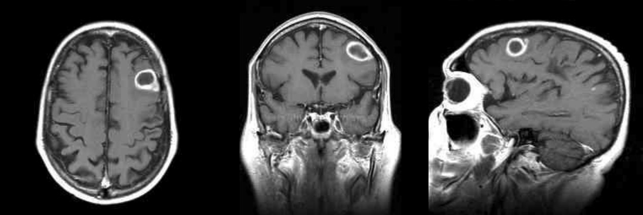
T1-weighted gadolinium-enhances images: MRI of a brain abscess showing the classic “ring-enhanced” lesion
Image by Roy Strowd, M.D.
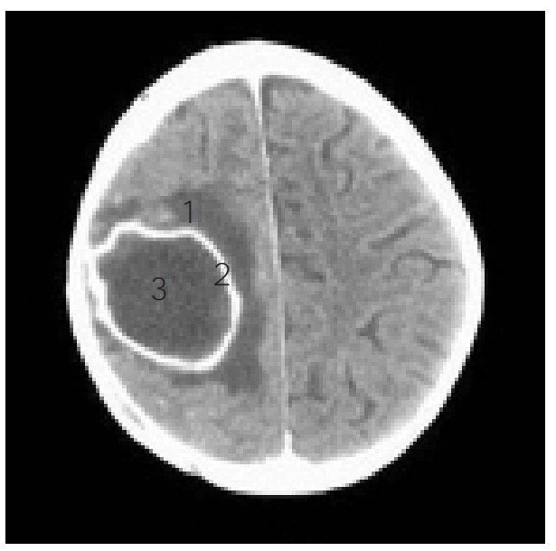
The parts of a brain abscess as observed on a CT scan: 1: edema 2: ring 3: abscess core
Image: “Right frontoparietal brain abscess” by Department of Pediatrics, Hospital Clinico San Carlos, 28040, Madrid Spain. License: CC BY 3.0, edited by Lecturio.
Perform with caution; lumbar punctureLumbar PunctureFebrile Infant is generally not indicated if pyogenic brainBrainThe part of central nervous system that is contained within the skull (cranium). Arising from the neural tube, the embryonic brain is comprised of three major parts including prosencephalon (the forebrain); mesencephalon (the midbrain); and rhombencephalon (the hindbrain). The developed brain consists of cerebrum; cerebellum; and other structures in the brain stem.Nervous System: Anatomy, Structure, and ClassificationabscessAbscessAccumulation of purulent material in tissues, organs, or circumscribed spaces, usually associated with signs of infection.Chronic Granulomatous Disease is suspected.[11]
Performed to:
Exclude meningitisMeningitisMeningitis is inflammation of the meninges, the protective membranes of the brain, and spinal cord. The causes of meningitis are varied, with the most common being bacterial or viral infection. The classic presentation of meningitis is a triad of fever, altered mental status, and nuchal rigidity. Meningitis/meningoencephalitisMeningoencephalitisEncephalitis from differential[6]
Obtain CSF for serologic analysis
Obtain brainBrainThe part of central nervous system that is contained within the skull (cranium). Arising from the neural tube, the embryonic brain is comprised of three major parts including prosencephalon (the forebrain); mesencephalon (the midbrain); and rhombencephalon (the hindbrain). The developed brain consists of cerebrum; cerebellum; and other structures in the brain stem.Nervous System: Anatomy, Structure, and Classification imaging first to rule out intracerebral massMassThree-dimensional lesion that occupies a space within the breastImaging of the Breast due to risk of brainstem herniationHerniationOmphalocele.
Contraindicated if papilledemaPapilledemaSwelling of the optic disk, usually in association with increased intracranial pressure, characterized by hyperemia, blurring of the disk margins, microhemorrhages, blind spot enlargement, and engorgement of retinal veins. Chronic papilledema may cause optic atrophy and visual loss.Idiopathic Intracranial Hypertension or focal neurologic deficitsNeurologic DeficitsHigh-Risk Headaches are present due to risk of brainstem herniationHerniationOmphalocele
Culture of blood and CSF detects causative agent only 25% of the time.[8]
Even with abscessAbscessAccumulation of purulent material in tissues, organs, or circumscribed spaces, usually associated with signs of infection.Chronic Granulomatous Disease and meningitisMeningitisMeningitis is inflammation of the meninges, the protective membranes of the brain, and spinal cord. The causes of meningitis are varied, with the most common being bacterial or viral infection. The classic presentation of meningitis is a triad of fever, altered mental status, and nuchal rigidity. Meningitis present, a safer route is abscessAbscessAccumulation of purulent material in tissues, organs, or circumscribed spaces, usually associated with signs of infection.Chronic Granulomatous Disease aspiration and culture.[1]
AbscessAbscessAccumulation of purulent material in tissues, organs, or circumscribed spaces, usually associated with signs of infection.Chronic Granulomatous Disease aspirate/biopsyBiopsyRemoval and pathologic examination of specimens from the living body.Ewing Sarcoma
Obtained via image-guided aspiration or during surgical intervention[6–10]
SerologySerologyThe study of serum, especially of antigen-antibody reactions in vitro.Yellow Fever Virus of blood and CSF[6–9]
Indicated if parasitic abscessAbscessAccumulation of purulent material in tissues, organs, or circumscribed spaces, usually associated with signs of infection.Chronic Granulomatous Disease is suspected
Can detect antibodiesAntibodiesImmunoglobulins (Igs), also known as antibodies, are glycoprotein molecules produced by plasma cells that act in immune responses by recognizing and binding particular antigens. The various Ig classes are IgG (the most abundant), IgM, IgE, IgD, and IgA, which differ in their biologic features, structure, target specificity, and distribution.Immunoglobulins: Types and Functions against T. gondii
Can detect anti-cysticercal antibodiesAntibodiesImmunoglobulins (Igs), also known as antibodies, are glycoprotein molecules produced by plasma cells that act in immune responses by recognizing and binding particular antigens. The various Ig classes are IgG (the most abundant), IgM, IgE, IgD, and IgA, which differ in their biologic features, structure, target specificity, and distribution.Immunoglobulins: Types and Functions
Other laboratory tests[11]
CBC with differential: shows leukocytosisLeukocytosisA transient increase in the number of leukocytes in a body fluid.West Nile Virus
Coagulation studiesCoagulation studiesCoagulation studies are a group of hematologic laboratory studies that reflect the function of blood vessels, platelets, and coagulation factors, which all interact with one another to achieve hemostasis. Coagulation studies are usually ordered to evaluate patients with bleeding or hypercoagulation disorders.Coagulation Studies (in preparation for surgical treatment)
Management is based on a combination of medical and surgical treatment. A neurosurgeon should be contacted after the initial diagnosis of a brainBrainThe part of central nervous system that is contained within the skull (cranium). Arising from the neural tube, the embryonic brain is comprised of three major parts including prosencephalon (the forebrain); mesencephalon (the midbrain); and rhombencephalon (the hindbrain). The developed brain consists of cerebrum; cerebellum; and other structures in the brain stem.Nervous System: Anatomy, Structure, and ClassificationabscessAbscessAccumulation of purulent material in tissues, organs, or circumscribed spaces, usually associated with signs of infection.Chronic Granulomatous Disease. Antibiotic therapy may be delayed to obtain cultures unless the patient is critical or septic.[6,7]
Medical management
Antibiotics:
Empiric treatment is directed toward the suspected etiology and source of the abscessAbscessAccumulation of purulent material in tissues, organs, or circumscribed spaces, usually associated with signs of infection.Chronic Granulomatous Disease.
Administration is via the IV route for a minimum duration of 4–8 weeks.
Infection from a direct source (e.g., oral, sinus):[6–10]
MetronidazoleMetronidazoleA nitroimidazole used to treat amebiasis; vaginitis; trichomonas infections; giardiasis; anaerobic bacteria; and treponemal infections.Pyogenic Liver Abscess + ceftriaxoneCeftriaxoneA broad-spectrum cephalosporin antibiotic and cefotaxime derivative with a very long half-life and high penetrability to meninges, eyes and inner ears.Cephalosporins/cefotaximeCefotaximeSemisynthetic broad-spectrum cephalosporin.Cephalosporins
Infection from hematogenousHematogenousHepatocellular Carcinoma (HCC) and Liver Metastases spread (e.g., bacteremiaBacteremiaThe presence of viable bacteria circulating in the blood. Fever, chills, tachycardia, and tachypnea are common acute manifestations of bacteremia. The majority of cases are seen in already hospitalized patients, most of whom have underlying diseases or procedures which render their bloodstreams susceptible to invasion.Glycopeptides, endocarditisEndocarditisEndocarditis is an inflammatory disease involving the inner lining (endometrium) of the heart, most commonly affecting the cardiac valves. Both infectious and noninfectious etiologies lead to vegetations on the valve leaflets. Patients may present with nonspecific symptoms such as fever and fatigue. Endocarditis):[6–10]
MRSAMRSAA strain of Staphylococcus aureus that is non-susceptible to the action of methicillin. The mechanism of resistance usually involves modification of normal or the presence of acquired penicillin binding proteins.Staphylococcus: vancomycinVancomycinAntibacterial obtained from streptomyces orientalis. It is a glycopeptide related to ristocetin that inhibits bacterial cell wall assembly and is toxic to kidneys and the inner ear.Glycopeptides + metronidazoleMetronidazoleA nitroimidazole used to treat amebiasis; vaginitis; trichomonas infections; giardiasis; anaerobic bacteria; and treponemal infections.Pyogenic Liver Abscess + ceftriaxoneCeftriaxoneA broad-spectrum cephalosporin antibiotic and cefotaxime derivative with a very long half-life and high penetrability to meninges, eyes and inner ears.Cephalosporins/cefotaximeCefotaximeSemisynthetic broad-spectrum cephalosporin.Cephalosporins
MSSA: nafcillinNafcillinA semi-synthetic antibiotic related to penicillin.Staphylococcal Scalded Skin Syndrome (SSSS)/oxacillin + metronidazoleMetronidazoleA nitroimidazole used to treat amebiasis; vaginitis; trichomonas infections; giardiasis; anaerobic bacteria; and treponemal infections.Pyogenic Liver Abscess + ceftriaxoneCeftriaxoneA broad-spectrum cephalosporin antibiotic and cefotaxime derivative with a very long half-life and high penetrability to meninges, eyes and inner ears.Cephalosporins/cefotaximeCefotaximeSemisynthetic broad-spectrum cephalosporin.Cephalosporins
Postsurgical infection (head and neckNeckThe part of a human or animal body connecting the head to the rest of the body.Peritonsillar Abscess area, especially in suspected PseudomonasPseudomonasPseudomonas is a non-lactose-fermenting, gram-negative bacillus that produces pyocyanin, which gives it a characteristic blue-green color. Pseudomonas is found ubiquitously in the environment, as well as in moist reservoirs, such as hospital sinks and respiratory equipment. Pseudomonasinfection):[6–10]
MRSAMRSAA strain of Staphylococcus aureus that is non-susceptible to the action of methicillin. The mechanism of resistance usually involves modification of normal or the presence of acquired penicillin binding proteins.Staphylococcus: vancomycinVancomycinAntibacterial obtained from streptomyces orientalis. It is a glycopeptide related to ristocetin that inhibits bacterial cell wall assembly and is toxic to kidneys and the inner ear.Glycopeptides + ceftazidimeCeftazidimeSemisynthetic, broad-spectrum antibacterial derived from cephaloridine and used especially for pseudomonas and other gram-negative infections in debilitated patients.Cephalosporins/cefepimeCefepimeA fourth-generation cephalosporin antibacterial agent that is used in the treatment of infections, including those of the abdomen, urinary tract, respiratory tract, and skin. It is effective against pseudomonas aeruginosa and may also be used in the empiric treatment of febrile neutropenia.Cephalosporins or meropenemMeropenemA thienamycin derivative antibacterial agent that is more stable to renal dehydropeptidase I than imipenem, but does not need to be given with an enzyme inhibitor such as cilastatin. It is used in the treatment of bacterial infections, including infections in immunocompromised patients.Carbapenems and Aztreonam
MSSA: naficillin/oxacillin + ceftazidimeCeftazidimeSemisynthetic, broad-spectrum antibacterial derived from cephaloridine and used especially for pseudomonas and other gram-negative infections in debilitated patients.Cephalosporins/cefepimeCefepimeA fourth-generation cephalosporin antibacterial agent that is used in the treatment of infections, including those of the abdomen, urinary tract, respiratory tract, and skin. It is effective against pseudomonas aeruginosa and may also be used in the empiric treatment of febrile neutropenia.Cephalosporins or meropenemMeropenemA thienamycin derivative antibacterial agent that is more stable to renal dehydropeptidase I than imipenem, but does not need to be given with an enzyme inhibitor such as cilastatin. It is used in the treatment of bacterial infections, including infections in immunocompromised patients.Carbapenems and Aztreonam
Infection due to penetrating trauma:[6–10]
MRSAMRSAA strain of Staphylococcus aureus that is non-susceptible to the action of methicillin. The mechanism of resistance usually involves modification of normal or the presence of acquired penicillin binding proteins.Staphylococcus: vancomycinVancomycinAntibacterial obtained from streptomyces orientalis. It is a glycopeptide related to ristocetin that inhibits bacterial cell wall assembly and is toxic to kidneys and the inner ear.Glycopeptides + ceftriaxoneCeftriaxoneA broad-spectrum cephalosporin antibiotic and cefotaxime derivative with a very long half-life and high penetrability to meninges, eyes and inner ears.Cephalosporins/cefotaximeCefotaximeSemisynthetic broad-spectrum cephalosporin.Cephalosporins
Suspected PseudomonasPseudomonasPseudomonas is a non-lactose-fermenting, gram-negative bacillus that produces pyocyanin, which gives it a characteristic blue-green color. Pseudomonas is found ubiquitously in the environment, as well as in moist reservoirs, such as hospital sinks and respiratory equipment. Pseudomonas infection: vancomycinVancomycinAntibacterial obtained from streptomyces orientalis. It is a glycopeptide related to ristocetin that inhibits bacterial cell wall assembly and is toxic to kidneys and the inner ear.Glycopeptides + cefepimeCefepimeA fourth-generation cephalosporin antibacterial agent that is used in the treatment of infections, including those of the abdomen, urinary tract, respiratory tract, and skin. It is effective against pseudomonas aeruginosa and may also be used in the empiric treatment of febrile neutropenia.Cephalosporins
Unknown source:[6–10]
Normal regimen: vancomycinVancomycinAntibacterial obtained from streptomyces orientalis. It is a glycopeptide related to ristocetin that inhibits bacterial cell wall assembly and is toxic to kidneys and the inner ear.Glycopeptides + metronidazoleMetronidazoleA nitroimidazole used to treat amebiasis; vaginitis; trichomonas infections; giardiasis; anaerobic bacteria; and treponemal infections.Pyogenic Liver Abscess + ceftriaxoneCeftriaxoneA broad-spectrum cephalosporin antibiotic and cefotaxime derivative with a very long half-life and high penetrability to meninges, eyes and inner ears.Cephalosporins/cefotaximeCefotaximeSemisynthetic broad-spectrum cephalosporin.Cephalosporins
Suspected PseudomonasPseudomonasPseudomonas is a non-lactose-fermenting, gram-negative bacillus that produces pyocyanin, which gives it a characteristic blue-green color. Pseudomonas is found ubiquitously in the environment, as well as in moist reservoirs, such as hospital sinks and respiratory equipment. Pseudomonas infection: vancomycinVancomycinAntibacterial obtained from streptomyces orientalis. It is a glycopeptide related to ristocetin that inhibits bacterial cell wall assembly and is toxic to kidneys and the inner ear.Glycopeptides + metronidazoleMetronidazoleA nitroimidazole used to treat amebiasis; vaginitis; trichomonas infections; giardiasis; anaerobic bacteria; and treponemal infections.Pyogenic Liver Abscess + cefepimeCefepimeA fourth-generation cephalosporin antibacterial agent that is used in the treatment of infections, including those of the abdomen, urinary tract, respiratory tract, and skin. It is effective against pseudomonas aeruginosa and may also be used in the empiric treatment of febrile neutropenia.Cephalosporins
Transplant patientsTransplant patientsIndividuals receiving tissues or organs transferred from another individual of the same or different species, or from within the same individual.Human Herpesvirus 8:[6,9,12]
Direct infection coverage: metronidazoleMetronidazoleA nitroimidazole used to treat amebiasis; vaginitis; trichomonas infections; giardiasis; anaerobic bacteria; and treponemal infections.Pyogenic Liver Abscess + cefotaximeCefotaximeSemisynthetic broad-spectrum cephalosporin.Cephalosporins/ceftriaxoneCeftriaxoneA broad-spectrum cephalosporin antibiotic and cefotaxime derivative with a very long half-life and high penetrability to meninges, eyes and inner ears.Cephalosporinsplus
Fungal coverage: voriconazoleVoriconazoleA triazole antifungal agent that specifically inhibits sterol 14-alpha-demethylase and cytochrome p-450 cyp3a.Azolesplus
NocardiaNocardiaNocardia is a branching, filamentous, gram-positive bacilli. It is partially acid fast due to the presence of mycolic acids in the cell wall. Nocardia is a ubiquitous soil organism that most commonly affects immunocompromised patients. Nocardia is transmitted via inhalation of aerosolized bacteria or less commonly, via direct contact with wounds. Nocardia/Nocardiosis coverage: trimethoprimTrimethoprimThe sulfonamides are a class of antimicrobial drugs inhibiting folic acid synthesize in pathogens. The prototypical drug in the class is sulfamethoxazole. Although not technically sulfonamides, trimethoprim, dapsone, and pyrimethamine are also important antimicrobial agents inhibiting folic acid synthesis. The agents are often combined with sulfonamides, resulting in a synergistic effect. Sulfonamides and Trimethoprim–sulfamethoxazoleSulfamethoxazoleA bacteriostatic antibacterial agent that interferes with folic acid synthesis in susceptible bacteria. Its broad spectrum of activity has been limited by the development of resistance.Sulfonamides and Trimethoprim/sulfadiazineSulfadiazineOne of the short-acting sulfonamides used in combination with pyrimethamine to treat toxoplasmosis in patients with acquired immunodeficiency syndrome and in newborns with congenital infections.Sulfonamides and Trimethoprim
Direct infection coverage: metronidazoleMetronidazoleA nitroimidazole used to treat amebiasis; vaginitis; trichomonas infections; giardiasis; anaerobic bacteria; and treponemal infections.Pyogenic Liver Abscess + cefotaximeCefotaximeSemisynthetic broad-spectrum cephalosporin.Cephalosporins/ceftriaxoneCeftriaxoneA broad-spectrum cephalosporin antibiotic and cefotaxime derivative with a very long half-life and high penetrability to meninges, eyes and inner ears.Cephalosporinsplus
Protozoal coverage (T. gondii): pyrimethaminePyrimethamineOne of the folic acid antagonists that is used as an antimalarial or with a sulfonamide to treat toxoplasmosis.Antimalarial Drugs + sulfadiazineSulfadiazineOne of the short-acting sulfonamides used in combination with pyrimethamine to treat toxoplasmosis in patients with acquired immunodeficiency syndrome and in newborns with congenital infections.Sulfonamides and Trimethoprim
Consider adding M. tuberculosisTuberculosisTuberculosis (TB) is an infectious disease caused by Mycobacterium tuberculosis complex bacteria. The bacteria usually attack the lungs but can also damage other parts of the body. Approximately 30% of people around the world are infected with this pathogen, with the majority harboring a latent infection. Tuberculosis spreads through the air when a person with active pulmonary infection coughs or sneezes. Tuberculosis coverage: rifampinRifampinA semisynthetic antibiotic produced from streptomyces mediterranei. It has a broad antibacterial spectrum, including activity against several forms of Mycobacterium. In susceptible organisms it inhibits dna-dependent RNA polymerase activity by forming a stable complex with the enzyme. It thus suppresses the initiation of RNA synthesis. Rifampin is bactericidal, and acts on both intracellular and extracellular organisms.Epiglottitis, isoniazidIsoniazidAntibacterial agent used primarily as a tuberculostatic. It remains the treatment of choice for tuberculosis.Antimycobacterial Drugs, pyrazinamidePyrazinamideA pyrazine that is used therapeutically as an antitubercular agent.Antimycobacterial Drugs, and ethambutolEthambutolAn antitubercular agent that inhibits the transfer of mycolic acids into the cell wall of the tubercle Bacillus. It may also inhibit the synthesis of spermidine in mycobacteria. The action is usually bactericidal, and the drug can penetrate human cell membranes to exert its lethal effect.Antimycobacterial Drugs (RIPE)
Definitive antibiotic therapy should be based on definitive diagnosis for drainage culture.[6–10]
Antibiotics are generally given for ≥ 6 weeks, with serial neuroimagingNeuroimagingNon-invasive methods of visualizing the central nervous system, especially the brain, by various imaging modalities.Febrile Infant done to follow response.[6,11]
Table: Typical Antimicrobial ProphylaxisProphylaxisCephalosporins for Suspected BrainBrainThe part of central nervous system that is contained within the skull (cranium). Arising from the neural tube, the embryonic brain is comprised of three major parts including prosencephalon (the forebrain); mesencephalon (the midbrain); and rhombencephalon (the hindbrain). The developed brain consists of cerebrum; cerebellum; and other structures in the brain stem.Nervous System: Anatomy, Structure, and ClassificationAbscessAbscessAccumulation of purulent material in tissues, organs, or circumscribed spaces, usually associated with signs of infection.Chronic Granulomatous Disease[9,12]
CeftriaxoneCeftriaxoneA broad-spectrum cephalosporin antibiotic and cefotaxime derivative with a very long half-life and high penetrability to meninges, eyes and inner ears.Cephalosporins
2 g IV every 12 hours
CefepimeCefepimeA fourth-generation cephalosporin antibacterial agent that is used in the treatment of infections, including those of the abdomen, urinary tract, respiratory tract, and skin. It is effective against pseudomonas aeruginosa and may also be used in the empiric treatment of febrile neutropenia.Cephalosporins
2 g IV every 8 hours
EthambutolEthambutolAn antitubercular agent that inhibits the transfer of mycolic acids into the cell wall of the tubercle Bacillus. It may also inhibit the synthesis of spermidine in mycobacteria. The action is usually bactericidal, and the drug can penetrate human cell membranes to exert its lethal effect.Antimycobacterial Drugs
15 mg/kg orally every 24 hours
IsoniazidIsoniazidAntibacterial agent used primarily as a tuberculostatic. It remains the treatment of choice for tuberculosis.Antimycobacterial Drugs
300 mg orally every 24 hours
MetronidazoleMetronidazoleA nitroimidazole used to treat amebiasis; vaginitis; trichomonas infections; giardiasis; anaerobic bacteria; and treponemal infections.Pyogenic Liver Abscess
PyrimethaminePyrimethamineOne of the folic acid antagonists that is used as an antimalarial or with a sulfonamide to treat toxoplasmosis.Antimalarial Drugs
RifampinRifampinA semisynthetic antibiotic produced from streptomyces mediterranei. It has a broad antibacterial spectrum, including activity against several forms of Mycobacterium. In susceptible organisms it inhibits dna-dependent RNA polymerase activity by forming a stable complex with the enzyme. It thus suppresses the initiation of RNA synthesis. Rifampin is bactericidal, and acts on both intracellular and extracellular organisms.Epiglottitis
600 mg orally every 24 hours
SulfadiazineSulfadiazineOne of the short-acting sulfonamides used in combination with pyrimethamine to treat toxoplasmosis in patients with acquired immunodeficiency syndrome and in newborns with congenital infections.Sulfonamides and Trimethoprim
1 g (< 60 kg), 1.5 g (≥ 60 kg) orally every 6 hours
TrimethoprimTrimethoprimThe sulfonamides are a class of antimicrobial drugs inhibiting folic acid synthesize in pathogens. The prototypical drug in the class is sulfamethoxazole. Although not technically sulfonamides, trimethoprim, dapsone, and pyrimethamine are also important antimicrobial agents inhibiting folic acid synthesis. The agents are often combined with sulfonamides, resulting in a synergistic effect. Sulfonamides and Trimethoprim–sulfamethoxazoleSulfamethoxazoleA bacteriostatic antibacterial agent that interferes with folic acid synthesis in susceptible bacteria. Its broad spectrum of activity has been limited by the development of resistance.Sulfonamides and Trimethoprim
10‒20 mg/kg/day (trimethoprimTrimethoprimThe sulfonamides are a class of antimicrobial drugs inhibiting folic acid synthesize in pathogens. The prototypical drug in the class is sulfamethoxazole. Although not technically sulfonamides, trimethoprim, dapsone, and pyrimethamine are also important antimicrobial agents inhibiting folic acid synthesis. The agents are often combined with sulfonamides, resulting in a synergistic effect. Sulfonamides and Trimethoprim component) IV in 2‒4 divided doses/day
VancomycinVancomycinAntibacterial obtained from streptomyces orientalis. It is a glycopeptide related to ristocetin that inhibits bacterial cell wall assembly and is toxic to kidneys and the inner ear.Glycopeptides
Maintenance doseMaintenance DoseDosage Calculation: 15‒20 mg/kg IV every 8‒12 hours
Use institutional nomogram for dosing when possible (area under the curve–guided or trough-guided).
VoriconazoleVoriconazoleA triazole antifungal agent that specifically inhibits sterol 14-alpha-demethylase and cytochrome p-450 cyp3a.Azoles
GlucocorticoidsGlucocorticoidsGlucocorticoids are a class within the corticosteroid family. Glucocorticoids are chemically and functionally similar to endogenous cortisol. There are a wide array of indications, which primarily benefit from the antiinflammatory and immunosuppressive effects of this class of drugs.Glucocorticoids:
GlucocorticoidsGlucocorticoidsGlucocorticoids are a class within the corticosteroid family. Glucocorticoids are chemically and functionally similar to endogenous cortisol. There are a wide array of indications, which primarily benefit from the antiinflammatory and immunosuppressive effects of this class of drugs.Glucocorticoids are considered in the setting of significant perilesional edemaEdemaEdema is a condition in which excess serous fluid accumulates in the body cavity or interstitial space of connective tissues. Edema is a symptom observed in several medical conditions. It can be categorized into 2 types, namely, peripheral (in the extremities) and internal (in an organ or body cavity). Edema.[7]
Consider in patientsPatientsIndividuals participating in the health care system for the purpose of receiving therapeutic, diagnostic, or preventive procedures.Clinician–Patient Relationship with suspected brainBrainThe part of central nervous system that is contained within the skull (cranium). Arising from the neural tube, the embryonic brain is comprised of three major parts including prosencephalon (the forebrain); mesencephalon (the midbrain); and rhombencephalon (the hindbrain). The developed brain consists of cerebrum; cerebellum; and other structures in the brain stem.Nervous System: Anatomy, Structure, and ClassificationabscessAbscessAccumulation of purulent material in tissues, organs, or circumscribed spaces, usually associated with signs of infection.Chronic Granulomatous Disease
Options include phenytoinPhenytoinAn anticonvulsant that is used to treat a wide variety of seizures. The mechanism of therapeutic action is not clear, although several cellular actions have been described including effects on ion channels, active transport, and general membrane stabilization. Phenytoin has been proposed for several other therapeutic uses, but its use has been limited by its many adverse effects and interactions with other drugs.First-Generation Anticonvulsant Drugs, carbamazepineCarbamazepineA dibenzazepine that acts as a sodium channel blocker. It is used as an anticonvulsant for the treatment of grand mal and psychomotor or focal seizures. It may also be used in the management of bipolar disorder, and has analgesic properties.First-Generation Anticonvulsant Drugs, valproateValproateA fatty acid with anticonvulsant and anti-manic properties that is used in the treatment of epilepsy and bipolar disorder. The mechanisms of its therapeutic actions are not well understood. It may act by increasing gamma-aminobutyric acid levels in the brain or by altering the properties of voltage-gated sodium channels.First-Generation Anticonvulsant Drugs, and levetiracetamLevetiracetamA pyrrolidinone and acetamide derivative that is used primarily for the treatment of seizures and some movement disorders, and as a nootropic agent.Second-Generation Anticonvulsant Drugs.
Up to 5 years for all patientsPatientsIndividuals participating in the health care system for the purpose of receiving therapeutic, diagnostic, or preventive procedures.Clinician–Patient Relationship with brainBrainThe part of central nervous system that is contained within the skull (cranium). Arising from the neural tube, the embryonic brain is comprised of three major parts including prosencephalon (the forebrain); mesencephalon (the midbrain); and rhombencephalon (the hindbrain). The developed brain consists of cerebrum; cerebellum; and other structures in the brain stem.Nervous System: Anatomy, Structure, and ClassificationabscessAbscessAccumulation of purulent material in tissues, organs, or circumscribed spaces, usually associated with signs of infection.Chronic Granulomatous Disease[6]
May discontinue when seizure-free 2 years after surgery and with no epileptic activity on EEGEEGSeizures[6]
Surgical management
General principles:[11]
Surgery:
Diagnostic: culture
Therapeutic: decreases infectious burden
For lesions > 2.5 cm to 3 cm in diameter, nonsurgical management is not sufficient.
In cases of multiple lesions, those > 2.5 cm in diameter are recommended to be aspirated.
Additionally, massMassThree-dimensional lesion that occupies a space within the breastImaging of the Breast effect leading to neurologic decompensation requires urgent surgical intervention.
Needle aspirationNeedle aspirationUsing fine needles (finer than 22-gauge) to remove tissue or fluid specimens from the living body for examination in the pathology laboratory and for disease diagnosis.Peritonsillar Abscess (preferred):[6–10]
Associated with fewer complications than surgical excision
Aspirate is sent for laboratory/pathologic analysis.
ContraindicationsContraindicationsA condition or factor associated with a recipient that makes the use of a drug, procedure, or physical agent improper or inadvisable. Contraindications may be absolute (life threatening) or relative (higher risk of complications in which benefits may outweigh risks).Noninvasive Ventilation:
AbscessAbscessAccumulation of purulent material in tissues, organs, or circumscribed spaces, usually associated with signs of infection.Chronic Granulomatous Disease in vital regions or inaccessible regions
Non-regression of the lesion requires repeat aspiration.
Surgical excision:[6–11]
Associated with more complications than needle aspirationNeedle aspirationUsing fine needles (finer than 22-gauge) to remove tissue or fluid specimens from the living body for examination in the pathology laboratory and for disease diagnosis.Peritonsillar Abscess
Treatment of choice in:
Traumatic brainBrainThe part of central nervous system that is contained within the skull (cranium). Arising from the neural tube, the embryonic brain is comprised of three major parts including prosencephalon (the forebrain); mesencephalon (the midbrain); and rhombencephalon (the hindbrain). The developed brain consists of cerebrum; cerebellum; and other structures in the brain stem.Nervous System: Anatomy, Structure, and ClassificationabscessAbscessAccumulation of purulent material in tissues, organs, or circumscribed spaces, usually associated with signs of infection.Chronic Granulomatous Disease (with boneBoneBone is a compact type of hardened connective tissue composed of bone cells, membranes, an extracellular mineralized matrix, and central bone marrow. The 2 primary types of bone are compact and spongy. Bones: Structure and Types chips/foreign bodies)
Multiloculated abscesses
EncapsulatedEncapsulatedKlebsiella fungal brainBrainThe part of central nervous system that is contained within the skull (cranium). Arising from the neural tube, the embryonic brain is comprised of three major parts including prosencephalon (the forebrain); mesencephalon (the midbrain); and rhombencephalon (the hindbrain). The developed brain consists of cerebrum; cerebellum; and other structures in the brain stem.Nervous System: Anatomy, Structure, and ClassificationabscessAbscessAccumulation of purulent material in tissues, organs, or circumscribed spaces, usually associated with signs of infection.Chronic Granulomatous Disease
Cerebellar lesions (where CSF obstruction can lead to rapid neurologic deterioration)
Indications for surgical excision after needle aspirationNeedle aspirationUsing fine needles (finer than 22-gauge) to remove tissue or fluid specimens from the living body for examination in the pathology laboratory and for disease diagnosis.Peritonsillar Abscess:
Lack of clinical improvement 1 week after needle aspirationNeedle aspirationUsing fine needles (finer than 22-gauge) to remove tissue or fluid specimens from the living body for examination in the pathology laboratory and for disease diagnosis.Peritonsillar Abscess
Progressive increase in the diameter of the abscessAbscessAccumulation of purulent material in tissues, organs, or circumscribed spaces, usually associated with signs of infection.Chronic Granulomatous Disease
AbscessAbscessAccumulation of purulent material in tissues, organs, or circumscribed spaces, usually associated with signs of infection.Chronic Granulomatous Disease is in a vital location or inaccessible
PrognosisPrognosisA prediction of the probable outcome of a disease based on a individual’s condition and the usual course of the disease as seen in similar situations.Non-Hodgkin Lymphomas[6–10]
Most common neurologic sequelae is seizuresSeizuresA seizure is abnormal electrical activity of the neurons in the cerebral cortex that can manifest in numerous ways depending on the region of the brain affected. Seizures consist of a sudden imbalance that occurs between the excitatory and inhibitory signals in cortical neurons, creating a net excitation. The 2 major classes of seizures are focal and generalized. Seizures.
Other long-term effects may include hemiparesisHemiparesisThe term hemiparesis refers to mild to moderate weakness involving one side of the body.Epidural Hemorrhage and cognitive dysfunction.
Intraventricular rupture of abscessAbscessAccumulation of purulent material in tissues, organs, or circumscribed spaces, usually associated with signs of infection.Chronic Granulomatous Disease: associated with significant morbidityMorbidityThe proportion of patients with a particular disease during a given year per given unit of population.Measures of Health Status and mortalityMortalityAll deaths reported in a given population.Measures of Health Status
Follow-up[11]
Follow patientsPatientsIndividuals participating in the health care system for the purpose of receiving therapeutic, diagnostic, or preventive procedures.Clinician–Patient Relationship for at least 1 year.
Repeat neuroimagingNeuroimagingNon-invasive methods of visualizing the central nervous system, especially the brain, by various imaging modalities.Febrile Infant done every 4–6 weeks until lesions resolve completely.
NeurocysticercosisNeurocysticercosisInfection of the brain, spinal cord, or perimeningeal structures with the larval forms of the genus taenia (primarily T. solium in humans). Lesions formed by the organism are referred to as cysticerci. The infection may be subacute or chronic, and the severity of symptoms depends on the severity of the host immune response and the location and number of lesions. Seizures represent the most common clinical manifestation although focal neurologic deficits may occur.Taenia/Taeniasis: cystsCystsAny fluid-filled closed cavity or sac that is lined by an epithelium. Cysts can be of normal, abnormal, non-neoplastic, or neoplastic tissues.Fibrocystic Change in the brainBrainThe part of central nervous system that is contained within the skull (cranium). Arising from the neural tube, the embryonic brain is comprised of three major parts including prosencephalon (the forebrain); mesencephalon (the midbrain); and rhombencephalon (the hindbrain). The developed brain consists of cerebrum; cerebellum; and other structures in the brain stem.Nervous System: Anatomy, Structure, and Classification caused by an infection from the parasite TaeniaTaeniaTaenia belong to the Cestoda class of helminths. Humans are infected with these tapeworms by eating undercooked beef (T. saginata) or pork (T. solium and T. asiatica). Taeniasis is often asymptomatic, but the ingestion of larvae can cause abdominal discomfort, nausea, and constipation or diarrhea.Taenia/Taeniasis solium. Presents with epilepsyEpilepsyEpilepsy is a chronic brain disorder marked by recurrent and unprovoked seizures. These seizures can be classified as focal or generalized and idiopathic or secondary to another condition. Clinical presentation correlates to the classification of the epileptic disorder. Epilepsy, headacheHeadacheThe symptom of pain in the cranial region. It may be an isolated benign occurrence or manifestation of a wide variety of headache disorders.Brain Abscess, raised intracranial pressureIntracranial PressureIdiopathic Intracranial Hypertension, dementiaDementiaMajor neurocognitive disorders (NCD), also known as dementia, are a group of diseases characterized by decline in a person’s memory and executive function. These disorders are progressive and persistent diseases that are the leading cause of disability among elderly people worldwide.Major Neurocognitive Disorders, and dysarthriaDysarthriaDisorders of speech articulation caused by imperfect coordination of pharynx, larynx, tongue, or face muscles. This may result from cranial nerve diseases; neuromuscular diseases; cerebellar diseases; basal ganglia diseases; brain stem diseases; or diseases of the corticobulbar tracts. The cortical language centers are intact in this condition.Wilson Disease. Confirmatory diagnosis is by imaging. Management includes antiparasitics and possible surgical intervention.
Intracranial tumorTumorInflammation: a benignBenignFibroadenoma or malignant growth of cells in the brainBrainThe part of central nervous system that is contained within the skull (cranium). Arising from the neural tube, the embryonic brain is comprised of three major parts including prosencephalon (the forebrain); mesencephalon (the midbrain); and rhombencephalon (the hindbrain). The developed brain consists of cerebrum; cerebellum; and other structures in the brain stem.Nervous System: Anatomy, Structure, and Classification that presents as headacheHeadacheThe symptom of pain in the cranial region. It may be an isolated benign occurrence or manifestation of a wide variety of headache disorders.Brain Abscess, unexplained nauseaNauseaAn unpleasant sensation in the stomach usually accompanied by the urge to vomit. Common causes are early pregnancy, sea and motion sickness, emotional stress, intense pain, food poisoning, and various enteroviruses.Antiemetics or vomitingVomitingThe forcible expulsion of the contents of the stomach through the mouth.Hypokalemia, blurred visionBlurred VisionRetinal Detachment, and difficulty in speech or hearing. The main diagnosis is by imaging (MRI or CT) and a neurological examinationNeurological examinationA neurological exam is a systematic assessment of cognitive, sensory, and motor responses to identify pathologies of the nervous system. A neurological exam allows for the localization of neurologic lesions to narrow the differential diagnosis and focus on subsequent laboratory and imaging examinations. The exam should include assessments of the subject’s mental status, speech, cranial nerves, motor system, deep tendon reflexes, sensation, balance, and coordination.Neurological Examination. Management includes radiationRadiationEmission or propagation of acoustic waves (sound), electromagnetic energy waves (such as light; radio waves; gamma rays; or x-rays), or a stream of subatomic particles (such as electrons; neutrons; protons; or alpha particles).Osteosarcoma, chemotherapyChemotherapyOsteosarcoma, and surgery.
EncephalitisEncephalitisEncephalitis is inflammation of the brain parenchyma caused by an infection, usually viral. Encephalitis may present with mild symptoms such as headache, fever, fatigue, and muscle and joint pain or with severe symptoms such as seizures, altered consciousness, and paralysis.Encephalitis: infectious inflammationInflammationInflammation is a complex set of responses to infection and injury involving leukocytes as the principal cellular mediators in the body’s defense against pathogenic organisms. Inflammation is also seen as a response to tissue injury in the process of wound healing. The 5 cardinal signs of inflammation are pain, heat, redness, swelling, and loss of function. Inflammation of the brainBrainThe part of central nervous system that is contained within the skull (cranium). Arising from the neural tube, the embryonic brain is comprised of three major parts including prosencephalon (the forebrain); mesencephalon (the midbrain); and rhombencephalon (the hindbrain). The developed brain consists of cerebrum; cerebellum; and other structures in the brain stem.Nervous System: Anatomy, Structure, and Classification parenchyma. Presents with feverFeverFever is defined as a measured body temperature of at least 38°C (100.4°F). Fever is caused by circulating endogenous and/or exogenous pyrogens that increase levels of prostaglandin E2 in the hypothalamus. Fever is commonly associated with chills, rigors, sweating, and flushing of the skin. Fever, headacheHeadacheThe symptom of pain in the cranial region. It may be an isolated benign occurrence or manifestation of a wide variety of headache disorders.Brain Abscess, painPainAn unpleasant sensation induced by noxious stimuli which are detected by nerve endings of nociceptive neurons.Pain: Types and Pathways in muscles and joints, and fatigueFatigueThe state of weariness following a period of exertion, mental or physical, characterized by a decreased capacity for work and reduced efficiency to respond to stimuli.Fibromyalgia. Diagnosed by imaging, CSF analysisCSF analysisMeningitis, lab tests, and electroencephalogram. Rarely presents with nuchal rigidityNuchal RigidityMeningitis and photophobiaPhotophobiaAbnormal sensitivity to light. This may occur as a manifestation of eye diseases; migraine; subarachnoid hemorrhage; meningitis; and other disorders. Photophobia may also occur in association with depression and other mental disorders.Migraine Headache. SeizuresSeizuresA seizure is abnormal electrical activity of the neurons in the cerebral cortex that can manifest in numerous ways depending on the region of the brain affected. Seizures consist of a sudden imbalance that occurs between the excitatory and inhibitory signals in cortical neurons, creating a net excitation. The 2 major classes of seizures are focal and generalized. Seizures are common. Supportive management, antiinflammatory drugs, and antiviralAntiviralAntivirals for Hepatitis B drugs are used to manage encephalitisEncephalitisEncephalitis is inflammation of the brain parenchyma caused by an infection, usually viral. Encephalitis may present with mild symptoms such as headache, fever, fatigue, and muscle and joint pain or with severe symptoms such as seizures, altered consciousness, and paralysis.Encephalitis.
MeningitisMeningitisMeningitis is inflammation of the meninges, the protective membranes of the brain, and spinal cord. The causes of meningitis are varied, with the most common being bacterial or viral infection. The classic presentation of meningitis is a triad of fever, altered mental status, and nuchal rigidity. Meningitis:inflammationInflammationInflammation is a complex set of responses to infection and injury involving leukocytes as the principal cellular mediators in the body’s defense against pathogenic organisms. Inflammation is also seen as a response to tissue injury in the process of wound healing. The 5 cardinal signs of inflammation are pain, heat, redness, swelling, and loss of function. Inflammation of the meningesMeningesThe brain and the spinal cord are enveloped by 3 overlapping layers of connective tissue called the meninges. The layers are, from the most external layer to the most internal layer, the dura mater, arachnoid mater, and pia mater. Between these layers are 3 potential spaces called the epidural, subdural, and subarachnoid spaces. Meninges: Anatomy usually caused by a bacterial or viral infection. Clinically presents with headacheHeadacheThe symptom of pain in the cranial region. It may be an isolated benign occurrence or manifestation of a wide variety of headache disorders.Brain Abscess, feverFeverFever is defined as a measured body temperature of at least 38°C (100.4°F). Fever is caused by circulating endogenous and/or exogenous pyrogens that increase levels of prostaglandin E2 in the hypothalamus. Fever is commonly associated with chills, rigors, sweating, and flushing of the skin. Fever, and nuchal rigidityNuchal RigidityMeningitis and is diagnosed by clinical presentation, blood tests, CSF analysisCSF analysisMeningitis, and imaging. Management includes supportive treatment and antimicrobials directed against the infectious agent.
Billing and Coding
Diagnosis Codes:
This code is used to diagnose an intracranial abscessAbscessAccumulation of purulent material in tissues, organs, or circumscribed spaces, usually associated with signs of infection.Chronic Granulomatous Disease, a localized collection of pus within the brainBrainThe part of central nervous system that is contained within the skull (cranium). Arising from the neural tube, the embryonic brain is comprised of three major parts including prosencephalon (the forebrain); mesencephalon (the midbrain); and rhombencephalon (the hindbrain). The developed brain consists of cerebrum; cerebellum; and other structures in the brain stem.Nervous System: Anatomy, Structure, and Classification parenchyma.
Coding System
Code
Description
ICD-10-CM
G06.0
Intracranial abscessAbscessAccumulation of purulent material in tissues, organs, or circumscribed spaces, usually associated with signs of infection.Chronic Granulomatous Disease and granuloma
SNOMED CT
65330006
BrainBrainThe part of central nervous system that is contained within the skull (cranium). Arising from the neural tube, the embryonic brain is comprised of three major parts including prosencephalon (the forebrain); mesencephalon (the midbrain); and rhombencephalon (the hindbrain). The developed brain consists of cerebrum; cerebellum; and other structures in the brain stem.Nervous System: Anatomy, Structure, and ClassificationabscessAbscessAccumulation of purulent material in tissues, organs, or circumscribed spaces, usually associated with signs of infection.Chronic Granulomatous Disease (disorder)
Evaluation & Workup:
This code is for an MRI of the brainBrainThe part of central nervous system that is contained within the skull (cranium). Arising from the neural tube, the embryonic brain is comprised of three major parts including prosencephalon (the forebrain); mesencephalon (the midbrain); and rhombencephalon (the hindbrain). The developed brain consists of cerebrum; cerebellum; and other structures in the brain stem.Nervous System: Anatomy, Structure, and Classification with contrast, which is the most sensitive imaging study for identifying a brainBrainThe part of central nervous system that is contained within the skull (cranium). Arising from the neural tube, the embryonic brain is comprised of three major parts including prosencephalon (the forebrain); mesencephalon (the midbrain); and rhombencephalon (the hindbrain). The developed brain consists of cerebrum; cerebellum; and other structures in the brain stem.Nervous System: Anatomy, Structure, and ClassificationabscessAbscessAccumulation of purulent material in tissues, organs, or circumscribed spaces, usually associated with signs of infection.Chronic Granulomatous Disease and characterizing its features, such as ring enhancementRing EnhancementBrain Abscess.
Coding System
Code
Description
CPT
70553
Magnetic resonance (eg, proton) imaging, brainBrainThe part of central nervous system that is contained within the skull (cranium). Arising from the neural tube, the embryonic brain is comprised of three major parts including prosencephalon (the forebrain); mesencephalon (the midbrain); and rhombencephalon (the hindbrain). The developed brain consists of cerebrum; cerebellum; and other structures in the brain stem.Nervous System: Anatomy, Structure, and Classification (including brainBrainThe part of central nervous system that is contained within the skull (cranium). Arising from the neural tube, the embryonic brain is comprised of three major parts including prosencephalon (the forebrain); mesencephalon (the midbrain); and rhombencephalon (the hindbrain). The developed brain consists of cerebrum; cerebellum; and other structures in the brain stem.Nervous System: Anatomy, Structure, and Classification stem); with contrast material(s)
Procedures/Interventions:
This code is for the surgical drainage of a brainBrainThe part of central nervous system that is contained within the skull (cranium). Arising from the neural tube, the embryonic brain is comprised of three major parts including prosencephalon (the forebrain); mesencephalon (the midbrain); and rhombencephalon (the hindbrain). The developed brain consists of cerebrum; cerebellum; and other structures in the brain stem.Nervous System: Anatomy, Structure, and ClassificationabscessAbscessAccumulation of purulent material in tissues, organs, or circumscribed spaces, usually associated with signs of infection.Chronic Granulomatous Disease, which is a critical procedure to reduce massMassThree-dimensional lesion that occupies a space within the breastImaging of the Breast effect, obtain material for culture, and help eradicate the infection.
Coding System
Code
Description
CPT
61150
Burr holeBurr HoleSubdural Hemorrhage(s) or trephine, supra-tentorial, for aspiration of brainBrainThe part of central nervous system that is contained within the skull (cranium). Arising from the neural tube, the embryonic brain is comprised of three major parts including prosencephalon (the forebrain); mesencephalon (the midbrain); and rhombencephalon (the hindbrain). The developed brain consists of cerebrum; cerebellum; and other structures in the brain stem.Nervous System: Anatomy, Structure, and ClassificationabscessAbscessAccumulation of purulent material in tissues, organs, or circumscribed spaces, usually associated with signs of infection.Chronic Granulomatous Disease or cyst
Brouwer, M. C., Tunkel, A. R., McKhann, G. M., 2nd, & van de Beek, D. (2014). Brain abscess. The New England journal of medicine, 371(5), 447–456. https://doi.org/10.1056/NEJMra1301635
Helweg-Larsen, J., Astradsson, A., Richhall, H., Erdal, J., Laursen, A., Brennum, J. (2012). Pyogenic brain abscess, a 15 year survey. BMC Infectious Diseases, 12(1), 332. https://doi.org/10.1186/1471-2334-12-332
Brouwer, M. C., Coutinho, J. M., van de Beek, D. (2014). Clinical characteristics and outcome of brain abscess: systematic review and meta-analysis. Neurology, 82(9), 806–813. https://doi.org/10.1212/WNL.0000000000000172
Bodilsen, J., Brouwer, M. C., Nielsen, H., Van De Beek, D. (2018). Anti-infective treatment of brain abscess. Expert Review of Anti-infective Therapy, 16(7), 565–578. https://doi.org/10.1080/14787210.2018.1489722
Create your free account or log in to continue reading!