Achieve Mastery of Medical Concepts

Study for medical school and boards with Lecturio

Dyspnea

Dyspnea is the subjective sensation of breathing discomfort. Dyspnea is a normal manifestation of heavy physical or psychologic exertion, but also may be caused by underlying conditions (both pulmonary and extrapulmonary). The medical history and physical exam of a patient presenting with dyspnea should be used to rule out certain conditions or suggest the most likely cause of the respiratory discomfort. Management focuses on treating the underlying condition.

Last updated: Jul 11, 2022

Editorial responsibility: Stanley Oiseth, Lindsay Jones, Evelin Maza

Definition and Epidemiology

Definition

Dyspnea is the subjective sensation of breathing discomfort that derives from interactions among multiple physiologic, psychologic, social, and environmental factors. 

  • Also referred to as chest tightness or shortness of breath
  • Experienced and described differently among patients Patients Individuals participating in the health care system for the purpose of receiving therapeutic, diagnostic, or preventive procedures. Clinician–Patient Relationship depending on the cause
  • Types:
    • Acute dyspnea: occurring within minutes of the triggering event
    • Chronic dyspnea: lasting longer than 1 month

Epidemiology

  • Very common symptom, present in: 
    • 50% of inpatients
    • 25% of ambulatory patients Patients Individuals participating in the health care system for the purpose of receiving therapeutic, diagnostic, or preventive procedures. Clinician–Patient Relationship
    • 9%–13% of the entire population
    • 37% of people aged ≥ 70 years
  • In chronic obstructive pulmonary disease Chronic obstructive pulmonary disease Chronic obstructive pulmonary disease (COPD) is a lung disease characterized by progressive, largely irreversible airflow obstruction. The condition usually presents in middle-aged or elderly persons with a history of cigarette smoking. Signs and symptoms include prolonged expiration, wheezing, diminished breath sounds, progressive dyspnea, and chronic cough. Chronic Obstructive Pulmonary Disease (COPD) ( COPD COPD Chronic obstructive pulmonary disease (COPD) is a lung disease characterized by progressive, largely irreversible airflow obstruction. The condition usually presents in middle-aged or elderly persons with a history of cigarette smoking. Signs and symptoms include prolonged expiration, wheezing, diminished breath sounds, progressive dyspnea, and chronic cough. Chronic Obstructive Pulmonary Disease (COPD)) and other pathologic lung and heart conditions, dyspnea may be a better predictor of outcome than traditional assessments.

Related videos

Etiology

There are 5 basic categories of causes that can lead to dyspnea. The causes can present individually or as a combination:

  1. Respiratory conditions
  2. Cardiac conditions
  3. Neuromuscular conditions
  4. Psychogenic conditions
  5. Systemic illnesses

General causes of dyspnea

Table: Pulmonary causes of dyspnea
Etiology Clinical presentation Associated conditions
Obstructive lung disease Characterized by air trapping in the lungs Lungs Lungs are the main organs of the respiratory system. Lungs are paired viscera located in the thoracic cavity and are composed of spongy tissue. The primary function of the lungs is to oxygenate blood and eliminate CO2. Lungs: Anatomy:
  • ↑ FRC and TLC
  • ↓↓ FEV₁
  • ↓ FVC
  • ↓ FEV₁/FVC
Findings:
  • Wheezing Wheezing Wheezing is an abnormal breath sound characterized by a whistling noise that can be relatively high-pitched and shrill (more common) or coarse. Wheezing is produced by the movement of air through narrowed or compressed small (intrathoracic) airways. Wheezing
  • Dyspnea is not worsened by a change in body position.
  • COPD COPD Chronic obstructive pulmonary disease (COPD) is a lung disease characterized by progressive, largely irreversible airflow obstruction. The condition usually presents in middle-aged or elderly persons with a history of cigarette smoking. Signs and symptoms include prolonged expiration, wheezing, diminished breath sounds, progressive dyspnea, and chronic cough. Chronic Obstructive Pulmonary Disease (COPD)
  • Chronic bronchitis Chronic bronchitis A subcategory of chronic obstructive pulmonary disease. The disease is characterized by hypersecretion of mucus accompanied by a chronic (more than 3 months in 2 consecutive years) productive cough. Infectious agents are a major cause of chronic bronchitis. Rhinovirus
  • Bronchiectasis Bronchiectasis Bronchiectasis is a chronic disease of the airways that results from permanent bronchial distortion. This results from a continuous cycle of inflammation, bronchial damage and dilation, impaired clearance of secretions, and recurrent infections. Bronchiectasis
  • Emphysema Emphysema Enlargement of air spaces distal to the terminal bronchioles where gas-exchange normally takes place. This is usually due to destruction of the alveolar wall. Pulmonary emphysema can be classified by the location and distribution of the lesions. Chronic Obstructive Pulmonary Disease (COPD)
  • Asthma Asthma Asthma is a chronic inflammatory respiratory condition characterized by bronchial hyperresponsiveness and airflow obstruction. The disease is believed to result from the complex interaction of host and environmental factors that increase disease predisposition, with inflammation causing symptoms and structural changes. Patients typically present with wheezing, cough, and dyspnea. Asthma
Restrictive lung disease Characterized by decreased lung compliance Compliance Distensibility measure of a chamber such as the lungs (lung compliance) or bladder. Compliance is expressed as a change in volume per unit change in pressure. Veins: Histology:
  • FVC and TLC
  • FEV₁/FVC ≥ 80%
  • Short, shallow breaths
Findings:
  • Short, shallow breaths
Poor breathing mechanics:
  • Polio
  • Kyphoscoliosis Kyphoscoliosis Osteomalacia and Rickets
  • Morbid obesity Obesity Obesity is a condition associated with excess body weight, specifically with the deposition of excessive adipose tissue. Obesity is considered a global epidemic. Major influences come from the western diet and sedentary lifestyles, but the exact mechanisms likely include a mixture of genetic and environmental factors. Obesity

ILDs:
  • Pneumoconiosis Pneumoconiosis Pneumoconiosis is an occupational disease that results from the inhalation and deposition of mineral dusts and other inorganic particles in the lung. It can be categorized according to the type of causative particle involved or by the type of response provoked. Pneumoconiosis
  • Sarcoidosis Sarcoidosis Sarcoidosis is a multisystem inflammatory disease that causes noncaseating granulomas. The exact etiology is unknown. Sarcoidosis usually affects the lungs and thoracic lymph nodes, but it can also affect almost every system in the body, including the skin, heart, and eyes, most commonly. Sarcoidosis
  • Idiopathic Idiopathic Dermatomyositis pulmonary fibrosis Pulmonary Fibrosis Idiopathic pulmonary fibrosis is a specific entity of the major idiopathic interstitial pneumonia classification of interstitial lung diseases. As implied by the name, the exact causes are poorly understood. Patients often present in the moderate to advanced stage with progressive dyspnea and nonproductive cough. Pulmonary Fibrosis
  • Drug toxicity Toxicity Dosage Calculation:
    • Bleomycin Bleomycin A complex of related glycopeptide antibiotics from streptomyces verticillus consisting of bleomycin a2 and b2. It inhibits DNA metabolism and is used as an antineoplastic, especially for solid tumors. Antitumor Antibiotics
    • Amiodarone Amiodarone An antianginal and class III antiarrhythmic drug. It increases the duration of ventricular and atrial muscle action by inhibiting potassium channels and voltage-gated sodium channels. There is a resulting decrease in heart rate and in vascular resistance. Pulmonary Fibrosis
    • Methotrexate Methotrexate An antineoplastic antimetabolite with immunosuppressant properties. It is an inhibitor of tetrahydrofolate dehydrogenase and prevents the formation of tetrahydrofolate, necessary for synthesis of thymidylate, an essential component of DNA. Antimetabolite Chemotherapy
Upper airway obstruction Airway obstruction Airway obstruction is a partial or complete blockage of the airways that impedes airflow. An airway obstruction can be classified as upper, central, or lower depending on location. Lower airway obstruction (LAO) is usually a manifestation of chronic disease, such as asthma or chronic obstructive pulmonary disease (COPD). Airway Obstruction Findings:
  • Epiglottitis Epiglottitis Epiglottitis (or “supraglottitis”) is an inflammation of the epiglottis and adjacent supraglottic structures. The majority of cases are caused by bacterial infection. Symptoms are rapid in onset and severe. Epiglottitis
  • Foreign body Foreign Body Foreign Body Aspiration obstruction
  • Croup Croup Croup, also known as laryngotracheobronchitis, is a disease most commonly caused by a viral infection that leads to severe inflammation of the upper airway. It usually presents in children < 5 years of age. Patients develop a hoarse, "seal-like" barking cough and inspiratory stridor. Croup
Others Clinical presentation and findings vary with the cause.
  • Pulmonary embolism Pulmonary Embolism Pulmonary embolism (PE) is a potentially fatal condition that occurs as a result of intraluminal obstruction of the main pulmonary artery or its branches. The causative factors include thrombi, air, amniotic fluid, and fat. In PE, gas exchange is impaired due to the decreased return of deoxygenated blood to the lungs. Pulmonary Embolism
  • Pneumothorax Pneumothorax A pneumothorax is a life-threatening condition in which air collects in the pleural space, causing partial or full collapse of the lung. A pneumothorax can be traumatic or spontaneous. Patients present with a sudden onset of sharp chest pain, dyspnea, and diminished breath sounds on exam. Pneumothorax
  • Pleural effusions
  • Metastatic disease
  • Pulmonary edema Pulmonary edema Pulmonary edema is a condition caused by excess fluid within the lung parenchyma and alveoli as a consequence of a disease process. Based on etiology, pulmonary edema is classified as cardiogenic or noncardiogenic. Patients may present with progressive dyspnea, orthopnea, cough, or respiratory failure. Pulmonary Edema
FRC: functional residual capacity
TLC: total lung capacity
FEV₁: forced expiratory capacity in 1 second
FVC: forced vital capacity
ILD: interstitial lung disease
Table: Extrapulmonary causes of dyspnea
EtiologyClinical presentationAssociated conditions
Cardiac Often a complication of left-sided heart failure Heart Failure A heterogeneous condition in which the heart is unable to pump out sufficient blood to meet the metabolic need of the body. Heart failure can be caused by structural defects, functional abnormalities (ventricular dysfunction), or a sudden overload beyond its capacity. Chronic heart failure is more common than acute heart failure which results from sudden insult to cardiac function, such as myocardial infarction. Total Anomalous Pulmonary Venous Return (TAPVR)
Findings:
  • Dyspnea varies with body position.
  • Orthopnea Orthopnea Pulmonary Edema
  • Paroxysmal nocturnal dyspnea Paroxysmal nocturnal dyspnea A disorder characterized by sudden attacks of respiratory distress in at rest patients with heart failure and pulmonary edema. It usually occurs at night after several hours of sleep in a reclining position. Patients awaken with a feeling of suffocation, coughing, a cold sweat, and tachycardia. When there is significant wheezing, it is called cardiac asthma. Pulmonary Edema
  • Bibasilar crackles
  • Hypertensive or valvular heart disease
  • Cardiomyopathy Cardiomyopathy Cardiomyopathy refers to a group of myocardial diseases associated with structural changes of the heart muscles (myocardium) and impaired systolic and/or diastolic function in the absence of other heart disorders (coronary artery disease, hypertension, valvular disease, and congenital heart disease). Cardiomyopathy: Overview and Types
  • Ischemic heart disease Ischemic heart disease Coronary heart disease (CHD), or ischemic heart disease, describes a situation in which an inadequate supply of blood to the myocardium exists due to a stenosis of the coronary arteries, typically from atherosclerosis. Coronary Heart Disease:
    • Stable/ unstable angina Unstable angina Precordial pain at rest, which may precede a myocardial infarction. Stable and Unstable Angina
    • Acute MI MI MI is ischemia and death of an area of myocardial tissue due to insufficient blood flow and oxygenation, usually from thrombus formation on a ruptured atherosclerotic plaque in the epicardial arteries. Clinical presentation is most commonly with chest pain, but women and patients with diabetes may have atypical symptoms. Myocardial Infarction
    • Coronary heart disease Coronary heart disease Coronary heart disease (CHD), or ischemic heart disease, describes a situation in which an inadequate supply of blood to the myocardium exists due to a stenosis of the coronary arteries, typically from atherosclerosis. Coronary Heart Disease
  • Heart failure Heart Failure A heterogeneous condition in which the heart is unable to pump out sufficient blood to meet the metabolic need of the body. Heart failure can be caused by structural defects, functional abnormalities (ventricular dysfunction), or a sudden overload beyond its capacity. Chronic heart failure is more common than acute heart failure which results from sudden insult to cardiac function, such as myocardial infarction. Total Anomalous Pulmonary Venous Return (TAPVR) with a reduced or preserved EF EF Cardiac Cycle
  • Pericarditis Pericarditis Pericarditis is an inflammation of the pericardium, often with fluid accumulation. It can be caused by infection (often viral), myocardial infarction, drugs, malignancies, metabolic disorders, autoimmune disorders, or trauma. Acute, subacute, and chronic forms exist. Pericarditis
  • Arrhythmia
Anemia Anemia Anemia is a condition in which individuals have low Hb levels, which can arise from various causes. Anemia is accompanied by a reduced number of RBCs and may manifest with fatigue, shortness of breath, pallor, and weakness. Subtypes are classified by the size of RBCs, chronicity, and etiology. Anemia: Overview and Types Due to impaired tissue oxygenation
Findings:
  • Dyspnea, persistent or on exertion
  • Pale skin Skin The skin, also referred to as the integumentary system, is the largest organ of the body. The skin is primarily composed of the epidermis (outer layer) and dermis (deep layer). The epidermis is primarily composed of keratinocytes that undergo rapid turnover, while the dermis contains dense layers of connective tissue. Skin: Structure and Functions and conjunctivae
  • Tachycardia Tachycardia Abnormally rapid heartbeat, usually with a heart rate above 100 beats per minute for adults. Tachycardia accompanied by disturbance in the cardiac depolarization (cardiac arrhythmia) is called tachyarrhythmia. Sepsis in Children
  • Hypovolemic shock Shock Shock is a life-threatening condition associated with impaired circulation that results in tissue hypoxia. The different types of shock are based on the underlying cause: distributive (↑ cardiac output (CO), ↓ systemic vascular resistance (SVR)), cardiogenic (↓ CO, ↑ SVR), hypovolemic (↓ CO, ↑ SVR), obstructive (↓ CO), and mixed. Types of Shock
  • Acute hemorrhage
Psychogenic Hyperventilation Hyperventilation A pulmonary ventilation rate faster than is metabolically necessary for the exchange of gases. It is the result of an increased frequency of breathing, an increased tidal volume, or a combination of both. It causes an excess intake of oxygen and the blowing off of carbon dioxide. Respiratory Alkalosis may lead to acute respiratory alkalosis Alkalosis A pathological condition that removes acid or adds base to the body fluids. Respiratory Alkalosis manifested as:
  • Panic disorder Panic disorder Panic disorder is a condition marked by recurrent and episodic panic attacks that occur abruptly and without a trigger. These episodes are time-limited and present with cardiorespiratory (palpitations, shortness of breath, choking), GI (nausea, abdominal distress), and neurologic (paresthesias, lightheadedness) symptoms. Panic Disorder
  • Anxiety Anxiety Feelings or emotions of dread, apprehension, and impending disaster but not disabling as with anxiety disorders. Generalized Anxiety Disorder
  • Intense pain Pain An unpleasant sensation induced by noxious stimuli which are detected by nerve endings of nociceptive neurons. Pain: Types and Pathways
  • Somatization Somatization Somatic Symptom Disorder disorder
Endocrine Changes in pH pH The quantitative measurement of the acidity or basicity of a solution. Acid-Base Balance and PaCO₂ stimulate the brainstem:
  • Alveolar hyperventilation Hyperventilation A pulmonary ventilation rate faster than is metabolically necessary for the exchange of gases. It is the result of an increased frequency of breathing, an increased tidal volume, or a combination of both. It causes an excess intake of oxygen and the blowing off of carbon dioxide. Respiratory Alkalosis
  • Deep breathing
  • Compensates acidosis Acidosis A pathologic condition of acid accumulation or depletion of base in the body. The two main types are respiratory acidosis and metabolic acidosis, due to metabolic acid build up. Respiratory Acidosis by dilating cerebral arterioles Arterioles The smallest divisions of the arteries located between the muscular arteries and the capillaries. Arteries: Histology, to increase “washout” of CO₂ from the brainstem
Metabolic acidosis Acidosis A pathologic condition of acid accumulation or depletion of base in the body. The two main types are respiratory acidosis and metabolic acidosis, due to metabolic acid build up. Respiratory Acidosis:
  • Ketoacidosis Ketoacidosis A life-threatening complication of diabetes mellitus, primarily of type 1 diabetes mellitus with severe insulin deficiency and extreme hyperglycemia. It is characterized by ketosis; dehydration; and depressed consciousness leading to coma. Metabolic Acidosis in diabetes Diabetes Diabetes mellitus (DM) is a metabolic disease characterized by hyperglycemia and dysfunction of the regulation of glucose metabolism by insulin. Type 1 DM is diagnosed mostly in children and young adults as the result of autoimmune destruction of β cells in the pancreas and the resulting lack of insulin. Type 2 DM has a significant association with obesity and is characterized by insulin resistance. Diabetes Mellitus
  • Acidosis Acidosis A pathologic condition of acid accumulation or depletion of base in the body. The two main types are respiratory acidosis and metabolic acidosis, due to metabolic acid build up. Respiratory Acidosis in renal insufficiency
Medications:
Central dyspnea Clinical presentation and findings vary with the cause. Neuromuscular disease
  • Myasthenia gravis Myasthenia Gravis Myasthenia gravis (MG) is an autoimmune neuromuscular disorder characterized by weakness and fatigability of skeletal muscles caused by dysfunction/destruction of acetylcholine receptors at the neuromuscular junction. MG presents with fatigue, ptosis, diplopia, dysphagia, respiratory difficulties, and progressive weakness in the limbs, leading to difficulty in movement. Myasthenia Gravis
  • Guillain-Barré
  • Amyotrophic lateral sclerosis Amyotrophic lateral sclerosis Amyotrophic lateral sclerosis (ALS), also known as Lou Gehrig’s disease, is a sporadic or inherited neurodegenerative disease of upper motor neurons (UMNs) and lower motor neurons (LMNs). ALS is the most common progressive motor neuron disease in North America, primarily affecting men and individuals of Caucasian ethnicity. Amyotrophic Lateral Sclerosis
MI: myocardial infarction
EF: ejection fraction

Acute dyspnea

Common causes per affected organ system:

  • Conditions affecting the cardiovascular system:
    • Acute myocardial ischemia Myocardial ischemia A disorder of cardiac function caused by insufficient blood flow to the muscle tissue of the heart. The decreased blood flow may be due to narrowing of the coronary arteries (coronary artery disease), to obstruction by a thrombus (coronary thrombosis), or less commonly, to diffuse narrowing of arterioles and other small vessels within the heart. Coronary Heart Disease
    • Heart failure Heart Failure A heterogeneous condition in which the heart is unable to pump out sufficient blood to meet the metabolic need of the body. Heart failure can be caused by structural defects, functional abnormalities (ventricular dysfunction), or a sudden overload beyond its capacity. Chronic heart failure is more common than acute heart failure which results from sudden insult to cardiac function, such as myocardial infarction. Total Anomalous Pulmonary Venous Return (TAPVR)
    • Cardiac tamponade Cardiac tamponade Compression of the heart by accumulated fluid (pericardial effusion) or blood (hemopericardium) in the pericardium surrounding the heart. The affected cardiac functions and cardiac output can range from minimal to total hemodynamic collapse. Pericardial Effusion and Cardiac Tamponade
  • Conditions affecting the respiratory system:
    • Asthma Asthma Asthma is a chronic inflammatory respiratory condition characterized by bronchial hyperresponsiveness and airflow obstruction. The disease is believed to result from the complex interaction of host and environmental factors that increase disease predisposition, with inflammation causing symptoms and structural changes. Patients typically present with wheezing, cough, and dyspnea. Asthma 
    • Pulmonary infection/bronchitis
    • Pulmonary embolism Pulmonary Embolism Pulmonary embolism (PE) is a potentially fatal condition that occurs as a result of intraluminal obstruction of the main pulmonary artery or its branches. The causative factors include thrombi, air, amniotic fluid, and fat. In PE, gas exchange is impaired due to the decreased return of deoxygenated blood to the lungs. Pulmonary Embolism
    • Pneumothorax Pneumothorax A pneumothorax is a life-threatening condition in which air collects in the pleural space, causing partial or full collapse of the lung. A pneumothorax can be traumatic or spontaneous. Patients present with a sudden onset of sharp chest pain, dyspnea, and diminished breath sounds on exam. Pneumothorax
    • Upper airway obstruction Airway obstruction Airway obstruction is a partial or complete blockage of the airways that impedes airflow. An airway obstruction can be classified as upper, central, or lower depending on location. Lower airway obstruction (LAO) is usually a manifestation of chronic disease, such as asthma or chronic obstructive pulmonary disease (COPD). Airway Obstruction

Chronic or recurrent dyspnea

The 5 most common causes:

  • Asthma Asthma Asthma is a chronic inflammatory respiratory condition characterized by bronchial hyperresponsiveness and airflow obstruction. The disease is believed to result from the complex interaction of host and environmental factors that increase disease predisposition, with inflammation causing symptoms and structural changes. Patients typically present with wheezing, cough, and dyspnea. Asthma
  • COPD COPD Chronic obstructive pulmonary disease (COPD) is a lung disease characterized by progressive, largely irreversible airflow obstruction. The condition usually presents in middle-aged or elderly persons with a history of cigarette smoking. Signs and symptoms include prolonged expiration, wheezing, diminished breath sounds, progressive dyspnea, and chronic cough. Chronic Obstructive Pulmonary Disease (COPD)
  • Interstitial lung disease ( ILD ILD Interstitial lung diseases are a heterogeneous group of disorders characterized by the inflammation and fibrosis of lung parenchyma, especially the pulmonary connective tissue in the alveolar walls. It may be idiopathic (e.g., idiopathic pulmonary fibrosis) or secondary to connective tissue diseases, medications, malignancies, occupational exposure, or allergens. Interstitial Lung Diseases)
  • Myocardial dysfunction 
  • Obesity Obesity Obesity is a condition associated with excess body weight, specifically with the deposition of excessive adipose tissue. Obesity is considered a global epidemic. Major influences come from the western diet and sedentary lifestyles, but the exact mechanisms likely include a mixture of genetic and environmental factors. Obesity/physical deconditioning Deconditioning Psychotherapy 

Pulmonary causes of chronic dyspnea can originate from disturbances in any of the following:

  • Central respiratory controller: the efferent signals from the brain to the ventilatory muscles, which produce “air hunger” when stimulated
  • Ventilatory pump Pump ACES and RUSH: Resuscitation Ultrasound Protocols: the muscles, peripheral nerves Peripheral Nerves The nerves outside of the brain and spinal cord, including the autonomic, cranial, and spinal nerves. Peripheral nerves contain non-neuronal cells and connective tissue as well as axons. The connective tissue layers include, from the outside to the inside, the epineurium, the perineurium, and the endoneurium. Nervous System: Histology, airways, and chest wall Chest wall The chest wall consists of skin, fat, muscles, bones, and cartilage. The bony structure of the chest wall is composed of the ribs, sternum, and thoracic vertebrae. The chest wall serves as armor for the vital intrathoracic organs and provides the stability necessary for the movement of the shoulders and arms. Chest Wall: Anatomy used to move air into and out of the lungs Lungs Lungs are the main organs of the respiratory system. Lungs are paired viscera located in the thoracic cavity and are composed of spongy tissue. The primary function of the lungs is to oxygenate blood and eliminate CO2. Lungs: Anatomy
  • Gas exchange Gas exchange Human cells are primarily reliant on aerobic metabolism. The respiratory system is involved in pulmonary ventilation and external respiration, while the circulatory system is responsible for transport and internal respiration. Pulmonary ventilation (breathing) represents movement of air into and out of the lungs. External respiration, or gas exchange, is represented by the O2 and CO2 exchange between the lungs and the blood. Gas Exchange: the alveolar membranes:

Extrapulmonary causes of chronic dyspnea:

  • Heart failure Heart Failure A heterogeneous condition in which the heart is unable to pump out sufficient blood to meet the metabolic need of the body. Heart failure can be caused by structural defects, functional abnormalities (ventricular dysfunction), or a sudden overload beyond its capacity. Chronic heart failure is more common than acute heart failure which results from sudden insult to cardiac function, such as myocardial infarction. Total Anomalous Pulmonary Venous Return (TAPVR)
  • Anemia Anemia Anemia is a condition in which individuals have low Hb levels, which can arise from various causes. Anemia is accompanied by a reduced number of RBCs and may manifest with fatigue, shortness of breath, pallor, and weakness. Subtypes are classified by the size of RBCs, chronicity, and etiology. Anemia: Overview and Types
  • Physical deconditioning Deconditioning Psychotherapy

Pathophysiology

  • Dyspnea is a poorly understood phenomenon. 
  • Appears to be a mismatch of afferent Afferent Neurons which conduct nerve impulses to the central nervous system. Nervous System: Histology and efferent Efferent Neurons which send impulses peripherally to activate muscles or secretory cells. Nervous System: Histology signaling between the respiratory and central nervous system Central nervous system The main information-processing organs of the nervous system, consisting of the brain, spinal cord, and meninges. Nervous System: Anatomy, Structure, and Classification (CNS) (“neuromechanical uncoupling”): 
    • Afferent Afferent Neurons which conduct nerve impulses to the central nervous system. Nervous System: Histology signals from:
    • Efferent Efferent Neurons which send impulses peripherally to activate muscles or secretory cells. Nervous System: Histology signals from the CNS ( motor cortex Motor cortex Area of the frontal lobe concerned with primary motor control located in the dorsal precentral gyrus immediately anterior to the central sulcus. It is comprised of three areas: the primary motor cortex located on the anterior paracentral lobule on the medial surface of the brain; the premotor cortex located anterior to the primary motor cortex; and the supplementary motor area located on the midline surface of the hemisphere anterior to the primary motor cortex. Cerebral Cortex: Anatomy, brainstem) to the following: 
      • Respiratory muscles
      • Sensory Sensory Neurons which conduct nerve impulses to the central nervous system. Nervous System: Histology cortex (by corollary discharge), which cause sensation of work of breathing Work of breathing Respiratory muscle contraction during inhalation. The work is accomplished in three phases: lung compliance work, that required to expand the lungs against its elastic forces; tissue resistance work, that required to overcome the viscosity of the lung and chest wall structures; and airway resistance work, that required to overcome airway resistance during the movement of air into the lungs. Work of breathing does not refer to expiration, which is entirely a passive process caused by elastic recoil of the lung and chest cage. Pulmonary Examination and air hunger 
    • Autonomic input by fear and anxiety Anxiety Feelings or emotions of dread, apprehension, and impending disaster but not disabling as with anxiety disorders. Generalized Anxiety Disorder contribute to the dyspneic state.
  • Changes in lung physiology can also contribute to the sensation of dyspnea:
    • Increased airway Airway ABCDE Assessment resistance Resistance Physiologically, the opposition to flow of air caused by the forces of friction. As a part of pulmonary function testing, it is the ratio of driving pressure to the rate of air flow. Ventilation: Mechanics of Breathing
    • Increased dead space Dead space That part of the respiratory tract or the air within the respiratory tract that does not exchange oxygen and carbon dioxide with pulmonary capillary blood. Ventilation: Mechanics of Breathing
    • Ventilation-perfusion mismatch
    • Decrease in lung/ chest wall Chest wall The chest wall consists of skin, fat, muscles, bones, and cartilage. The bony structure of the chest wall is composed of the ribs, sternum, and thoracic vertebrae. The chest wall serves as armor for the vital intrathoracic organs and provides the stability necessary for the movement of the shoulders and arms. Chest Wall: Anatomy compliance Compliance Distensibility measure of a chamber such as the lungs (lung compliance) or bladder. Compliance is expressed as a change in volume per unit change in pressure. Veins: Histology
    • Impaired oxygen delivery/utilization:

Diagnosis

Important elements in the clinical history

  • Features of the dyspnea:
    • Duration
    • Onset
    • Severity
    • Progression
  • Triggers Triggers Hereditary Angioedema (C1 Esterase Inhibitor Deficiency):
  • Associated symptoms:
    • Cough
    • Sputum production
    • Orthopnea Orthopnea Pulmonary Edema
    • Paroxysmal nocturnal dyspnea Paroxysmal nocturnal dyspnea A disorder characterized by sudden attacks of respiratory distress in at rest patients with heart failure and pulmonary edema. It usually occurs at night after several hours of sleep in a reclining position. Patients awaken with a feeling of suffocation, coughing, a cold sweat, and tachycardia. When there is significant wheezing, it is called cardiac asthma. Pulmonary Edema
    • Chest pain Pain An unpleasant sensation induced by noxious stimuli which are detected by nerve endings of nociceptive neurons. Pain: Types and Pathways
    • Peripheral edema Edema Edema is a condition in which excess serous fluid accumulates in the body cavity or interstitial space of connective tissues. Edema is a symptom observed in several medical conditions. It can be categorized into 2 types, namely, peripheral (in the extremities) and internal (in an organ or body cavity). Edema
    • Palpitations Palpitations Ebstein’s Anomaly
  • Effects of medications (e.g., albuterol Albuterol A short-acting beta-2 adrenergic agonist that is primarily used as a bronchodilator agent to treat asthma. Sympathomimetic Drugs, beta blockers)
  • Tobacco history
  • Thrombosis Thrombosis Formation and development of a thrombus or blood clot in the blood vessel. Epidemic Typhus risk factors
Table: Important elements in the physical exam
Elements of the physical examImportant signsRed-flag signs
Vital signs
  • Stable versus unstable
  • Is the respiratory rate Respiratory rate The number of times an organism breathes with the lungs (respiration) per unit time, usually per minute. Pulmonary Examination increasing?
  • Is the work of breathing Work of breathing Respiratory muscle contraction during inhalation. The work is accomplished in three phases: lung compliance work, that required to expand the lungs against its elastic forces; tissue resistance work, that required to overcome the viscosity of the lung and chest wall structures; and airway resistance work, that required to overcome airway resistance during the movement of air into the lungs. Work of breathing does not refer to expiration, which is entirely a passive process caused by elastic recoil of the lung and chest cage. Pulmonary Examination increasing?
Red-flag vital signs:
  • Heart rate Heart rate The number of times the heart ventricles contract per unit of time, usually per minute. Cardiac Physiology > 120/min
  • Respiratory rate Respiratory rate The number of times an organism breathes with the lungs (respiration) per unit time, usually per minute. Pulmonary Examination > 30/min
  • Pulse oximetry < 90%
  • Hypotension Hypotension Hypotension is defined as low blood pressure, specifically < 90/60 mm Hg, and is most commonly a physiologic response. Hypotension may be mild, serious, or life threatening, depending on the cause. Hypotension
Pulmonary Red-flag exam findings:
  • Stridor Stridor Laryngomalacia and Tracheomalacia or airway obstruction Airway obstruction Airway obstruction is a partial or complete blockage of the airways that impedes airflow. An airway obstruction can be classified as upper, central, or lower depending on location. Lower airway obstruction (LAO) is usually a manifestation of chronic disease, such as asthma or chronic obstructive pulmonary disease (COPD). Airway Obstruction
  • Periods of apnea
  • Use of accessory muscles, chest retractions, or tracheal deviation Tracheal Deviation Pneumothorax
  • Cyanosis Cyanosis A bluish or purplish discoloration of the skin and mucous membranes due to an increase in the amount of deoxygenated hemoglobin in the blood or a structural defect in the hemoglobin molecule. Pulmonary Examination
  • Altered mental status Altered Mental Status Sepsis in Children
  • Fatigue Fatigue The state of weariness following a period of exertion, mental or physical, characterized by a decreased capacity for work and reduced efficiency to respond to stimuli. Fibromyalgia
  • Reduced or asymmetric breath sounds
Cardiovascular
  • Jugular venous distention
  • Distant heart sounds Heart sounds Heart sounds are brief, transient sounds produced by valve opening and closure and by movement of blood in the heart. They are divided into systolic and diastolic sounds. In most cases, only the first (S1) and second (S2) heart sounds are heard. These are high-frequency sounds and arise from aortic and pulmonary valve closure (S1), as well as mitral and tricuspid valve closure (S2). Heart Sounds
  • Tachycardia Tachycardia Abnormally rapid heartbeat, usually with a heart rate above 100 beats per minute for adults. Tachycardia accompanied by disturbance in the cardiac depolarization (cardiac arrhythmia) is called tachyarrhythmia. Sepsis in Children
  • Arrhythmia
  • Murmurs
  • Gallops
  • Peripheral edema Edema Edema is a condition in which excess serous fluid accumulates in the body cavity or interstitial space of connective tissues. Edema is a symptom observed in several medical conditions. It can be categorized into 2 types, namely, peripheral (in the extremities) and internal (in an organ or body cavity). Edema
Abdominal
  • Protruding abdomen
Musculoskeletal

Labs

  • Complete blood count and basic metabolic profile, especially if patients Patients Individuals participating in the health care system for the purpose of receiving therapeutic, diagnostic, or preventive procedures. Clinician–Patient Relationship present with fever Fever Fever is defined as a measured body temperature of at least 38°C (100.4°F). Fever is caused by circulating endogenous and/or exogenous pyrogens that increase levels of prostaglandin E2 in the hypothalamus. Fever is commonly associated with chills, rigors, sweating, and flushing of the skin. Fever, sputum production, or anemia Anemia Anemia is a condition in which individuals have low Hb levels, which can arise from various causes. Anemia is accompanied by a reduced number of RBCs and may manifest with fatigue, shortness of breath, pallor, and weakness. Subtypes are classified by the size of RBCs, chronicity, and etiology. Anemia: Overview and Types
  • Brain natriuretic peptide Brain natriuretic peptide A peptide that is secreted by the brain and the heart atria, stored mainly in cardiac ventricular myocardium. It can cause natriuresis; diuresis; vasodilation; and inhibits secretion of renin and aldosterone. It improves heart function. It contains 32 amino acids. Renal Sodium and Water Regulation ( BNP BNP A peptide that is secreted by the brain and the heart atria, stored mainly in cardiac ventricular myocardium. It can cause natriuresis; diuresis; vasodilation; and inhibits secretion of renin and aldosterone. It improves heart function. It contains 32 amino acids. Renal Sodium and Water Regulation) levels to assist in the diagnosis of heart failure Heart Failure A heterogeneous condition in which the heart is unable to pump out sufficient blood to meet the metabolic need of the body. Heart failure can be caused by structural defects, functional abnormalities (ventricular dysfunction), or a sudden overload beyond its capacity. Chronic heart failure is more common than acute heart failure which results from sudden insult to cardiac function, such as myocardial infarction. Total Anomalous Pulmonary Venous Return (TAPVR) 
  • D-dimer D-dimer Deep Vein Thrombosis, in conjunction with Well’s criteria, to evaluate for pulmonary embolism Pulmonary Embolism Pulmonary embolism (PE) is a potentially fatal condition that occurs as a result of intraluminal obstruction of the main pulmonary artery or its branches. The causative factors include thrombi, air, amniotic fluid, and fat. In PE, gas exchange is impaired due to the decreased return of deoxygenated blood to the lungs. Pulmonary Embolism
  • Troponin to assist in the diagnosis of MI MI MI is ischemia and death of an area of myocardial tissue due to insufficient blood flow and oxygenation, usually from thrombus formation on a ruptured atherosclerotic plaque in the epicardial arteries. Clinical presentation is most commonly with chest pain, but women and patients with diabetes may have atypical symptoms. Myocardial Infarction
  • Arterial blood gas Arterial blood gas Respiratory Alkalosis

Imaging and other tests

  • Chest X-ray Chest X-ray X-ray visualization of the chest and organs of the thoracic cavity. It is not restricted to visualization of the lungs. Pulmonary Function Tests to evaluate the presence of cardiopulmonary disease (e.g., congestive heart failure Congestive heart failure Congestive heart failure refers to the inability of the heart to supply the body with normal cardiac output to meet metabolic needs. Echocardiography can confirm the diagnosis and give information about the ejection fraction. Heart Failure, COPD COPD Chronic obstructive pulmonary disease (COPD) is a lung disease characterized by progressive, largely irreversible airflow obstruction. The condition usually presents in middle-aged or elderly persons with a history of cigarette smoking. Signs and symptoms include prolonged expiration, wheezing, diminished breath sounds, progressive dyspnea, and chronic cough. Chronic Obstructive Pulmonary Disease (COPD), cor pulmonale Cor Pulmonale Cor pulmonale is right ventricular (RV) dysfunction caused by lung disease that results in pulmonary artery hypertension. The most common cause of cor pulmonale is chronic obstructive pulmonary disease. Dyspnea is the usual presenting symptom. Cor Pulmonale, pulmonary hypertension Hypertension Hypertension, or high blood pressure, is a common disease that manifests as elevated systemic arterial pressures. Hypertension is most often asymptomatic and is found incidentally as part of a routine physical examination or during triage for an unrelated medical encounter. Hypertension)
  • Lateral neck Neck The part of a human or animal body connecting the head to the rest of the body. Peritonsillar Abscess radiography and direct visualization by endoscopy Endoscopy Procedures of applying endoscopes for disease diagnosis and treatment. Endoscopy involves passing an optical instrument through a small incision in the skin i.e., percutaneous; or through a natural orifice and along natural body pathways such as the digestive tract; and/or through an incision in the wall of a tubular structure or organ, i.e. Transluminal, to examine or perform surgery on the interior parts of the body. Gastroesophageal Reflux Disease (GERD), if upper airway obstruction Airway obstruction Airway obstruction is a partial or complete blockage of the airways that impedes airflow. An airway obstruction can be classified as upper, central, or lower depending on location. Lower airway obstruction (LAO) is usually a manifestation of chronic disease, such as asthma or chronic obstructive pulmonary disease (COPD). Airway Obstruction is suspected 
  • Electrocardiogram Electrocardiogram An electrocardiogram (ECG) is a graphic representation of the electrical activity of the heart plotted against time. Adhesive electrodes are affixed to the skin surface allowing measurement of cardiac impulses from many angles. The ECG provides 3-dimensional information about the conduction system of the heart, the myocardium, and other cardiac structures. Electrocardiogram (ECG) ( ECG ECG An electrocardiogram (ECG) is a graphic representation of the electrical activity of the heart plotted against time. Adhesive electrodes are affixed to the skin surface allowing measurement of cardiac impulses from many angles. The ECG provides 3-dimensional information about the conduction system of the heart, the myocardium, and other cardiac structures. Electrocardiogram (ECG)) may detect ischemia Ischemia A hypoperfusion of the blood through an organ or tissue caused by a pathologic constriction or obstruction of its blood vessels, or an absence of blood circulation. Ischemic Cell Damage/infarction, arrhythmia, or ventricular hypertrophy Ventricular Hypertrophy Tetralogy of Fallot.
  • Computed tomography (CT) pulmonary angiogram or ventilation-perfusion (VQ) lung scan if pulmonary embolism Pulmonary Embolism Pulmonary embolism (PE) is a potentially fatal condition that occurs as a result of intraluminal obstruction of the main pulmonary artery or its branches. The causative factors include thrombi, air, amniotic fluid, and fat. In PE, gas exchange is impaired due to the decreased return of deoxygenated blood to the lungs. Pulmonary Embolism is suspected 
  • Spirometry Spirometry Measurement of volume of air inhaled or exhaled by the lung. Pulmonary Function Tests can differentiate the following:
    • Obstructive versus restrictive lung disease
    • Upper versus lower airway obstruction Airway obstruction Airway obstruction is a partial or complete blockage of the airways that impedes airflow. An airway obstruction can be classified as upper, central, or lower depending on location. Lower airway obstruction (LAO) is usually a manifestation of chronic disease, such as asthma or chronic obstructive pulmonary disease (COPD). Airway Obstruction
    • Asthma Asthma Asthma is a chronic inflammatory respiratory condition characterized by bronchial hyperresponsiveness and airflow obstruction. The disease is believed to result from the complex interaction of host and environmental factors that increase disease predisposition, with inflammation causing symptoms and structural changes. Patients typically present with wheezing, cough, and dyspnea. Asthma versus COPD COPD Chronic obstructive pulmonary disease (COPD) is a lung disease characterized by progressive, largely irreversible airflow obstruction. The condition usually presents in middle-aged or elderly persons with a history of cigarette smoking. Signs and symptoms include prolonged expiration, wheezing, diminished breath sounds, progressive dyspnea, and chronic cough. Chronic Obstructive Pulmonary Disease (COPD), if used with a bronchodilator 
  • Echocardiography Echocardiography Ultrasonic recording of the size, motion, and composition of the heart and surrounding tissues. The standard approach is transthoracic. Tricuspid Valve Atresia (TVA) and cardiac stress testing if cardiac conditions are suspected (e.g., MI MI MI is ischemia and death of an area of myocardial tissue due to insufficient blood flow and oxygenation, usually from thrombus formation on a ruptured atherosclerotic plaque in the epicardial arteries. Clinical presentation is most commonly with chest pain, but women and patients with diabetes may have atypical symptoms. Myocardial Infarction, heart failure Heart Failure A heterogeneous condition in which the heart is unable to pump out sufficient blood to meet the metabolic need of the body. Heart failure can be caused by structural defects, functional abnormalities (ventricular dysfunction), or a sudden overload beyond its capacity. Chronic heart failure is more common than acute heart failure which results from sudden insult to cardiac function, such as myocardial infarction. Total Anomalous Pulmonary Venous Return (TAPVR))
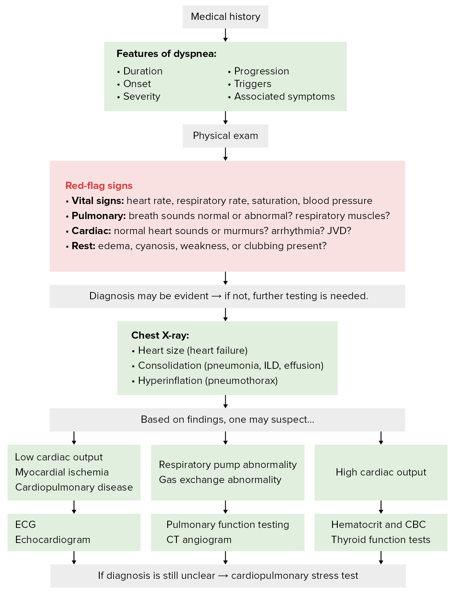
Diagnostic algorithm for dyspnea

Diagnostic algorithm for dyspnea

Image by Lecturio.

Management

  • Symptomatic relief can be achieved with supplemental oxygen Supplemental Oxygen Respiratory Failure, but one must identify and correct the underlying condition for long-term treatment:
    • Asthma Asthma Asthma is a chronic inflammatory respiratory condition characterized by bronchial hyperresponsiveness and airflow obstruction. The disease is believed to result from the complex interaction of host and environmental factors that increase disease predisposition, with inflammation causing symptoms and structural changes. Patients typically present with wheezing, cough, and dyspnea. Asthma:
      • Inhaled bronchodilators Bronchodilators Asthma Drugs, corticosteroids Corticosteroids Chorioretinitis, other medications
      • 5-step treatment strategy from the Global Initiative for Asthma Asthma Asthma is a chronic inflammatory respiratory condition characterized by bronchial hyperresponsiveness and airflow obstruction. The disease is believed to result from the complex interaction of host and environmental factors that increase disease predisposition, with inflammation causing symptoms and structural changes. Patients typically present with wheezing, cough, and dyspnea. Asthma Group
    • COPD COPD Chronic obstructive pulmonary disease (COPD) is a lung disease characterized by progressive, largely irreversible airflow obstruction. The condition usually presents in middle-aged or elderly persons with a history of cigarette smoking. Signs and symptoms include prolonged expiration, wheezing, diminished breath sounds, progressive dyspnea, and chronic cough. Chronic Obstructive Pulmonary Disease (COPD) exacerbations: 
      • Bronchodilator therapy and a course of oral glucocorticoids Glucocorticoids Glucocorticoids are a class within the corticosteroid family. Glucocorticoids are chemically and functionally similar to endogenous cortisol. There are a wide array of indications, which primarily benefit from the antiinflammatory and immunosuppressive effects of this class of drugs. Glucocorticoids
      • Possible oral antibiotics (antivirals if there is evidence of influenza Influenza Influenza viruses are members of the Orthomyxoviridae family and the causative organisms of influenza, a highly contagious febrile respiratory disease. There are 3 primary influenza viruses (A, B, and C) and various subtypes, which are classified based on their virulent surface antigens, hemagglutinin (HA) and neuraminidase (NA). Influenza typically presents with a fever, myalgia, headache, and symptoms of an upper respiratory infection. Influenza Viruses/Influenza)
      • Supplemental oxygen Supplemental Oxygen Respiratory Failure if hypoxic and pulmonary rehabilitation program
    • Heart failure Heart Failure A heterogeneous condition in which the heart is unable to pump out sufficient blood to meet the metabolic need of the body. Heart failure can be caused by structural defects, functional abnormalities (ventricular dysfunction), or a sudden overload beyond its capacity. Chronic heart failure is more common than acute heart failure which results from sudden insult to cardiac function, such as myocardial infarction. Total Anomalous Pulmonary Venous Return (TAPVR)
      • Treatment depends on the type of heart failure Heart Failure A heterogeneous condition in which the heart is unable to pump out sufficient blood to meet the metabolic need of the body. Heart failure can be caused by structural defects, functional abnormalities (ventricular dysfunction), or a sudden overload beyond its capacity. Chronic heart failure is more common than acute heart failure which results from sudden insult to cardiac function, such as myocardial infarction. Total Anomalous Pulmonary Venous Return (TAPVR) and severity.
      • Follow published guidelines for treatment.
      • Heart failure Heart Failure A heterogeneous condition in which the heart is unable to pump out sufficient blood to meet the metabolic need of the body. Heart failure can be caused by structural defects, functional abnormalities (ventricular dysfunction), or a sudden overload beyond its capacity. Chronic heart failure is more common than acute heart failure which results from sudden insult to cardiac function, such as myocardial infarction. Total Anomalous Pulmonary Venous Return (TAPVR) and angina medication Angina medication Angina is defined as chest pain or discomfort resulting from myocardial ischemia. Therapy for angina is targeted at limiting platelet aggregation and adhesion (with antiplatelet agents), reducing O2 demand (with beta-blockers), reducing preload (with nitrates), and preventing the progression of atherosclerotic disease (using statin therapy), along with the management of comorbidities. Heart Failure and Angina Medication
    • Pneumonia Pneumonia Pneumonia or pulmonary inflammation is an acute or chronic inflammation of lung tissue. Causes include infection with bacteria, viruses, or fungi. In more rare cases, pneumonia can also be caused through toxic triggers through inhalation of toxic substances, immunological processes, or in the course of radiotherapy. Pneumonia/ infectious Infectious Febrile Infant causes: 
      • Use the appropriate antibiotic for the causative pathogen.
    • Pulmonary embolism Pulmonary Embolism Pulmonary embolism (PE) is a potentially fatal condition that occurs as a result of intraluminal obstruction of the main pulmonary artery or its branches. The causative factors include thrombi, air, amniotic fluid, and fat. In PE, gas exchange is impaired due to the decreased return of deoxygenated blood to the lungs. Pulmonary Embolism:
      • Thrombolytics Thrombolytics Thrombolytics, also known as fibrinolytics, include recombinant tissue plasminogen activator (TPa) (i.e., alteplase, reteplase, and tenecteplase), urokinase, and streptokinase. The agents promote the breakdown of a blood clot by converting plasminogen to plasmin, which then degrades fibrin. Thrombolytics ( anticoagulants Anticoagulants Anticoagulants are drugs that retard or interrupt the coagulation cascade. The primary classes of available anticoagulants include heparins, vitamin K-dependent antagonists (e.g., warfarin), direct thrombin inhibitors, and factor Xa inhibitors. Anticoagulants and antiplatelet agents Antiplatelet agents Antiplatelet agents are medications that inhibit platelet aggregation, a critical step in the formation of the initial platelet plug. Abnormal, or inappropriate, platelet aggregation is a key step in the pathophysiology of arterial ischemic events. The primary categories of antiplatelet agents include aspirin, ADP inhibitors, phosphodiesterase/adenosine uptake inhibitors, and glycoprotein IIb/IIIa inhibitors. Antiplatelet Drugs)
      • Embolectomy and/or a vena cava filter if thrombolytics Thrombolytics Thrombolytics, also known as fibrinolytics, include recombinant tissue plasminogen activator (TPa) (i.e., alteplase, reteplase, and tenecteplase), urokinase, and streptokinase. The agents promote the breakdown of a blood clot by converting plasminogen to plasmin, which then degrades fibrin. Thrombolytics are contraindicated
  • If complete relief of dyspnea is not possible, then attempts at lessening the intensity of dyspnea should follow with other therapy:
    • Oxygen supplementation if O₂ saturation O₂ saturation Gas Exchange < 88% at rest, with activity, or while sleeping
    • Opioids Opioids Opiates are drugs that are derived from the sap of the opium poppy. Opiates have been used since antiquity for the relief of acute severe pain. Opioids are synthetic opiates with properties that are substantially similar to those of opiates. Opioid Analgesics can reduce air hunger, but one must:
      • Consider the risk-benefit profile.
      • Monitor the patient carefully because of opioids Opioids Opiates are drugs that are derived from the sap of the opium poppy. Opiates have been used since antiquity for the relief of acute severe pain. Opioids are synthetic opiates with properties that are substantially similar to those of opiates. Opioid Analgesics‘ ability to cause respiratory depression. 
    • Inhaled furosemide Furosemide A benzoic-sulfonamide-furan. It is a diuretic with fast onset and short duration that is used for edema and chronic renal insufficiency. Loop Diuretics appears to be effective (large-scale studies are pending).

Clinical Relevance

The following conditions are the most common causes of dyspnea by organ system:

Cardiovascular conditions

  • Acute MI MI MI is ischemia and death of an area of myocardial tissue due to insufficient blood flow and oxygenation, usually from thrombus formation on a ruptured atherosclerotic plaque in the epicardial arteries. Clinical presentation is most commonly with chest pain, but women and patients with diabetes may have atypical symptoms. Myocardial Infarction: ischemia Ischemia A hypoperfusion of the blood through an organ or tissue caused by a pathologic constriction or obstruction of its blood vessels, or an absence of blood circulation. Ischemic Cell Damage of the myocardial tissue due to a complete obstruction or drastic constriction of the coronary artery Coronary Artery Truncus Arteriosus. Acute MI MI MI is ischemia and death of an area of myocardial tissue due to insufficient blood flow and oxygenation, usually from thrombus formation on a ruptured atherosclerotic plaque in the epicardial arteries. Clinical presentation is most commonly with chest pain, but women and patients with diabetes may have atypical symptoms. Myocardial Infarction is usually accompanied by an increase in cardiac enzymes Enzymes Enzymes are complex protein biocatalysts that accelerate chemical reactions without being consumed by them. Due to the body’s constant metabolic needs, the absence of enzymes would make life unsustainable, as reactions would occur too slowly without these molecules. Basics of Enzymes, typical ECG ECG An electrocardiogram (ECG) is a graphic representation of the electrical activity of the heart plotted against time. Adhesive electrodes are affixed to the skin surface allowing measurement of cardiac impulses from many angles. The ECG provides 3-dimensional information about the conduction system of the heart, the myocardium, and other cardiac structures. Electrocardiogram (ECG) changes, and chest pain Pain An unpleasant sensation induced by noxious stimuli which are detected by nerve endings of nociceptive neurons. Pain: Types and Pathways
  • Heart failure Heart Failure A heterogeneous condition in which the heart is unable to pump out sufficient blood to meet the metabolic need of the body. Heart failure can be caused by structural defects, functional abnormalities (ventricular dysfunction), or a sudden overload beyond its capacity. Chronic heart failure is more common than acute heart failure which results from sudden insult to cardiac function, such as myocardial infarction. Total Anomalous Pulmonary Venous Return (TAPVR): the inability of the heart to supply the body with normal cardiac output Cardiac output The volume of blood passing through the heart per unit of time. It is usually expressed as liters (volume) per minute so as not to be confused with stroke volume (volume per beat). Cardiac Mechanics to meet metabolic needs. Risk factors include hypertension Hypertension Hypertension, or high blood pressure, is a common disease that manifests as elevated systemic arterial pressures. Hypertension is most often asymptomatic and is found incidentally as part of a routine physical examination or during triage for an unrelated medical encounter. Hypertension, diabetes mellitus Diabetes mellitus Diabetes mellitus (DM) is a metabolic disease characterized by hyperglycemia and dysfunction of the regulation of glucose metabolism by insulin. Type 1 DM is diagnosed mostly in children and young adults as the result of autoimmune destruction of β cells in the pancreas and the resulting lack of insulin. Type 2 DM has a significant association with obesity and is characterized by insulin resistance. Diabetes Mellitus, and coronary artery Coronary Artery Truncus Arteriosus disease. Echocardiography Echocardiography Ultrasonic recording of the size, motion, and composition of the heart and surrounding tissues. The standard approach is transthoracic. Tricuspid Valve Atresia (TVA) can confirm the reduced EF EF Cardiac Cycle
  • Cardiac tamponade Cardiac tamponade Compression of the heart by accumulated fluid (pericardial effusion) or blood (hemopericardium) in the pericardium surrounding the heart. The affected cardiac functions and cardiac output can range from minimal to total hemodynamic collapse. Pericardial Effusion and Cardiac Tamponade: the condition of having an abnormal amount of fluid in the pericardial cavity Pericardial cavity Heart: Anatomy of the heart that restricts cardiac filling and leads to acute heart failure Heart Failure A heterogeneous condition in which the heart is unable to pump out sufficient blood to meet the metabolic need of the body. Heart failure can be caused by structural defects, functional abnormalities (ventricular dysfunction), or a sudden overload beyond its capacity. Chronic heart failure is more common than acute heart failure which results from sudden insult to cardiac function, such as myocardial infarction. Total Anomalous Pulmonary Venous Return (TAPVR). Treated by pericardiocentesis Pericardiocentesis Puncture and aspiration of fluid from the pericardium. Cardiac Surgery, removing the fluid with a needle.

Respiratory conditions

  • Asthma Asthma Asthma is a chronic inflammatory respiratory condition characterized by bronchial hyperresponsiveness and airflow obstruction. The disease is believed to result from the complex interaction of host and environmental factors that increase disease predisposition, with inflammation causing symptoms and structural changes. Patients typically present with wheezing, cough, and dyspnea. Asthma: a chronic inflammatory condition characterized by reversible obstruction to airflow in the lower airways. Presents with intermittent or persistent wheezing Wheezing Wheezing is an abnormal breath sound characterized by a whistling noise that can be relatively high-pitched and shrill (more common) or coarse. Wheezing is produced by the movement of air through narrowed or compressed small (intrathoracic) airways. Wheezing, cough, and dyspnea. Diagnosis is confirmed with pulmonary function testing Pulmonary Function Testing Pulmonary Function Tests that shows a reversible obstructive pattern. 
  • Pneumonia Pneumonia Pneumonia or pulmonary inflammation is an acute or chronic inflammation of lung tissue. Causes include infection with bacteria, viruses, or fungi. In more rare cases, pneumonia can also be caused through toxic triggers through inhalation of toxic substances, immunological processes, or in the course of radiotherapy. Pneumonia: acute or chronic inflammation Inflammation Inflammation is a complex set of responses to infection and injury involving leukocytes as the principal cellular mediators in the body’s defense against pathogenic organisms. Inflammation is also seen as a response to tissue injury in the process of wound healing. The 5 cardinal signs of inflammation are pain, heat, redness, swelling, and loss of function. Inflammation of lung tissue caused by infection with bacteria Bacteria Bacteria are prokaryotic single-celled microorganisms that are metabolically active and divide by binary fission. Some of these organisms play a significant role in the pathogenesis of diseases. Bacteriology, viruses Viruses Minute infectious agents whose genomes are composed of DNA or RNA, but not both. They are characterized by a lack of independent metabolism and the inability to replicate outside living host cells. Virology, or fungi Fungi A kingdom of eukaryotic, heterotrophic organisms that live parasitically as saprobes, including mushrooms; yeasts; smuts, molds, etc. They reproduce either sexually or asexually, and have life cycles that range from simple to complex. Filamentous fungi, commonly known as molds, refer to those that grow as multicellular colonies. Mycology. Pneumonia Pneumonia Pneumonia or pulmonary inflammation is an acute or chronic inflammation of lung tissue. Causes include infection with bacteria, viruses, or fungi. In more rare cases, pneumonia can also be caused through toxic triggers through inhalation of toxic substances, immunological processes, or in the course of radiotherapy. Pneumonia can also be due to toxic triggers Triggers Hereditary Angioedema (C1 Esterase Inhibitor Deficiency) through inhalation of toxic substances, immunologic processes, or radiotherapy.
  • Pulmonary embolism Pulmonary Embolism Pulmonary embolism (PE) is a potentially fatal condition that occurs as a result of intraluminal obstruction of the main pulmonary artery or its branches. The causative factors include thrombi, air, amniotic fluid, and fat. In PE, gas exchange is impaired due to the decreased return of deoxygenated blood to the lungs. Pulmonary Embolism: a potentially fatal clinical condition that occurs as a result of mechanical obstruction Mechanical Obstruction Any impairment, arrest, or reversal of the normal flow of intestinal contents toward the anal canal. Imaging of the Intestines of the pulmonary artery Pulmonary artery The short wide vessel arising from the conus arteriosus of the right ventricle and conveying unaerated blood to the lungs. Lungs: Anatomy or its branches by a thrombus, air, or fat. Diagnosis is established by CT pulmonary angiogram CT pulmonary angiogram Pulmonary Function Tests.
  • Pneumothorax Pneumothorax A pneumothorax is a life-threatening condition in which air collects in the pleural space, causing partial or full collapse of the lung. A pneumothorax can be traumatic or spontaneous. Patients present with a sudden onset of sharp chest pain, dyspnea, and diminished breath sounds on exam. Pneumothorax: a collection of air in the pleural space Pleural space The thin serous membrane enveloping the lungs (lung) and lining the thoracic cavity. Pleura consist of two layers, the inner visceral pleura lying next to the pulmonary parenchyma and the outer parietal pleura. Between the two layers is the pleural cavity which contains a thin film of liquid. Pleuritis that causes the lung to collapse due to the loss of negative pressure. Presents with pleuritic chest pain Pain An unpleasant sensation induced by noxious stimuli which are detected by nerve endings of nociceptive neurons. Pain: Types and Pathways, dyspnea, tachycardia Tachycardia Abnormally rapid heartbeat, usually with a heart rate above 100 beats per minute for adults. Tachycardia accompanied by disturbance in the cardiac depolarization (cardiac arrhythmia) is called tachyarrhythmia. Sepsis in Children, and reduced breath sounds on the ipsilateral side.
  • Airway obstruction Airway obstruction Airway obstruction is a partial or complete blockage of the airways that impedes airflow. An airway obstruction can be classified as upper, central, or lower depending on location. Lower airway obstruction (LAO) is usually a manifestation of chronic disease, such as asthma or chronic obstructive pulmonary disease (COPD). Airway Obstruction: a partial or complete blockage of airflow in the respiratory tract, which can be classified into upper or lower airway Airway ABCDE Assessment obstructions. Usually associated with foreign body Foreign Body Foreign Body Aspiration aspirations, but can be caused by other conditions including mucous plug, epiglottitis Epiglottitis Epiglottitis (or “supraglottitis”) is an inflammation of the epiglottis and adjacent supraglottic structures. The majority of cases are caused by bacterial infection. Symptoms are rapid in onset and severe. Epiglottitis, infections Infections Invasion of the host organism by microorganisms or their toxins or by parasites that can cause pathological conditions or diseases. Chronic Granulomatous Disease, smoke inhalation Smoke Inhalation Carbon Monoxide Poisoning, goiter Goiter A goiter is a chronic enlargement of the thyroid gland due to nonneoplastic growth occurring in the setting of hypothyroidism, hyperthyroidism, or euthyroidism. Morphologically, thyroid enlargement can be diffuse (smooth consistency) or nodular (uninodular or multinodular). Goiter, etc ETC The electron transport chain (ETC) sends electrons through a series of proteins, which generate an electrochemical proton gradient that produces energy in the form of adenosine triphosphate (ATP). Electron Transport Chain (ETC).
  • COPD COPD Chronic obstructive pulmonary disease (COPD) is a lung disease characterized by progressive, largely irreversible airflow obstruction. The condition usually presents in middle-aged or elderly persons with a history of cigarette smoking. Signs and symptoms include prolonged expiration, wheezing, diminished breath sounds, progressive dyspnea, and chronic cough. Chronic Obstructive Pulmonary Disease (COPD): a spectrum of conditions characterized by irreversible airflow limitation correlated to smoking Smoking Willful or deliberate act of inhaling and exhaling smoke from burning substances or agents held by hand. Interstitial Lung Diseases. Results from obstructive inflammation Inflammation Inflammation is a complex set of responses to infection and injury involving leukocytes as the principal cellular mediators in the body’s defense against pathogenic organisms. Inflammation is also seen as a response to tissue injury in the process of wound healing. The 5 cardinal signs of inflammation are pain, heat, redness, swelling, and loss of function. Inflammation of the small airways as well as changes in the lung parenchyma and pulmonary vasculature. 
  • ILD ILD Interstitial lung diseases are a heterogeneous group of disorders characterized by the inflammation and fibrosis of lung parenchyma, especially the pulmonary connective tissue in the alveolar walls. It may be idiopathic (e.g., idiopathic pulmonary fibrosis) or secondary to connective tissue diseases, medications, malignancies, occupational exposure, or allergens. Interstitial Lung Diseases: a heterogeneous group of disorders characterized by the inflammation Inflammation Inflammation is a complex set of responses to infection and injury involving leukocytes as the principal cellular mediators in the body’s defense against pathogenic organisms. Inflammation is also seen as a response to tissue injury in the process of wound healing. The 5 cardinal signs of inflammation are pain, heat, redness, swelling, and loss of function. Inflammation and fibrosis Fibrosis Any pathological condition where fibrous connective tissue invades any organ, usually as a consequence of inflammation or other injury. Bronchiolitis Obliterans of the lung parenchyma, especially the pulmonary connective tissue Connective tissue Connective tissues originate from embryonic mesenchyme and are present throughout the body except inside the brain and spinal cord. The main function of connective tissues is to provide structural support to organs. Connective tissues consist of cells and an extracellular matrix. Connective Tissue: Histology in the alveolar walls. May be idiopathic Idiopathic Dermatomyositis or secondary to connective tissue Connective tissue Connective tissues originate from embryonic mesenchyme and are present throughout the body except inside the brain and spinal cord. The main function of connective tissues is to provide structural support to organs. Connective tissues consist of cells and an extracellular matrix. Connective Tissue: Histology diseases, medications, malignancies, occupational exposure, or allergens.

References

  1. Parshall, M.B., Schwartzstein, R.M., et al. (2012). American Thoracic Society Committee on Dyspnea. Am J Respir Crit Care Med. 185(4):435. 
  2. Schwartzstein, R.M. (2020). Approach to the patient with dyspnea. UpToDate. Retrieved September 16, 2020, from https://www.uptodate.com/contents/approach-to-the-patient-with-dyspnea?search=dyspnea&source=search_result&selectedTitle=1~150&usage_type=default&display_rank=1
  3. Baron, R.M. (2018). In Jameson, J.L., et al. (Ed.), Harrison’s Principles of Internal Medicine (20th ed. Vol 1, pp. 226–230 and 1943–1947). 
  4. Pasterkamp, H. (2012). Kendig & Chernick’s Disorders of the Respiratory Tract in Children (8th ed.), pp. 110–130.

Create your free account or log in to continue reading!

Sign up now and get free access to Lecturio with concept pages, medical videos, and questions for your medical education.

User Reviews

Details