Epidemiology and Etiology
Definition and types
Pneumoconiosis is the classic term used to describe the non-neoplastic lung reaction to chronic inhalation of mineral dusts encountered at the workplace. Some lung experts believe that the term “pneumoconiosis” should also include diseases induced by chemical fumes and vapors but this is not a widespread practice and will not be followed here.
Most common causes:
- Silicosis: caused by crystalline silica dust (< 5 microns)
- Asbestosis: caused by asbestos fibers (crystalline hydrated silicates) < 10 microns
- Coal worker’s pneumoconiosis (CWP) or anthracosis: caused by coal dust
- Berylliosis: also called chronic beryllium disease, caused by beryllium (alkali earth metal, lighter than aluminum, stronger than steel)
Less common causes, not discussed further in this monograph:
- Siderosis: caused by iron oxide, seen in welders
- Stannosis: caused by tin oxide, affects workers who mine it
- Baritosis: caused by barium sulfate, affects workers who mine it
- Hard metal pneumoconiosis:
- Caused by cobalt with or without tungsten carbide
- Affecting workers who process it and diamond polishers
- Talcosis:
- Caused by talc powder, which is composed of crystalline hydrous magnesium silicate
- Affects workers in industries that make or process ceramics, paper, plastics, rubber, paint, and cosmetics
Epidemiology
- Silicosis:
- Most common form of occupational lung disease
- Currently, > 2 million workers are exposed to silica dust worldwide.
- In 2016, the Global Burden of Disease Study estimated 10,400 deaths per year, and 210,000 years of life lost due to silicosis.
- Incidence and mortality have decreased in the past several decades due to better workplace protection and regulations.
- Asbestosis:
- 150,000 cases and 3,600 deaths in 2015
- Exposure has greatly decreased due to the enforcement of environmental rules and regulations.
- Asbestos is the most commonly encountered carcinogen.
- CWP:
- Common condition among coal miners
- Estimated prevalence of 30%
- Berylliosis:
- Rare form of pneumoconiosis
- Overall prevalence among exposed workers ranges from 1%–5%
Risk factors
- Silicosis:
- Occupational: mining, sandblasting, quarry, ceramics, and foundry workers; and in grinding, stone cutting, fiberglass, and glass manufacturing
- Asbestosis:
- Occupational: shipbuilding, roofing, plumbing, and demolition
- Nonoccupational: family members of exposed workers, geological sources, smoking (accelerates disease progression)
- CWP: coal miners
- Berylliosis:
- Occupational: aerospace, ceramics, and metallurgical industries; electronics repair; jewelry makers; and dentists
- Nonoccupational: family members of workers, or those living 5 miles or less from a beryllium manufacturing facility
Related videos
Pathophysiology
- Inhaled toxic fibers cannot be metabolized by the body and accumulate in the alveolar ducts.
- The most dangerous particles measure 1 to 5 µm, since they are able to be transported all the way into the terminal small airways and air sacs and settle there, provoking inflammation and fibrosis.
- Asbestos occurs in two distinct geometric forms, 90% as curled serpentine forms like chrysotiles and 10% as needle-like amphibole forms.
- Amphibole asbestos fibers are more pathogenic (especially for the development of mesothelioma) than chrysotiles because they can travel farther and penetrate deeper into tissues.
- The fibers are engulfed by macrophages, which undergo lysis and release cytokines.
- Cytokines induce an inflammatory reaction, producing airway obstruction and stimulating fibroblasts.
- Fibrotic scarring leads to thickening of the airways, reduced elasticity, and impaired gas exchange.
Fiber-specific pathogenesis:
- Progressive massive fibrosis (PMF)
- Can occur in both silicosis and CWP
- Represents an evolution from the simple form of pneumoconiosis with small nodules to the complicated form when the nodules coalesce to form larger nodules that can evolve into PMF
- Asbestosis affects the lower lobes first and the pleura (unusual in other interstitial lung disorders)
- Silicosis, CWP, and berylliosis affect the upper lobes first.
- Berylliosis leads to the development of noncaseating granulomas (delayed hypersensitivity reaction)
- Direct contact with beryllium fumes or dusts may injure the exposed areas of the body, such as the eyes or the skin, and sensitization may occur
Factors of disease progression:
- Size, type, and physiochemical reactivity of inhaled fibers
- Duration of exposure (disease may continue to progress even after the end of exposure)
- Intensity of exposure (dose-response relationship)
- Genetic susceptibility of the individual is likely since only a small percentage of exposed people develop occupational exposure respiratory diseases: Mutations of the HLA-DPB1 gene produce a higher susceptibility to beryllium.
- Smoking habits and exposure to other toxins/pollutants
Clinical Presentation
General signs and symptoms
- Progressive exertional dyspnea
- Dry cough
- May be productive in the morning, with clear-to-white sputum, or yellow, purulent, in the case of concomitant bacterial pneumonia
- Rare in cases of asbestosis in the absence of concomitant cigarette smoking
- Inspiratory rales or crackles
- Digital clubbing in advanced disease
- Berylliosis also presents less commonly with fever, chest pain, arthralgias, weight loss, and nodules on the areas of exposed skin.
Onset
Onset depends on the intensity and duration of exposure and of the type of dust inhaled.
- Silicosis usually presents after > 10 years of exposure but can manifest after several months of intense daily exposure.
- Asbestosis presents after > 20 years of exposure.
- CWP presents after prolonged exposure (> 10 years).
- Berylliosis presents within weeks to decades after initial exposure. The rate of progression from sensitization to disease may vary from 6%–8%.
Complications
- Tuberculosis (increased risk in silicosis)
- Bronchogenic carcinoma (most common neoplastic complication)
- Develops > 15 years after exposure
- Increased amount of exposure is associated with a higher risk of lung cancer
- Mesothelioma (especially with asbestosis)
- Can develop as early as 1–2 years after intense asbestos exposure or later, up to 40 years after exposure
- Pulmonary hypertension, cor pulmonale, respiratory failure, and right-sided heart failure
- Caplan’s syndrome (a type of pneumoconiosis with intrapulmonary nodules in combination with rheumatoid arthritis)
Diagnosis
A diagnosis of occupational lung disease relies upon 4 essential criteria:
- Documented exposure history to a known toxic agent
- A typical latent period between exposure and symptoms
- Clinicoradiological features compatible with the known features of the disease
- Exclusion of any other disease that could explain the findings
Chest X-ray
Silicosis | Asbestosis | CWP | Berylliosis |
---|---|---|---|
|
|
|
|
Pulmonary function tests
Most cases of pneumoconiosis show a restrictive lung disease pattern by spirometry.
- ↓ functional vital capacity (FVC) and total lung capacity
- ↓ diffusion capacity (DLCO)
- Normal forced expiratory volume in 1 second (FEV1)/FVC ratio (may be decreased if the patient is also a smoker = mixed restrictive and obstructive lung disease)
Laboratory tests
- Usually not helpful in the confirmation of the diagnosis but can exclude infectious complications
- Beryllium lymphocyte proliferation test (BePLT): test of choice to diagnose beryllium sensitization or berylliosis
- Mononuclear cells from the patient (from blood or obtained by bronchoalveolar lavage) exposed to different concentrations of beryllium salts
- Increased lymphocyte proliferation is a positive result.
Confirmatory tests
- High-resolution computed tomography
- Bronchoscopy with bronchoalveolar lavage (BAL): to exclude infection or malignancy
- Video-assisted transthoracic or open biopsy:
- Gold standard for diagnosis but rarely needed; should be limited to exclude malignancy or when there is no known history of exposure to mineral dust
- Silicosis shows nodules with silica particles usually in a central hyalinized region surrounded by concentric collagen fibers.
- Asbestosis may rarely show asbestos bodies that stain positive with Prussian blue and appear as dumbbell-shaped and golden-brown fusiform rods.
- CWP shows 1–2 mm nodular aggregations of anthracotic macrophages supported by a fine collagen network in the upper regions of the lungs around respiratory bronchioles.
- Berylliosis shows noncaseating granulomas and/or mononuclear cell interstitial infiltrates.
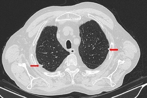
Thoracic high resonance CT scan of a patient with asbestosis reveals pleural plaques (arrows)
Image: “Thoracic HRCT” by Consultant in Rheumatology, Rheumatology Division, Hospital of Prato, Prato, Italy. License: CC BY 4.0Close up of a chest X-ray of a patient with asbestosis which shows signs of interstitial fibrosis
Image: “Asbestosis” by DrSHaber. License: CC0 1.0
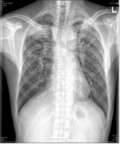
Chest X-ray of a patient with progressive pneumoconiosis (silicosis) with signs of massive fibrosis
Image: “Chest X-ray” by Department of Occupational & Environmental Medicine, College of Medicine, Dong-A University, Busan, Korea. License: CC BY 2.0, edited by Lecturio.
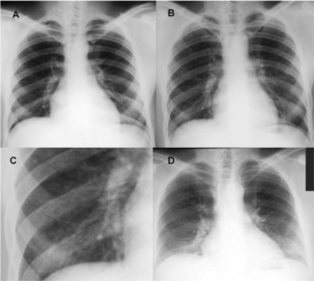
Chest X-ray of a patient with berylliosis due to occupational exposure
Image: “Chest radiographs of case 1” by Division of Respiratory Disease Studies, National Institute for Occupational Safety and Health, Centers for Disease Control and Prevention, Morgantown, West Virginia 26505, USA. License: Public Domain
(A) Taken pre-hire in March 1979, showing normal lung fields
(B) Taken during the second episode of acute work-related illness in March 1981, showing a mild diffuse nodular infiltrate
(C) Close-up of the right lower lung field from image B
(D) Taken at follow-up in February 1997, showing reduced lung volumes and bilateral interstitial infiltrates
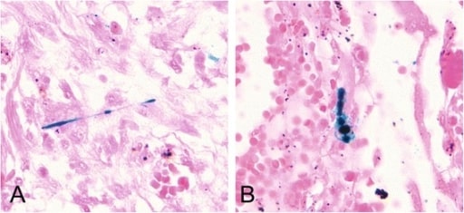
Histologic slide shows scattered asbestos bodies showing a fibrous core coated by iron-containing material. Prussian blue staining (400×).
Image: “Scattered asbestos bodies showing a fibrous core” by Institute and Outpatient Clinic for Occupational and Social Medicine, University Medical Center Giessen, Aulweg 129/III, D-35385 Giessen, Germany. License: CC BY 4.0
Management
- There is no definitive treatment for pneumoconiosis, regardless of the clinical stage.
- Immediate cessation of exposure is recommended as well as cessation of tobacco smoking habits, if applicable.
- Stopping exposure may not stop the progression of the disease.
- Management is largely supportive and focuses on the prevention of further progression and complications.
- Prophylactic vaccination against influenza and pneumococcal pneumonia
- If there is evidence of underlying obstructive disease, bronchodilator therapy can be administered.
- Oxygen therapy is often necessary to relieve dyspnea.
- Early systemic oral glucocorticoid therapy may relieve symptoms, improve lung appearance on imaging, and normalize pulmonary function testing scores.
- Immunosuppressants, (e.g., methotrexate, azathioprine) are an option for patients who are refractory to glucocorticoids, or those experiencing adverse effects.
- Lung transplantation has been performed for advanced pneumoconiosis with respiratory failure, but evidence on patient outcomes is lacking.
- Pulmonary rehabilitation can improve quality of life; includes patient education, exercise/breathing training, nutrition advice, and psychosocial support.
Differential Diagnosis
The differential diagnoses of pneumoconioses caused by mineral dusts include the following conditions:
- Idiopathic pulmonary fibrosis: most common interstitial lung disease, characterized by irreversible pulmonary fibrosis and impaired pulmonary function. Presents with exertional dyspnea, persistent dry cough, and fatigue
- Hypersensitivity pneumonitis: pulmonary disease characterized by an immune-mediated inflammatory response in the small airways as a result of exposure to inhaled antigens
- Sarcoidosis: chronic inflammatory disease characterized by the formation of noncaseating granulomas, typically in the lungs and, less commonly, the liver, eyes, and skin.
- Tuberculosis: disease caused by Mycobacterium tuberculosis. The bacteria usually attack the lungs, but can also damage other parts of the body.
References
- Stark, P. Imaging of occupational lung diseases. UpToDate Evidence-Based Medicine. Retrieved August 13, 2020, from https://www.uptodate.com/contents/imaging-of-occupational-lung-diseases?search=pneumoconiosis&source=search_result&selectedTitle=1~125&usage_type=default&display_rank=1#H365264570
- Rose, C. Silicosis. UpToDate Evidence-Based Medicine. Retrieved August 13, 2020, from https://www.uptodate.com/contents/silicosis?search=pneumoconiosis&source=search_result&selectedTitle=2~125&usage_type=default&display_rank=2
- King, T.E. Asbestos-related pleuropulmonary disease. UpToDate Evidence-Based Medicine. Retrieved August 13, 2020, from https://www.uptodate.com/contents/asbestos-related-pleuropulmonary-disease?search=pneumoconiosis&source=search_result&selectedTitle=3~125&usage_type=default&display_rank=3
- Hu, C. X., Chen, W. H., He at al. (2019). Lung transplantation in China between 2015 and 2018. Chinese medical journal, 132(23), 2783–2789. https://doi.org/10.1097/CM9.0000000000000543