Achieve Mastery of Medical Concepts

Study for medical school and boards with Lecturio

Strongyloidiasis

Strongyloidiasis is a common parasitic disease caused by infection with the roundworm Strongyloides stercoralis. Transmission occurs through skin Skin The skin, also referred to as the integumentary system, is the largest organ of the body. The skin is primarily composed of the epidermis (outer layer) and dermis (deep layer). The epidermis is primarily composed of keratinocytes that undergo rapid turnover, while the dermis contains dense layers of connective tissue. Skin: Structure and Functions penetration Penetration X-rays, most commonly from walking barefoot. Strongyloides has a unique life cycle that can be entirely completed in the human host, migrating from the skin Skin The skin, also referred to as the integumentary system, is the largest organ of the body. The skin is primarily composed of the epidermis (outer layer) and dermis (deep layer). The epidermis is primarily composed of keratinocytes that undergo rapid turnover, while the dermis contains dense layers of connective tissue. Skin: Structure and Functions to the pulmonary system and then to the GI system. Symptoms include cutaneous irritation, constipation Constipation Constipation is common and may be due to a variety of causes. Constipation is generally defined as bowel movement frequency < 3 times per week. Patients who are constipated often strain to pass hard stools. The condition is classified as primary (also known as idiopathic or functional constipation) or secondary, and as acute or chronic. Constipation, diarrhea Diarrhea Diarrhea is defined as ≥ 3 watery or loose stools in a 24-hour period. There are a multitude of etiologies, which can be classified based on the underlying mechanism of disease. The duration of symptoms (acute or chronic) and characteristics of the stools (e.g., watery, bloody, steatorrheic, mucoid) can help guide further diagnostic evaluation. Diarrhea, dry cough, and wheezing Wheezing Wheezing is an abnormal breath sound characterized by a whistling noise that can be relatively high-pitched and shrill (more common) or coarse. Wheezing is produced by the movement of air through narrowed or compressed small (intrathoracic) airways. Wheezing, depending on where the parasite is in its life cycle. Effective eradication of the parasite can be obtained with anthelmintic medications, usually ivermectin Ivermectin A mixture of mostly avermectin h2b1a (rn 71827-03-7) with some avermectin h2b1b (rn 70209-81-3), which are macrolides from streptomyces avermitilis. It binds glutamate-gated chloride channel to cause increased permeability and hyperpolarization of nerve and muscle cells. It also interacts with other chloride channels. It is a broad spectrum antiparasitic that is active against microfilariae of onchocerca volvulus but not the adult form. Anthelmintic Drugs.

Last updated: Nov 7, 2022

Editorial responsibility: Stanley Oiseth, Lindsay Jones, Evelin Maza

Epidemiology and Etiology

Epidemiology

  • Worldwide prevalence Prevalence The total number of cases of a given disease in a specified population at a designated time. It is differentiated from incidence, which refers to the number of new cases in the population at a given time. Measures of Disease Frequency: 100 million cases currently
  • Most common in tropical and warm temperate regions, with ¾ of cases in Southeast Asia ASIA Spinal Cord Injuries, Africa, and Western Pacific countries

Etiology

Strongyloidiasis is caused by a parasitic infection by the roundworm ( nematode Nematode A phylum of unsegmented helminths with fundamental bilateral symmetry and secondary triradiate symmetry of the oral and esophageal structures. Many species are parasites. Toxocariasis) Strongyloides stercoralis. Roundworms Roundworms Anthelmintic Drugs are nonsegmented worms that can infect the GI tract, blood, or skin Skin The skin, also referred to as the integumentary system, is the largest organ of the body. The skin is primarily composed of the epidermis (outer layer) and dermis (deep layer). The epidermis is primarily composed of keratinocytes that undergo rapid turnover, while the dermis contains dense layers of connective tissue. Skin: Structure and Functions.

  • The ability to “auto-infect” its host leads to chronic cases.
  • Adult males: 0.9 mm MM Multiple myeloma (MM) is a malignant condition of plasma cells (activated B lymphocytes) primarily seen in the elderly. Monoclonal proliferation of plasma cells results in cytokine-driven osteoclastic activity and excessive secretion of IgG antibodies. Multiple Myeloma (0.04 in) long, can be distinguished from females by size and presence of spicules (needle-like mating structure)
  • Adult females: 2.0–2.5 mm MM Multiple myeloma (MM) is a malignant condition of plasma cells (activated B lymphocytes) primarily seen in the elderly. Monoclonal proliferation of plasma cells results in cytokine-driven osteoclastic activity and excessive secretion of IgG antibodies. Multiple Myeloma (0.08–0.10 in) long
Strongyloides sp.

Strongyloides sp.:
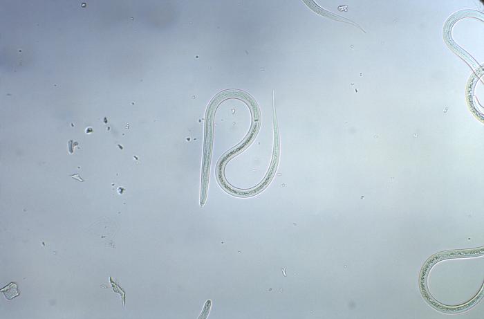
Photomicrograph with a magnification of 128X of a Strongyloides sp. filariform, a larval-staged organism

Image: “1547” by Dr. Mae Melvin. License: Public Domain

Pathophysiology

Life cycle of S. stercoralis

  1. Filariform larvae Filariform Larvae Hookworm Infections in contaminated soil penetrate human skin Skin The skin, also referred to as the integumentary system, is the largest organ of the body. The skin is primarily composed of the epidermis (outer layer) and dermis (deep layer). The epidermis is primarily composed of keratinocytes that undergo rapid turnover, while the dermis contains dense layers of connective tissue. Skin: Structure and Functions.
  2. Larvae Larvae Wormlike or grublike stage, following the egg in the life cycle of insects, worms, and other metamorphosing animals. Ascaris/Ascariasis migrate through bloodstream and lymphatics to lungs Lungs Lungs are the main organs of the respiratory system. Lungs are paired viscera located in the thoracic cavity and are composed of spongy tissue. The primary function of the lungs is to oxygenate blood and eliminate CO2. Lungs: Anatomy.
    • In lung parenchyma, larvae Larvae Wormlike or grublike stage, following the egg in the life cycle of insects, worms, and other metamorphosing animals. Ascaris/Ascariasis penetrate alveoli Alveoli Small polyhedral outpouchings along the walls of the alveolar sacs, alveolar ducts and terminal bronchioles through the walls of which gas exchange between alveolar air and pulmonary capillary blood takes place. Acute Respiratory Distress Syndrome (ARDS) and migrate up trachea Trachea The trachea is a tubular structure that forms part of the lower respiratory tract. The trachea is continuous superiorly with the larynx and inferiorly becomes the bronchial tree within the lungs. The trachea consists of a support frame of semicircular, or C-shaped, rings made out of hyaline cartilage and reinforced by collagenous connective tissue. Trachea: Anatomy and larynx Larynx The larynx, also commonly called the voice box, is a cylindrical space located in the neck at the level of the C3-C6 vertebrae. The major structures forming the framework of the larynx are the thyroid cartilage, cricoid cartilage, and epiglottis. The larynx serves to produce sound (phonation), conducts air to the trachea, and prevents large molecules from reaching the lungs. Larynx: Anatomy.
    • Larvae Larvae Wormlike or grublike stage, following the egg in the life cycle of insects, worms, and other metamorphosing animals. Ascaris/Ascariasis are then swallowed by the host.
  3. Swallowed larvae Larvae Wormlike or grublike stage, following the egg in the life cycle of insects, worms, and other metamorphosing animals. Ascaris/Ascariasis enter GI tract.
    • Larvae Larvae Wormlike or grublike stage, following the egg in the life cycle of insects, worms, and other metamorphosing animals. Ascaris/Ascariasis mature within mucosa of small intestine Small intestine The small intestine is the longest part of the GI tract, extending from the pyloric orifice of the stomach to the ileocecal junction. The small intestine is the major organ responsible for chemical digestion and absorption of nutrients. It is divided into 3 segments: the duodenum, the jejunum, and the ileum. Small Intestine: Anatomy.
    • Adult larvae Larvae Wormlike or grublike stage, following the egg in the life cycle of insects, worms, and other metamorphosing animals. Ascaris/Ascariasis produce eggs, which yield noninfectious, rhabditiform larvae Rhabditiform Larvae Hookworm Infections that migrate to intestinal lumen and are excreted in feces.
  4. Autoinfection Autoinfection Enterobius/Enterobiasis can occur in hosts with compromised immune systems:
    • Noninfectious, rhabditiform larvae Rhabditiform Larvae Hookworm Infections mature into infectious, filariform larvae Filariform Larvae Hookworm Infections within the intestine.
    • Filariform larvae Filariform Larvae Hookworm Infections penetrate intestinal mucosa Intestinal Mucosa Lining of the intestines, consisting of an inner epithelium, a middle lamina propria, and an outer muscularis mucosae. In the small intestine, the mucosa is characterized by a series of folds and abundance of absorptive cells (enterocytes) with microvilli. Small Intestine: Anatomy or perianal skin Skin The skin, also referred to as the integumentary system, is the largest organ of the body. The skin is primarily composed of the epidermis (outer layer) and dermis (deep layer). The epidermis is primarily composed of keratinocytes that undergo rapid turnover, while the dermis contains dense layers of connective tissue. Skin: Structure and Functions to migrate through bloodstream and lymphatics to restart infectious cycle.

Reservoirs

  • Humans (most common)
  • Cats and dogs

Transmission

  • Most common: larvae Larvae Wormlike or grublike stage, following the egg in the life cycle of insects, worms, and other metamorphosing animals. Ascaris/Ascariasis found in contaminated sand or soil penetrate skin Skin The skin, also referred to as the integumentary system, is the largest organ of the body. The skin is primarily composed of the epidermis (outer layer) and dermis (deep layer). The epidermis is primarily composed of keratinocytes that undergo rapid turnover, while the dermis contains dense layers of connective tissue. Skin: Structure and Functions 
  • Fecal-oral transmission (less common)

Host risk factors

  • Walking barefoot on contaminated soil 
  • Contact with human waste or sewage
  • Occupations that increase contact with contaminated soil (farming and coal mining)
  • Immunosuppression
The life cycle of strongyloides stercoralis

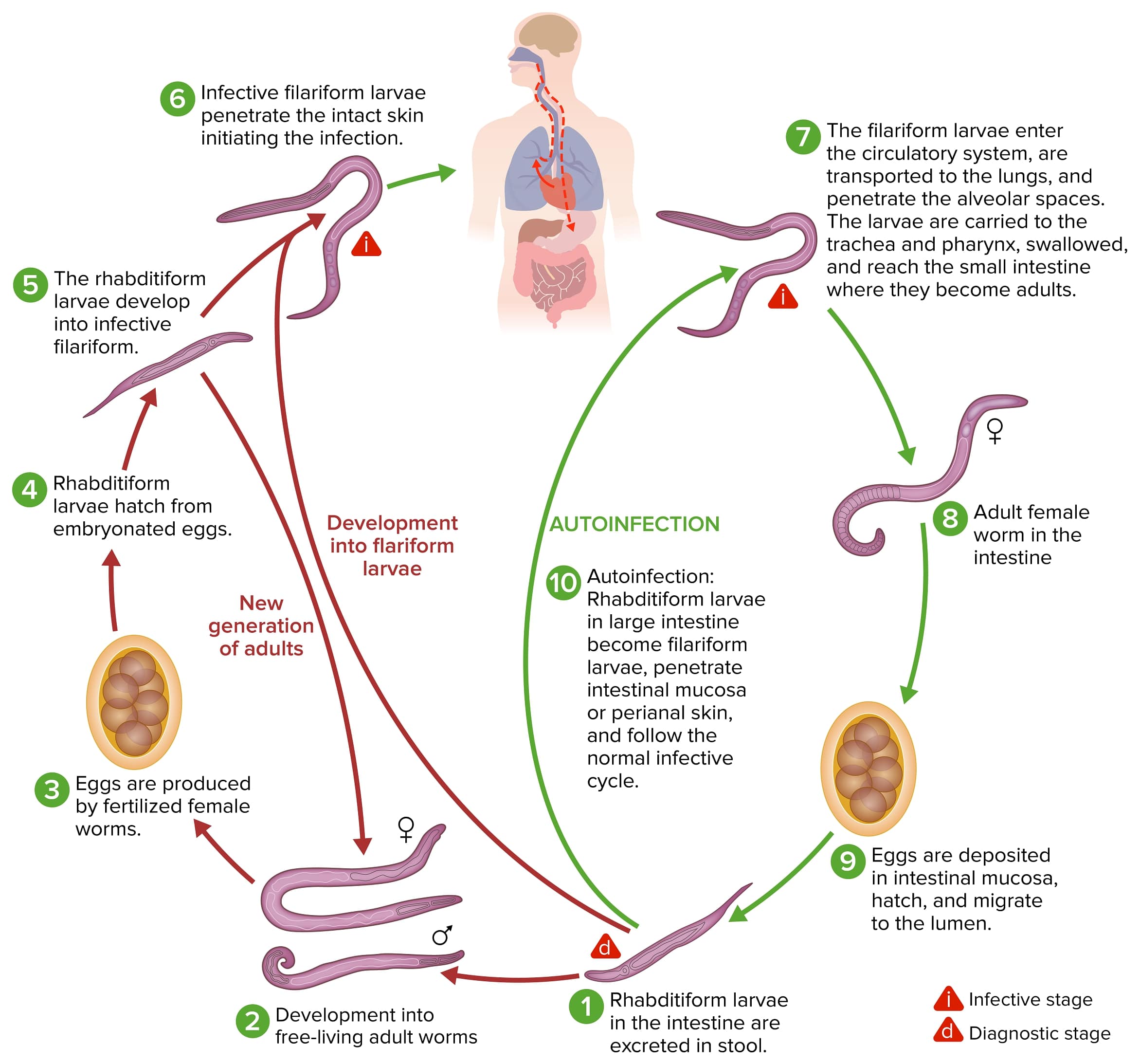
The life cycle of Strongyloides stercoralis:
The roundworm S. stercoralis has a unique lifecycle that can be completed within its definitive human host. The parasite is transmitted to other hosts through excreted feces, but in immunodeficient patients, autoinfection can occur, leading to more severe, chronic forms of the disease.

Image by Lecturio.

Clinical Presentation

Phases of strongyloidiasis infection

Strongyloidiasis can cause both acute and chronic infection. Symptoms correlate with the migration of the larvae Larvae Wormlike or grublike stage, following the egg in the life cycle of insects, worms, and other metamorphosing animals. Ascaris/Ascariasis from the skin Skin The skin, also referred to as the integumentary system, is the largest organ of the body. The skin is primarily composed of the epidermis (outer layer) and dermis (deep layer). The epidermis is primarily composed of keratinocytes that undergo rapid turnover, while the dermis contains dense layers of connective tissue. Skin: Structure and Functions, lung, and intestine.

  • Cutaneous phase: secondary to penetration Penetration X-rays of larvae Larvae Wormlike or grublike stage, following the egg in the life cycle of insects, worms, and other metamorphosing animals. Ascaris/Ascariasis into skin Skin The skin, also referred to as the integumentary system, is the largest organ of the body. The skin is primarily composed of the epidermis (outer layer) and dermis (deep layer). The epidermis is primarily composed of keratinocytes that undergo rapid turnover, while the dermis contains dense layers of connective tissue. Skin: Structure and Functions
  • Pulmonary phase: secondary to migration of larvae Larvae Wormlike or grublike stage, following the egg in the life cycle of insects, worms, and other metamorphosing animals. Ascaris/Ascariasis through bronchial tree Bronchial tree The collective term “bronchial tree” refers to the bronchi and all of their subsequent branches. The bronchi are the airways of the lower respiratory tract. At the level of the 3rd or 4th thoracic vertebra, the trachea bifurcates into the left and right main bronchi. Both of these bronchi continue to divide into secondary or lobar bronchi that bifurcate further and further. Bronchial Tree: Anatomy and alveoli Alveoli Small polyhedral outpouchings along the walls of the alveolar sacs, alveolar ducts and terminal bronchioles through the walls of which gas exchange between alveolar air and pulmonary capillary blood takes place. Acute Respiratory Distress Syndrome (ARDS)
    • 1 week after skin Skin The skin, also referred to as the integumentary system, is the largest organ of the body. The skin is primarily composed of the epidermis (outer layer) and dermis (deep layer). The epidermis is primarily composed of keratinocytes that undergo rapid turnover, while the dermis contains dense layers of connective tissue. Skin: Structure and Functions penetration Penetration X-rays
    • Löffler syndrome Löffler Syndrome Ascaris/Ascariasis (pulmonary eosinophilia Eosinophilia Abnormal increase of eosinophils in the blood, tissues or organs. Autosomal Dominant Hyperimmunoglobulin E Syndrome): transient respiratory disorder characterized by accumulation of eosinophils Eosinophils Granular leukocytes with a nucleus that usually has two lobes connected by a slender thread of chromatin, and cytoplasm containing coarse, round granules that are uniform in size and stainable by eosin. Innate Immunity: Phagocytes and Antigen Presentation in lungs Lungs Lungs are the main organs of the respiratory system. Lungs are paired viscera located in the thoracic cavity and are composed of spongy tissue. The primary function of the lungs is to oxygenate blood and eliminate CO2. Lungs: Anatomy due to parasitic infection
      • Dry cough
      • Throat Throat The pharynx is a component of the digestive system that lies posterior to the nasal cavity, oral cavity, and larynx. The pharynx can be divided into the oropharynx, nasopharynx, and laryngopharynx. Pharyngeal muscles play an integral role in vital processes such as breathing, swallowing, and speaking. Pharynx: Anatomy irritation
      • Wheezing Wheezing Wheezing is an abnormal breath sound characterized by a whistling noise that can be relatively high-pitched and shrill (more common) or coarse. Wheezing is produced by the movement of air through narrowed or compressed small (intrathoracic) airways. Wheezing
  • GI phase GI Phase Trichinella/Trichinellosis: secondary to adult larvae Larvae Wormlike or grublike stage, following the egg in the life cycle of insects, worms, and other metamorphosing animals. Ascaris/Ascariasis within mucosa of small intestine Small intestine The small intestine is the longest part of the GI tract, extending from the pyloric orifice of the stomach to the ileocecal junction. The small intestine is the major organ responsible for chemical digestion and absorption of nutrients. It is divided into 3 segments: the duodenum, the jejunum, and the ileum. Small Intestine: Anatomy
    • 3–4 weeks after skin Skin The skin, also referred to as the integumentary system, is the largest organ of the body. The skin is primarily composed of the epidermis (outer layer) and dermis (deep layer). The epidermis is primarily composed of keratinocytes that undergo rapid turnover, while the dermis contains dense layers of connective tissue. Skin: Structure and Functions penetration Penetration X-rays
    • Symptoms:
      • Abdominal pain Abdominal Pain Acute Abdomen
      • Diarrhea Diarrhea Diarrhea is defined as ≥ 3 watery or loose stools in a 24-hour period. There are a multitude of etiologies, which can be classified based on the underlying mechanism of disease. The duration of symptoms (acute or chronic) and characteristics of the stools (e.g., watery, bloody, steatorrheic, mucoid) can help guide further diagnostic evaluation. Diarrhea or constipation Constipation Constipation is common and may be due to a variety of causes. Constipation is generally defined as bowel movement frequency < 3 times per week. Patients who are constipated often strain to pass hard stools. The condition is classified as primary (also known as idiopathic or functional constipation) or secondary, and as acute or chronic. Constipation
      • Anorexia Anorexia The lack or loss of appetite accompanied by an aversion to food and the inability to eat. It is the defining characteristic of the disorder anorexia nervosa. Anorexia Nervosa
      • Nausea Nausea An unpleasant sensation in the stomach usually accompanied by the urge to vomit. Common causes are early pregnancy, sea and motion sickness, emotional stress, intense pain, food poisoning, and various enteroviruses. Antiemetics/ vomiting Vomiting The forcible expulsion of the contents of the stomach through the mouth. Hypokalemia

Severe manifestations

  • Hyperinfection syndrome: occurs due to acceleration of Strongyloides life cycle
    • Excessive worm burden within traditional reproductive route ( skin Skin The skin, also referred to as the integumentary system, is the largest organ of the body. The skin is primarily composed of the epidermis (outer layer) and dermis (deep layer). The epidermis is primarily composed of keratinocytes that undergo rapid turnover, while the dermis contains dense layers of connective tissue. Skin: Structure and Functions, lungs Lungs Lungs are the main organs of the respiratory system. Lungs are paired viscera located in the thoracic cavity and are composed of spongy tissue. The primary function of the lungs is to oxygenate blood and eliminate CO2. Lungs: Anatomy, and intestines)
    • Occurs most often in immunosuppressed patients Patients Individuals participating in the health care system for the purpose of receiving therapeutic, diagnostic, or preventive procedures. Clinician–Patient Relationship
  • Disseminated strongyloidiasis: due to widespread dissemination of larvae Larvae Wormlike or grublike stage, following the egg in the life cycle of insects, worms, and other metamorphosing animals. Ascaris/Ascariasis outside traditional reproductive route, often involving liver Liver The liver is the largest gland in the human body. The liver is found in the superior right quadrant of the abdomen and weighs approximately 1.5 kilograms. Its main functions are detoxification, metabolism, nutrient storage (e.g., iron and vitamins), synthesis of coagulation factors, formation of bile, filtration, and storage of blood. Liver: Anatomy, brain Brain The part of central nervous system that is contained within the skull (cranium). Arising from the neural tube, the embryonic brain is comprised of three major parts including prosencephalon (the forebrain); mesencephalon (the midbrain); and rhombencephalon (the hindbrain). The developed brain consists of cerebrum; cerebellum; and other structures in the brain stem. Nervous System: Anatomy, Structure, and Classification, heart, and urinary tract Urinary tract The urinary tract is located in the abdomen and pelvis and consists of the kidneys, ureters, urinary bladder, and urethra. The structures permit the excretion of urine from the body. Urine flows from the kidneys through the ureters to the urinary bladder and out through the urethra. Urinary Tract: Anatomy
    • CNS: 
    • Respiratory: 
      • Choking
      • Hemoptysis Hemoptysis Hemoptysis is defined as the expectoration of blood originating in the lower respiratory tract. Hemoptysis is a consequence of another disease process and can be classified as either life threatening or non-life threatening. Hemoptysis can result in significant morbidity and mortality due to both drowning (reduced gas exchange as the lungs fill with blood) and hemorrhagic shock. Hemoptysis
      • Chest pain Pain An unpleasant sensation induced by noxious stimuli which are detected by nerve endings of nociceptive neurons. Pain: Types and Pathways
      • Palpitation
      • Dyspnea Dyspnea Dyspnea is the subjective sensation of breathing discomfort. Dyspnea is a normal manifestation of heavy physical or psychological exertion, but also may be caused by underlying conditions (both pulmonary and extrapulmonary). Dyspnea (can progress to acute respiratory distress syndrome Acute Respiratory Distress Syndrome Acute respiratory distress syndrome is characterized by the sudden onset of hypoxemia and bilateral pulmonary edema without cardiac failure. Sepsis is the most common cause of ARDS. The underlying mechanism and histologic correlate is diffuse alveolar damage (DAD). Acute Respiratory Distress Syndrome (ARDS))
    • Immune: 
      • Sepsis Sepsis Systemic inflammatory response syndrome with a proven or suspected infectious etiology. When sepsis is associated with organ dysfunction distant from the site of infection, it is called severe sepsis. When sepsis is accompanied by hypotension despite adequate fluid infusion, it is called septic shock. Sepsis and Septic Shock
      • Anaphylaxis Anaphylaxis An acute hypersensitivity reaction due to exposure to a previously encountered antigen. The reaction may include rapidly progressing urticaria, respiratory distress, vascular collapse, systemic shock, and death. Type I Hypersensitivity Reaction
      • Shock Shock Shock is a life-threatening condition associated with impaired circulation that results in tissue hypoxia. The different types of shock are based on the underlying cause: distributive (↑ cardiac output (CO), ↓ systemic vascular resistance (SVR)), cardiogenic (↓ CO, ↑ SVR), hypovolemic (↓ CO, ↑ SVR), obstructive (↓ CO), and mixed. Types of Shock
    • GI: 
      • Malabsorption Malabsorption General term for a group of malnutrition syndromes caused by failure of normal intestinal absorption of nutrients. Malabsorption and Maldigestion
      • Abdominal pain Abdominal Pain Acute Abdomen 
      • Distension
      • GI inflammation Inflammation Inflammation is a complex set of responses to infection and injury involving leukocytes as the principal cellular mediators in the body’s defense against pathogenic organisms. Inflammation is also seen as a response to tissue injury in the process of wound healing. The 5 cardinal signs of inflammation are pain, heat, redness, swelling, and loss of function. Inflammation and hemorrhage
      • Small bowel obstruction Bowel obstruction Any impairment, arrest, or reversal of the normal flow of intestinal contents toward the anal canal. Ascaris/Ascariasis
Intestinal strongyloidiasis

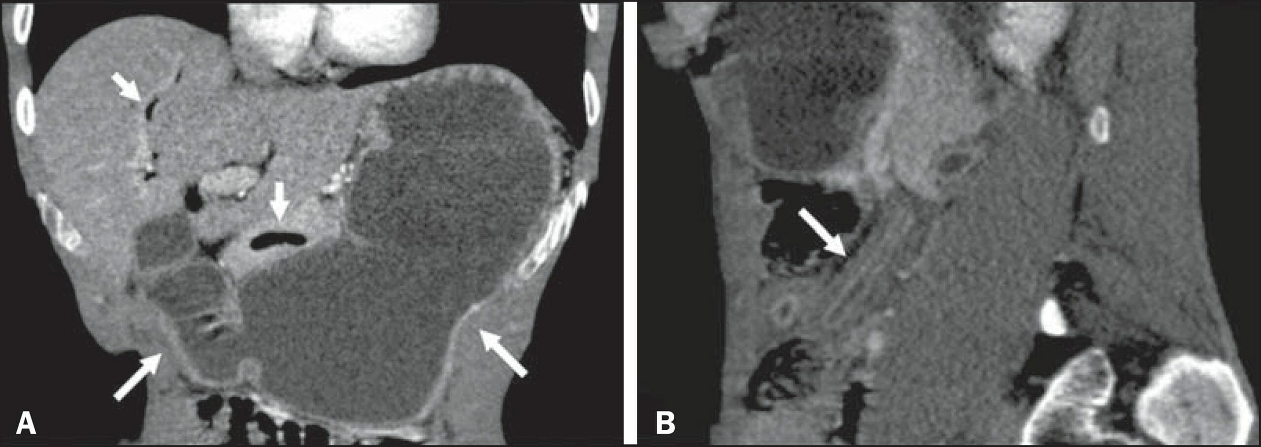
Intestinal strongyloidiasis:
A) Contrast-enhanced CT in the coronal plane shows accentuated duodenal and gastric distension (long arrows) and gas in the biliary tract and pancreas (short arrows).
B) CT in the sagittal plane shows the lead pipe sign, characterized by thickening of the walls, rigidity, and luminal narrowing of the small intestine (long arrow).

Image: “Intravenous contrast-enhanced CT scan” by José Henrique Frota Júnior et al. License: CC BY 4.0

Diagnosis and Management

Diagnosis

 Diagnosis requires a high clinical suspicion test, based on symptoms. 

  • Eosinophilia Eosinophilia Abnormal increase of eosinophils in the blood, tissues or organs. Autosomal Dominant Hyperimmunoglobulin E Syndrome 
    • Common finding in chronic infections Infections Invasion of the host organism by microorganisms or their toxins or by parasites that can cause pathological conditions or diseases. Chronic Granulomatous Disease
    • Nonspecific finding
  • Skin biopsy Skin Biopsy Secondary Skin Lesions: may provide microscopic identification Identification Defense Mechanisms of mobile rhabditiform larvae Rhabditiform Larvae Hookworm Infections
  • Serologic studies 
    • ELISA ELISA An immunoassay utilizing an antibody labeled with an enzyme marker such as horseradish peroxidase. While either the enzyme or the antibody is bound to an immunosorbent substrate, they both retain their biologic activity; the change in enzyme activity as a result of the enzyme-antibody-antigen reaction is proportional to the concentration of the antigen and can be measured spectrophotometrically or with the naked eye. Many variations of the method have been developed. St. Louis Encephalitis Virus
    • Indirect immunofluorescence microscopy
    • Immunoblot
  • Stool studies: identify parasite in feces
  • Endoscopy Endoscopy Procedures of applying endoscopes for disease diagnosis and treatment. Endoscopy involves passing an optical instrument through a small incision in the skin i.e., percutaneous; or through a natural orifice and along natural body pathways such as the digestive tract; and/or through an incision in the wall of a tubular structure or organ, i.e. Transluminal, to examine or perform surgery on the interior parts of the body. Gastroesophageal Reflux Disease (GERD) not routinely performed
Strongyloides larvae

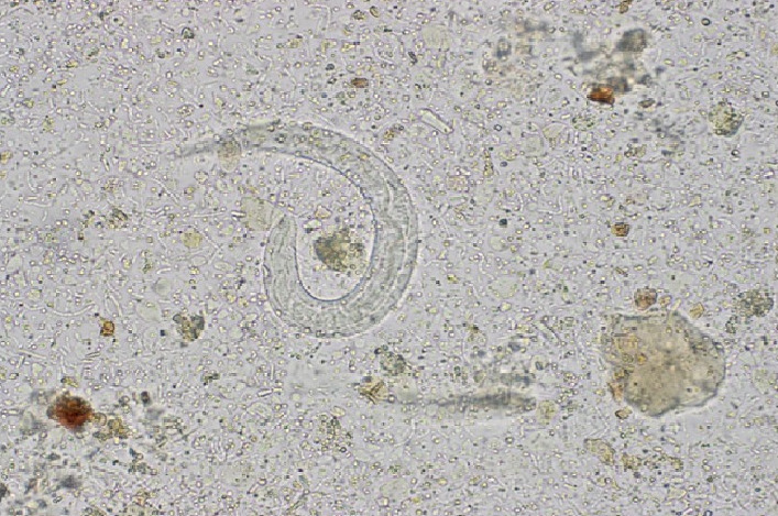
Wet mount of stool showing Strongyloides larvae

Image: “Strongyloides Hyperinfection in a Renal Transplant Patient: Always Be on the Lookout” by Case Reports in Infectious Diseases. License: CC BY 4.0

Management

  • Anthelmintic medication:
    • 1st line: ivermectin Ivermectin A mixture of mostly avermectin h2b1a (rn 71827-03-7) with some avermectin h2b1b (rn 70209-81-3), which are macrolides from streptomyces avermitilis. It binds glutamate-gated chloride channel to cause increased permeability and hyperpolarization of nerve and muscle cells. It also interacts with other chloride channels. It is a broad spectrum antiparasitic that is active against microfilariae of onchocerca volvulus but not the adult form. Anthelmintic Drugs
    • For complicated infections Infections Invasion of the host organism by microorganisms or their toxins or by parasites that can cause pathological conditions or diseases. Chronic Granulomatous Disease in immunosuppressed individuals, albendazole Albendazole A benzimidazole broad-spectrum anthelmintic structurally related to mebendazole that is effective against many diseases. Anthelmintic Drugs may be added.
  • Treatment may need to be repeated for persistent infections Infections Invasion of the host organism by microorganisms or their toxins or by parasites that can cause pathological conditions or diseases. Chronic Granulomatous Disease.

Prevention

  • Use of footwear when walking outdoors, especially on soil or sand in endemic regions
  • Proper sewage disposal and fecal management

Differential Diagnosis

  • Pulmonary phase:
    • Tuberculosis Tuberculosis Tuberculosis (TB) is an infectious disease caused by Mycobacterium tuberculosis complex bacteria. The bacteria usually attack the lungs but can also damage other parts of the body. Approximately 30% of people around the world are infected with this pathogen, with the majority harboring a latent infection. Tuberculosis spreads through the air when a person with active pulmonary infection coughs or sneezes. Tuberculosis: pulmonary bacterial infection caused by Mycobacterium tuberculosis Mycobacterium tuberculosis Tuberculosis (TB) is an infectious disease caused by Mycobacterium tuberculosis complex bacteria. The bacteria usually attack the lungs but can also damage other parts of the body. Approximately 30% of people around the world are infected with this pathogen, with the majority harboring a latent infection. Tuberculosis spreads through the air when a person with active pulmonary infection coughs or sneezes. Tuberculosis, which may present asymptomatically or with fever Fever Fever is defined as a measured body temperature of at least 38°C (100.4°F). Fever is caused by circulating endogenous and/or exogenous pyrogens that increase levels of prostaglandin E2 in the hypothalamus. Fever is commonly associated with chills, rigors, sweating, and flushing of the skin. Fever, weight loss Weight loss Decrease in existing body weight. Bariatric Surgery, night sweats Night sweats Tuberculosis, hemoptysis Hemoptysis Hemoptysis is defined as the expectoration of blood originating in the lower respiratory tract. Hemoptysis is a consequence of another disease process and can be classified as either life threatening or non-life threatening. Hemoptysis can result in significant morbidity and mortality due to both drowning (reduced gas exchange as the lungs fill with blood) and hemorrhagic shock. Hemoptysis, and productive cough.
    • Ascariasis Ascariasis Ascariasis is most often caused by A. lumbricoides. If symptomatic, characteristics typically follow 2 phases, which correlate with the migration of the parasite through the body. The early phase may include cough, dyspnea, and wheezing. The late phase typically includes abdominal discomfort, bloating, nausea, and intermittent diarrhea. Ascaris/Ascariasis: infection caused by a parasitic roundworm, Ascaris Ascaris Ascaris is a genus of parasitic nematodes. The infection, ascariasis, is most often caused by A. lumbricoides. Transmission occurs primarily via ingestion of water or food contaminated with Ascaris eggs. Most patients with ascariasis are asymptomatic. Ascaris/Ascariasis lumbricoides. May present asymptomatically or with abdominal discomfort, bowel obstruction Bowel obstruction Any impairment, arrest, or reversal of the normal flow of intestinal contents toward the anal canal. Ascaris/Ascariasis, and Löffler syndrome Löffler Syndrome Ascaris/Ascariasis.
  • GI phase GI Phase Trichinella/Trichinellosis:
    • Crohn’s disease: inflammatory bowel disease characterized by inflammation Inflammation Inflammation is a complex set of responses to infection and injury involving leukocytes as the principal cellular mediators in the body’s defense against pathogenic organisms. Inflammation is also seen as a response to tissue injury in the process of wound healing. The 5 cardinal signs of inflammation are pain, heat, redness, swelling, and loss of function. Inflammation of GI tract, presenting with signs of malabsorption Malabsorption General term for a group of malnutrition syndromes caused by failure of normal intestinal absorption of nutrients. Malabsorption and Maldigestion, abdominal pain Abdominal Pain Acute Abdomen, and non-bloody, chronic diarrhea Diarrhea Diarrhea is defined as ≥ 3 watery or loose stools in a 24-hour period. There are a multitude of etiologies, which can be classified based on the underlying mechanism of disease. The duration of symptoms (acute or chronic) and characteristics of the stools (e.g., watery, bloody, steatorrheic, mucoid) can help guide further diagnostic evaluation. Diarrhea.
    • Peptic ulcer Peptic ulcer Peptic ulcer disease (PUD) refers to the full-thickness ulcerations of duodenal or gastric mucosa. The ulcerations form when exposure to acid and digestive enzymes overcomes mucosal defense mechanisms. The most common etiologies include Helicobacter pylori (H. pylori) infection and prolonged use of non-steroidal anti-inflammatory drugs (NSAIDs). Peptic Ulcer Disease disease: presence of 1 or more ulcerative lesions in stomach Stomach The stomach is a muscular sac in the upper left portion of the abdomen that plays a critical role in digestion. The stomach develops from the foregut and connects the esophagus with the duodenum. Structurally, the stomach is C-shaped and forms a greater and lesser curvature and is divided grossly into regions: the cardia, fundus, body, and pylorus. Stomach: Anatomy or lining of duodenum Duodenum The shortest and widest portion of the small intestine adjacent to the pylorus of the stomach. It is named for having the length equal to about the width of 12 fingers. Small Intestine: Anatomy, presenting with dyspepsia Dyspepsia Impaired digestion, especially after eating. Lactose Intolerance. May progress with signs of GI bleeding.

References

  1. La Hoz RM, Morris MI; AST Infectious Diseases Community of Practice. (2019). Intestinal parasites including Cryptosporidium, Cyclospora, Giardia, and Microsporidia, Entamoeba histolytica, Strongyloides, Schistosomiasis, and Echinococcus: Guidelines from the American Society of Transplantation Infectious Diseases Community of Practice. Clin Transplant. 2019 Sep;33(9):e13618. doi: 10.1111/ctr.13618. Epub 2019 Jun 23. Erratum in: Clin Transplant. 2020 Mar;34(3):e13807. PMID: 31145496. Retrieved from https://pubmed.ncbi.nlm.nih.gov/31145496/.
  2. Chu E, Whitlock WL, Dietrich RA. (1990). Pulmonary hyperinfection syndrome with Strongyloides stercoralis. Chest. 1990 Jun;97(6):1475-7. doi: 10.1378/chest.97.6.1475. PMID: 2347234. Retrieved from https://pubmed.ncbi.nlm.nih.gov/2347234/.
  3. Krolewiecki A, Nutman TB. (2019). Strongyloidiasis: A Neglected Tropical Disease. Infect Dis Clin North Am. 2019 Mar;33(1):135-151. doi: 10.1016/j.idc.2018.10.006. PMID: 30712758; PMCID: PMC6367705. Retrieved from https://pubmed.ncbi.nlm.nih.gov/30712758/.
  4. Mehta RK, Shah N, Scott DG, Grattan CE, Barker TH. (2002). Case 4. Chronic urticaria due to strongyloidiasis. Clin Exp Dermatol. 2002 Jan;27(1):84-5. doi: 10.1046/j.0307-6938.2001.00951.x. PMID: 11952686. Retrieved from https://pubmed.ncbi.nlm.nih.gov/11952686/.

Create your free account or log in to continue reading!

Sign up now and get free access to Lecturio with concept pages, medical videos, and questions for your medical education.

User Reviews

Details