Nephrolithiasis is the formation of a stone, or calculus, caused by the precipitation of solutes in the urine anywhere along the urinary tract. The most common type of kidney stone is calcium oxalate; other types include calcium phosphate, struvite (ammonium magnesium phosphate), uric acid, and cystine stones. Nephrolithiasis presents with severe, colicky flank pain that often radiates to the groin, and hematuria due to ureteral damage. Diagnosis is made by noncontrast CT of the abdomen and pelvis or by renal ultrasound, and urinalysis is needed to exclude concomitant urinary tract infection (UTI). Management depends on the size of the stone. Small stones (< 5 mm) are likely to pass on their own and are managed expectantly with hydration and analgesics. Large stones unlikely to pass spontaneously are treated with extracorporeal shock wave lithotripsy (ESWL), ureterorenoscopy, or percutaneous nephrolithotomy. Nephrolithiasis can be complicated by hydronephrosis or acute pyelonephritis. Adequate hydration is the best prophylactic intervention to prevent kidney stones.
Last updated: Dec 15, 2025
Nephrolithiasis Nephrolithiasis Nephrolithiasis is the formation of a stone, or calculus, anywhere along the urinary tract caused by precipitations of solutes in the urine. The most common type of kidney stone is the calcium oxalate stone, but other types include calcium phosphate, struvite (ammonium magnesium phosphate), uric acid, and cystine stones. Nephrolithiasis (also known as kidney stones Kidney stones Nephrolithiasis is the formation of a stone, or calculus, anywhere along the urinary tract caused by precipitations of solutes in the urine. The most common type of kidney stone is the calcium oxalate stone, but other types include calcium phosphate, struvite (ammonium magnesium phosphate), uric acid, and cystine stones. Nephrolithiasis, urolithiasis, or urinary calculi) is the formation of stones anywhere along the urinary tract Urinary tract The urinary tract is located in the abdomen and pelvis and consists of the kidneys, ureters, urinary bladder, and urethra. The structures permit the excretion of urine from the body. Urine flows from the kidneys through the ureters to the urinary bladder and out through the urethra. Urinary Tract: Anatomy.
There are 5 main types of kidney stones Kidney stones Nephrolithiasis is the formation of a stone, or calculus, anywhere along the urinary tract caused by precipitations of solutes in the urine. The most common type of kidney stone is the calcium oxalate stone, but other types include calcium phosphate, struvite (ammonium magnesium phosphate), uric acid, and cystine stones. Nephrolithiasis:
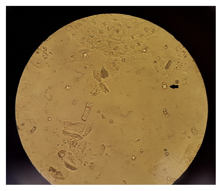
Black arrow showing an envelope, or dumbbell-shaped, calcium oxalate crystal under microscopy
Image: “Ethylene Glycol Poisoning: An Unusual Cause of Altered Mental Status and the Lessons Learned from Management of the Disease in the Acute Setting” by Case Reports in Critical Care. License: CC BY 4.0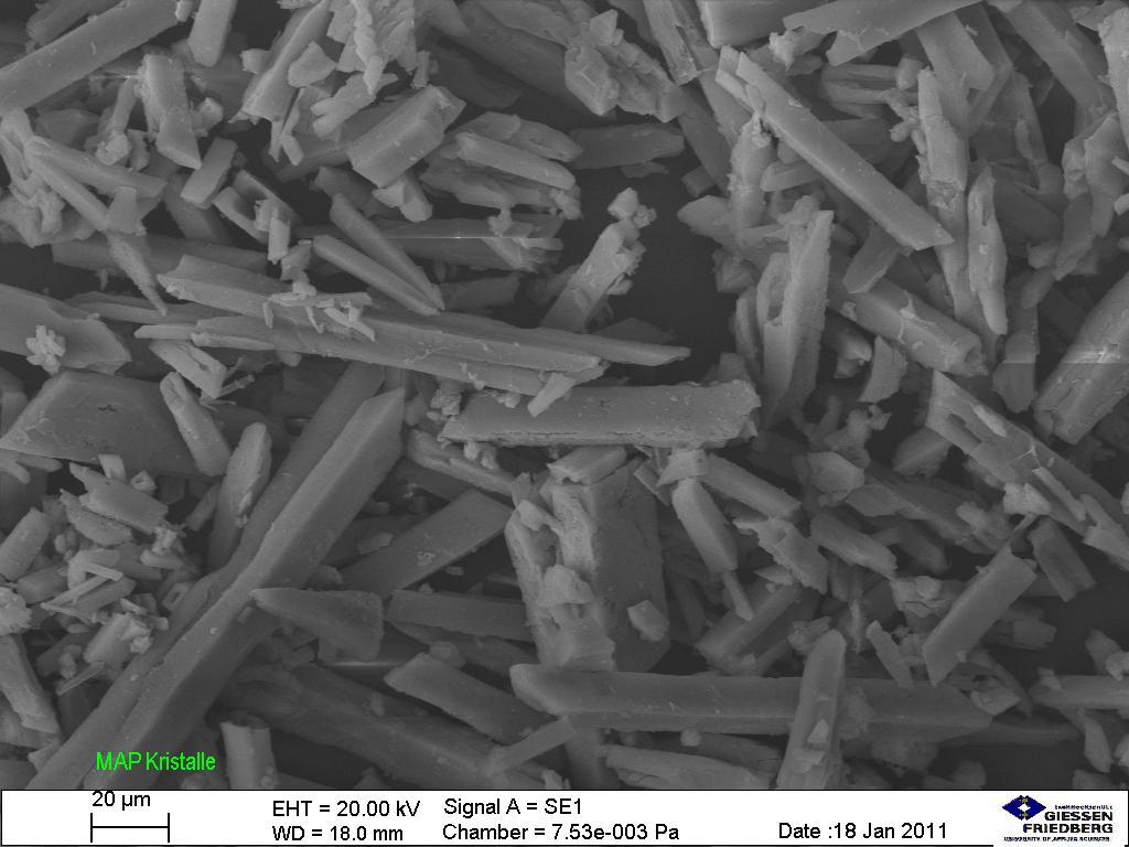
Struvite stone crystals under microscopy
Image: “Struvite under the microscope (5534899028)” by Sustainable Sanitation Alliance. License: CC BY 2.0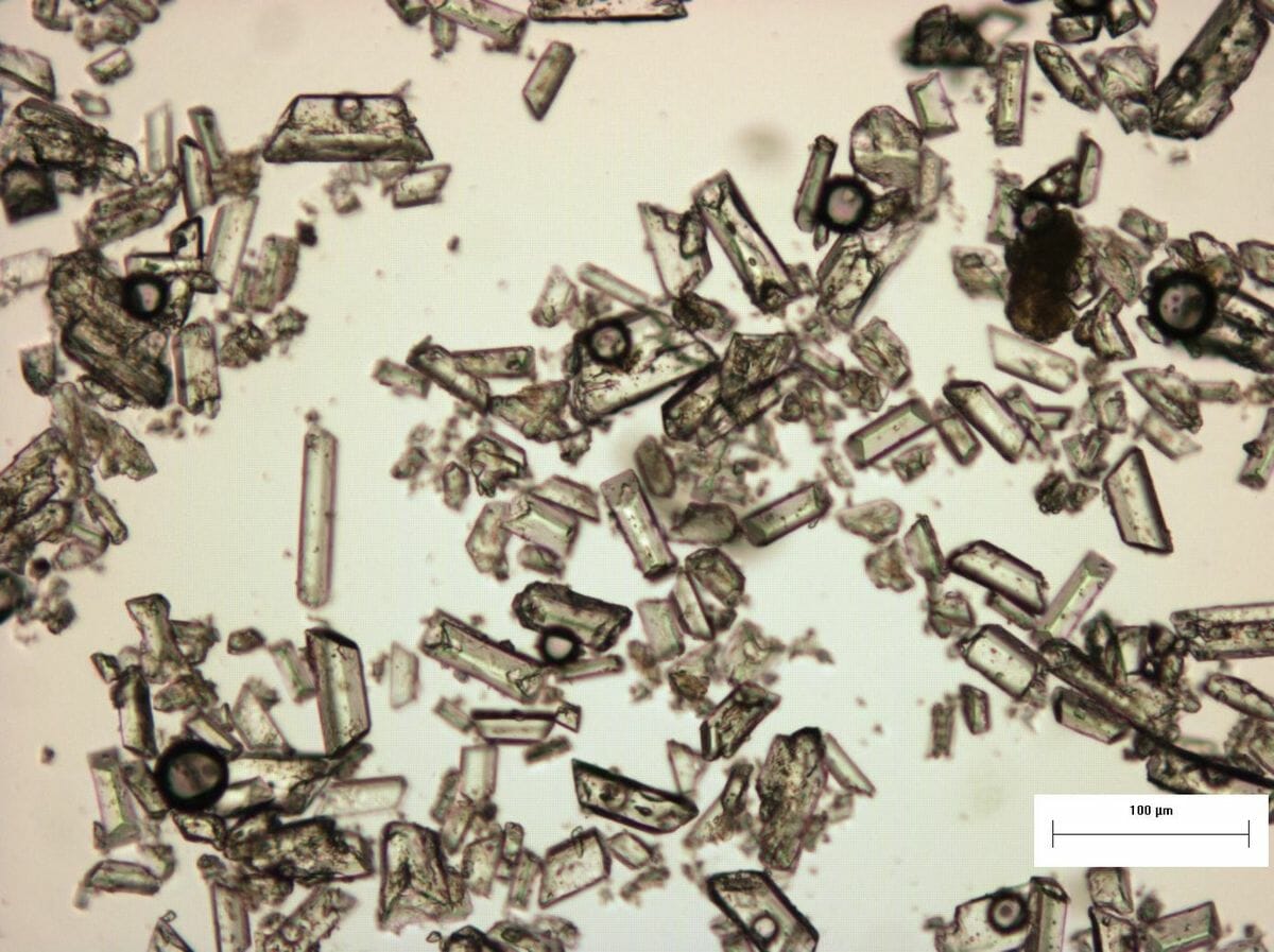
Struvite crystals and their specific structures under a microscope.
Image: “Struvite under the microscope (5267841405)” by SuSanA Secretariat. License: CC BY 2.0Nephrolithiasis Nephrolithiasis Nephrolithiasis is the formation of a stone, or calculus, anywhere along the urinary tract caused by precipitations of solutes in the urine. The most common type of kidney stone is the calcium oxalate stone, but other types include calcium phosphate, struvite (ammonium magnesium phosphate), uric acid, and cystine stones. Nephrolithiasis occurs when normally soluble material supersaturates the urine and crystal formation begins.
Underlying medical (including genetic) and environmental factors affect mechanisms that lead to formation of stones.[15]
| Type of stone | % | Causes | Crystals | Urine pH pH The quantitative measurement of the acidity or basicity of a solution. Acid-Base Balance |
|---|---|---|---|---|
| Calcium Calcium A basic element found in nearly all tissues. It is a member of the alkaline earth family of metals with the atomic symbol ca, atomic number 20, and atomic weight 40. Calcium is the most abundant mineral in the body and combines with phosphorus to form calcium phosphate in the bones and teeth. It is essential for the normal functioning of nerves and muscles and plays a role in blood coagulation (as factor IV) and in many enzymatic processes. Electrolytes oxalate | 70–80 |
|
Envelope Envelope Bilayer lipid membrane acquired by viral particles during viral morphogenesis. Although the lipids of the viral envelope are host derived, various virus-encoded integral membrane proteins, i.e. Viral envelope proteins are incorporated there. Virology or dumbbell shaped | ↓ |
| Uric acid Uric acid An oxidation product, via xanthine oxidase, of oxypurines such as xanthine and hypoxanthine. It is the final oxidation product of purine catabolism in humans and primates, whereas in most other mammals urate oxidase further oxidizes it to allantoin. Nephrolithiasis | 8 |
|
Rhomboid or rosette shaped | ↓ |
| Struvite Struvite The mineral magnesium ammonium phosphate with the formula NH4mgpo4. It is associated with urea-splitting organisms in a high magnesium, high phosphate, alkaline environment. Accumulation of crystallized struvite is found in the urinary tract as struvite calculi and as scale on sewage system equipment and wastewater pipes. Nephrolithiasis (ammonium magnesium Magnesium A metallic element that has the atomic symbol mg, atomic number 12, and atomic weight 24. 31. It is important for the activity of many enzymes, especially those involved in oxidative phosphorylation. Electrolytes phosphate Phosphate Inorganic salts of phosphoric acid. Electrolytes) | Approximately 1 | UTI UTI Urinary tract infections (UTIs) represent a wide spectrum of diseases, from self-limiting simple cystitis to severe pyelonephritis that can result in sepsis and death. Urinary tract infections are most commonly caused by Escherichia coli, but may also be caused by other bacteria and fungi. Urinary Tract Infections (UTIs) with urease-positive Urease-positive Helicobacter bacteria Bacteria Bacteria are prokaryotic single-celled microorganisms that are metabolically active and divide by binary fission. Some of these organisms play a significant role in the pathogenesis of diseases. Bacteriology | Coffin-lid shaped | ↑ |
| Calcium Calcium A basic element found in nearly all tissues. It is a member of the alkaline earth family of metals with the atomic symbol ca, atomic number 20, and atomic weight 40. Calcium is the most abundant mineral in the body and combines with phosphorus to form calcium phosphate in the bones and teeth. It is essential for the normal functioning of nerves and muscles and plays a role in blood coagulation (as factor IV) and in many enzymatic processes. Electrolytes phosphate Phosphate Inorganic salts of phosphoric acid. Electrolytes | 15 | Increased urine pH pH The quantitative measurement of the acidity or basicity of a solution. Acid-Base Balance | Wedge-shaped prism | ↑ |
| Cystine Cystine A covalently linked dimeric nonessential amino acid formed by the oxidation of cysteine. Two molecules of cysteine are joined together by a disulfide bridge to form cystine. Nephrolithiasis | < 1–2 | Cystinuria Cystinuria An inherited disorder due to defective reabsorption of cystine and other basic amino acids by the proximal renal tubules. This form of aminoaciduria is characterized by the abnormally high urinary levels of cystine; lysine; arginine; and ornithine. Mutations involve the amino acid transport protein gene slc3a1. Nephrolithiasis | Hexagonal | ↓ |
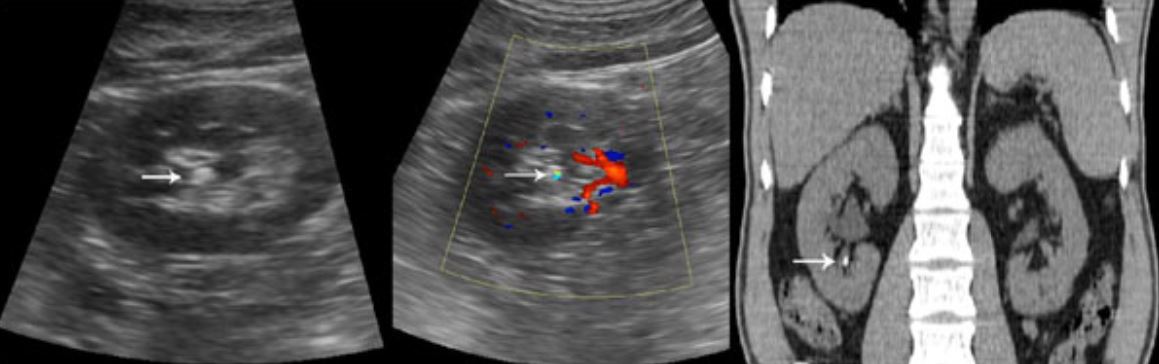
Left: White arrow shows renal calculus on ultrasound.
Right: White arrow shows renal calculus on CT scan.
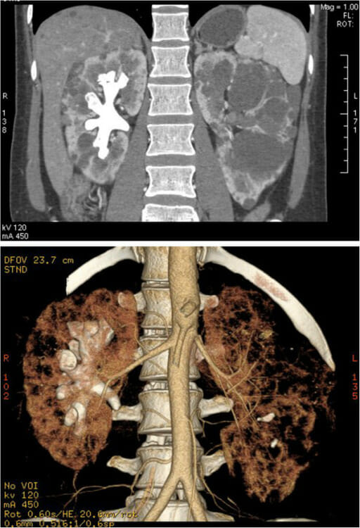
CT images of a complete staghorn calculus in the right kidney of a woman with autosomal dominant polycystic kidney disease:
Above: staghorn calculus and polycystic kidney in the coronal plane
Below: 3D image of the reconstructed calculus and kidney
General supportive care:
Medical expulsive therapy ( MET MET Preoperative Care):[6,9,10,16,17]
Indications:
Procedures:
Diagnosis Codes:
These codes are used to diagnose nephrolithiasis Nephrolithiasis Nephrolithiasis is the formation of a stone, or calculus, anywhere along the urinary tract caused by precipitations of solutes in the urine. The most common type of kidney stone is the calcium oxalate stone, but other types include calcium phosphate, struvite (ammonium magnesium phosphate), uric acid, and cystine stones. Nephrolithiasis ( kidney stones Kidney stones Nephrolithiasis is the formation of a stone, or calculus, anywhere along the urinary tract caused by precipitations of solutes in the urine. The most common type of kidney stone is the calcium oxalate stone, but other types include calcium phosphate, struvite (ammonium magnesium phosphate), uric acid, and cystine stones. Nephrolithiasis), with specificity for the location of the stone (kidney, ureter, etc ETC The electron transport chain (ETC) sends electrons through a series of proteins, which generate an electrochemical proton gradient that produces energy in the form of adenosine triphosphate (ATP). Electron Transport Chain (ETC).).
| Coding System | Code | Description |
|---|---|---|
| ICD-10-CM | N20.0 | Calculus of kidney |
| ICD-10-CM | N20.1 | Calculus of ureter |
| ICD-10-CM | N20.9 | Urinary calculus, unspecified |
Evaluation & Workup:
These codes are for the primary diagnostic tests Diagnostic tests Diagnostic tests are important aspects in making a diagnosis. Some of the most important epidemiological values of diagnostic tests include sensitivity and specificity, false positives and false negatives, positive and negative predictive values, likelihood ratios, and pre-test and post-test probabilities. Epidemiological Values of Diagnostic Tests for kidney stones Kidney stones Nephrolithiasis is the formation of a stone, or calculus, anywhere along the urinary tract caused by precipitations of solutes in the urine. The most common type of kidney stone is the calcium oxalate stone, but other types include calcium phosphate, struvite (ammonium magnesium phosphate), uric acid, and cystine stones. Nephrolithiasis. A non-contrast CT scan is the gold standard for identifying the size and location of stones, while a urinalysis Urinalysis Examination of urine by chemical, physical, or microscopic means. Routine urinalysis usually includes performing chemical screening tests, determining specific gravity, observing any unusual color or odor, screening for bacteriuria, and examining the sediment microscopically. Urinary Tract Infections (UTIs) in Children checks for blood and signs of infection.
| Coding System | Code | Description |
|---|---|---|
| CPT | 74176 | Computed tomography, abdomen and pelvis Pelvis The pelvis consists of the bony pelvic girdle, the muscular and ligamentous pelvic floor, and the pelvic cavity, which contains viscera, vessels, and multiple nerves and muscles. The pelvic girdle, composed of 2 “hip” bones and the sacrum, is a ring-like bony structure of the axial skeleton that links the vertebral column with the lower extremities. Pelvis: Anatomy; without contrast material |
| CPT | 76770 | Ultrasound, retroperitoneal Retroperitoneal Peritoneum: Anatomy (eg, renal, aorta Aorta The main trunk of the systemic arteries. Mediastinum and Great Vessels: Anatomy, nodes), real time with image documentation Documentation Systematic organization, storage, retrieval, and dissemination of specialized information, especially of a scientific or technical nature. It often involves authenticating or validating information. Advance Directives; complete |
| CPT | 81000 | Urinalysis Urinalysis Examination of urine by chemical, physical, or microscopic means. Routine urinalysis usually includes performing chemical screening tests, determining specific gravity, observing any unusual color or odor, screening for bacteriuria, and examining the sediment microscopically. Urinary Tract Infections (UTIs) in Children, by dip stick or tablet reagent, automated, with microscopy |
Procedures/Interventions:
These codes represent common procedures to treat kidney stones Kidney stones Nephrolithiasis is the formation of a stone, or calculus, anywhere along the urinary tract caused by precipitations of solutes in the urine. The most common type of kidney stone is the calcium oxalate stone, but other types include calcium phosphate, struvite (ammonium magnesium phosphate), uric acid, and cystine stones. Nephrolithiasis that do not pass on their own, including non-invasive Extracorporeal Shock Shock Shock is a life-threatening condition associated with impaired circulation that results in tissue hypoxia. The different types of shock are based on the underlying cause: distributive (↑ cardiac output (CO), ↓ systemic vascular resistance (SVR)), cardiogenic (↓ CO, ↑ SVR), hypovolemic (↓ CO, ↑ SVR), obstructive (↓ CO), and mixed. Types of Shock Wave Lithotripsy (ESWL) and minimally invasive ureteroscopy with stone removal.
| Coding System | Code | Description |
|---|---|---|
| CPT | 50590 | Lithotripsy, extracorporeal shock Shock Shock is a life-threatening condition associated with impaired circulation that results in tissue hypoxia. The different types of shock are based on the underlying cause: distributive (↑ cardiac output (CO), ↓ systemic vascular resistance (SVR)), cardiogenic (↓ CO, ↑ SVR), hypovolemic (↓ CO, ↑ SVR), obstructive (↓ CO), and mixed. Types of Shock wave |
| CPT | 52356 | Cystourethroscopy, with ureteroscopy and/or pyeloscopy; with lithotripsy including insertion of indwelling ureteral stent |
Medications:
These codes are for medications used to manage kidney stones Kidney stones Nephrolithiasis is the formation of a stone, or calculus, anywhere along the urinary tract caused by precipitations of solutes in the urine. The most common type of kidney stone is the calcium oxalate stone, but other types include calcium phosphate, struvite (ammonium magnesium phosphate), uric acid, and cystine stones. Nephrolithiasis, including tamsulosin Tamsulosin A sulfonamide derivative and adrenergic alpha-1 receptor antagonist that is used to relieve symptoms of urinary obstruction caused by benign prostatic hyperplasia. Antiadrenergic Drugs for medical expulsive therapy to help pass the stone, and NSAIDs NSAIDS Primary vs Secondary Headaches like ibuprofen Ibuprofen A nonsteroidal anti-inflammatory agent with analgesic properties used in the treatment of rheumatism and arthritis. Nonsteroidal Antiinflammatory Drugs (NSAIDs) for pain Pain An unpleasant sensation induced by noxious stimuli which are detected by nerve endings of nociceptive neurons. Pain: Types and Pathways control.
| Coding System | Code | Description |
|---|---|---|
| RxNorm | 41133 | Tamsulosin Tamsulosin A sulfonamide derivative and adrenergic alpha-1 receptor antagonist that is used to relieve symptoms of urinary obstruction caused by benign prostatic hyperplasia. Antiadrenergic Drugs (ingredient) |
| RxNorm | 5640 | Ibuprofen Ibuprofen A nonsteroidal anti-inflammatory agent with analgesic properties used in the treatment of rheumatism and arthritis. Nonsteroidal Antiinflammatory Drugs (NSAIDs) (ingredient) |
| ATC | G04CA02 | Tamsulosin Tamsulosin A sulfonamide derivative and adrenergic alpha-1 receptor antagonist that is used to relieve symptoms of urinary obstruction caused by benign prostatic hyperplasia. Antiadrenergic Drugs |