Conjunctivitis is a common inflammation of the bulbar and/or palpebral conjunctiva. It can be classified into infectious (mostly viral) and noninfectious conjunctivitis, which includes allergic causes. Patients commonly present with red eyes, increased tearing, burning, foreign body sensation, and photophobia. Itching is a chief symptom in allergic conjunctivitis. Treatment depends on the underlying cause and includes antibiotic and antiviral therapy, glucocorticoids, and antihistamines.
Last updated: Dec 15, 2025
Infectious conjunctivitis Infectious Conjunctivitis Conjunctivitis
Noninfectious conjunctivitis Noninfectious Conjunctivitis Conjunctivitis
Patients Patients Individuals participating in the health care system for the purpose of receiving therapeutic, diagnostic, or preventive procedures. Clinician–Patient Relationship can present with some or all of the following symptoms:[1 –5]
| Viral conjunctivitis Viral Conjunctivitis Conjunctivitis | Bacterial conjunctivitis Bacterial Conjunctivitis Conjunctivitis | Allergic conjunctivitis Allergic Conjunctivitis Conjunctivitis | |
|---|---|---|---|
| Discharge | Clear, watery discharge | Thick, purulent yellow, white, or green discharge with severe crusting (eyes glued shut upon awakening) | Clear, watery discharge |
| Eye involvement | Begins unilateral, but usually progresses to bilateral (highly infectious) | Unilateral, can rarely progress to bilateral | Bilateral |
| Conjunctival appearance | Mostly peripheral injection with conjunctival follicles (small swollen papules usually on the palpebral and bulbar conjunctiva Conjunctiva The mucous membrane that covers the posterior surface of the eyelids and the anterior pericorneal surface of the eyeball. Eye: Anatomy) | Diffuse injection, non-follicular | Diffuse injection with chemosis Chemosis Conjunctivitis and conjunctival follicles |
| Other symptoms | Extraocular signs of viral infection ( fever Fever Fever is defined as a measured body temperature of at least 38°C (100.4°F). Fever is caused by circulating endogenous and/or exogenous pyrogens that increase levels of prostaglandin E2 in the hypothalamus. Fever is commonly associated with chills, rigors, sweating, and flushing of the skin. Fever, lymphadenopathy Lymphadenopathy Lymphadenopathy is lymph node enlargement (> 1 cm) and is benign and self-limited in most patients. Etiologies include malignancy, infection, and autoimmune disorders, as well as iatrogenic causes such as the use of certain medications. Generalized lymphadenopathy often indicates underlying systemic disease. Lymphadenopathy, pharyngitis Pharyngitis Pharyngitis is an inflammation of the back of the throat (pharynx). Pharyngitis is usually caused by an upper respiratory tract infection, which is viral in most cases. It typically results in a sore throat and fever. Other symptoms may include a runny nose, cough, headache, and hoarseness. Pharyngitis, rash Rash Rocky Mountain Spotted Fever) | Signs of bacterial infection, depending on etiology | Sneezing Sneezing The sudden, forceful, involuntary expulsion of air from the nose and mouth caused by irritation to the mucous membranes of the upper respiratory tract. Rhinovirus, itching, atopic dermatitis Dermatitis Any inflammation of the skin. Atopic Dermatitis (Eczema) |
| Complications |
|
|
|
Viral conjunctivitis Viral Conjunctivitis Conjunctivitis:
Bacterial conjunctivitis Bacterial Conjunctivitis Conjunctivitis:

Patient with bacterial conjunctivitis presenting with characteristic thick yellow purulent discharge
Image: “A swollen, pus-filled eye with conjunctivitis” by Tanalai at English Wkipedia. License: CC BY 3.0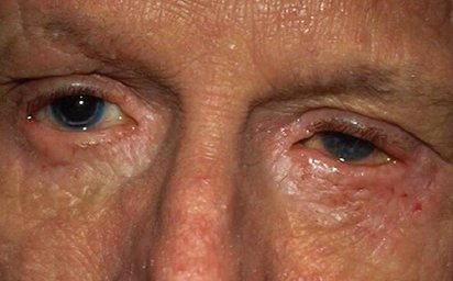
Patient with allergic conjunctivitis presenting with clear, watery discharge and chemosis
Image: “Atopic keratoconjunctivitis” by U.S. National Library of Medicine. License: CC BY 2.0, edited by Lecturio.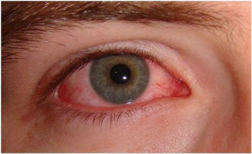
Patient with viral conjunctivitis presenting with significant peripheral injection and no visible discharge
Image: “Acute viral conjunctivitis” by Wikimedia Creative Commons. License: CC BY 4.0
Newborn presenting with gonococcal ophthalmia neonatorum, caused by a maternally transmitted gonococcal infection
Image: “Gonococcal ophthalmia neonatorum” by CDC/J. Pledger. License: Public DomainThe diagnosis is mainly clinical, based on history and physical exam. Testing is warranted only in certain cases.[1–3,9]
Management for antimicrobial treatment will vary based on locale and access to medications. The following recommendations are based on US and UK guidelines.
| Conjunctivitis Conjunctivitis Conjunctivitis is a common inflammation of the bulbar and/or palpebral conjunctiva. It can be classified into infectious (mostly viral) and noninfectious conjunctivitis, which includes allergic causes. Patients commonly present with red eyes, increased tearing, burning, foreign body sensation, and photophobia. Conjunctivitis type | Causes | Treatment |
|---|---|---|
| Viral (general) | Variety of viruses Viruses Minute infectious agents whose genomes are composed of DNA or RNA, but not both. They are characterized by a lack of independent metabolism and the inability to replicate outside living host cells. Virology but majority of cases caused by adenovirus Adenovirus Adenovirus (member of the family Adenoviridae) is a nonenveloped, double-stranded DNA virus. Adenovirus is transmitted in a variety of ways, and it can have various presentations based on the site of entry. Presentation can include febrile pharyngitis, conjunctivitis, acute respiratory disease, atypical pneumonia, and gastroenteritis. Adenovirus |
|
| Herpes simplex Herpes Simplex A group of acute infections caused by herpes simplex virus type 1 or type 2 that is characterized by the development of one or more small fluid-filled vesicles with a raised erythematous base on the skin or mucous membrane. It occurs as a primary infection or recurs due to a reactivation of a latent infection. Congenital TORCH Infections | Herpes simplex Herpes Simplex A group of acute infections caused by herpes simplex virus type 1 or type 2 that is characterized by the development of one or more small fluid-filled vesicles with a raised erythematous base on the skin or mucous membrane. It occurs as a primary infection or recurs due to a reactivation of a latent infection. Congenital TORCH Infections virus Virus Viruses are infectious, obligate intracellular parasites composed of a nucleic acid core surrounded by a protein capsid. Viruses can be either naked (non-enveloped) or enveloped. The classification of viruses is complex and based on many factors, including type and structure of the nucleoid and capsid, the presence of an envelope, the replication cycle, and the host range. Virology |
|
| Herpes zoster Herpes Zoster Varicella-zoster virus (VZV) is a linear, double-stranded DNA virus in the Herpesviridae family. Shingles (also known as herpes zoster) is more common in adults and occurs due to the reactivation of VZV. Varicella-Zoster Virus/Chickenpox | Varicella zoster virus Virus Viruses are infectious, obligate intracellular parasites composed of a nucleic acid core surrounded by a protein capsid. Viruses can be either naked (non-enveloped) or enveloped. The classification of viruses is complex and based on many factors, including type and structure of the nucleoid and capsid, the presence of an envelope, the replication cycle, and the host range. Virology | Topical agents alone are not enough:
|
| Conjunctivitis Conjunctivitis Conjunctivitis is a common inflammation of the bulbar and/or palpebral conjunctiva. It can be classified into infectious (mostly viral) and noninfectious conjunctivitis, which includes allergic causes. Patients commonly present with red eyes, increased tearing, burning, foreign body sensation, and photophobia. Conjunctivitis type | Causative organism | Treatment |
|---|---|---|
| Bacterial (general) | Available
broad-spectrum
Broad-Spectrum
Fluoroquinolones topical agents are usually effective (base choices on cost-effectiveness and local
resistance
Resistance
Physiologically, the opposition to flow of air caused by the forces of friction. As a part of pulmonary function testing, it is the ratio of driving pressure to the rate of air flow.
Ventilation: Mechanics of Breathing patterns):
|
|
| Gonococcal infection (Treat for chlamydial infection concurrently.) | Neisseria gonorrhoeae Neisseria gonorrhoeae A species of gram-negative, aerobic bacteria primarily found in purulent venereal discharges. It is the causative agent of gonorrhea. Neisseria |
|
| Chlamydial infection | Chlamydia trachomatis Chlamydia trachomatis Type species of Chlamydia causing a variety of ocular and urogenital diseases. Chlamydia | Topical treatment is not effective.
|
| Ophthalmia neonatorum Ophthalmia Neonatorum Acute conjunctival inflammation in the newborn, usually caused by maternal gonococcal infection. The causative agent is Neisseria gonorrhoeae. The baby’s eyes are contaminated during passage through the birth canal. Gonorrhea | Neisseria gonorrhoeae Neisseria gonorrhoeae A species of gram-negative, aerobic bacteria primarily found in purulent venereal discharges. It is the causative agent of gonorrhea. Neisseria |
|
| Chlamydia trachomatis Chlamydia trachomatis Type species of Chlamydia causing a variety of ocular and urogenital diseases. Chlamydia |
|
Options include the following:
| Medication class | Medication options |
|---|---|
| Antihistamine/vasoconstrictor agents | Over-the-counter options, generally given at 1–2 drops 4 times/day (≤ 2 weeks or episodic use only):
|
| Antihistamines Antihistamines Antihistamines are drugs that target histamine receptors, particularly H1 and H2 receptors. H1 antagonists are competitive and reversible inhibitors of H1 receptors. First-generation antihistamines cross the blood-brain barrier and can cause sedation. Antihistamines (2nd-generation) |
|
| Mast cell stabilizers Mast Cell Stabilizers Compounds that prevent the release of inflammatory mediators from mast cells. Asthma Drugs |
|
| Antihistamine with mast cell Mast cell Granulated cells that are found in almost all tissues, most abundantly in the skin and the gastrointestinal tract. Like the basophils, mast cells contain large amounts of histamine and heparin. Unlike basophils, mast cells normally remain in the tissues and do not circulate in the blood. Mast cells, derived from the bone marrow stem cells, are regulated by the stem cell factor. Angioedema stabilizing activity |
|
| NSAIDs NSAIDS Primary vs Secondary Headaches |
|
The following conditions enter into the differential diagnoses of conjunctivitis Conjunctivitis Conjunctivitis is a common inflammation of the bulbar and/or palpebral conjunctiva. It can be classified into infectious (mostly viral) and noninfectious conjunctivitis, which includes allergic causes. Patients commonly present with red eyes, increased tearing, burning, foreign body sensation, and photophobia. Conjunctivitis: