Pulmonary embolism (PE) is a potentially fatal condition that occurs as a result of intraluminal obstruction of the main pulmonary artery or its branches. The causative factors include thrombi, air, amniotic fluid, and fat. In PE, gas exchange is impaired due to the decreased return of deoxygenated blood to the lungs. More than 90% of PEs are complications of a deep vein thrombosis (DVT) of the lower extremity. Some individuals are asymptomatic, but the most common presenting symptom is dyspnea. The symptoms can be acute or chronic, and diagnosis is usually based on radiographic findings, typically a CT pulmonary angiogram. Initial management is supportive and focuses on restoring oxygenation and hemodynamic stability. Both medical (systemic anticoagulants and/or thrombolytics) and interventional therapies (catheter-based thrombolysis, surgery) are used to reestablish vessel patency.Note: The following page will focus on thrombotic PE. See Nonthrombotic embolism for information on air, amniotic fluid, and fat emboli.
Pulmonary embolismPulmonary EmbolismPulmonary embolism (PE) is a potentially fatal condition that occurs as a result of intraluminal obstruction of the main pulmonary artery or its branches. The causative factors include thrombi, air, amniotic fluid, and fat. In PE, gas exchange is impaired due to the decreased return of deoxygenated blood to the lungs. Pulmonary Embolism (PE) is the intraluminal obstruction of a main pulmonary arteryPulmonary arteryThe short wide vessel arising from the conus arteriosus of the right ventricle and conveying unaerated blood to the lungs.Lungs: Anatomy or any of its branches by a thrombus, air, amniotic fluidAmniotic fluidA clear, yellowish liquid that envelopes the fetus inside the sac of amnion. In the first trimester, it is likely a transudate of maternal or fetal plasma. In the second trimester, amniotic fluid derives primarily from fetal lung and kidney. Cells or substances in this fluid can be removed for prenatal diagnostic tests (amniocentesis).Placenta, Umbilical Cord, and Amniotic Cavity, or fat. When thrombotic PE is considered together with DVTDVTDeep vein thrombosis (DVT) usually occurs in the deep veins of the lower extremities. The affected veins include the femoral, popliteal, iliofemoral, and pelvic veins. Proximal DVT is more likely to cause a pulmonary embolism (PE) and is generally considered more serious. Deep Vein Thrombosis, the condition is known as venous thromboembolic (VTEVTEObstruction of a vein or veins (embolism) by a blood clot (thrombus) in the bloodstream.Hypercoagulable States) disease.
Epidemiology[3]
IncidenceIncidenceThe number of new cases of a given disease during a given period in a specified population. It also is used for the rate at which new events occur in a defined population. It is differentiated from prevalence, which refers to all cases in the population at a given time.Measures of Disease Frequency: approximately 112 per 100,000 in the United States
Common in the elderly (> 500 per 100,000 in individuals > 75 years of age)
Slight male predominance
Relationship of PE with DVTDVTDeep vein thrombosis (DVT) usually occurs in the deep veins of the lower extremities. The affected veins include the femoral, popliteal, iliofemoral, and pelvic veins. Proximal DVT is more likely to cause a pulmonary embolism (PE) and is generally considered more serious. Deep Vein Thrombosis:
50% of untreated proximal DVTs lead to PE within 3 months.
> 90% of PEsPESRemoval of plasma and replacement with various fluids, e.g., fresh frozen plasma, plasma protein fractions (ppf), albumin preparations, dextran solutions, saline. Used in treatment of autoimmune diseases, immune complex diseases, diseases of excess plasma factors, and other conditions.Thrombotic Thrombocytopenic Purpura are due to DVTs of the lower legLegThe lower leg, or just “leg” in anatomical terms, is the part of the lower limb between the knee and the ankle joint. The bony structure is composed of the tibia and fibula bones, and the muscles of the leg are grouped into the anterior, lateral, and posterior compartments by extensions of fascia.Leg: Anatomy.
Etiology and risk factors[1]
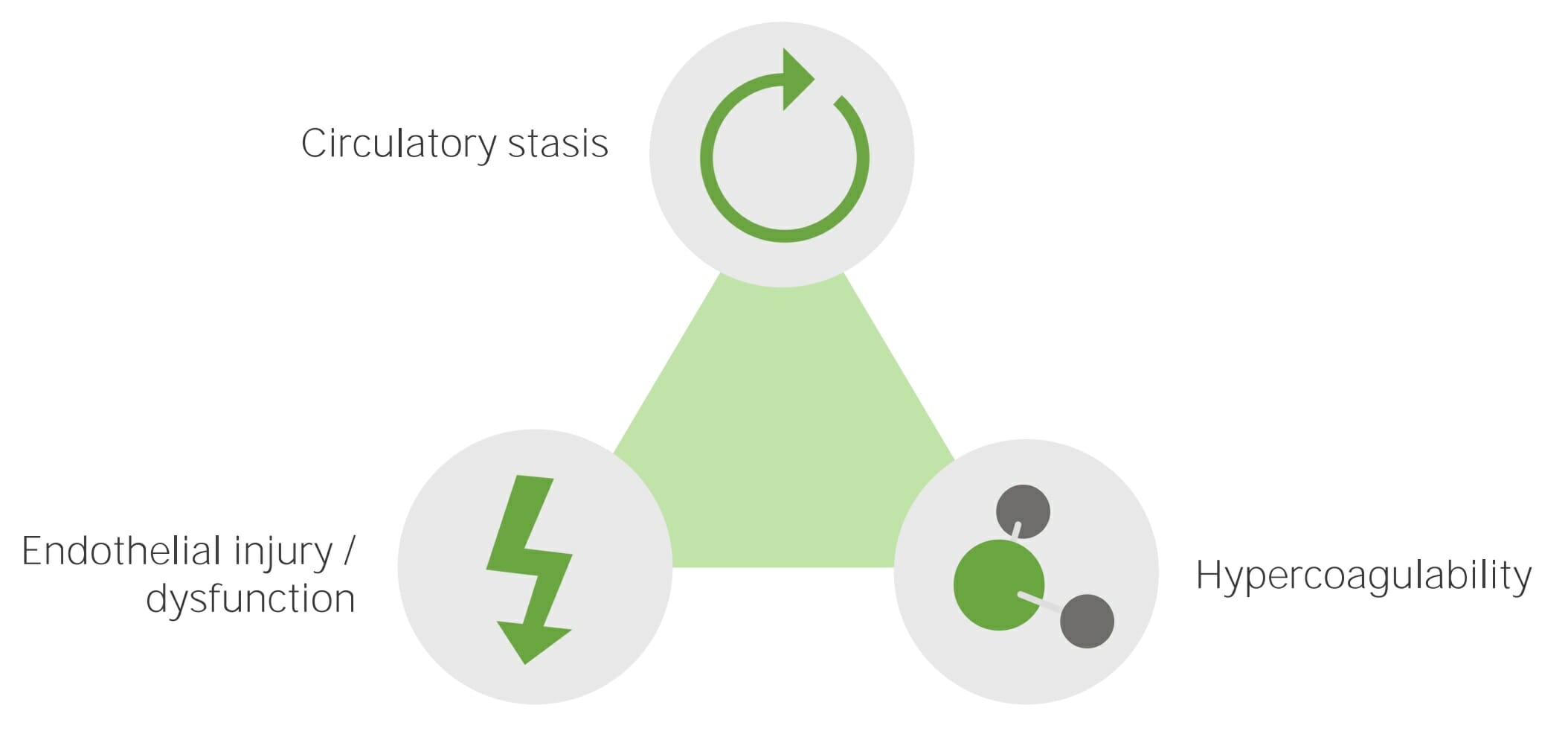
The 3 primary factors that contribute to VTEVTEObstruction of a vein or veins (embolism) by a blood clot (thrombus) in the bloodstream.Hypercoagulable States (known as Virchow’s triadVirchow’s triadDeep Vein Thrombosis) include venous stasis, hypercoagulabilityHypercoagulabilityHypercoagulable States, and vascular endothelial damage. Any condition that worsens 1 (or more) of the 3 factors increases the risk of DVTDVTDeep vein thrombosis (DVT) usually occurs in the deep veins of the lower extremities. The affected veins include the femoral, popliteal, iliofemoral, and pelvic veins. Proximal DVT is more likely to cause a pulmonary embolism (PE) and is generally considered more serious. Deep Vein Thrombosis formation and, thus, PE.
Virchow’s triadVirchow’s triadDeep Vein Thrombosis(predictive for developing DVTDVTDeep vein thrombosis (DVT) usually occurs in the deep veins of the lower extremities. The affected veins include the femoral, popliteal, iliofemoral, and pelvic veins. Proximal DVT is more likely to cause a pulmonary embolism (PE) and is generally considered more serious. Deep Vein Thrombosis):
Stasis
Endothelial injury
HypercoagulableHypercoagulableHypercoagulable states (also referred to as thrombophilias) are a group of hematologic diseases defined by an increased risk of clot formation (i.e., thrombosis) due to either an increase in procoagulants, a decrease in anticoagulants, or a decrease in fibrinolysis. Hypercoagulable States state
Genetic:
Factor V LeidenFactor V LeidenHypercoagulable StatesmutationMutationGenetic mutations are errors in DNA that can cause protein misfolding and dysfunction. There are various types of mutations, including chromosomal, point, frameshift, and expansion mutations. Types of Mutations
ProthrombinProthrombinA plasma protein that is the inactive precursor of thrombin. It is converted to thrombin by a prothrombin activator complex consisting of factor Xa, factor V, phospholipid, and calcium ions.Hemostasisgene mutationGene MutationMyotonic Dystrophies
Protein C and Protein SProtein SProtein S augments the activity of protein C.Hemostasis deficiencies
ObesityObesityObesity is a condition associated with excess body weight, specifically with the deposition of excessive adipose tissue. Obesity is considered a global epidemic. Major influences come from the western diet and sedentary lifestyles, but the exact mechanisms likely include a mixture of genetic and environmental factors. Obesity
SmokingSmokingWillful or deliberate act of inhaling and exhaling smoke from burning substances or agents held by hand.Interstitial Lung Diseases
EstrogenEstrogenCompounds that interact with estrogen receptors in target tissues to bring about the effects similar to those of estradiol. Estrogens stimulate the female reproductive organs, and the development of secondary female sex characteristics. Estrogenic chemicals include natural, synthetic, steroidal, or non-steroidal compounds.Ovaries: Anatomy exposure:
PregnancyPregnancyThe status during which female mammals carry their developing young (embryos or fetuses) in utero before birth, beginning from fertilization to birth.Pregnancy: Diagnosis, Physiology, and Care
Hormonal contraception
Hormone replacement therapyHormone Replacement TherapyHormone replacement therapy (HRT) is used to treat symptoms associated with female menopause and in combination to suppress ovulation. Risks and side effects include uterine bleeding, predisposition to cancer, breast tenderness, hyperpigmentation, migraine headaches, hypertension, bloating, and mood changes.Noncontraceptive Estrogen and Progestins
Medical conditions:
HypertensionHypertensionHypertension, or high blood pressure, is a common disease that manifests as elevated systemic arterial pressures. Hypertension is most often asymptomatic and is found incidentally as part of a routine physical examination or during triage for an unrelated medical encounter. Hypertension
Congestive heart failureHeart FailureA heterogeneous condition in which the heart is unable to pump out sufficient blood to meet the metabolic need of the body. Heart failure can be caused by structural defects, functional abnormalities (ventricular dysfunction), or a sudden overload beyond its capacity. Chronic heart failure is more common than acute heart failure which results from sudden insult to cardiac function, such as myocardial infarction.Total Anomalous Pulmonary Venous Return (TAPVR)
Autoimmune disease
Nephrotic syndromeNephrotic syndromeNephrotic syndrome is characterized by severe proteinuria, hypoalbuminemia, and peripheral edema. In contrast, the nephritic syndromes present with hematuria, variable loss of renal function, and hypertension, although there is sometimes overlap of > 1 glomerular disease in the same individual. Nephrotic Syndrome
Inflammatory bowel disease
Paroxysmal nocturnal hemoglobinuriaHemoglobinuriaThe presence of free hemoglobin in the urine, indicating hemolysis of erythrocytes within the vascular system. After saturating the hemoglobin-binding proteins (haptoglobins), free hemoglobin begins to appear in the urine.Transfusion Reactions
Severe liverLiverThe liver is the largest gland in the human body. The liver is found in the superior right quadrant of the abdomen and weighs approximately 1.5 kilograms. Its main functions are detoxification, metabolism, nutrient storage (e.g., iron and vitamins), synthesis of coagulation factors, formation of bile, filtration, and storage of blood. Liver: Anatomy disease
Virchow’s triad:
Circulatory stasis, endothelial injury or dysfunction, and hypercoagulability are the primary etiologic factors that cause venous thromboembolic disease.
May extend into the right or left main pulmonary arteryPulmonary arteryThe short wide vessel arising from the conus arteriosus of the right ventricle and conveying unaerated blood to the lungs.Lungs: Anatomy
Most emboli move beyond the bifurcation to smaller branches of a pulmonary arteryPulmonary arteryThe short wide vessel arising from the conus arteriosus of the right ventricle and conveying unaerated blood to the lungs.Lungs: Anatomy:
Lobar branches
Segmental branches
Subsegmental branches
Cases usually have multiple emboli
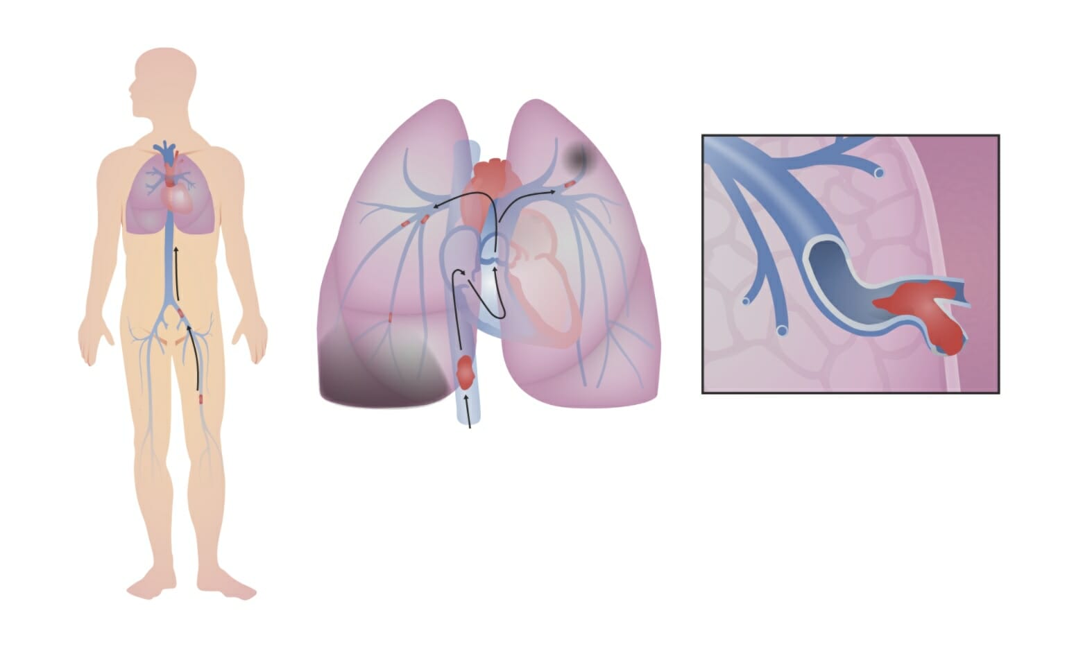
Venous thromboembolism:
A deep vein thrombosis (DVT) becomes dislodged in the leg (in this image, the thrombus forms in the left femoral vein). The DVT travels up the inferior vena cava (IVC) and through the right side of the heart. In this case, the DVT breaks into smaller fragments, which become lodged in smaller branches of the pulmonary arteries. The obstruction in blood flow through the lungs decreases the ability of the lungs to oxygenate the blood and, if large enough, puts strain on the right side of the heart.
Obstruction of vascular flowFlowBlood flows through the heart, arteries, capillaries, and veins in a closed, continuous circuit. Flow is the movement of volume per unit of time. Flow is affected by the pressure gradient and the resistance fluid encounters between 2 points. Vascular resistance is the opposition to flow, which is caused primarily by blood friction against vessel walls.Vascular Resistance, Flow, and Mean Arterial Pressure → results in dead spaceDead spaceThat part of the respiratory tract or the air within the respiratory tract that does not exchange oxygen and carbon dioxide with pulmonary capillary blood.Ventilation: Mechanics of Breathing (ventilationVentilationThe total volume of gas inspired or expired per unit of time, usually measured in liters per minute.Ventilation: Mechanics of Breathing without perfusion)
Inflammatory response, which leads to:
VasoconstrictionVasoconstrictionThe physiological narrowing of blood vessels by contraction of the vascular smooth muscle.Vascular Resistance, Flow, and Mean Arterial Pressure and bronchoconstriction in unaffected nearby areas → further decreases blood flowBlood flowBlood flow refers to the movement of a certain volume of blood through the vasculature over a given unit of time (e.g., mL per minute).Vascular Resistance, Flow, and Mean Arterial Pressure (Q) and air flowFlowBlood flows through the heart, arteries, capillaries, and veins in a closed, continuous circuit. Flow is the movement of volume per unit of time. Flow is affected by the pressure gradient and the resistance fluid encounters between 2 points. Vascular resistance is the opposition to flow, which is caused primarily by blood friction against vessel walls.Vascular Resistance, Flow, and Mean Arterial Pressure (V)
Decreased surfactantSurfactantSubstances and drugs that lower the surface tension of the mucoid layer lining the pulmonary alveoli.Acute Respiratory Distress Syndrome (ARDS) production + atelectasisAtelectasisAtelectasis is the partial or complete collapse of a part of the lung. Atelectasis is almost always a secondary phenomenon from conditions causing bronchial obstruction, external compression, surfactant deficiency, or scarring. Atelectasis → shunting (perfusion without ventilationVentilationThe total volume of gas inspired or expired per unit of time, usually measured in liters per minute.Ventilation: Mechanics of Breathing) → worsens V/Q mismatch
Abnormal gas exchangeGas exchangeHuman cells are primarily reliant on aerobic metabolism. The respiratory system is involved in pulmonary ventilation and external respiration, while the circulatory system is responsible for transport and internal respiration. Pulmonary ventilation (breathing) represents movement of air into and out of the lungs. External respiration, or gas exchange, is represented by the O2 and CO2 exchange between the lungs and the blood.Gas Exchange (due to dead spaceDead spaceThat part of the respiratory tract or the air within the respiratory tract that does not exchange oxygen and carbon dioxide with pulmonary capillary blood.Ventilation: Mechanics of Breathing and shunting), resulting in:
↑ Respiratory drive → hypocapnia and respiratory alkalosisAlkalosisA pathological condition that removes acid or adds base to the body fluids.Respiratory Alkalosis
Note: ShockShockShock is a life-threatening condition associated with impaired circulation that results in tissue hypoxia. The different types of shock are based on the underlying cause: distributive (↑ cardiac output (CO), ↓ systemic vascular resistance (SVR)), cardiogenic (↓ CO, ↑ SVR), hypovolemic (↓ CO, ↑ SVR), obstructive (↓ CO), and mixed. Types of Shock may lead to hypercapniaHypercapniaA clinical manifestation of abnormal increase in the amount of carbon dioxide in arterial blood.Neonatal Respiratory Distress Syndrome and acidosisAcidosisA pathologic condition of acid accumulation or depletion of base in the body. The two main types are respiratory acidosis and metabolic acidosis, due to metabolic acid build up.Respiratory Acidosis.
Increased pulmonary vascular resistanceResistancePhysiologically, the opposition to flow of air caused by the forces of friction. As a part of pulmonary function testing, it is the ratio of driving pressure to the rate of air flow.Ventilation: Mechanics of Breathing (PVR)
Decreased ejection from the right ventricle (RV) with subsequent increase in central venous pressureCentral venous pressureThe blood pressure in the central large veins of the body. It is distinguished from peripheral venous pressure which occurs in an extremity.Central Venous Catheter (CVPCVPThe blood pressure in the central large veins of the body. It is distinguished from peripheral venous pressure which occurs in an extremity.Central Venous Catheter)
Ultimately leads to prolonged RV strain, distention, and decreased contractility
Left-sided heart failureHeart FailureA heterogeneous condition in which the heart is unable to pump out sufficient blood to meet the metabolic need of the body. Heart failure can be caused by structural defects, functional abnormalities (ventricular dysfunction), or a sudden overload beyond its capacity. Chronic heart failure is more common than acute heart failure which results from sudden insult to cardiac function, such as myocardial infarction.Total Anomalous Pulmonary Venous Return (TAPVR):
“Right heart failureHeart FailureA heterogeneous condition in which the heart is unable to pump out sufficient blood to meet the metabolic need of the body. Heart failure can be caused by structural defects, functional abnormalities (ventricular dysfunction), or a sudden overload beyond its capacity. Chronic heart failure is more common than acute heart failure which results from sudden insult to cardiac function, such as myocardial infarction.Total Anomalous Pulmonary Venous Return (TAPVR) causes left heart failureHeart FailureA heterogeneous condition in which the heart is unable to pump out sufficient blood to meet the metabolic need of the body. Heart failure can be caused by structural defects, functional abnormalities (ventricular dysfunction), or a sudden overload beyond its capacity. Chronic heart failure is more common than acute heart failure which results from sudden insult to cardiac function, such as myocardial infarction.Total Anomalous Pulmonary Venous Return (TAPVR).”
Timing:
Occurs acutely with larger (i.e., central) emboli
Occurs later with emboli lodged in more peripheral pulmonary vessels
↓ Cardiac outputCardiac outputThe volume of blood passing through the heart per unit of time. It is usually expressed as liters (volume) per minute so as not to be confused with stroke volume (volume per beat).Cardiac Mechanics (CO), which manifests clinically as hypotensionHypotensionHypotension is defined as low blood pressure, specifically < 90/60 mm Hg, and is most commonly a physiologic response. Hypotension may be mild, serious, or life threatening, depending on the cause. Hypotension and tachycardiaTachycardiaAbnormally rapid heartbeat, usually with a heart rate above 100 beats per minute for adults. Tachycardia accompanied by disturbance in the cardiac depolarization (cardiac arrhythmia) is called tachyarrhythmia.Sepsis in Children
Pulmonary infarction:
Occurs in about 10% of patientsPatientsIndividuals participating in the health care system for the purpose of receiving therapeutic, diagnostic, or preventive procedures.Clinician–Patient Relationship
Associated with small emboli in the segmental and subsegmental branches, causing ischemiaIschemiaA hypoperfusion of the blood through an organ or tissue caused by a pathologic constriction or obstruction of its blood vessels, or an absence of blood circulation.Ischemic Cell Damage of lung tissue
Produces:
An intense inflammatory response
Intra-alveolar hemorrhage (possible)
Clinical Presentation
Timeline[3–5]
Acute: immediate development of symptoms
Subacute: development of symptoms within days to weeks
Chronic:
No immediate symptoms
PatientsPatientsIndividuals participating in the health care system for the purpose of receiving therapeutic, diagnostic, or preventive procedures.Clinician–Patient Relationship gradually develop pulmonary hypertensionPulmonary HypertensionPulmonary hypertension (PH) or pulmonary arterial hypertension (PAH) is characterized by elevated pulmonary arterial pressure, which can lead to chronic progressive right heart failure. Pulmonary hypertension is grouped into 5 categories based on etiology, which include primary PAH, and PH due to cardiac disease, lung or hypoxic disease, chronic thromboembolic disease, and multifactorial or unclear etiologies. Pulmonary Hypertension (over years).
Symptoms[3–5,11]
Presentation varies significantly. High suspicion must be maintained given the risks of complications and mortalityMortalityAll deaths reported in a given population.Measures of Health Status.
Can be asymptomatic (incidentally found on imaging)
Massive PE presents with hemodynamic instability/shockShockShock is a life-threatening condition associated with impaired circulation that results in tissue hypoxia. The different types of shock are based on the underlying cause: distributive (↑ cardiac output (CO), ↓ systemic vascular resistance (SVR)), cardiogenic (↓ CO, ↑ SVR), hypovolemic (↓ CO, ↑ SVR), obstructive (↓ CO), and mixed. Types of Shock.
Most common symptom: dyspneaDyspneaDyspnea is the subjective sensation of breathing discomfort. Dyspnea is a normal manifestation of heavy physical or psychological exertion, but also may be caused by underlying conditions (both pulmonary and extrapulmonary). Dyspneaat rest and/or during exertion
Pleuritic chest painPainAn unpleasant sensation induced by noxious stimuli which are detected by nerve endings of nociceptive neurons.Pain: Types and Pathways
Cough
HemoptysisHemoptysisHemoptysis is defined as the expectoration of blood originating in the lower respiratory tract. Hemoptysis is a consequence of another disease process and can be classified as either life threatening or non-life threatening. Hemoptysis can result in significant morbidity and mortality due to both drowning (reduced gas exchange as the lungs fill with blood) and hemorrhagic shock. Hemoptysis
Symptoms of DVTDVTDeep vein thrombosis (DVT) usually occurs in the deep veins of the lower extremities. The affected veins include the femoral, popliteal, iliofemoral, and pelvic veins. Proximal DVT is more likely to cause a pulmonary embolism (PE) and is generally considered more serious. Deep Vein Thrombosis:
Calf painPainAn unpleasant sensation induced by noxious stimuli which are detected by nerve endings of nociceptive neurons.Pain: Types and Pathways and/or tenderness
TachycardiaTachycardiaAbnormally rapid heartbeat, usually with a heart rate above 100 beats per minute for adults. Tachycardia accompanied by disturbance in the cardiac depolarization (cardiac arrhythmia) is called tachyarrhythmia.Sepsis in Children
HypotensionHypotensionHypotension is defined as low blood pressure, specifically < 90/60 mm Hg, and is most commonly a physiologic response. Hypotension may be mild, serious, or life threatening, depending on the cause. Hypotension
Note: Variation in diagnostic approach may occur based on availability of resources and practice location. Detailed information for different regions(US[26] ,UK[28], Europe[29]) is available for review.
Diagnosis is primarily via imaging. The decision to obtain imaging is based on clinical suspicion, pretest probabilityProbabilityProbability is a mathematical tool used to study randomness and provide predictions about the likelihood of something happening. There are several basic rules of probability that can be used to help determine the probability of multiple events happening together, separately, or sequentially.Basics of Probability assessment (typically using the Modified Wells criteria), and D-dimerD-dimerDeep Vein Thrombosis levels.
High-sensitivity D dimer (< 500 ng/mL is normal):[11,12,16]
By-product of cross-linked fibrinFibrinA protein derived from fibrinogen in the presence of thrombin, which forms part of the blood clot.Rapidly Progressive Glomerulonephritis degradation → elevated levels indicate thrombus breakdown
> 95% sensitivity when negative (i.e., normal values) → a negative test effectively rules out VTEVTEObstruction of a vein or veins (embolism) by a blood clot (thrombus) in the bloodstream.Hypercoagulable States in most low-risk and moderate-risk cases
Low specificity → a positive test (elevated value) does not confirm VTEVTEObstruction of a vein or veins (embolism) by a blood clot (thrombus) in the bloodstream.Hypercoagulable States since any condition that causes clots to form can elevate D-dimerD-dimerDeep Vein Thrombosis levels (e.g., recent surgery, cancer, or sepsisSepsisSystemic inflammatory response syndrome with a proven or suspected infectious etiology. When sepsis is associated with organ dysfunction distant from the site of infection, it is called severe sepsis. When sepsis is accompanied by hypotension despite adequate fluid infusion, it is called septic shock.Sepsis and Septic Shock)
Considered in those with low to low-intermediate probabilityProbabilityProbability is a mathematical tool used to study randomness and provide predictions about the likelihood of something happening. There are several basic rules of probability that can be used to help determine the probability of multiple events happening together, separately, or sequentially.Basics of Probability of PE only (NOT in those with high-intermediate and high probabilityProbabilityProbability is a mathematical tool used to study randomness and provide predictions about the likelihood of something happening. There are several basic rules of probability that can be used to help determine the probability of multiple events happening together, separately, or sequentially.Basics of Probability)
Proposed adjustment: age (if > 50 years) x 10 = cutoff value in ng/mL (fibrinogenFibrinogenPlasma glycoprotein clotted by thrombin, composed of a dimer of three non-identical pairs of polypeptide chains (alpha, beta, gamma) held together by disulfide bonds. Fibrinogen clotting is a sol-gel change involving complex molecular arrangements: whereas fibrinogen is cleaved by thrombin to form polypeptides a and b, the proteolytic action of other enzymes yields different fibrinogen degradation products.Hemostasis equivalent units)
An example: age (60 years) x 10 = 600 ng/mL (instead of the fixed cutoff level of 500 ng/mL)
Respiratory alkalosisAlkalosisA pathological condition that removes acid or adds base to the body fluids.Respiratory Alkalosis
Wide alveolar-arterial (A-a) gradient for oxygen (calculator: A-a O2 gradient)
CBC: LeukocytosisLeukocytosisA transient increase in the number of leukocytes in a body fluid.West Nile Virus may be present.
Troponins:[5,29]
May be elevated in 50% of patientsPatientsIndividuals participating in the health care system for the purpose of receiving therapeutic, diagnostic, or preventive procedures.Clinician–Patient Relationship with moderate-to-large emboli.
Normal results on highly sensitive assays have high negative predictive valueNegative predictive valueThe NPV is the percentage of people with a negative test result who are actually disease free, among all people with a negative result regardless of whether or not they have the disease.Epidemiological Values of Diagnostic Tests for excluding adverse in-hospital outcomes
BrainBrainThe part of central nervous system that is contained within the skull (cranium). Arising from the neural tube, the embryonic brain is comprised of three major parts including prosencephalon (the forebrain); mesencephalon (the midbrain); and rhombencephalon (the hindbrain). The developed brain consists of cerebrum; cerebellum; and other structures in the brain stem.Nervous System: Anatomy, Structure, and Classification natriuretic peptide (BNPBNPA peptide that is secreted by the brain and the heart atria, stored mainly in cardiac ventricular myocardium. It can cause natriuresis; diuresis; vasodilation; and inhibits secretion of renin and aldosterone. It improves heart function. It contains 32 amino acids.Renal Sodium and Water Regulation) or proBNP:[5,16,29]
Assists in risk stratification → ↑ levels are associated with ↑ mortalityMortalityAll deaths reported in a given population.Measures of Health Status risk
Frequently increased in those with RV dysfunction (in those with PE)
Prothrombin timeProthrombin timeClotting time of plasma recalcified in the presence of excess tissue thromboplastin. Factors measured are fibrinogen; prothrombin; factor V; factor VII; and factor X.Hemostasis (PT), activated partial thromboplastin timePartial thromboplastin timeThe time required for the appearance of fibrin strands following the mixing of plasma with phospholipid platelet substitute (e.g., crude cephalins, soybean phosphatides). It is a test of the intrinsic pathway (factors VIII, IX, XI, and XII) and the common pathway (fibrinogen, prothrombin, factors V and X) of blood coagulation.Hemostasis (aPTT)
ElectrocardiographyElectrocardiographyRecording of the moment-to-moment electromotive forces of the heart as projected onto various sites on the body’s surface, delineated as a scalar function of time. The recording is monitored by a tracing on slow moving chart paper or by observing it on a cardioscope, which is a cathode ray tube display.Electrocardiogram (ECG) (ECGECGAn electrocardiogram (ECG) is a graphic representation of the electrical activity of the heart plotted against time. Adhesive electrodes are affixed to the skin surface allowing measurement of cardiac impulses from many angles. The ECG provides 3-dimensional information about the conduction system of the heart, the myocardium, and other cardiac structures. Electrocardiogram (ECG)) may show:[5,29]
Arrhythmia:
Sinus tachycardiaTachycardiaAbnormally rapid heartbeat, usually with a heart rate above 100 beats per minute for adults. Tachycardia accompanied by disturbance in the cardiac depolarization (cardiac arrhythmia) is called tachyarrhythmia.Sepsis in Children is most common.
Atrial fibrillationAtrial fibrillationAtrial fibrillation (AF or Afib) is a supraventricular tachyarrhythmia and the most common kind of arrhythmia. It is caused by rapid, uncontrolled atrial contractions and uncoordinated ventricular responses. Atrial Fibrillation
These prediction tools (see tables below) are part of the preliminary assessment and help determine the probabilityProbabilityProbability is a mathematical tool used to study randomness and provide predictions about the likelihood of something happening. There are several basic rules of probability that can be used to help determine the probability of multiple events happening together, separately, or sequentially.Basics of Probability of PE and the subsequent testing needed. These tools are not intended for use in pregnant women in whom a PE is suspected.
Modified Wells criteria→ rates PE as either likely or unlikely (2-tiered system)[5,19,28]
Traditional Wells criteria score:[5,29]
Uses the same parameters as the Modified Wells criteria, but the interpretation rates PE as either low, intermediate, or high probabilityProbabilityProbability is a mathematical tool used to study randomness and provide predictions about the likelihood of something happening. There are several basic rules of probability that can be used to help determine the probability of multiple events happening together, separately, or sequentially.Basics of Probability (3-tiered system)
Considers clinical variables such as age, medical and surgical history, and examination findings
PE rule out criteria (PERC) rule:[5,18,28,29]
A list of 8 criteria that, if metMETPreoperative Care in a patient with low probabilityProbabilityProbability is a mathematical tool used to study randomness and provide predictions about the likelihood of something happening. There are several basic rules of probability that can be used to help determine the probability of multiple events happening together, separately, or sequentially.Basics of Probabilityfor PE, rules out PE or recommends further testing
Best used in the emergency department for those with low probabilityProbabilityProbability is a mathematical tool used to study randomness and provide predictions about the likelihood of something happening. There are several basic rules of probability that can be used to help determine the probability of multiple events happening together, separately, or sequentially.Basics of Probability of PE to determine if D-dimerD-dimerDeep Vein Thrombosis or imaging is needed
Based on the YEARS study, which uses variableVariableVariables represent information about something that can change. The design of the measurement scales, or of the methods for obtaining information, will determine the data gathered and the characteristics of that data. As a result, a variable can be qualitative or quantitative, and may be further classified into subgroups.Types of VariablesD-dimerD-dimerDeep Vein Thrombosis thresholds + select clinical items (found in Wells criteria) to determine the need for imaging
Signs/symptoms of DVTDVTDeep vein thrombosis (DVT) usually occurs in the deep veins of the lower extremities. The affected veins include the femoral, popliteal, iliofemoral, and pelvic veins. Proximal DVT is more likely to cause a pulmonary embolism (PE) and is generally considered more serious. Deep Vein Thrombosis (requires a minimum of legLegThe lower leg, or just “leg” in anatomical terms, is the part of the lower limb between the knee and the ankle joint. The bony structure is composed of the tibia and fibula bones, and the muscles of the leg are grouped into the anterior, lateral, and posterior compartments by extensions of fascia.Leg: AnatomyswellingSwellingInflammation and tenderness on palpationPalpationApplication of fingers with light pressure to the surface of the body to determine consistency of parts beneath in physical diagnosis; includes palpation for determining the outlines of organs.Dermatologic Examination)
3.0
PE clinically more likely than other diagnoses
3.0
TachycardiaTachycardiaAbnormally rapid heartbeat, usually with a heart rate above 100 beats per minute for adults. Tachycardia accompanied by disturbance in the cardiac depolarization (cardiac arrhythmia) is called tachyarrhythmia.Sepsis in Children (> 100/min)
1.5
Prolonged immobilizationImmobilizationDelirium (≥ 3 days) or recent surgery (within the last 30 days)
1.5
History of PE or DVTDVTDeep vein thrombosis (DVT) usually occurs in the deep veins of the lower extremities. The affected veins include the femoral, popliteal, iliofemoral, and pelvic veins. Proximal DVT is more likely to cause a pulmonary embolism (PE) and is generally considered more serious. Deep Vein Thrombosis
1.5
HemoptysisHemoptysisHemoptysis is defined as the expectoration of blood originating in the lower respiratory tract. Hemoptysis is a consequence of another disease process and can be classified as either life threatening or non-life threatening. Hemoptysis can result in significant morbidity and mortality due to both drowning (reduced gas exchange as the lungs fill with blood) and hemorrhagic shock. Hemoptysis
Interpretation: Modified Wells criteria scoring:
Score > 4: PE likely
Score ≤ 4: PE unlikely Traditional Wells criteria scoring:
0–1: Low risk of PE
2–6: Intermediate risk of PE
≥ 6: High risk of PE
DVTDVTDeep vein thrombosis (DVT) usually occurs in the deep veins of the lower extremities. The affected veins include the femoral, popliteal, iliofemoral, and pelvic veins. Proximal DVT is more likely to cause a pulmonary embolism (PE) and is generally considered more serious. Deep Vein Thrombosis: deep vein thrombosisThrombosisFormation and development of a thrombus or blood clot in the blood vessel.Epidemic Typhus PE: pulmonary embolismPulmonary EmbolismPulmonary embolism (PE) is a potentially fatal condition that occurs as a result of intraluminal obstruction of the main pulmonary artery or its branches. The causative factors include thrombi, air, amniotic fluid, and fat. In PE, gas exchange is impaired due to the decreased return of deoxygenated blood to the lungs. Pulmonary Embolism
Table: Revised Geneva scoring for PE[5]
VariableVariableVariables represent information about something that can change. The design of the measurement scales, or of the methods for obtaining information, will determine the data gathered and the characteristics of that data. As a result, a variable can be qualitative or quantitative, and may be further classified into subgroups.Types of Variables
Surgery requiring general anesthesiaGeneral anesthesiaProcedure in which patients are induced into an unconscious state through use of various medications so that they do not feel pain during surgery.Anesthesiology: History and Basic Concepts or fractureFractureA fracture is a disruption of the cortex of any bone and periosteum and is commonly due to mechanical stress after an injury or accident. Open fractures due to trauma can be a medical emergency. Fractures are frequently associated with automobile accidents, workplace injuries, and trauma.Overview of Bone Fractures of lower limb in the past month
Unilateral legLegThe lower leg, or just “leg” in anatomical terms, is the part of the lower limb between the knee and the ankle joint. The bony structure is composed of the tibia and fibula bones, and the muscles of the leg are grouped into the anterior, lateral, and posterior compartments by extensions of fascia.Leg: AnatomypainPainAn unpleasant sensation induced by noxious stimuli which are detected by nerve endings of nociceptive neurons.Pain: Types and Pathways
+3
HemoptysisHemoptysisHemoptysis is defined as the expectoration of blood originating in the lower respiratory tract. Hemoptysis is a consequence of another disease process and can be classified as either life threatening or non-life threatening. Hemoptysis can result in significant morbidity and mortality due to both drowning (reduced gas exchange as the lungs fill with blood) and hemorrhagic shock. Hemoptysis
+2
Unilateral legLegThe lower leg, or just “leg” in anatomical terms, is the part of the lower limb between the knee and the ankle joint. The bony structure is composed of the tibia and fibula bones, and the muscles of the leg are grouped into the anterior, lateral, and posterior compartments by extensions of fascia.Leg: AnatomyedemaEdemaEdema is a condition in which excess serous fluid accumulates in the body cavity or interstitial space of connective tissues. Edema is a symptom observed in several medical conditions. It can be categorized into 2 types, namely, peripheral (in the extremities) and internal (in an organ or body cavity). Edema and painPainAn unpleasant sensation induced by noxious stimuli which are detected by nerve endings of nociceptive neurons.Pain: Types and Pathways on deep venous palpationPalpationApplication of fingers with light pressure to the surface of the body to determine consistency of parts beneath in physical diagnosis; includes palpation for determining the outlines of organs.Dermatologic Examination
+4
Heart rateHeart rateThe number of times the heart ventricles contract per unit of time, usually per minute.Cardiac Physiology 75‒94/min
+3
Heart rateHeart rateThe number of times the heart ventricles contract per unit of time, usually per minute.Cardiac Physiology ≥ 95/min
+5
Interpretation (score):
≤ 3: low probabilityProbabilityProbability is a mathematical tool used to study randomness and provide predictions about the likelihood of something happening. There are several basic rules of probability that can be used to help determine the probability of multiple events happening together, separately, or sequentially.Basics of Probability
4–10: intermediate probabilityProbabilityProbability is a mathematical tool used to study randomness and provide predictions about the likelihood of something happening. There are several basic rules of probability that can be used to help determine the probability of multiple events happening together, separately, or sequentially.Basics of Probability
≥ 11: high probabilityProbabilityProbability is a mathematical tool used to study randomness and provide predictions about the likelihood of something happening. There are several basic rules of probability that can be used to help determine the probability of multiple events happening together, separately, or sequentially.Basics of Probability
Table: PERC Criteria[5,18,28]
Criteria
Age < 50 years
Heart rateHeart rateThe number of times the heart ventricles contract per unit of time, usually per minute.Cardiac Physiology <100/min
Oxyhemoglobin saturation ≥ 95%
No hemoptysisHemoptysisHemoptysis is defined as the expectoration of blood originating in the lower respiratory tract. Hemoptysis is a consequence of another disease process and can be classified as either life threatening or non-life threatening. Hemoptysis can result in significant morbidity and mortality due to both drowning (reduced gas exchange as the lungs fill with blood) and hemorrhagic shock. Hemoptysis
No estrogenEstrogenCompounds that interact with estrogen receptors in target tissues to bring about the effects similar to those of estradiol. Estrogens stimulate the female reproductive organs, and the development of secondary female sex characteristics. Estrogenic chemicals include natural, synthetic, steroidal, or non-steroidal compounds.Ovaries: Anatomy use
No prior DVTDVTDeep vein thrombosis (DVT) usually occurs in the deep veins of the lower extremities. The affected veins include the femoral, popliteal, iliofemoral, and pelvic veins. Proximal DVT is more likely to cause a pulmonary embolism (PE) and is generally considered more serious. Deep Vein Thrombosis or PE
No unilateral legLegThe lower leg, or just “leg” in anatomical terms, is the part of the lower limb between the knee and the ankle joint. The bony structure is composed of the tibia and fibula bones, and the muscles of the leg are grouped into the anterior, lateral, and posterior compartments by extensions of fascia.Leg: AnatomyswellingSwellingInflammation
No surgery requiring anesthesiaAnesthesiaA state characterized by loss of feeling or sensation. This depression of nerve function is usually the result of pharmacologic action and is induced to allow performance of surgery or other painful procedures.Anesthesiology: History and Basic Concepts or trauma requiring hospitalizationHospitalizationThe confinement of a patient in a hospital.Delirium within the previous 4 weeks
Interpretation:
All 8 criteria are metMETPreoperative Care in individuals with low pretest probabilityProbabilityProbability is a mathematical tool used to study randomness and provide predictions about the likelihood of something happening. There are several basic rules of probability that can be used to help determine the probability of multiple events happening together, separately, or sequentially.Basics of Probability of PE (estimated to be < 15%): no further imaging or testing indicated given the low likelihood of PE
Clinical signs of DVTDVTDeep vein thrombosis (DVT) usually occurs in the deep veins of the lower extremities. The affected veins include the femoral, popliteal, iliofemoral, and pelvic veins. Proximal DVT is more likely to cause a pulmonary embolism (PE) and is generally considered more serious. Deep Vein Thrombosis (e.g., legLegThe lower leg, or just “leg” in anatomical terms, is the part of the lower limb between the knee and the ankle joint. The bony structure is composed of the tibia and fibula bones, and the muscles of the leg are grouped into the anterior, lateral, and posterior compartments by extensions of fascia.Leg: AnatomyswellingSwellingInflammation, tenderness when palpating deep legLegThe lower leg, or just “leg” in anatomical terms, is the part of the lower limb between the knee and the ankle joint. The bony structure is composed of the tibia and fibula bones, and the muscles of the leg are grouped into the anterior, lateral, and posterior compartments by extensions of fascia.Leg: AnatomyveinsVeinsVeins are tubular collections of cells, which transport deoxygenated blood and waste from the capillary beds back to the heart. Veins are classified into 3 types: small veins/venules, medium veins, and large veins. Each type contains 3 primary layers: tunica intima, tunica media, and tunica adventitia. Veins: Histology)
HemoptysisHemoptysisHemoptysis is defined as the expectoration of blood originating in the lower respiratory tract. Hemoptysis is a consequence of another disease process and can be classified as either life threatening or non-life threatening. Hemoptysis can result in significant morbidity and mortality due to both drowning (reduced gas exchange as the lungs fill with blood) and hemorrhagic shock. Hemoptysis
AtelectasisAtelectasisAtelectasis is the partial or complete collapse of a part of the lung. Atelectasis is almost always a secondary phenomenon from conditions causing bronchial obstruction, external compression, surfactant deficiency, or scarring. Atelectasis or parenchymal abnormality
Effusion
CardiomegalyCardiomegalyEnlargement of the heart, usually indicated by a cardiothoracic ratio above 0. 50. Heart enlargement may involve the right, the left, or both heart ventricles or heart atria. Cardiomegaly is a nonspecific symptom seen in patients with chronic systolic heart failure (heart failure) or several forms of cardiomyopathies.Ebstein’s Anomaly
Westermark’s sign: cutoff of pulmonary vessels causes hyperlucency distally (due to oligemia)
Chest CT pulmonary angiographyAngiographyRadiography of blood vessels after injection of a contrast medium.Cardiac Surgery (CTPACTPAPulmonary Function Tests) with contrast or spiralSpiralComputed tomography where there is continuous x-ray exposure to the patient while being transported in a spiral or helical pattern through the beam of irradiation. This provides improved three-dimensional contrast and spatial resolution compared to conventional computed tomography, where data is obtained and computed from individual sequential exposures.Computed Tomography (CT) CT:
Also provides information regarding other pulmonary structures that could be contributing to the individual’s signs and symptoms
Ventilation-perfusion (V/Q) scan
2nd-line diagnostic modality
Used if CT scan is contraindicated, not available, or inconclusive
Compares distribution of air in the lungsLungsLungs are the main organs of the respiratory system. Lungs are paired viscera located in the thoracic cavity and are composed of spongy tissue. The primary function of the lungs is to oxygenate blood and eliminate CO2. Lungs: Anatomy via inhalation of radioactive xenon gas (the ventilationVentilationThe total volume of gas inspired or expired per unit of time, usually measured in liters per minute.Ventilation: Mechanics of Breathing scan) and perfusion via labeled serum markers (the perfusion scan)
PE shows areas of perfusion defects with normal ventilationVentilationThe total volume of gas inspired or expired per unit of time, usually measured in liters per minute.Ventilation: Mechanics of Breathing
Scored as normal, low-, intermediate-, or high-probability of PE
Major limitation: most individuals have indeterminate scans
A normal chest X-rayX-rayPenetrating electromagnetic radiation emitted when the inner orbital electrons of an atom are excited and release radiant energy. X-ray wavelengths range from 1 pm to 10 nm. Hard x-rays are the higher energy, shorter wavelength x-rays. Soft x-rays or grenz rays are less energetic and longer in wavelength. The short wavelength end of the x-ray spectrum overlaps the gamma rays wavelength range. The distinction between gamma rays and x-rays is based on their radiation source.Pulmonary Function Tests is required (otherwise, there is increased false positiveFalse positiveAn FP test result indicates that a person has the disease when they do not.Epidemiological Values of Diagnostic Tests results).
Pulmonary angiographyAngiographyRadiography of blood vessels after injection of a contrast medium.Cardiac Surgery:
Fluoroscopic image is obtained during direct injection of iodineIodineA nonmetallic element of the halogen group that is represented by the atomic symbol I, atomic number 53, and atomic weight of 126. 90. It is a nutritionally essential element, especially important in thyroid hormone synthesis. In solution, it has anti-infective properties and is used topically.Thyroid Hormones contrast into the main pulmonary arteryPulmonary arteryThe short wide vessel arising from the conus arteriosus of the right ventricle and conveying unaerated blood to the lungs.Lungs: Anatomy via a central catheter.
QualityQualityActivities and programs intended to assure or improve the quality of care in either a defined medical setting or a program. The concept includes the assessment or evaluation of the quality of care; identification of problems or shortcomings in the delivery of care; designing activities to overcome these deficiencies; and follow-up monitoring to ensure effectiveness of corrective steps.Quality Measurement and Improvement of images is affected by multiple factors; therefore, MRPA can be used only at sites with expertise in this imaging.
Tests in hemodynamically unstable individuals: Bedside imaging can be used to make a presumptive diagnosis of PE, which justifies the initiation of treatment.
Bedside venous ultrasonography (US):
Used to look for DVTDVTDeep vein thrombosis (DVT) usually occurs in the deep veins of the lower extremities. The affected veins include the femoral, popliteal, iliofemoral, and pelvic veins. Proximal DVT is more likely to cause a pulmonary embolism (PE) and is generally considered more serious. Deep Vein Thrombosis → if present, makes a presumptive diagnosis of PE
Findings:
Loss of normal venous compressibility detected using ultrasound probeProbeA device placed on the patient’s body to visualize a targetUltrasound (Sonography)
Turbulent or retrograde flowRetrograde flowVeins: Histology on DopplerDopplerUltrasonography applying the doppler effect, with frequency-shifted ultrasound reflections produced by moving targets (usually red blood cells) in the bloodstream along the ultrasound axis in direct proportion to the velocity of movement of the targets, to determine both direction and velocity of blood flow.Ultrasound (Sonography) imaging
Bedside echocardiographyEchocardiographyUltrasonic recording of the size, motion, and composition of the heart and surrounding tissues. The standard approach is transthoracic.Tricuspid Valve Atresia (TVA)→ findings to make a presumptive diagnosis of PE include:
New RV strain
Can show cardiac thrombus (“in transit” embolus)
Note:
EchocardiographyEchocardiographyUltrasonic recording of the size, motion, and composition of the heart and surrounding tissues. The standard approach is transthoracic.Tricuspid Valve Atresia (TVA) also provides prognostic information in patientsPatientsIndividuals participating in the health care system for the purpose of receiving therapeutic, diagnostic, or preventive procedures.Clinician–Patient Relationship with confirmed PE.
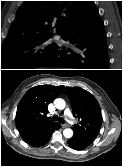
CT pulmonary angiography images confirming the presence of a saddle embolus and substantial thrombus burden in the lobar branches of both main pulmonary arteries
Image: “Pulmonary embolism CTPA” by Aung Myat and Arif Ahsan. License: CC BY 2.0
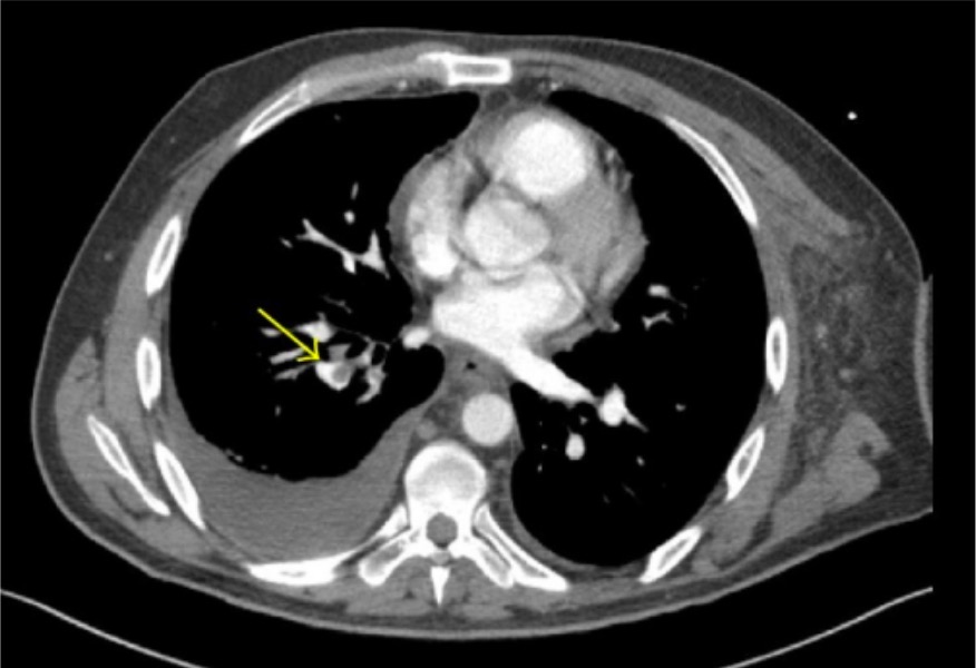
CT angiography demonstrating lobar and segmental pulmonary emboli (arrow) in the right lower lobe
Image: “CT angiography” by Thomas Jefferson University, 1025 Walnut Street, Philadelphia, PA 19107, USA. License: CC BY 3.0
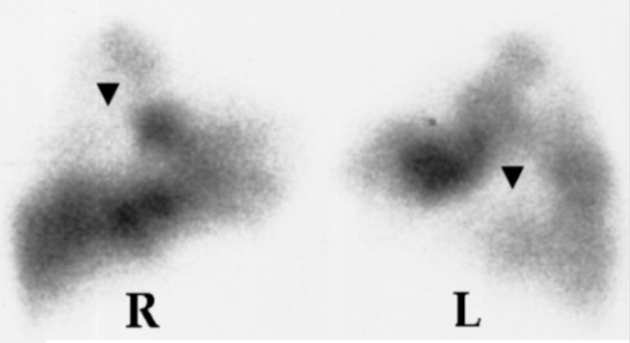
V/Q scan showing perfusion defects (arrows) in the right (R) and left (L) lungs
Image: “Lung scan” by Department of Cardiology, Sotiria Chest Diseases Hospital, Athens, Greece. License: CC BY 2.5
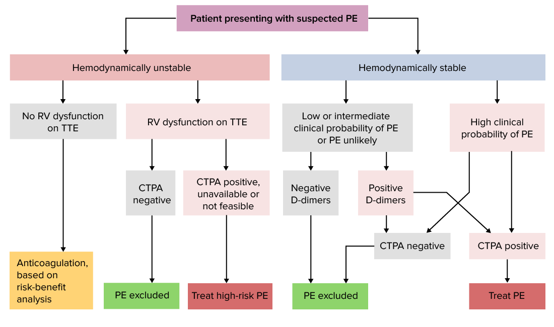
The diagnostic process based on hemodynamic stability
The Wells criteria (traditional and modified), Geneva score, and YEARS criteria (which uses items from Wells criteria) are validated. The choice of a particular scoring system is influenced by clinical training and site of practice. The following approach integrates the use of the pretest probabilityProbabilityProbability is a mathematical tool used to study randomness and provide predictions about the likelihood of something happening. There are several basic rules of probability that can be used to help determine the probability of multiple events happening together, separately, or sequentially.Basics of Probability with clinical stability.
Hemodynamically stable:[5,28,29]
Most cases of PE are hemodynamically stable (e.g., no signs of shockShockShock is a life-threatening condition associated with impaired circulation that results in tissue hypoxia. The different types of shock are based on the underlying cause: distributive (↑ cardiac output (CO), ↓ systemic vascular resistance (SVR)), cardiogenic (↓ CO, ↑ SVR), hypovolemic (↓ CO, ↑ SVR), obstructive (↓ CO), and mixed. Types of Shock), allowing time to assess the pretest probabilityProbabilityProbability is a mathematical tool used to study randomness and provide predictions about the likelihood of something happening. There are several basic rules of probability that can be used to help determine the probability of multiple events happening together, separately, or sequentially.Basics of Probability, as well as utilize PERC and D-dimerD-dimerDeep Vein Thrombosis testing.
Low probabilityProbabilityProbability is a mathematical tool used to study randomness and provide predictions about the likelihood of something happening. There are several basic rules of probability that can be used to help determine the probability of multiple events happening together, separately, or sequentially.Basics of Probability:
Intermediate probabilityProbabilityProbability is a mathematical tool used to study randomness and provide predictions about the likelihood of something happening. There are several basic rules of probability that can be used to help determine the probability of multiple events happening together, separately, or sequentially.Basics of Probability:
High probabilityProbabilityProbability is a mathematical tool used to study randomness and provide predictions about the likelihood of something happening. There are several basic rules of probability that can be used to help determine the probability of multiple events happening together, separately, or sequentially.Basics of Probability:
HypotensionHypotensionHypotension is defined as low blood pressure, specifically < 90/60 mm Hg, and is most commonly a physiologic response. Hypotension may be mild, serious, or life threatening, depending on the cause. Hypotension and/or overt shockShockShock is a life-threatening condition associated with impaired circulation that results in tissue hypoxia. The different types of shock are based on the underlying cause: distributive (↑ cardiac output (CO), ↓ systemic vascular resistance (SVR)), cardiogenic (↓ CO, ↑ SVR), hypovolemic (↓ CO, ↑ SVR), obstructive (↓ CO), and mixed. Types of Shock (e.g., systolic blood pressure (SBPSBPAscites) < 90 mm Hg for > 15 minutes) can occur in a minority of cases on initial presentation or during the course of treatment. Diagnosis and management occur simultaneously in hemodynamically unstable patientsHemodynamically Unstable PatientsBlunt Chest Trauma with a high possibility of PE.
Restoring hemodynamic stability should be prioritized:
Adequate perfusion (e.g., IV fluidsIV fluidsIntravenous fluids are one of the most common interventions administered in medicine to approximate physiologic bodily fluids. Intravenous fluids are divided into 2 categories: crystalloid and colloid solutions. Intravenous fluids have a wide variety of indications, including intravascular volume expansion, electrolyte manipulation, and maintenance fluids. Intravenous Fluids, vasopressorsVasopressorsSepsis in Children)
Oxygenation
Once the individual is stable:
Those with high probabilityProbabilityProbability is a mathematical tool used to study randomness and provide predictions about the likelihood of something happening. There are several basic rules of probability that can be used to help determine the probability of multiple events happening together, separately, or sequentially.Basics of Probability of PE:
Those with low to intermediate probabilityProbabilityProbability is a mathematical tool used to study randomness and provide predictions about the likelihood of something happening. There are several basic rules of probability that can be used to help determine the probability of multiple events happening together, separately, or sequentially.Basics of Probability, evaluation proceeds:
AnticoagulationAnticoagulationPulmonary Hypertension Drugs is started if imaging will take > 4 hours for patient at intermediate risk or > 24 hours for patientsPatientsIndividuals participating in the health care system for the purpose of receiving therapeutic, diagnostic, or preventive procedures.Clinician–Patient Relationship at low risk.2
Persistently unstable:
Diagnostic approach relies on the resources (such as cardiothoracic surgery, interventional radiologyInterventional radiologySubspecialty of radiology that combines organ system radiography, catheter techniques and sectional imaging.Penetrating Abdominal Injury, intensive care) in the institution.
Bedside imaging (e.g., lower-extremity US) is done, and empiric treatment can be given after risk–benefit analysis.
PE diagnostic algorithm
Algorithm for diagnosing pulmonary embolism.
CTPA: computed tomography pulmonary angiography; PE: pulmonary embolism; RV: right ventricle; TTE: transthoracic echocardiography
Note: Variation in management approaches may occur based on experience, availability of resources, and practice location. Detailed information for different regions (US,UK, Europe) is available for review.
Address the ABCs[29]
Assess and treat any hemodynamic instability. A majority of individuals will either present in stable condition, or respond to initial resuscitationResuscitationThe restoration to life or consciousness of one apparently dead. .Neonatal Respiratory Distress Syndrome efforts.
CirculationCirculationThe movement of the blood as it is pumped through the cardiovascular system.ABCDE Assessment/hemodynamic support:
Small amount of IV fluidsIV fluidsIntravenous fluids are one of the most common interventions administered in medicine to approximate physiologic bodily fluids. Intravenous fluids are divided into 2 categories: crystalloid and colloid solutions. Intravenous fluids have a wide variety of indications, including intravascular volume expansion, electrolyte manipulation, and maintenance fluids. Intravenous Fluids (avoid fluid overload, which worsens the effects of right-sided heart failureRight-Sided Heart FailureEbstein’s Anomaly)
VasopressorsVasopressorsSepsis in Children (typically norepinephrineNorepinephrinePrecursor of epinephrine that is secreted by the adrenal medulla and is a widespread central and autonomic neurotransmitter. Norepinephrine is the principal transmitter of most postganglionic sympathetic fibers, and of the diffuse projection system in the brain that arises from the locus ceruleus.Receptors and Neurotransmitters of the CNS)
SelectionSelectionLymphocyte activation by a specific antigen thus triggering clonal expansion of lymphocytes already capable of mounting an immune response to the antigen.B cells: Types and Functions of cardiac drugs should be based on the presence and severity of heart failureHeart FailureA heterogeneous condition in which the heart is unable to pump out sufficient blood to meet the metabolic need of the body. Heart failure can be caused by structural defects, functional abnormalities (ventricular dysfunction), or a sudden overload beyond its capacity. Chronic heart failure is more common than acute heart failure which results from sudden insult to cardiac function, such as myocardial infarction.Total Anomalous Pulmonary Venous Return (TAPVR) and hemodynamicsHemodynamicsThe movement and the forces involved in the movement of the blood through the cardiovascular system.Vascular Resistance, Flow, and Mean Arterial Pressure.
DobutamineDobutamineA catecholamine derivative with specificity for beta-1 adrenergic receptors.Sympathomimetic Drugs (2–20 µg/kg/min) improves myocardial contractility.[2,29]
NorepinephrineNorepinephrinePrecursor of epinephrine that is secreted by the adrenal medulla and is a widespread central and autonomic neurotransmitter. Norepinephrine is the principal transmitter of most postganglionic sympathetic fibers, and of the diffuse projection system in the brain that arises from the locus ceruleus.Receptors and Neurotransmitters of the CNS (0.2–1.0 µg/kg/min) is given with dobutamineDobutamineA catecholamine derivative with specificity for beta-1 adrenergic receptors.Sympathomimetic Drugs to counter the vasodilatory effect of the latter.
Bleeding risk:AnticoagulationAnticoagulationPulmonary Hypertension Drugs is the mainstay of treatment for PE, however, the risk of bleeding must be evaluated prior to starting therapy.[24]
There is no particular index that reliably predicts bleeding risk; thus, clinical judgmentJudgmentThe process of discovering or asserting an objective or intrinsic relation between two objects or concepts; a faculty or power that enables a person to make judgments; the process of bringing to light and asserting the implicit meaning of a concept; a critical evaluation of a person or situation.Psychiatric Assessment is needed.
Among the tools that can be used:
HAS-BLED score (calculator) developed to estimate 1-year risk of major bleeding in patientsPatientsIndividuals participating in the health care system for the purpose of receiving therapeutic, diagnostic, or preventive procedures.Clinician–Patient Relationship with atrial fibrillationAtrial fibrillationAtrial fibrillation (AF or Afib) is a supraventricular tachyarrhythmia and the most common kind of arrhythmia. It is caused by rapid, uncontrolled atrial contractions and uncoordinated ventricular responses. Atrial Fibrillation[23,25,29]
VTE-BLEED score (calculator) a tool developed to estimate VTE-specific bleeding risk; classifies patientsPatientsIndividuals participating in the health care system for the purpose of receiving therapeutic, diagnostic, or preventive procedures.Clinician–Patient Relationship as “high” or “low” risk of bleeding.[25]
For those with moderate risk of bleeding: individualize treatment (assess risk–benefit ratio)
For those with contraindicationsContraindicationsA condition or factor associated with a recipient that makes the use of a drug, procedure, or physical agent improper or inadvisable. Contraindications may be absolute (life threatening) or relative (higher risk of complications in which benefits may outweigh risks).Noninvasive Ventilation to anticoagulationAnticoagulationPulmonary Hypertension Drugs or unacceptable risk of bleeding: inferior vena cavaInferior vena cavaThe venous trunk which receives blood from the lower extremities and from the pelvic and abdominal organs.Mediastinum and Great Vessels: Anatomy (IVCIVCThe venous trunk which receives blood from the lower extremities and from the pelvic and abdominal organs.Mediastinum and Great Vessels: Anatomy) filter placement
AnticoagulantsAnticoagulantsAnticoagulants are drugs that retard or interrupt the coagulation cascade. The primary classes of available anticoagulants include heparins, vitamin K-dependent antagonists (e.g., warfarin), direct thrombin inhibitors, and factor Xa inhibitors. Anticoagulants:
Empiric anticoagulationAnticoagulationPulmonary Hypertension Drugs (while awaiting results of diagnostic testsDiagnostic testsDiagnostic tests are important aspects in making a diagnosis. Some of the most important epidemiological values of diagnostic tests include sensitivity and specificity, false positives and false negatives, positive and negative predictive values, likelihood ratios, and pre-test and post-test probabilities. Epidemiological Values of Diagnostic Tests) should be:[2]
Started in individuals with a low risk for bleeding and:
High-probability of PE
Intermediate probabilityProbabilityProbability is a mathematical tool used to study randomness and provide predictions about the likelihood of something happening. There are several basic rules of probability that can be used to help determine the probability of multiple events happening together, separately, or sequentially.Basics of Probability of PE, in which diagnostic evaluation will likely be > 4 hours
Low probabilityProbabilityProbability is a mathematical tool used to study randomness and provide predictions about the likelihood of something happening. There are several basic rules of probability that can be used to help determine the probability of multiple events happening together, separately, or sequentially.Basics of Probability of PE, in which diagnostic evaluation will likely be > 24 hours
Not given to those at unacceptably high risk for bleeding
Given on an individual basis for those at moderate risk for bleeding
Options:
Low-molecular-weight heparin (LMWH) given SC for at least 5 days
EnoxaparinEnoxaparinLow-molecular-weight fragment of heparin, having a 4-enopyranosuronate sodium structure at the non-reducing end of the chain. It is prepared by depolymerization of the benzylic ester of porcine mucosal heparin. Therapeutically, it is used as an antithrombotic agent.Anticoagulants 1 mg/kg twice daily (preferred) or 1.5 mg/kg once daily
DalteparinDalteparinA low-molecular-weight fragment of heparin, prepared by nitrous acid depolymerization of porcine mucosal heparin. The mean molecular weight is 4000-6000 daltons. It is used therapeutically as an antithrombotic agent.Anticoagulants 200 units/kg once daily or 100 units/kg twice daily
Unfractionated heparinUnfractionated heparinA highly acidic mucopolysaccharide formed of equal parts of sulfated d-glucosamine and d-glucuronic acid with sulfaminic bridges. The molecular weight ranges from six to twenty thousand. Heparin occurs in and is obtained from liver, lung, mast cells, etc. , of vertebrates. Its function is unknown, but it is used to prevent blood clotting in vivo and vitro, in the form of many different salts.Anticoagulants (UFHUFHA highly acidic mucopolysaccharide formed of equal parts of sulfated d-glucosamine and d-glucuronic acid with sulfaminic bridges. The molecular weight ranges from six to twenty thousand. Heparin occurs in and is obtained from liver, lung, mast cells, etc. , of vertebrates. Its function is unknown, but it is used to prevent blood clotting in vivo and vitro, in the form of many different salts.Anticoagulants):
Initial bolus, followed by a maintenance rate that is adjusted based on results of monitoring
Example regimen 1: 80 units/kg bolus, then 18 units/kg/hr
Example regimen 2: 5000-unit bolus followed by 1333 units/hour
Initial monitoring is typically every 6 hours with aPTT and/or anti–factor Xa activity
Indirect factor Xa inhibitorIndirect Factor Xa InhibitorAnticoagulants: fondaparinuxFondaparinuxSynthetic pentasaccharide that mediates the interaction of heparin with antithrombins and inhibits factor Xa; it is used for prevention of venous thromboembolism after surgery.Anticoagulants (dosed to patient weight and given SC)
5 mg once daily (< 50 kg)
7.5 mg once daily (50–100 kg)
10 mg (> 100 kg)
Direct factor Xa inhibitors: rivaroxabanRivaroxabanA morpholine and thiophene derivative that functions as a factor Xa inhibitor and is used in the treatment and prevention of deep-vein thrombosis and pulmonary embolism. It is also used for the prevention of stroke and systemic embolization in patients with non-valvular atrial fibrillation, and for the prevention of atherothrombotic events in patients after an acute coronary syndrome.Anticoagulants, apixabanApixabanAnticoagulants (both approved for monotherapy for VTEVTEObstruction of a vein or veins (embolism) by a blood clot (thrombus) in the bloodstream.Hypercoagulable States), edoxabanEdoxabanAnticoagulants
RivaroxabanRivaroxabanA morpholine and thiophene derivative that functions as a factor Xa inhibitor and is used in the treatment and prevention of deep-vein thrombosis and pulmonary embolism. It is also used for the prevention of stroke and systemic embolization in patients with non-valvular atrial fibrillation, and for the prevention of atherothrombotic events in patients after an acute coronary syndrome.Anticoagulants 15 mg by mouth twice daily (initial dose)
Direct thrombin inhibitorDirect Thrombin InhibitorAnticoagulants: dabigatranDabigatranA thrombin inhibitor which acts by binding and blocking thrombogenic activity and the prevention of thrombus formation. It is used to reduce the risk of stroke and systemic embolism in patients with nonvalvular atrial fibrillation.Anticoagulants
Note: WarfarinWarfarinAn anticoagulant that acts by inhibiting the synthesis of vitamin K-dependent coagulation factors. Warfarin is indicated for the prophylaxis and/or treatment of venous thrombosis and its extension, pulmonary embolism, and atrial fibrillation with embolization. It is also used as an adjunct in the prophylaxis of systemic embolism after myocardial infarction. Warfarin is also used as a rodenticide.Anticoagulants should not be used as initial treatment owing to the brief period of hypercoagulabilityHypercoagulabilityHypercoagulable States that occurs when starting it.
Medication is selected based on clinicianClinicianA physician, nurse practitioner, physician assistant, or another health professional who is directly involved in patient care and has a professional relationship with patients.Clinician–Patient Relationship experience, patient comorbiditiesComorbiditiesThe presence of co-existing or additional diseases with reference to an initial diagnosis or with reference to the index condition that is the subject of study. Comorbidity may affect the ability of affected individuals to function and also their survival; it may be used as a prognostic indicator for length of hospital stay, cost factors, and outcome or survival.St. Louis Encephalitis Virus, and patient preferences (including cost); preferred agents in specific populations include:
General population (hemodynamically stable, not pregnant, without cancer or creatinine clearanceCreatinine clearanceKidney Function Tests [CrCl] < 30 mL/min):[2,24]
LMWH
FondaparinuxFondaparinuxSynthetic pentasaccharide that mediates the interaction of heparin with antithrombins and inhibits factor Xa; it is used for prevention of venous thromboembolism after surgery.Anticoagulants
RivaroxabanRivaroxabanA morpholine and thiophene derivative that functions as a factor Xa inhibitor and is used in the treatment and prevention of deep-vein thrombosis and pulmonary embolism. It is also used for the prevention of stroke and systemic embolization in patients with non-valvular atrial fibrillation, and for the prevention of atherothrombotic events in patients after an acute coronary syndrome.Anticoagulants or apixabanApixabanAnticoagulants
EdoxabanEdoxabanAnticoagulants or dabigatranDabigatranA thrombin inhibitor which acts by binding and blocking thrombogenic activity and the prevention of thrombus formation. It is used to reduce the risk of stroke and systemic embolism in patients with nonvalvular atrial fibrillation.Anticoagulants (started after initial parenteral anticoagulationAnticoagulationPulmonary Hypertension Drugs)
PregnancyPregnancyThe status during which female mammals carry their developing young (embryos or fetuses) in utero before birth, beginning from fertilization to birth.Pregnancy: Diagnosis, Physiology, and Care: LMWH (dose-adjusted)[2,16,24]
Cancer:[2,16]
LMWH
Factor Xa inhibitors: apixabanApixabanAnticoagulants, rivaroxabanRivaroxabanA morpholine and thiophene derivative that functions as a factor Xa inhibitor and is used in the treatment and prevention of deep-vein thrombosis and pulmonary embolism. It is also used for the prevention of stroke and systemic embolization in patients with non-valvular atrial fibrillation, and for the prevention of atherothrombotic events in patients after an acute coronary syndrome.Anticoagulants
WarfarinWarfarinAn anticoagulant that acts by inhibiting the synthesis of vitamin K-dependent coagulation factors. Warfarin is indicated for the prophylaxis and/or treatment of venous thrombosis and its extension, pulmonary embolism, and atrial fibrillation with embolization. It is also used as an adjunct in the prophylaxis of systemic embolism after myocardial infarction. Warfarin is also used as a rodenticide.Anticoagulants(after initial parenteral anticoagulationAnticoagulationPulmonary Hypertension Drugs)
Factor Xa inhibitors: apixabanApixabanAnticoagulants, rivaroxabanRivaroxabanA morpholine and thiophene derivative that functions as a factor Xa inhibitor and is used in the treatment and prevention of deep-vein thrombosis and pulmonary embolism. It is also used for the prevention of stroke and systemic embolization in patients with non-valvular atrial fibrillation, and for the prevention of atherothrombotic events in patients after an acute coronary syndrome.Anticoagulants
Heparin-induced thrombocytopeniaHeparin-Induced ThrombocytopeniaThrombocytopenia (HIT): non-heparin anticoagulantsAnticoagulantsAnticoagulants are drugs that retard or interrupt the coagulation cascade. The primary classes of available anticoagulants include heparins, vitamin K-dependent antagonists (e.g., warfarin), direct thrombin inhibitors, and factor Xa inhibitors. Anticoagulants[24]
Renal failureRenal failureConditions in which the kidneys perform below the normal level in the ability to remove wastes, concentrate urine, and maintain electrolyte balance; blood pressure; and calcium metabolism. Renal insufficiency can be classified by the degree of kidney damage (as measured by the level of proteinuria) and reduction in glomerular filtration rate.Crush Syndrome: IV UFHUFHA highly acidic mucopolysaccharide formed of equal parts of sulfated d-glucosamine and d-glucuronic acid with sulfaminic bridges. The molecular weight ranges from six to twenty thousand. Heparin occurs in and is obtained from liver, lung, mast cells, etc. , of vertebrates. Its function is unknown, but it is used to prevent blood clotting in vivo and vitro, in the form of many different salts.Anticoagulants[24]
Extensive DVTDVTDeep vein thrombosis (DVT) usually occurs in the deep veins of the lower extremities. The affected veins include the femoral, popliteal, iliofemoral, and pelvic veins. Proximal DVT is more likely to cause a pulmonary embolism (PE) and is generally considered more serious. Deep Vein Thrombosis or with phlegmasia cerulea dolensPhlegmasia cerulea dolensNear-total occlusion of the deep venous system resulting in venous gangrene.Acute Limb Ischemia, or massive PE: IV UFHUFHA highly acidic mucopolysaccharide formed of equal parts of sulfated d-glucosamine and d-glucuronic acid with sulfaminic bridges. The molecular weight ranges from six to twenty thousand. Heparin occurs in and is obtained from liver, lung, mast cells, etc. , of vertebrates. Its function is unknown, but it is used to prevent blood clotting in vivo and vitro, in the form of many different salts.Anticoagulants[24]
Antiphospholipid syndromeAntiphospholipid syndromeAntiphospholipid syndrome (APLS) is an acquired autoimmune disorder characterized by the persistent presence of antiphospholipid antibodies, which create a hypercoagulable state. These antibodies are most commonly discovered during a workup for a thrombotic event or recurrent pregnancy loss, which are the 2 most common clinical manifestations.Antiphospholipid Syndrome: heparin, then warfarinWarfarinAn anticoagulant that acts by inhibiting the synthesis of vitamin K-dependent coagulation factors. Warfarin is indicated for the prophylaxis and/or treatment of venous thrombosis and its extension, pulmonary embolism, and atrial fibrillation with embolization. It is also used as an adjunct in the prophylaxis of systemic embolism after myocardial infarction. Warfarin is also used as a rodenticide.Anticoagulants (preferred for long-term anticoagulationAnticoagulationPulmonary Hypertension Drugs)[16]
In other cases of PE:
Subsegmental PE: Proximal pulmonary arteriesArteriesArteries are tubular collections of cells that transport oxygenated blood and nutrients from the heart to the tissues of the body. The blood passes through the arteries in order of decreasing luminal diameter, starting in the largest artery (the aorta) and ending in the small arterioles. Arteries are classified into 3 types: large elastic arteries, medium muscular arteries, and small arteries and arterioles. Arteries: Histology are not involved.
CHEST guidelines recommend surveillanceSurveillanceDevelopmental Milestones and Normal Growth (over anticoagulationAnticoagulationPulmonary Hypertension Drugs) in those without DVTDVTDeep vein thrombosis (DVT) usually occurs in the deep veins of the lower extremities. The affected veins include the femoral, popliteal, iliofemoral, and pelvic veins. Proximal DVT is more likely to cause a pulmonary embolism (PE) and is generally considered more serious. Deep Vein Thrombosis and without recurrent VTEVTEObstruction of a vein or veins (embolism) by a blood clot (thrombus) in the bloodstream.Hypercoagulable States risk.[26]
ContraindicationsContraindicationsA condition or factor associated with a recipient that makes the use of a drug, procedure, or physical agent improper or inadvisable. Contraindications may be absolute (life threatening) or relative (higher risk of complications in which benefits may outweigh risks).Noninvasive Ventilation:
Active bleeding
Acute intracranial hemorrhageIntracranial hemorrhageSubarachnoid hemorrhage (SAH) is a type of cerebrovascular accident (stroke) resulting from intracranial hemorrhage into the subarachnoid space between the arachnoid and the pia mater layers of the meninges surrounding the brain. Most sahs originate from a saccular aneurysm in the circle of willis but may also occur as a result of trauma, uncontrolled hypertension, vasculitis, anticoagulant use, or stimulant use.Subarachnoid Hemorrhage
Major trauma
Severe bleeding disordersBleeding disordersHypocoagulable conditions, also known as bleeding disorders or bleeding diathesis, are a diverse group of diseases that result in abnormal hemostasis. Physiologic hemostasis is dependent on the integrity of endothelial cells, subendothelial matrix, platelets, and coagulation factors. The hypocoagulable states result from abnormalities in one or more of these contributors, resulting in ineffective thrombosis and bleeding.Hypocoagulable Conditions
Infusion of a lytic agent (usually tissue plasminogen activatorTissue plasminogen activatorA proteolytic enzyme in the serine protease family found in many tissues which converts plasminogen to fibrinolysin. It has fibrin-binding activity and is immunologically different from urokinase-type plasminogen activator. The primary sequence, composed of 527 amino acids, is identical in both the naturally occurring and synthetic proteases.Hemostasis [tPAtPAIschemic Stroke])
Routes:
Systemic therapy
Catheter directed: a catheter tip directed at the embolus in an attempt to lyse the thrombus
Individuals unresponsive to systemic anticoagulantsAnticoagulantsAnticoagulants are drugs that retard or interrupt the coagulation cascade. The primary classes of available anticoagulants include heparins, vitamin K-dependent antagonists (e.g., warfarin), direct thrombin inhibitors, and factor Xa inhibitors. Anticoagulants with clear deterioration[26]
PrognosisPrognosisA prediction of the probable outcome of a disease based on a individual’s condition and the usual course of the disease as seen in similar situations.Non-Hodgkin Lymphomas assessment[2,29]
PatientsPatientsIndividuals participating in the health care system for the purpose of receiving therapeutic, diagnostic, or preventive procedures.Clinician–Patient Relationship should be assessed for their risk of early mortalityMortalityAll deaths reported in a given population.Measures of Health Status (within 30 days):
All patientsPatientsIndividuals participating in the health care system for the purpose of receiving therapeutic, diagnostic, or preventive procedures.Clinician–Patient Relationship with initial hemodynamic instability are at high risk, which can be defined as at least one of the following:
Cardiac arrestCardiac arrestCardiac arrest is the sudden, complete cessation of cardiac output with hemodynamic collapse. Patients present as pulseless, unresponsive, and apneic. Rhythms associated with cardiac arrest are ventricular fibrillation/tachycardia, asystole, or pulseless electrical activity. Cardiac Arrest
Persistent hypotensionHypotensionHypotension is defined as low blood pressure, specifically < 90/60 mm Hg, and is most commonly a physiologic response. Hypotension may be mild, serious, or life threatening, depending on the cause. Hypotension
Original PE Severity Index (PESI): looks at 11 parameters to classify patientsPatientsIndividuals participating in the health care system for the purpose of receiving therapeutic, diagnostic, or preventive procedures.Clinician–Patient Relationship into 5 classes from “very low risk” to “very high risk”
Simplified PE severity index (sPESI calculator): looks at 6 parameters to classify patientsPatientsIndividuals participating in the health care system for the purpose of receiving therapeutic, diagnostic, or preventive procedures.Clinician–Patient Relationship simply as low or high risk[2,29]
Other poor prognostic indicators that elevate early mortalityMortalityAll deaths reported in a given population.Measures of Health Status risk:
Elevated BNPBNPA peptide that is secreted by the brain and the heart atria, stored mainly in cardiac ventricular myocardium. It can cause natriuresis; diuresis; vasodilation; and inhibits secretion of renin and aldosterone. It improves heart function. It contains 32 amino acids.Renal Sodium and Water Regulation
Management location based on prognostic assessment:
Low-risk PE cases can be managed on an outpatient basis as long as outpatient access and home resources are adequate.[24,26,28]
Clinical consideration must be given to those with renal failureRenal failureConditions in which the kidneys perform below the normal level in the ability to remove wastes, concentrate urine, and maintain electrolyte balance; blood pressure; and calcium metabolism. Renal insufficiency can be classified by the degree of kidney damage (as measured by the level of proteinuria) and reduction in glomerular filtration rate.Crush Syndrome and severe comorbiditiesComorbiditiesThe presence of co-existing or additional diseases with reference to an initial diagnosis or with reference to the index condition that is the subject of study. Comorbidity may affect the ability of affected individuals to function and also their survival; it may be used as a prognostic indicator for length of hospital stay, cost factors, and outcome or survival.St. Louis Encephalitis Virus (as sPESI will not be applicable to them).
Administered for 3‒6 months to prevent recurrence (some patientsPatientsIndividuals participating in the health care system for the purpose of receiving therapeutic, diagnostic, or preventive procedures.Clinician–Patient Relationship may require lifelong therapy)
Duration of therapy depends on risk factors for recurrence and bleeding.
Risk of VTEVTEObstruction of a vein or veins (embolism) by a blood clot (thrombus) in the bloodstream.Hypercoagulable States recurrence: primarily based on whether PE was provoked or unprovoked and on whether risk factors are transient or persistent:
Unprovoked VTEVTEObstruction of a vein or veins (embolism) by a blood clot (thrombus) in the bloodstream.Hypercoagulable States: proceed with extended therapy
Persistent risk factor: proceed with extended therapy
Transient risk factor: no extended therapy
Risk of bleeding
Patient values and preference
Options:
LMWH
Vitamin KVitamin KA lipid cofactor that is required for normal blood clotting. Several forms of vitamin K have been identified: vitamin K 1 (phytomenadione) derived from plants, vitamin K 2 (menaquinone) from bacteria, and synthetic naphthoquinone provitamins, vitamin K 3 (menadione). Vitamin k 3 provitamins, after being alkylated in vivo, exhibit the antifibrinolytic activity of vitamin k. Green leafy vegetables, liver, cheese, butter, and egg yolk are good sources of vitamin k.Fat-soluble Vitamins and their Deficiencies antagonists: warfarinWarfarinAn anticoagulant that acts by inhibiting the synthesis of vitamin K-dependent coagulation factors. Warfarin is indicated for the prophylaxis and/or treatment of venous thrombosis and its extension, pulmonary embolism, and atrial fibrillation with embolization. It is also used as an adjunct in the prophylaxis of systemic embolism after myocardial infarction. Warfarin is also used as a rodenticide.Anticoagulants
Requires regularRegularInsulin monitoring of the prothrombin timeProthrombin timeClotting time of plasma recalcified in the presence of excess tissue thromboplastin. Factors measured are fibrinogen; prothrombin; factor V; factor VII; and factor X.Hemostasis (PT)
Numerous drug interactions
Contraindicated in pregnancyPregnancyThe status during which female mammals carry their developing young (embryos or fetuses) in utero before birth, beginning from fertilization to birth.Pregnancy: Diagnosis, Physiology, and Care (teratogenic)
Direct factor Xa inhibitors: rivaroxabanRivaroxabanA morpholine and thiophene derivative that functions as a factor Xa inhibitor and is used in the treatment and prevention of deep-vein thrombosis and pulmonary embolism. It is also used for the prevention of stroke and systemic embolization in patients with non-valvular atrial fibrillation, and for the prevention of atherothrombotic events in patients after an acute coronary syndrome.Anticoagulants, apixabanApixabanAnticoagulants, edoxabanEdoxabanAnticoagulants
Monitoring generally not required
More expensive
ThrombinThrombinAn enzyme formed from prothrombin that converts fibrinogen to fibrin.Hemostasis inhibitors: dabigatranDabigatranA thrombin inhibitor which acts by binding and blocking thrombogenic activity and the prevention of thrombus formation. It is used to reduce the risk of stroke and systemic embolism in patients with nonvalvular atrial fibrillation.Anticoagulants
Prolongs aPTT
Similar to the direct factor Xa inhibitors; an option for those with a history of HIT
Depending on the initial and secondary therapies, “bridging” may be required to ensure full anticoagulationAnticoagulationPulmonary Hypertension Drugs during transition of therapy (e.g., continuing LMWH until warfarinWarfarinAn anticoagulant that acts by inhibiting the synthesis of vitamin K-dependent coagulation factors. Warfarin is indicated for the prophylaxis and/or treatment of venous thrombosis and its extension, pulmonary embolism, and atrial fibrillation with embolization. It is also used as an adjunct in the prophylaxis of systemic embolism after myocardial infarction. Warfarin is also used as a rodenticide.Anticoagulants is effective based on the PT)
AspirinAspirinThe prototypical analgesic used in the treatment of mild to moderate pain. It has anti-inflammatory and antipyretic properties and acts as an inhibitor of cyclooxygenase which results in the inhibition of the biosynthesis of prostaglandins. Aspirin also inhibits platelet aggregation and is used in the prevention of arterial and venous thrombosis.Nonsteroidal Antiinflammatory Drugs (NSAIDs):[26]
CHEST guidelines indicate that anticoagulationAnticoagulationPulmonary Hypertension Drugs is superior to aspirinAspirinThe prototypical analgesic used in the treatment of mild to moderate pain. It has anti-inflammatory and antipyretic properties and acts as an inhibitor of cyclooxygenase which results in the inhibition of the biosynthesis of prostaglandins. Aspirin also inhibits platelet aggregation and is used in the prevention of arterial and venous thrombosis.Nonsteroidal Antiinflammatory Drugs (NSAIDs) in those with an unprovoked PE.
However, for those with an unprovoked PE who decide to stop anticoagulant therapy, aspirinAspirinThe prototypical analgesic used in the treatment of mild to moderate pain. It has anti-inflammatory and antipyretic properties and acts as an inhibitor of cyclooxygenase which results in the inhibition of the biosynthesis of prostaglandins. Aspirin also inhibits platelet aggregation and is used in the prevention of arterial and venous thrombosis.Nonsteroidal Antiinflammatory Drugs (NSAIDs) is suggested in order to prevent recurrence.
IVC filterIVC filterMechanical devices inserted in the inferior vena cava that prevent the migration of blood clots from deep venous thrombosis of the leg.Hypercoagulable States:[2,29]
Used to prevent recurrent PE in patientsPatientsIndividuals participating in the health care system for the purpose of receiving therapeutic, diagnostic, or preventive procedures.Clinician–Patient Relationship with DVTDVTDeep vein thrombosis (DVT) usually occurs in the deep veins of the lower extremities. The affected veins include the femoral, popliteal, iliofemoral, and pelvic veins. Proximal DVT is more likely to cause a pulmonary embolism (PE) and is generally considered more serious. Deep Vein Thrombosis
Barrier placed within the IVCIVCThe venous trunk which receives blood from the lower extremities and from the pelvic and abdominal organs.Mediastinum and Great Vessels: Anatomy to prevent passage of the migratory thrombus into the pulmonary arterial system
Not useful for DVTDVTDeep vein thrombosis (DVT) usually occurs in the deep veins of the lower extremities. The affected veins include the femoral, popliteal, iliofemoral, and pelvic veins. Proximal DVT is more likely to cause a pulmonary embolism (PE) and is generally considered more serious. Deep Vein Thrombosis in the upper extremities
Indications:
ContraindicationsContraindicationsA condition or factor associated with a recipient that makes the use of a drug, procedure, or physical agent improper or inadvisable. Contraindications may be absolute (life threatening) or relative (higher risk of complications in which benefits may outweigh risks).Noninvasive Ventilation to anticoagulantsAnticoagulantsAnticoagulants are drugs that retard or interrupt the coagulation cascade. The primary classes of available anticoagulants include heparins, vitamin K-dependent antagonists (e.g., warfarin), direct thrombin inhibitors, and factor Xa inhibitors. Anticoagulants
Complications from or failed systemic anticoagulantsAnticoagulantsAnticoagulants are drugs that retard or interrupt the coagulation cascade. The primary classes of available anticoagulants include heparins, vitamin K-dependent antagonists (e.g., warfarin), direct thrombin inhibitors, and factor Xa inhibitors. Anticoagulants
SmokingSmokingWillful or deliberate act of inhaling and exhaling smoke from burning substances or agents held by hand.Interstitial Lung Diseases cessation
Control of hypertensionHypertensionHypertension, or high blood pressure, is a common disease that manifests as elevated systemic arterial pressures. Hypertension is most often asymptomatic and is found incidentally as part of a routine physical examination or during triage for an unrelated medical encounter. Hypertension
Avoid estrogenEstrogenCompounds that interact with estrogen receptors in target tissues to bring about the effects similar to those of estradiol. Estrogens stimulate the female reproductive organs, and the development of secondary female sex characteristics. Estrogenic chemicals include natural, synthetic, steroidal, or non-steroidal compounds.Ovaries: Anatomy (e.g., combined oral contraceptive pillsCombined oral contraceptive pillsFixed drug combinations administered orally for contraceptive purposes.Hormonal Contraceptives) in high-risk patientsPatientsIndividuals participating in the health care system for the purpose of receiving therapeutic, diagnostic, or preventive procedures.Clinician–Patient Relationship
Long-term prognosisPrognosisA prediction of the probable outcome of a disease based on a individual’s condition and the usual course of the disease as seen in similar situations.Non-Hodgkin Lymphomas[3,10,29]
More likely in patientsPatientsIndividuals participating in the health care system for the purpose of receiving therapeutic, diagnostic, or preventive procedures.Clinician–Patient Relationship with unresolved risk factors (i.e., cancer)
Highest in the 1st 2 weeks after presentation
Chronic thromboembolic pulmonary hypertensionPulmonary HypertensionPulmonary hypertension (PH) or pulmonary arterial hypertension (PAH) is characterized by elevated pulmonary arterial pressure, which can lead to chronic progressive right heart failure. Pulmonary hypertension is grouped into 5 categories based on etiology, which include primary PAH, and PH due to cardiac disease, lung or hypoxic disease, chronic thromboembolic disease, and multifactorial or unclear etiologies. Pulmonary Hypertension (CTEPH) is a long-term sequela that rarely occurs:
PatientsPatientsIndividuals participating in the health care system for the purpose of receiving therapeutic, diagnostic, or preventive procedures.Clinician–Patient Relationship report progressive dyspneaDyspneaDyspnea is the subjective sensation of breathing discomfort. Dyspnea is a normal manifestation of heavy physical or psychological exertion, but also may be caused by underlying conditions (both pulmonary and extrapulmonary). Dyspnea and/or exercise intolerance.
May result from an undissolved thrombus or parenchymal damage
Summary
Review clinical presentation (history and examination) and determine if PE is a possible diagnosis. Determine hemodynamic stability and note that for hemodynamically unstable patientsHemodynamically Unstable PatientsBlunt Chest Trauma, diagnosis, and treatment occur simultaneously.
BNPBNPA peptide that is secreted by the brain and the heart atria, stored mainly in cardiac ventricular myocardium. It can cause natriuresis; diuresis; vasodilation; and inhibits secretion of renin and aldosterone. It improves heart function. It contains 32 amino acids.Renal Sodium and Water Regulation and/or proBNP
ECGECGAn electrocardiogram (ECG) is a graphic representation of the electrical activity of the heart plotted against time. Adhesive electrodes are affixed to the skin surface allowing measurement of cardiac impulses from many angles. The ECG provides 3-dimensional information about the conduction system of the heart, the myocardium, and other cardiac structures. Electrocardiogram (ECG)
Chest X-rayX-rayPenetrating electromagnetic radiation emitted when the inner orbital electrons of an atom are excited and release radiant energy. X-ray wavelengths range from 1 pm to 10 nm. Hard x-rays are the higher energy, shorter wavelength x-rays. Soft x-rays or grenz rays are less energetic and longer in wavelength. The short wavelength end of the x-ray spectrum overlaps the gamma rays wavelength range. The distinction between gamma rays and x-rays is based on their radiation source.Pulmonary Function Tests (can be performed bedside if patient is unstable)
For the hemodynamically unstable patient after initial resuscitationResuscitationThe restoration to life or consciousness of one apparently dead. .Neonatal Respiratory Distress Syndrome with high suspicion of PE:
Coordinate available resources: PE response team, intensive care, cardiothoracic surgery, interventional radiologyInterventional radiologySubspecialty of radiology that combines organ system radiography, catheter techniques and sectional imaging.Penetrating Abdominal Injury, etcETCThe electron transport chain (ETC) sends electrons through a series of proteins, which generate an electrochemical proton gradient that produces energy in the form of adenosine triphosphate (ATP).Electron Transport Chain (ETC).
Bedside imaging to make a presumptive diagnosis of PE:
Bedside US (look for lower-extremity DVTDVTDeep vein thrombosis (DVT) usually occurs in the deep veins of the lower extremities. The affected veins include the femoral, popliteal, iliofemoral, and pelvic veins. Proximal DVT is more likely to cause a pulmonary embolism (PE) and is generally considered more serious. Deep Vein Thrombosis)
Bedside echocardiographyEchocardiographyUltrasonic recording of the size, motion, and composition of the heart and surrounding tissues. The standard approach is transthoracic.Tricuspid Valve Atresia (TVA) (look for RV dysfunction and/or thrombus in the heart)
Empiric anticoagulationAnticoagulationPulmonary Hypertension Drugs (if no contraindicationsContraindicationsA condition or factor associated with a recipient that makes the use of a drug, procedure, or physical agent improper or inadvisable. Contraindications may be absolute (life threatening) or relative (higher risk of complications in which benefits may outweigh risks).Noninvasive Ventilation)
Prompt diagnostic imaging if possible (CTPACTPAPulmonary Function Tests, if no contraindicationsContraindicationsA condition or factor associated with a recipient that makes the use of a drug, procedure, or physical agent improper or inadvisable. Contraindications may be absolute (life threatening) or relative (higher risk of complications in which benefits may outweigh risks).Noninvasive Ventilation)
If PE is confirmed:
Ensure adequate perfusion and oxygenation.
Consider reperfusion therapy (thrombolytic therapy if no contraindicationsContraindicationsA condition or factor associated with a recipient that makes the use of a drug, procedure, or physical agent improper or inadvisable. Contraindications may be absolute (life threatening) or relative (higher risk of complications in which benefits may outweigh risks).Noninvasive Ventilation).
For hemodynamically stablepatientsPatientsIndividuals participating in the health care system for the purpose of receiving therapeutic, diagnostic, or preventive procedures.Clinician–Patient Relationship and those stabilized after initial resuscitationResuscitationThe restoration to life or consciousness of one apparently dead. .Neonatal Respiratory Distress Syndrome, calculate pretest probabilityProbabilityProbability is a mathematical tool used to study randomness and provide predictions about the likelihood of something happening. There are several basic rules of probability that can be used to help determine the probability of multiple events happening together, separately, or sequentially.Basics of Probability (e.g., Wells criteria):
Low probabilityProbabilityProbability is a mathematical tool used to study randomness and provide predictions about the likelihood of something happening. There are several basic rules of probability that can be used to help determine the probability of multiple events happening together, separately, or sequentially.Basics of Probability → calculate PERC (some go straight to D-dimerD-dimerDeep Vein Thrombosis):
Elevated D-dimerD-dimerDeep Vein Thrombosis → imaging (CTPACTPAPulmonary Function Tests if no contraindicationsContraindicationsA condition or factor associated with a recipient that makes the use of a drug, procedure, or physical agent improper or inadvisable. Contraindications may be absolute (life threatening) or relative (higher risk of complications in which benefits may outweigh risks).Noninvasive Ventilation)
Normal → no further testing (search for other etiology)
Intermediate probabilityProbabilityProbability is a mathematical tool used to study randomness and provide predictions about the likelihood of something happening. There are several basic rules of probability that can be used to help determine the probability of multiple events happening together, separately, or sequentially.Basics of Probability → look at D-dimerD-dimerDeep Vein Thrombosis
Elevated D-dimerD-dimerDeep Vein Thrombosis → imaging (CTPACTPAPulmonary Function Tests if no contraindicationsContraindicationsA condition or factor associated with a recipient that makes the use of a drug, procedure, or physical agent improper or inadvisable. Contraindications may be absolute (life threatening) or relative (higher risk of complications in which benefits may outweigh risks).Noninvasive Ventilation)
Normal → no further testing (search for other etiology)
High probabilityProbabilityProbability is a mathematical tool used to study randomness and provide predictions about the likelihood of something happening. There are several basic rules of probability that can be used to help determine the probability of multiple events happening together, separately, or sequentially.Basics of Probability → CTPACTPAPulmonary Function Tests if no contraindicationsContraindicationsA condition or factor associated with a recipient that makes the use of a drug, procedure, or physical agent improper or inadvisable. Contraindications may be absolute (life threatening) or relative (higher risk of complications in which benefits may outweigh risks).Noninvasive Ventilation
Assess PE severity/risk of early mortalityMortalityAll deaths reported in a given population.Measures of Health Status (comorbiditiesComorbiditiesThe presence of co-existing or additional diseases with reference to an initial diagnosis or with reference to the index condition that is the subject of study. Comorbidity may affect the ability of affected individuals to function and also their survival; it may be used as a prognostic indicator for length of hospital stay, cost factors, and outcome or survival.St. Louis Encephalitis Virus, sPESI, RV dysfunction, elevated troponins or BNPBNPA peptide that is secreted by the brain and the heart atria, stored mainly in cardiac ventricular myocardium. It can cause natriuresis; diuresis; vasodilation; and inhibits secretion of renin and aldosterone. It improves heart function. It contains 32 amino acids.Renal Sodium and Water Regulation)
Treatment:
Anticoagulate if no contraindicationsContraindicationsA condition or factor associated with a recipient that makes the use of a drug, procedure, or physical agent improper or inadvisable. Contraindications may be absolute (life threatening) or relative (higher risk of complications in which benefits may outweigh risks).Noninvasive Ventilation (option depends on underlying conditions and risk)
IVC filterIVC filterMechanical devices inserted in the inferior vena cava that prevent the migration of blood clots from deep venous thrombosis of the leg.Hypercoagulable States if patient has contraindicationsContraindicationsA condition or factor associated with a recipient that makes the use of a drug, procedure, or physical agent improper or inadvisable. Contraindications may be absolute (life threatening) or relative (higher risk of complications in which benefits may outweigh risks).Noninvasive Ventilation to anticoagulationAnticoagulationPulmonary Hypertension Drugs
Differential Diagnosis
Acute heart failureHeart FailureA heterogeneous condition in which the heart is unable to pump out sufficient blood to meet the metabolic need of the body. Heart failure can be caused by structural defects, functional abnormalities (ventricular dysfunction), or a sudden overload beyond its capacity. Chronic heart failure is more common than acute heart failure which results from sudden insult to cardiac function, such as myocardial infarction.Total Anomalous Pulmonary Venous Return (TAPVR): an acute reduction in cardiac function.Acute heart failureHeart FailureA heterogeneous condition in which the heart is unable to pump out sufficient blood to meet the metabolic need of the body. Heart failure can be caused by structural defects, functional abnormalities (ventricular dysfunction), or a sudden overload beyond its capacity. Chronic heart failure is more common than acute heart failure which results from sudden insult to cardiac function, such as myocardial infarction.Total Anomalous Pulmonary Venous Return (TAPVR) commonly presents with dyspneaDyspneaDyspnea is the subjective sensation of breathing discomfort. Dyspnea is a normal manifestation of heavy physical or psychological exertion, but also may be caused by underlying conditions (both pulmonary and extrapulmonary). Dyspnea, low room-air O2 saturation levels, and lower-extremity edemaEdemaEdema is a condition in which excess serous fluid accumulates in the body cavity or interstitial space of connective tissues. Edema is a symptom observed in several medical conditions. It can be categorized into 2 types, namely, peripheral (in the extremities) and internal (in an organ or body cavity). Edema, which is more likely to be bilateral as opposed to unilateral. Elevated BNPBNPA peptide that is secreted by the brain and the heart atria, stored mainly in cardiac ventricular myocardium. It can cause natriuresis; diuresis; vasodilation; and inhibits secretion of renin and aldosterone. It improves heart function. It contains 32 amino acids.Renal Sodium and Water Regulation and troponin levels are commonplace and echocardiographyEchocardiographyUltrasonic recording of the size, motion, and composition of the heart and surrounding tissues. The standard approach is transthoracic.Tricuspid Valve Atresia (TVA) findings aid in the diagnosis. Chest X-rayX-rayPenetrating electromagnetic radiation emitted when the inner orbital electrons of an atom are excited and release radiant energy. X-ray wavelengths range from 1 pm to 10 nm. Hard x-rays are the higher energy, shorter wavelength x-rays. Soft x-rays or grenz rays are less energetic and longer in wavelength. The short wavelength end of the x-ray spectrum overlaps the gamma rays wavelength range. The distinction between gamma rays and x-rays is based on their radiation source.Pulmonary Function Tests may show pulmonary edemaPulmonary edemaPulmonary edema is a condition caused by excess fluid within the lung parenchyma and alveoli as a consequence of a disease process. Based on etiology, pulmonary edema is classified as cardiogenic or noncardiogenic. Patients may present with progressive dyspnea, orthopnea, cough, or respiratory failure.Pulmonary Edema. Management relies on medical optimization.
Myocardial infarctionMyocardial infarctionMI is ischemia and death of an area of myocardial tissue due to insufficient blood flow and oxygenation, usually from thrombus formation on a ruptured atherosclerotic plaque in the epicardial arteries. Clinical presentation is most commonly with chest pain, but women and patients with diabetes may have atypical symptoms.Myocardial Infarction: acute ischemiaIschemiaA hypoperfusion of the blood through an organ or tissue caused by a pathologic constriction or obstruction of its blood vessels, or an absence of blood circulation.Ischemic Cell Damage of the myocardiumMyocardiumThe muscle tissue of the heart. It is composed of striated, involuntary muscle cells connected to form the contractile pump to generate blood flow.Heart: Anatomy. Myocardial infarctionMyocardial infarctionMI is ischemia and death of an area of myocardial tissue due to insufficient blood flow and oxygenation, usually from thrombus formation on a ruptured atherosclerotic plaque in the epicardial arteries. Clinical presentation is most commonly with chest pain, but women and patients with diabetes may have atypical symptoms.Myocardial Infarction can result in tachyarrhythmiaTachyarrhythmiaA tachyarrhythmia is a rapid heart rhythm, regular or irregular, with a rate > 100 beats/min. Tachyarrhythmia may or may not be accompanied by symptoms of hemodynamic change.Tachyarrhythmias, right- and/or left-sided heart failureHeart FailureA heterogeneous condition in which the heart is unable to pump out sufficient blood to meet the metabolic need of the body. Heart failure can be caused by structural defects, functional abnormalities (ventricular dysfunction), or a sudden overload beyond its capacity. Chronic heart failure is more common than acute heart failure which results from sudden insult to cardiac function, such as myocardial infarction.Total Anomalous Pulmonary Venous Return (TAPVR), and hypotensionHypotensionHypotension is defined as low blood pressure, specifically < 90/60 mm Hg, and is most commonly a physiologic response. Hypotension may be mild, serious, or life threatening, depending on the cause. Hypotension depending on the severity and location. The chest painPainAn unpleasant sensation induced by noxious stimuli which are detected by nerve endings of nociceptive neurons.Pain: Types and Pathways that occurs during a heart attackHeart attackMi is ischemia and death of an area of myocardial tissue due to insufficient blood flow and oxygenation, usually from thrombus formation on a ruptured atherosclerotic plaque in the epicardial arteries. Clinical presentation is most commonly with chest pain, but women and patients with diabetes may have atypical symptoms.Myocardial Infarction is most commonly substernal in location and not usually pleuritic. Serially elevated cardiac enzymesEnzymesEnzymes are complex protein biocatalysts that accelerate chemical reactions without being consumed by them. Due to the body’s constant metabolic needs, the absence of enzymes would make life unsustainable, as reactions would occur too slowly without these molecules. Basics of Enzymes and ST-segment changes on the ECGECGAn electrocardiogram (ECG) is a graphic representation of the electrical activity of the heart plotted against time. Adhesive electrodes are affixed to the skin surface allowing measurement of cardiac impulses from many angles. The ECG provides 3-dimensional information about the conduction system of the heart, the myocardium, and other cardiac structures. Electrocardiogram (ECG) are usually diagnostic. Management involves stabilization, anticoagulant, and reperfusion therapies.
PneumoniaPneumoniaPneumonia or pulmonary inflammation is an acute or chronic inflammation of lung tissue. Causes include infection with bacteria, viruses, or fungi. In more rare cases, pneumonia can also be caused through toxic triggers through inhalation of toxic substances, immunological processes, or in the course of radiotherapy.Pneumonia: an inflammationInflammationInflammation is a complex set of responses to infection and injury involving leukocytes as the principal cellular mediators in the body’s defense against pathogenic organisms. Inflammation is also seen as a response to tissue injury in the process of wound healing. The 5 cardinal signs of inflammation are pain, heat, redness, swelling, and loss of function. Inflammation of the lung parenchyma caused by respiratory infection.PneumoniaPneumoniaPneumonia or pulmonary inflammation is an acute or chronic inflammation of lung tissue. Causes include infection with bacteria, viruses, or fungi. In more rare cases, pneumonia can also be caused through toxic triggers through inhalation of toxic substances, immunological processes, or in the course of radiotherapy.Pneumonia may cause pleuritic chest painPainAn unpleasant sensation induced by noxious stimuli which are detected by nerve endings of nociceptive neurons.Pain: Types and Pathways with accompanying low room-air O2 saturation levels. Chest X-rayX-rayPenetrating electromagnetic radiation emitted when the inner orbital electrons of an atom are excited and release radiant energy. X-ray wavelengths range from 1 pm to 10 nm. Hard x-rays are the higher energy, shorter wavelength x-rays. Soft x-rays or grenz rays are less energetic and longer in wavelength. The short wavelength end of the x-ray spectrum overlaps the gamma rays wavelength range. The distinction between gamma rays and x-rays is based on their radiation source.Pulmonary Function Tests commonly reveals parenchymal lesions, which are usually absent in PE unless a parenchymal infarctInfarctArea of necrotic cells in an organ, arising mainly from hypoxia and ischemiaIschemic Cell Damage is present. Treatment is with antibiotics and respiratory support in advanced cases.
Chronic obstructive pulmonary diseasePulmonary diseaseDiseases involving the respiratory system.Blastomyces/Blastomycosis (COPDCOPDChronic obstructive pulmonary disease (COPD) is a lung disease characterized by progressive, largely irreversible airflow obstruction. The condition usually presents in middle-aged or elderly persons with a history of cigarette smoking. Signs and symptoms include prolonged expiration, wheezing, diminished breath sounds, progressive dyspnea, and chronic cough. Chronic Obstructive Pulmonary Disease (COPD)) exacerbation: an acute exacerbation of chronic disease of the lower respiratory tract causing respiratory impairment. COPDCOPDChronic obstructive pulmonary disease (COPD) is a lung disease characterized by progressive, largely irreversible airflow obstruction. The condition usually presents in middle-aged or elderly persons with a history of cigarette smoking. Signs and symptoms include prolonged expiration, wheezing, diminished breath sounds, progressive dyspnea, and chronic cough. Chronic Obstructive Pulmonary Disease (COPD) presents with marked dyspneaDyspneaDyspnea is the subjective sensation of breathing discomfort. Dyspnea is a normal manifestation of heavy physical or psychological exertion, but also may be caused by underlying conditions (both pulmonary and extrapulmonary). Dyspnea and is commonly accompanied by wheezingWheezingWheezing is an abnormal breath sound characterized by a whistling noise that can be relatively high-pitched and shrill (more common) or coarse. Wheezing is produced by the movement of air through narrowed or compressed small (intrathoracic) airways. Wheezing. Chest painPainAn unpleasant sensation induced by noxious stimuli which are detected by nerve endings of nociceptive neurons.Pain: Types and Pathways is an unlikely symptom in the absence of concomitant pneumoniaPneumoniaPneumonia or pulmonary inflammation is an acute or chronic inflammation of lung tissue. Causes include infection with bacteria, viruses, or fungi. In more rare cases, pneumonia can also be caused through toxic triggers through inhalation of toxic substances, immunological processes, or in the course of radiotherapy.Pneumonia. Diagnosis is established based on a combination of history, clinical symptoms, and pulmonary function tests. Symptoms generally improve with supplemental oxygenSupplemental OxygenRespiratory Failure therapy and a combination of steroidal treatments.
Billing and Coding
Diagnosis Codes:
These codes are used to diagnose a pulmonary embolismPulmonary EmbolismPulmonary embolism (PE) is a potentially fatal condition that occurs as a result of intraluminal obstruction of the main pulmonary artery or its branches. The causative factors include thrombi, air, amniotic fluid, and fat. In PE, gas exchange is impaired due to the decreased return of deoxygenated blood to the lungs. Pulmonary Embolism (PE), a blockage in one of the pulmonary arteriesArteriesArteries are tubular collections of cells that transport oxygenated blood and nutrients from the heart to the tissues of the body. The blood passes through the arteries in order of decreasing luminal diameter, starting in the largest artery (the aorta) and ending in the small arterioles. Arteries are classified into 3 types: large elastic arteries, medium muscular arteries, and small arteries and arterioles. Arteries: Histology. The codes can specify the presence of acute cor pulmonaleAcute cor pulmonaleCor Pulmonale (right heart strain).
Coding System
Code
Description
ICD-10-CM
I26.99
Other pulmonary embolismPulmonary EmbolismPulmonary embolism (PE) is a potentially fatal condition that occurs as a result of intraluminal obstruction of the main pulmonary artery or its branches. The causative factors include thrombi, air, amniotic fluid, and fat. In PE, gas exchange is impaired due to the decreased return of deoxygenated blood to the lungs. Pulmonary Embolism without acute cor pulmonaleAcute cor pulmonaleCor Pulmonale
ICD-10-CM
I26.09
Other pulmonary embolismPulmonary EmbolismPulmonary embolism (PE) is a potentially fatal condition that occurs as a result of intraluminal obstruction of the main pulmonary artery or its branches. The causative factors include thrombi, air, amniotic fluid, and fat. In PE, gas exchange is impaired due to the decreased return of deoxygenated blood to the lungs. Pulmonary Embolism with acute cor pulmonaleAcute cor pulmonaleCor Pulmonale
Evaluation & Workup:
This CPT code is for a CT pulmonary angiographyAngiographyRadiography of blood vessels after injection of a contrast medium.Cardiac Surgery (CTPACTPAPulmonary Function Tests), the primary imaging modality for diagnosing a PE, which provides direct visualization of the thrombus in the pulmonary arteriesArteriesArteries are tubular collections of cells that transport oxygenated blood and nutrients from the heart to the tissues of the body. The blood passes through the arteries in order of decreasing luminal diameter, starting in the largest artery (the aorta) and ending in the small arterioles. Arteries are classified into 3 types: large elastic arteries, medium muscular arteries, and small arteries and arterioles. Arteries: Histology.
Coding System
Code
Description
CPT
71275
Computed tomography angiographyAngiographyRadiography of blood vessels after injection of a contrast medium.Cardiac Surgery, chest (noncoronary), with contrast material(s)
Medications:
These codes are for anticoagulantsAnticoagulantsAnticoagulants are drugs that retard or interrupt the coagulation cascade. The primary classes of available anticoagulants include heparins, vitamin K-dependent antagonists (e.g., warfarin), direct thrombin inhibitors, and factor Xa inhibitors. Anticoagulants, the primary treatment for PE. Treatment is often initiated with heparin or a direct oral anticoagulant (DOAC) like apixabanApixabanAnticoagulants.
Cordeanu, E.M., Lambach, H., Heitz, M., et al. (2019). Pulmonary Embolism and Coexisting Deep Vein Thrombosis: A Detrimental Association? Journal of Clinical Medicine, 8(6), 899. https://doi.org/10.3390/jcm8060899
Sohaip, K.S, Singh, V., Alkassis, S. (2020). A Comprehensive Literature Review on the Management of Distal Deep Vein Thrombosis. Cureus. 12(5): e8048. https://doi.org/10.7759/cureus.8048
Pulivarthi, S., Gurram, M.K. Effectiveness of D-dimer as a screening test for thromboembolism: An Update. (2014). North American Journal of Medical Sciences, 6(10), 491–499. https://doi.org/10.4103/1947-2714.143278
Moore, A.J.E., Wachsmann, J., Chamarthy, M.R., et al. (2018). Imaging of acute PE: an update. Cardiovascular Diagnosis & Therapy, 225–243. https://doi.org/10.21037/cdt.2017.12.01
Malik, S., Bhardwaj, A., Eisen, M., et al. (2016). Advanced Management Options For Massive And Submassive Pulmonary Emboli. US Cardiology Review, 10(1), 30–35. https://doi.org/10.15420/usc.2016.10.1.30
Erythropoulou-Kaltsidou, A., Alkagiet, S., Tziomalos, K. (2020). New guidelines for the diagnosis and management of pulmonary embolism: key changes. World Journal of Cardiology, 12(5), 161–166. https://doi.org/10.4330/wjc.v12.i5.161
Lim, W., et al. (2018). American Society of Hematology 2018 guidelines for management of venous thromboembolism: diagnosis of venous thromboembolism. Blood Advances, 2 (22): 3226–3256. doi: https://doi.org/10.1182/bloodadvances.2018024828
Raja, A., et al. (2015) Evaluation of patients with suspected acute pulmonary embolism: best practice advice from the clinical guidelines committee of the American College of Physicians. Annals of Internal Medicine, 163(9), 701–711. https://doi.org/10.7326/M14-1772
van der Hulle, T., Cheung, W. Y., Kooij, S., et al. (2017). Simplified diagnostic management of suspected pulmonary embolism (the YEARS study): a prospective, multicentre, cohort study. Lancet, 390(10091), 289–297. https://doi.org/10.1016/S0140-6736(17)30885-1
Kooiman, J., van Hagen, N., Iglesias Del Sol, A., Planken, E. V., Lip, G. Y., van der Meer, F. J., Cannegieter, S. C., Klok, F. A., & Huisman, M. V. (2015). The HAS-BLED score identifies patients with acute venous thromboembolism at high risk of major nleeding complications during the first six months of anticoagulant treatment. PLoS One, 10(4), e0122520. https://doi.org/10.1371/journal.pone.0122520
Lip, G., Hull, R., Garcia, D. (2024). Selecting adult patients with lower extremity deep venous thrombosis and pulmonary embolism for indefinite anticoagulation. UpToDate. Retrieved January 26, 2025, from https://www.upt
Stevens, S., et al. (2021). Antithrombotic therapy for VTE disease: second update of the CHEST guideline and expert panel report. Chest, 160(6), E545–E608. https://doi.org/10.1016/j.chest.2021.07.055
Konstantinides, S., et al (ESC Scientific Document Group). (2020). 2019 ESC guidelines for the diagnosis and management of acute pulmonary embolism developed in collaboration with the European Respiratory Society (ERS): the task force for the diagnosis and management of acute pulmonary embolism of the European Society of Cardiology (ESC). European Heart Journal, 41(4), 543–603. https://doi.org/10.1093/eurheartj/ehz405
Create your free account or log in to continue reading!