Celiac disease (also known as celiac sprue or gluten-sensitive enteropathy) is an autoimmune disorder triggered by gluten (in wheat and other grains) in individuals with a genetic predisposition. Celiac disease is closely associated with HLA gene loci DR3-DQ2 and DR4-DQ8. The immune response is localized to the proximal small intestine and causes the characteristic histologic findings of villous atrophy, crypt hyperplasia, and intraepithelial lymphocytosis. Patients typically present with diarrhea and symptoms related to malabsorption (steatorrhea, weight loss, and nutritional deficiencies). The diagnosis is made by serologic antibody testing and confirmed by duodenal biopsy. Treatment requires a lifelong gluten-free diet.
Previously thought to affect mostly White population of European descent, but has been increasingly diagnosed in Northern Africa, Middle East, and AsiaASIASpinal Cord Injuries
PrevalencePrevalenceThe total number of cases of a given disease in a specified population at a designated time. It is differentiated from incidence, which refers to the number of new cases in the population at a given time.Measures of Disease Frequency worldwide: approximately 1%
More common in women
Bimodal age distribution, but more common in childhood:
99% of individuals with celiac diseaseCeliac diseaseCeliac disease (also known as celiac sprue or gluten enteropathy) is an autoimmune reaction to gliadin, which is a component of gluten. Celiac disease is closely associated with HLA-DQ2 and HLA-DQ8. The immune response is localized to the proximal small intestine and causes the characteristic histologic findings of villous atrophy, crypt hyperplasia, and intraepithelial lymphocytosis. Celiac Disease have HLA DR3-DQ2 and/or HLA-DR4-DQ8.
Celiac diseaseCeliac diseaseCeliac disease (also known as celiac sprue or gluten enteropathy) is an autoimmune reaction to gliadin, which is a component of gluten. Celiac disease is closely associated with HLA-DQ2 and HLA-DQ8. The immune response is localized to the proximal small intestine and causes the characteristic histologic findings of villous atrophy, crypt hyperplasia, and intraepithelial lymphocytosis. Celiac Disease is associated with:
Down and Turner syndromes
Moderately increased risk of small bowelSmall bowelThe small intestine is the longest part of the GI tract, extending from the pyloric orifice of the stomach to the ileocecal junction. The small intestine is the major organ responsible for chemical digestion and absorption of nutrients. It is divided into 3 segments: the duodenum, the jejunum, and the ileum.Small Intestine: AnatomylymphomaLymphomaA general term for various neoplastic diseases of the lymphoid tissue.Imaging of the Mediastinum
Autoimmune thyroidThyroidThe thyroid gland is one of the largest endocrine glands in the human body. The thyroid gland is a highly vascular, brownish-red gland located in the visceral compartment of the anterior region of the neck.Thyroid Gland: Anatomy disease
Type 1Type 1Spinal Muscular AtrophydiabetesDiabetesDiabetes mellitus (DM) is a metabolic disease characterized by hyperglycemia and dysfunction of the regulation of glucose metabolism by insulin. Type 1 DM is diagnosed mostly in children and young adults as the result of autoimmune destruction of β cells in the pancreas and the resulting lack of insulin. Type 2 DM has a significant association with obesity and is characterized by insulin resistance.Diabetes Mellitus
Underdiagnosed: Approximately 5% of patientsPatientsIndividuals participating in the health care system for the purpose of receiving therapeutic, diagnostic, or preventive procedures.Clinician–Patient Relationship diagnosed with irritable bowel syndromeIrritable bowel syndromeIrritable bowel syndrome (IBS) is a functional bowel disease characterized by chronic abdominal pain and altered bowel habits without an identifiable organic cause. The etiology and pathophysiology of this disease are not well understood, and there are many factors that may contribute. Irritable Bowel Syndrome or microscopic colitisColitisInflammation of the colon section of the large intestine, usually with symptoms such as diarrhea (often with blood and mucus), abdominal pain, and fever.Pseudomembranous Colitis have celiac diseaseCeliac diseaseCeliac disease (also known as celiac sprue or gluten enteropathy) is an autoimmune reaction to gliadin, which is a component of gluten. Celiac disease is closely associated with HLA-DQ2 and HLA-DQ8. The immune response is localized to the proximal small intestine and causes the characteristic histologic findings of villous atrophy, crypt hyperplasia, and intraepithelial lymphocytosis. Celiac Disease.[7]
Etiology[1,2,6,8]
Environmental, immunologic, and genetic factors contribute to the disease process:
Environmental
Triggered by gliadinGliadinSimple protein, one of the prolamins, derived from the gluten of wheat, rye, etc. May be separated into 4 discrete electrophoretic fractions. It is the toxic factor associated with celiac disease.Celiac Disease, which is a component of glutenGlutenProlamins in the endosperm of seeds from the triticeae tribe which includes species of wheat; barley; and rye.Celiac Disease (found in wheat, barley, and rye)
The only autoimmune disease in which the antigenAntigenSubstances that are recognized by the immune system and induce an immune reaction.Vaccination is known
Immunologic
Involves both innate and adaptive immune responseAdaptive immune responseImmune responses against pathogens are divided into the innate and adaptive immune response systems. The adaptive immune response, also called the acquired immune system, consists of 2 main mechanisms: the humoral- and cellular-mediated immune responses.Adaptive Immune Response (type 4Type 4Spinal Muscular Atrophy hypersensitivity reaction)
AutoantibodiesAutoantibodiesAntibodies that react with self-antigens (autoantigens) of the organism that produced them.Blotting Techniques:
Immunoglobulin A (IgAIgARepresents 15-20% of the human serum immunoglobulins, mostly as the 4-chain polymer in humans or dimer in other mammals. Secretory iga is the main immunoglobulin in secretions.Immunoglobulins: Types and Functions) anti-tissue transglutaminase
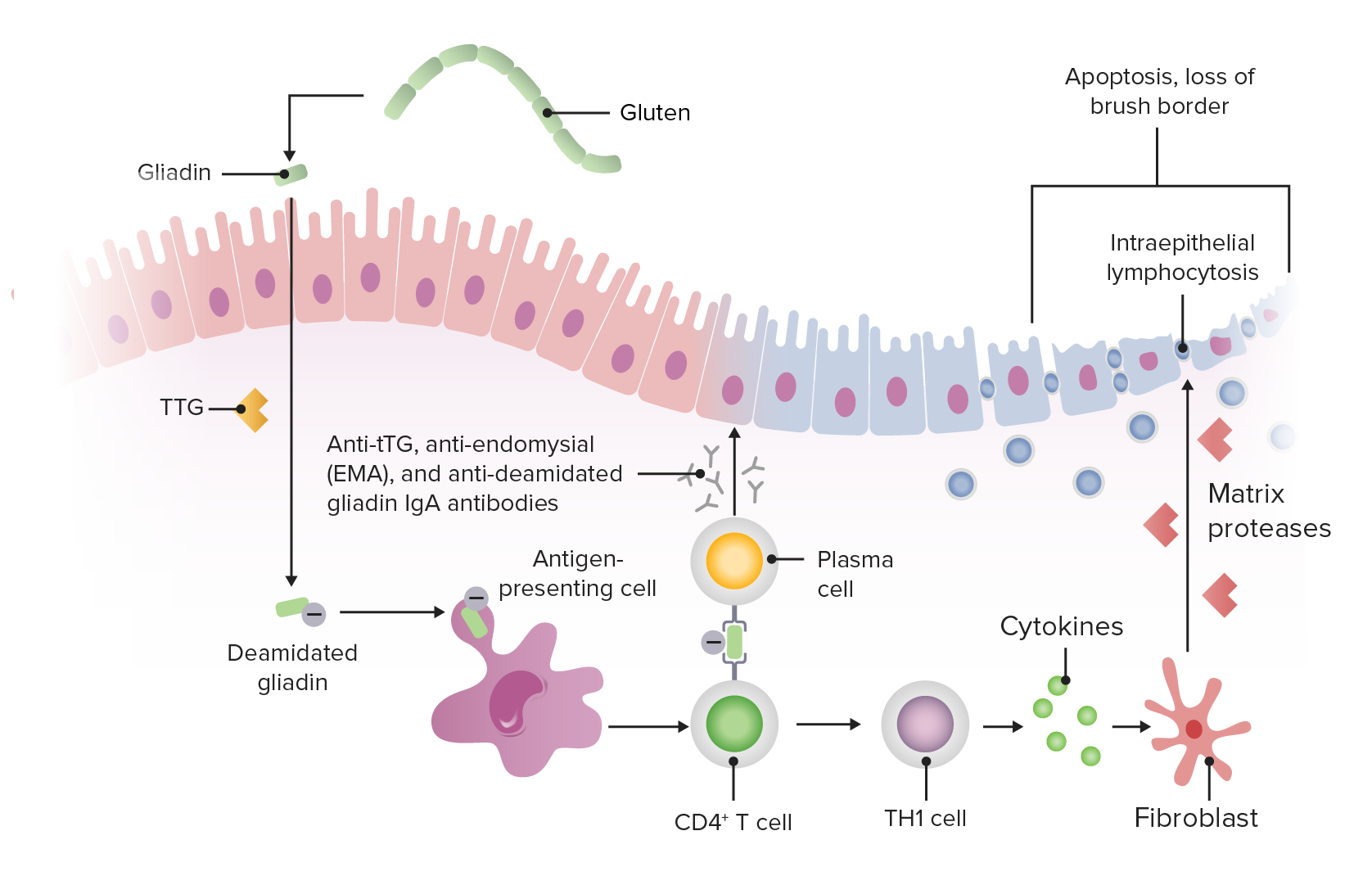
GlutenGlutenProlamins in the endosperm of seeds from the triticeae tribe which includes species of wheat; barley; and rye.Celiac Disease peptides triggerTriggerThe type of signal that initiates the inspiratory phase by the ventilatorInvasive Mechanical Ventilation the innate immune responseInnate Immune ResponseImmunity to pathogens is divided into innate and adaptive immune responses. The innate immune response is the 1st line of defense against a variety of pathogens, including bacteria, fungi, viruses, and parasites. In essentially the same form, the innate type of immunity is present in all multicellular organisms. Innate Immunity: Barriers, Complement, and Cytokines in intestinal epithelial cells, leading to T cell–mediated mucosal damage of the proximal small intestineSmall intestineThe small intestine is the longest part of the GI tract, extending from the pyloric orifice of the stomach to the ileocecal junction. The small intestine is the major organ responsible for chemical digestion and absorption of nutrients. It is divided into 3 segments: the duodenum, the jejunum, and the ileum. Small Intestine: Anatomy (distal duodenumDuodenumThe shortest and widest portion of the small intestine adjacent to the pylorus of the stomach. It is named for having the length equal to about the width of 12 fingers.Small Intestine: Anatomy and proximal jejunumJejunumThe middle portion of the small intestine, between duodenum and ileum. It represents about 2/5 of the remaining portion of the small intestine below duodenum.Small Intestine: Anatomy).[2,8]
Presence of gliadinGliadinSimple protein, one of the prolamins, derived from the gluten of wheat, rye, etc. May be separated into 4 discrete electrophoretic fractions. It is the toxic factor associated with celiac disease.Celiac Disease → release of tissue transglutaminase (tTG) (intracellular enzyme) → deaminated gliadinGliadinSimple protein, one of the prolamins, derived from the gluten of wheat, rye, etc. May be separated into 4 discrete electrophoretic fractions. It is the toxic factor associated with celiac disease.Celiac Disease
Antigen-presenting cellsAntigen-presenting cellsA heterogeneous group of immunocompetent cells that mediate the cellular immune response by processing and presenting antigens to the T-cells. Traditional antigen-presenting cells include macrophages; dendritic cells; langerhans cells; and B-lymphocytes. Follicular dendritic cells are not traditional antigen-presenting cells, but because they hold antigen on their cell surface in the form of immune complexes for b-cell recognition they are considered so by some authors.Adaptive Immune Response process and present this to T cellsT cellsLymphocytes responsible for cell-mediated immunity. Two types have been identified – cytotoxic (t-lymphocytes, cytotoxic) and helper T-lymphocytes (t-lymphocytes, helper-inducer). They are formed when lymphocytes circulate through the thymus gland and differentiate to thymocytes. When exposed to an antigen, they divide rapidly and produce large numbers of new T cells sensitized to that antigen.T cells: Types and Functions → T helper cell and plasmaPlasmaThe residual portion of blood that is left after removal of blood cells by centrifugation without prior blood coagulation.Transfusion Products cell activation
PlasmaPlasmaThe residual portion of blood that is left after removal of blood cells by centrifugation without prior blood coagulation.Transfusion Products cells release anti-tTG, anti-endomysialAnti-endomysialCeliac Disease, and anti-deamidated gliadinGliadinSimple protein, one of the prolamins, derived from the gluten of wheat, rye, etc. May be separated into 4 discrete electrophoretic fractions. It is the toxic factor associated with celiac disease.Celiac DiseaseantibodiesAntibodiesImmunoglobulins (Igs), also known as antibodies, are glycoprotein molecules produced by plasma cells that act in immune responses by recognizing and binding particular antigens. The various Ig classes are IgG (the most abundant), IgM, IgE, IgD, and IgA, which differ in their biologic features, structure, target specificity, and distribution.Immunoglobulins: Types and Functions.
T helper cells → cytokine release → intraepithelial lymphocytosisLymphocytosisWBCs develop from stem cells in the bone marrow and are called leukocytes when circulating in the bloodstream. Lymphocytes are 1 of the 5 subclasses of WBCs. Lymphocytosis is an increase in the number or proportion of the lymphocyte subclass of WBCs, often as a result of an immune response to infection (known as reactive lymphocytosis). Lymphocytosis and activation of fibroblastsFibroblastsConnective tissue cells which secrete an extracellular matrix rich in collagen and other macromolecules.Sarcoidosis → matrix proteasesProteasesProteins and Peptides → intestinal destruction, including loss of the brush borderBrush borderTubular System, crypt hyperplasiaCrypt hyperplasiaCeliac Disease, and villous atrophyVillous AtrophyGiardia/Giardiasis
This process causes impaired absorptionAbsorptionAbsorption involves the uptake of nutrient molecules and their transfer from the lumen of the GI tract across the enterocytes and into the interstitial space, where they can be taken up in the venous or lymphatic circulation.Digestion and Absorption of fat, fat-soluble vitamins, and mineralsMineralsElectrolytes → malabsorptionMalabsorptionGeneral term for a group of malnutrition syndromes caused by failure of normal intestinal absorption of nutrients.Malabsorption and Maldigestion
DiarrheaDiarrheaDiarrhea is defined as ≥ 3 watery or loose stools in a 24-hour period. There are a multitude of etiologies, which can be classified based on the underlying mechanism of disease. The duration of symptoms (acute or chronic) and characteristics of the stools (e.g., watery, bloody, steatorrheic, mucoid) can help guide further diagnostic evaluation. Diarrhea (most common symptom)
SteatorrheaSteatorrheaA condition that is characterized by chronic fatty diarrhea, a result of abnormal digestion and/or intestinal absorption of fats.Diarrhea (bulky, foul-smelling, floating stool)
FatigueFatigueThe state of weariness following a period of exertion, mental or physical, characterized by a decreased capacity for work and reduced efficiency to respond to stimuli.Fibromyalgia and weakness
Failure to thriveFailure to ThriveFailure to thrive (FTT), or faltering growth, describes suboptimal weight gain and growth in children. The majority of cases are due to inadequate caloric intake; however, genetic, infectious, and oncological etiologies are also common. Failure to Thrive in infants and children
Impaired ironIronA metallic element with atomic symbol fe, atomic number 26, and atomic weight 55. 85. It is an essential constituent of hemoglobins; cytochromes; and iron-binding proteins. It plays a role in cellular redox reactions and in the transport of oxygen.Trace Elements and folateFolateFolate and vitamin B12 are 2 of the most clinically important water-soluble vitamins. Deficiencies can present with megaloblastic anemia, GI symptoms, neuropsychiatric symptoms, and adverse pregnancy complications, including neural tube defects. Folate and Vitamin B12absorptionAbsorptionAbsorption involves the uptake of nutrient molecules and their transfer from the lumen of the GI tract across the enterocytes and into the interstitial space, where they can be taken up in the venous or lymphatic circulation.Digestion and Absorption → anemiaAnemiaAnemia is a condition in which individuals have low Hb levels, which can arise from various causes. Anemia is accompanied by a reduced number of RBCs and may manifest with fatigue, shortness of breath, pallor, and weakness. Subtypes are classified by the size of RBCs, chronicity, and etiology. Anemia: Overview and Types
Impaired vitamin KVitamin KA lipid cofactor that is required for normal blood clotting. Several forms of vitamin K have been identified: vitamin K 1 (phytomenadione) derived from plants, vitamin K 2 (menaquinone) from bacteria, and synthetic naphthoquinone provitamins, vitamin K 3 (menadione). Vitamin k 3 provitamins, after being alkylated in vivo, exhibit the antifibrinolytic activity of vitamin k. Green leafy vegetables, liver, cheese, butter, and egg yolk are good sources of vitamin k.Fat-soluble Vitamins and their Deficiencies (fat soluble) absorptionAbsorptionAbsorption involves the uptake of nutrient molecules and their transfer from the lumen of the GI tract across the enterocytes and into the interstitial space, where they can be taken up in the venous or lymphatic circulation.Digestion and Absorption → prothrombinProthrombinA plasma protein that is the inactive precursor of thrombin. It is converted to thrombin by a prothrombin activator complex consisting of factor Xa, factor V, phospholipid, and calcium ions.Hemostasis deficiency → bleeding diathesisBleeding diathesisWiskott-Aldrich Syndrome
HypocalcemiaHypocalcemiaHypocalcemia, a serum calcium < 8.5 mg/dL, can result from various conditions. The causes may include hypoparathyroidism, drugs, disorders leading to vitamin D deficiency, and more. Calcium levels are regulated and affected by different elements such as dietary intake, parathyroid hormone (PTH), vitamin D, pH, and albumin. Presentation can range from an asymptomatic (mild deficiency) to a life-threatening condition (acute, significant deficiency). Hypocalcemia and vitamin deficiencies → motorMotorNeurons which send impulses peripherally to activate muscles or secretory cells.Nervous System: Histology weakness, paresthesiasParesthesiasSubjective cutaneous sensations (e.g., cold, warmth, tingling, pressure, etc.) that are experienced spontaneously in the absence of stimulation.Posterior Cord Syndrome, sensorySensoryNeurons which conduct nerve impulses to the central nervous system.Nervous System: Histology loss
Dermatitis herpetiformisDermatitis herpetiformisRare, chronic, papulo-vesicular disease characterized by an intensely pruritic eruption consisting of various combinations of symmetrical, erythematous, papular, vesicular, or bullous lesions. The disease is strongly associated with the presence of hla-b8 and hla-dr3 antigens. A variety of different autoantibodies has been detected in small numbers in patients with dermatitis herpetiformis.Celiac Disease (pruritic papules, vesiclesVesiclesFemale Genitourinary Examination, and bullaeBullaeErythema Multiforme on the extensor surface of extremities)
Atrophic glossitis, oral mucosal lesions
Impaired amino acidAmino acidAmino acids (AAs) are composed of a central carbon atom attached to a carboxyl group, an amino group, a hydrogen atom, and a side chain (R group). Basics of Amino AcidsabsorptionAbsorptionAbsorption involves the uptake of nutrient molecules and their transfer from the lumen of the GI tract across the enterocytes and into the interstitial space, where they can be taken up in the venous or lymphatic circulation.Digestion and Absorption → peripheral edemaPeripheral edemaPeripheral edema is the swelling of the lower extremities, namely, legs, feet, and ankles.Edema
AmenorrheaAmenorrheaAbsence of menstruation.Congenital Malformations of the Female Reproductive System, delayed menarcheMenarcheThe first menstrual cycle marked by the initiation of menstruation.Menstrual Cycle, and infertilityInfertilityInfertility is the inability to conceive in the context of regular intercourse. The most common causes of infertility in women are related to ovulatory dysfunction or tubal obstruction, whereas, in men, abnormal sperm is a common cause. Infertility
BoneBoneBone is a compact type of hardened connective tissue composed of bone cells, membranes, an extracellular mineralized matrix, and central bone marrow. The 2 primary types of bone are compact and spongy. Bones: Structure and Types loss, ↑ risk of fractures caused by vitamin D deficiencyVitamin D DeficiencyA nutritional condition produced by a deficiency of vitamin D in the diet, insufficient production of vitamin D in the skin, inadequate absorption of vitamin D from the diet, or abnormal conversion of vitamin D to its bioactive metabolites. It is manifested clinically as rickets in children and osteomalacia in adults.Fat-soluble Vitamins and their Deficiencies
Table: Manifestations and laboratory findings of malabsorptionMalabsorptionGeneral term for a group of malnutrition syndromes caused by failure of normal intestinal absorption of nutrients.Malabsorption and Maldigestion syndrome
Manifestations
Laboratory finding
SteatorrheaSteatorrheaA condition that is characterized by chronic fatty diarrhea, a result of abnormal digestion and/or intestinal absorption of fats.Diarrhea (bulky, foul-smelling, light-colored stool)
Increased fecal fat due to fat malabsorptionMalabsorptionGeneral term for a group of malnutrition syndromes caused by failure of normal intestinal absorption of nutrients.Malabsorption and Maldigestion
DiarrheaDiarrheaDiarrhea is defined as ≥ 3 watery or loose stools in a 24-hour period. There are a multitude of etiologies, which can be classified based on the underlying mechanism of disease. The duration of symptoms (acute or chronic) and characteristics of the stools (e.g., watery, bloody, steatorrheic, mucoid) can help guide further diagnostic evaluation. Diarrhea (increased fecal content)
Increased stool osmolalityOsmolalityPlasma osmolality refers to the combined concentration of all solutes in the blood.Renal Sodium and Water Regulation gap due to unabsorbed fatsFatsThe glyceryl esters of a fatty acid, or of a mixture of fatty acids. They are generally odorless, colorless, and tasteless if pure, but they may be flavored according to origin. Fats are insoluble in water, soluble in most organic solvents. They occur in animal and vegetable tissue and are generally obtained by boiling or by extraction under pressure. They are important in the diet (dietary fats) as a source of energy.Energy Homeostasis and carbohydratesCarbohydratesA class of organic compounds composed of carbon, hydrogen, and oxygen in a ratio of cn(H2O)n. The largest class of organic compounds, including starch; glycogen; cellulose; polysaccharides; and simple monosaccharides.Basics of Carbohydrates
Weight lossWeight lossDecrease in existing body weight.Bariatric Surgery/failure to thriveFailure to ThriveFailure to thrive (FTT), or faltering growth, describes suboptimal weight gain and growth in children. The majority of cases are due to inadequate caloric intake; however, genetic, infectious, and oncological etiologies are also common. Failure to Thrive/muscle wastingMuscle WastingDuchenne Muscular Dystrophy
overall nutrient malabsorptionMalabsorptionGeneral term for a group of malnutrition syndromes caused by failure of normal intestinal absorption of nutrients.Malabsorption and Maldigestion (Decreased D-xylose test due to proximal small bowelSmall bowelThe small intestine is the longest part of the GI tract, extending from the pyloric orifice of the stomach to the ileocecal junction. The small intestine is the major organ responsible for chemical digestion and absorption of nutrients. It is divided into 3 segments: the duodenum, the jejunum, and the ileum.Small Intestine: Anatomy mucosal damage)
Bleeding/repeated ecchymosisEcchymosisExtravasation of blood into the skin, resulting in a nonelevated, rounded or irregular, blue or purplish patch, larger than a petechia.Orbital Fractures
Prolonged PT/INR due to inability to absorb vitamin KVitamin KA lipid cofactor that is required for normal blood clotting. Several forms of vitamin K have been identified: vitamin K 1 (phytomenadione) derived from plants, vitamin K 2 (menaquinone) from bacteria, and synthetic naphthoquinone provitamins, vitamin K 3 (menadione). Vitamin k 3 provitamins, after being alkylated in vivo, exhibit the antifibrinolytic activity of vitamin k. Green leafy vegetables, liver, cheese, butter, and egg yolk are good sources of vitamin k.Fat-soluble Vitamins and their Deficiencies
Microcytic anemiaMicrocytic anemiaConditions in which there is a generalized increase in the iron stores of body tissues, particularly of liver and the mononuclear phagocyte system, without demonstrable tissue damage. The name refers to the presence of stainable iron in the tissue in the form of hemosiderin.Anemia: Overview and Types
Low ferritinFerritinIron-containing proteins that are widely distributed in animals, plants, and microorganisms. Their major function is to store iron in a nontoxic bioavailable form. Each ferritin molecule consists of ferric iron in a hollow protein shell (apoferritins) made of 24 subunits of various sequences depending on the species and tissue types.Hereditary Hemochromatosis due to inability to absorb ironIronA metallic element with atomic symbol fe, atomic number 26, and atomic weight 55. 85. It is an essential constituent of hemoglobins; cytochromes; and iron-binding proteins. It plays a role in cellular redox reactions and in the transport of oxygen.Trace Elements
Macrocytic anemiaMacrocytic anemiaAnemia characterized by larger than normal erythrocytes, increased mean corpuscular volume (MCV) and increased mean corpuscular hemoglobin (mMCH).Anemia: Overview and Types
Low serum B12 or folic acid due to inability to absorb vitamin B12 and B9
BoneBoneBone is a compact type of hardened connective tissue composed of bone cells, membranes, an extracellular mineralized matrix, and central bone marrow. The 2 primary types of bone are compact and spongy. Bones: Structure and TypespainPainAn unpleasant sensation induced by noxious stimuli which are detected by nerve endings of nociceptive neurons.Pain: Types and Pathways/fractures on minimal trauma
OsteopeniaOsteopeniaOsteoporosis on plain film and osteoporosisOsteoporosisOsteoporosis refers to a decrease in bone mass and density leading to an increased number of fractures. There are 2 forms of osteoporosis: primary, which is commonly postmenopausal or senile; and secondary, which is a manifestation of immobilization, underlying medical disorders, or long-term use of certain medications. Osteoporosis on DEXADEXAOsteoporosis due to inability to absorb calciumCalciumA basic element found in nearly all tissues. It is a member of the alkaline earth family of metals with the atomic symbol ca, atomic number 20, and atomic weight 40. Calcium is the most abundant mineral in the body and combines with phosphorus to form calcium phosphate in the bones and teeth. It is essential for the normal functioning of nerves and muscles and plays a role in blood coagulation (as factor IV) and in many enzymatic processes.Electrolytes and vitamin DVitamin DA vitamin that includes both cholecalciferols and ergocalciferols, which have the common effect of preventing or curing rickets in animals. It can also be viewed as a hormone since it can be formed in skin by action of ultraviolet rays upon the precursors, 7-dehydrocholesterol and ergosterol, and acts on vitamin D receptors to regulate calcium in opposition to parathyroid hormone.Fat-soluble Vitamins and their Deficiencies
EdemaEdemaEdema is a condition in which excess serous fluid accumulates in the body cavity or interstitial space of connective tissues. Edema is a symptom observed in several medical conditions. It can be categorized into 2 types, namely, peripheral (in the extremities) and internal (in an organ or body cavity). Edema
Decreased serum protein and albuminAlbuminSerum albumin from humans. It is an essential carrier of both endogenous substances, such as fatty acids and bilirubin, and of xenobiotics in the blood.Liver Function Tests due to inability to absorb amino acidsAmino acidsOrganic compounds that generally contain an amino (-NH2) and a carboxyl (-COOH) group. Twenty alpha-amino acids are the subunits which are polymerized to form proteins.Basics of Amino Acids from the diet
PT/INR: prothrombin time/international normalized ratio DEXA: dual-energy X-ray absorptiometry
Dermatitis herpetiformis rash involving the extensor surface of the forearms, hands, and lower limbs in a patient with celiac disease
Image: “Skin lesions on dorsum of hand and legs” by Department of Surgery, The Aga Khan University Hospital (Stadium Road), Karachi (74800), Pakistan. License: CC BY 3.0
Associated conditions
Celiac diseaseCeliac diseaseCeliac disease (also known as celiac sprue or gluten enteropathy) is an autoimmune reaction to gliadin, which is a component of gluten. Celiac disease is closely associated with HLA-DQ2 and HLA-DQ8. The immune response is localized to the proximal small intestine and causes the characteristic histologic findings of villous atrophy, crypt hyperplasia, and intraepithelial lymphocytosis. Celiac Disease is also associated with:
DiabetesDiabetesDiabetes mellitus (DM) is a metabolic disease characterized by hyperglycemia and dysfunction of the regulation of glucose metabolism by insulin. Type 1 DM is diagnosed mostly in children and young adults as the result of autoimmune destruction of β cells in the pancreas and the resulting lack of insulin. Type 2 DM has a significant association with obesity and is characterized by insulin resistance.Diabetes Mellitus mellitus (type 1Type 1Spinal Muscular Atrophy)
Selective immunoglobulin A (IgAIgARepresents 15-20% of the human serum immunoglobulins, mostly as the 4-chain polymer in humans or dimer in other mammals. Secretory iga is the main immunoglobulin in secretions.Immunoglobulins: Types and Functions) deficiency
Autoimmune thyroidThyroidThe thyroid gland is one of the largest endocrine glands in the human body. The thyroid gland is a highly vascular, brownish-red gland located in the visceral compartment of the anterior region of the neck.Thyroid Gland: Anatomy diseases
Gastroesophageal reflux diseaseGastroesophageal Reflux DiseaseGastroesophageal reflux disease (GERD) occurs when the stomach acid frequently flows back into the esophagus. This backwash (acid reflux) can irritate the lining of the esophagus, causing symptoms such as retrosternal burning pain (heartburn). Gastroesophageal Reflux Disease (GERD)
Inflammatory bowel disease and microscopic colitisColitisInflammation of the colon section of the large intestine, usually with symptoms such as diarrhea (often with blood and mucus), abdominal pain, and fever.Pseudomembranous Colitis
IdiopathicIdiopathicDermatomyositisdilated cardiomyopathyDilated CardiomyopathyDilated cardiomyopathy (DCM) is the most common type of non-ischemic cardiomyopathy and a common cause of heart failure (HF). The cause may be idiopathic, familial, or secondary to a variety of underlying conditions. The disease is characterized by the enlargement of 1 or both ventricles and reduced systolic function. Dilated Cardiomyopathy
Diagnosis generally involves a combination of serologic testing and biopsyBiopsyRemoval and pathologic examination of specimens from the living body.Ewing Sarcoma in the setting of suggestive history and symptoms. Specialist referral (e.g., gastroenterology) is necessary to help confirm the diagnosis.
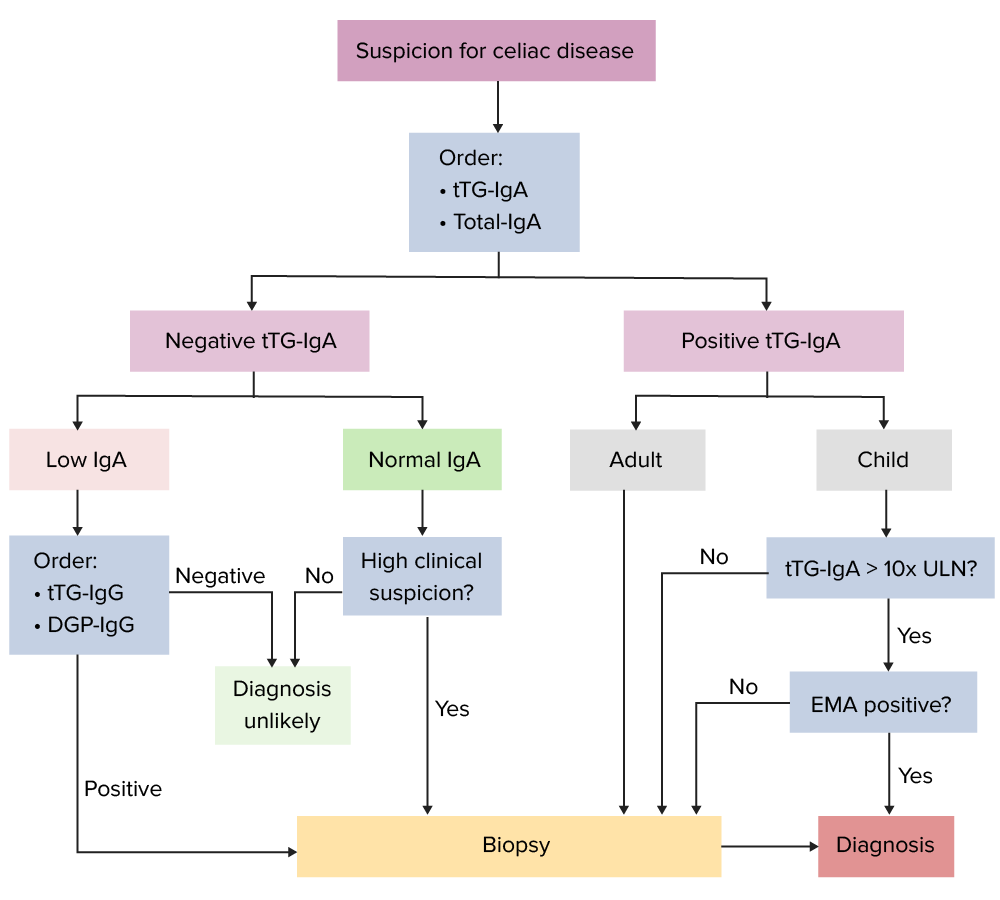
Simplified diagnostic algorithm for celiac disease:[4,6,12] Note: Serology-only diagnosis in children should be agreed upon by the family. Additionally, this is a potential option for adults who are unable or unwilling to undergo endoscopy and biopsy. This approach is currently not preferred in the UK guidelines. DGP: deamidated gliadin peptide EMA: anti-endomysial antibody tTG: tissue transglutaminase ULN: upper limit of normal Image by Lecturio.
Serologic testing
Tests:[4–6,12]
Tissue transglutaminase (tTG)-IgA antibody:
Single preferred test for diagnosis (96% sensitivity and specificitySensitivity and SpecificityBinary classification measures to assess test results. Sensitivity or recall rate is the proportion of true positives. Specificity is the probability of correctly determining the absence of a condition.Epidemiological Values of Diagnostic Tests)
Not accurate if the patient is on a gluten-free diet
Total IgAIgARepresents 15-20% of the human serum immunoglobulins, mostly as the 4-chain polymer in humans or dimer in other mammals. Secretory iga is the main immunoglobulin in secretions.Immunoglobulins: Types and Functions also recommended:
Rules out IgA deficiencyIgA deficiencyA dysgammaglobulinemia characterized by a deficiency of immunoglobulin a.Selective IgA Deficiency, which affects the accuracy of the above tests
If IgA-deficient → check IgG-deamidated gliadinGliadinSimple protein, one of the prolamins, derived from the gluten of wheat, rye, etc. May be separated into 4 discrete electrophoretic fractions. It is the toxic factor associated with celiac disease.Celiac Disease peptide (DGP-IgG) and tTG-IgG
Note: IgGIgGThe major immunoglobulin isotype class in normal human serum. There are several isotype subclasses of igg, for example, igg1, igg2a, and igg2b.Hypersensitivity Pneumonitis isotype testing is not specific in the absence of IgA deficiencyIgA deficiencyA dysgammaglobulinemia characterized by a deficiency of immunoglobulin a.Selective IgA Deficiency.
Other antibodiesAntibodiesImmunoglobulins (Igs), also known as antibodies, are glycoprotein molecules produced by plasma cells that act in immune responses by recognizing and binding particular antigens. The various Ig classes are IgG (the most abundant), IgM, IgE, IgD, and IgA, which differ in their biologic features, structure, target specificity, and distribution.Immunoglobulins: Types and Functions:
IgAIgARepresents 15-20% of the human serum immunoglobulins, mostly as the 4-chain polymer in humans or dimer in other mammals. Secretory iga is the main immunoglobulin in secretions.Immunoglobulins: Types and Functionsanti-endomysialAnti-endomysialCeliac Disease antibody (EMA)
IgAIgARepresents 15-20% of the human serum immunoglobulins, mostly as the 4-chain polymer in humans or dimer in other mammals. Secretory iga is the main immunoglobulin in secretions.Immunoglobulins: Types and Functions deamidated gliadinGliadinSimple protein, one of the prolamins, derived from the gluten of wheat, rye, etc. May be separated into 4 discrete electrophoretic fractions. It is the toxic factor associated with celiac disease.Celiac Disease peptide (DGP): almost as sensitive as tTG but detects antibodiesAntibodiesImmunoglobulins (Igs), also known as antibodies, are glycoprotein molecules produced by plasma cells that act in immune responses by recognizing and binding particular antigens. The various Ig classes are IgG (the most abundant), IgM, IgE, IgD, and IgA, which differ in their biologic features, structure, target specificity, and distribution.Immunoglobulins: Types and Functions to gliadinGliadinSimple protein, one of the prolamins, derived from the gluten of wheat, rye, etc. May be separated into 4 discrete electrophoretic fractions. It is the toxic factor associated with celiac disease.Celiac Disease instead of autoantibodiesAutoantibodiesAntibodies that react with self-antigens (autoantigens) of the organism that produced them.Blotting Techniques
Additional recommendations for serologic testing:[4–6,12]
Serologic tests are obtained with patientsPatientsIndividuals participating in the health care system for the purpose of receiving therapeutic, diagnostic, or preventive procedures.Clinician–Patient Relationship on a gluten-containing diet (typically equivalent to ≥1 slice of bread daily for 6–8 weeks).
In children < 2 years of age:
There has been concern that tTG and EMA antibodiesAntibodiesImmunoglobulins (Igs), also known as antibodies, are glycoprotein molecules produced by plasma cells that act in immune responses by recognizing and binding particular antigens. The various Ig classes are IgG (the most abundant), IgM, IgE, IgD, and IgA, which differ in their biologic features, structure, target specificity, and distribution.Immunoglobulins: Types and Functions may be less sensitive, but recent guidelines still indicate tTG-IgA as the initial test of choice (for children with sufficient IgAIgARepresents 15-20% of the human serum immunoglobulins, mostly as the 4-chain polymer in humans or dimer in other mammals. Secretory iga is the main immunoglobulin in secretions.Immunoglobulins: Types and Functions).
IF tTG-IgA is negative, DGP-IgG may be added in children <2 with high clinical suspicion.
For those with negative serologySerologyThe study of serum, especially of antigen-antibody reactions in vitro.Yellow Fever Virus but high likelihood of celiac diseaseCeliac diseaseCeliac disease (also known as celiac sprue or gluten enteropathy) is an autoimmune reaction to gliadin, which is a component of gluten. Celiac disease is closely associated with HLA-DQ2 and HLA-DQ8. The immune response is localized to the proximal small intestine and causes the characteristic histologic findings of villous atrophy, crypt hyperplasia, and intraepithelial lymphocytosis. Celiac Disease (e.g., type 1Type 1Spinal Muscular AtrophydiabetesDiabetesDiabetes mellitus (DM) is a metabolic disease characterized by hyperglycemia and dysfunction of the regulation of glucose metabolism by insulin. Type 1 DM is diagnosed mostly in children and young adults as the result of autoimmune destruction of β cells in the pancreas and the resulting lack of insulin. Type 2 DM has a significant association with obesity and is characterized by insulin resistance.Diabetes Mellitus, Down syndromeDown syndromeDown syndrome, or trisomy 21, is the most common chromosomal aberration and the most frequent genetic cause of developmental delay. Both boys and girls are affected and have characteristic craniofacial and musculoskeletal features, as well as multiple medical anomalies involving the cardiac, gastrointestinal, ocular, and auditory systems.Down syndrome (Trisomy 21), family historyFamily HistoryAdult Health Maintenance), perform duodenal biopsyBiopsyRemoval and pathologic examination of specimens from the living body.Ewing Sarcoma.
In children (and symptomatic patientsPatientsIndividuals participating in the health care system for the purpose of receiving therapeutic, diagnostic, or preventive procedures.Clinician–Patient Relationship who cannot, or choose not to, undergo biopsyBiopsyRemoval and pathologic examination of specimens from the living body.Ewing Sarcoma confirmation), a diagnosis can be made with both:
tTG > 10 times the upper limitLimitA value (e.g., pressure or time) that should not be exceeded and which is specified by the operator to protect the lungInvasive Mechanical Ventilation of normal
Positive EMA from a second blood sample
EndoscopyEndoscopyProcedures of applying endoscopes for disease diagnosis and treatment. Endoscopy involves passing an optical instrument through a small incision in the skin i.e., percutaneous; or through a natural orifice and along natural body pathways such as the digestive tract; and/or through an incision in the wall of a tubular structure or organ, i.e. Transluminal, to examine or perform surgery on the interior parts of the body.Gastroesophageal Reflux Disease (GERD) and duodenal biopsyBiopsyRemoval and pathologic examination of specimens from the living body.Ewing Sarcoma
EndoscopyEndoscopyProcedures of applying endoscopes for disease diagnosis and treatment. Endoscopy involves passing an optical instrument through a small incision in the skin i.e., percutaneous; or through a natural orifice and along natural body pathways such as the digestive tract; and/or through an incision in the wall of a tubular structure or organ, i.e. Transluminal, to examine or perform surgery on the interior parts of the body.Gastroesophageal Reflux Disease (GERD) and biopsies are recommended to confirm the diagnosis in children and adults.
If serologic testing is positive → proceed with upper endoscopyEndoscopyProcedures of applying endoscopes for disease diagnosis and treatment. Endoscopy involves passing an optical instrument through a small incision in the skin i.e., percutaneous; or through a natural orifice and along natural body pathways such as the digestive tract; and/or through an incision in the wall of a tubular structure or organ, i.e. Transluminal, to examine or perform surgery on the interior parts of the body.Gastroesophageal Reflux Disease (GERD) with duodenal biopsyBiopsyRemoval and pathologic examination of specimens from the living body.Ewing Sarcoma:[4–6,12]
Because histologic changes may be patchy, multiple biopsies are performed, requiring:
1 or 2 biopsies of the duodenal bulb
≥ 4 biopsies of the distal duodenumDuodenumThe shortest and widest portion of the small intestine adjacent to the pylorus of the stomach. It is named for having the length equal to about the width of 12 fingers.Small Intestine: Anatomy
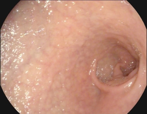
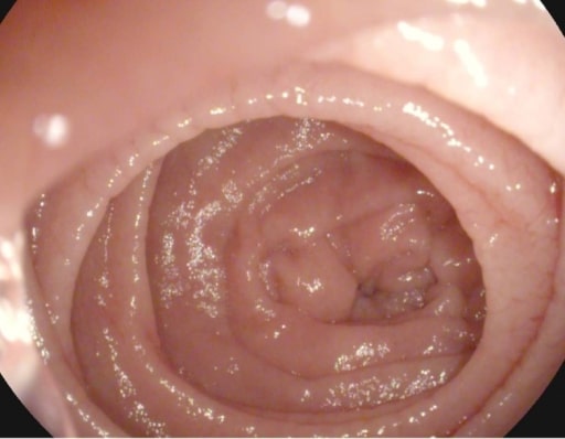
Endoscopic features:
Atrophic mucosa
Fissures
Scalloping
Submucosal vascularity
Histology:
Increased intraepithelial lymphocytesIntraepithelial lymphocytesT lymphocytes with limited diversity of receptors (e.g., alpha e integrins) in the epidermis of the skin and the mucosal linings. They recognize common microbes via t-cell receptors and pathogen-associated molecular pattern molecules and function as effector cells for innate immunity. Activation of intraepithelial lymphocytes is a marker for various gastrointestinal diseases (e.g., celiac disease; hairy cell leukemia; and enteropathy-associated t-cell lymphoma).Adaptive Cell-mediated Immunity
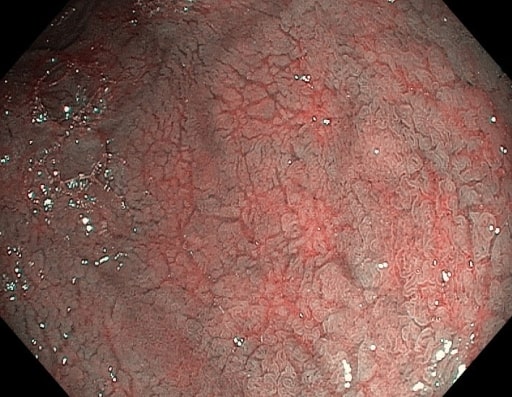
Mucosal atrophy and submucosal vascularity seen on endoscopy in a patient with celiac disease
Image: “Atrophy with visible vessel pattern in the duodenal bulb” by “Dr. Carol Davila” Central Military University Emergency Hospital, Bucharest, Romania ; “Carol Davila” University of Medicine and Pharmacy, Bucharest, Romania. License: CC BY 2.0
Scalloping of the Kerckring folds in a patient with celiac disease
Image: “Atrophy with visible vessel pattern in the duodenal bulb” by “Dr. Carol Davila” Central Military University Emergency Hospital, Bucharest, Romania; “Carol Davila” University of Medicine and Pharmacy, Bucharest, Romania. License: CC BY 2.0
Mucosal fissures and prominent submucosal vessels seen on endoscopy in a patient with celiac disease
Image: “NBI” by “Dr. Carol Davila” Central Military University Emergency Hospital, Bucharest, Romania. License: CC BY 2.0
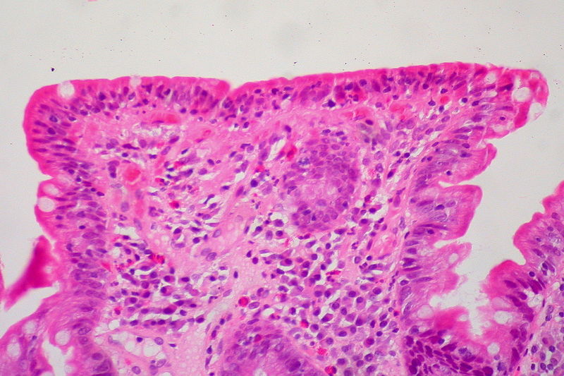
Small intestine biopsy showing crowding of lymphocytes (blue cells), loss of villi, and deepening (hyperplasia) of crypts
Image: “Celiac Sprue, Small Bowel Biopsy” by Ed Uthman from Houston, TX, USA. License: CC BY 2.0
Histologic classification: Marsh score[13,14]
The Marsh score categorizes histological findings in small-intestinal biopsies of celiac diseaseCeliac diseaseCeliac disease (also known as celiac sprue or gluten enteropathy) is an autoimmune reaction to gliadin, which is a component of gluten. Celiac disease is closely associated with HLA-DQ2 and HLA-DQ8. The immune response is localized to the proximal small intestine and causes the characteristic histologic findings of villous atrophy, crypt hyperplasia, and intraepithelial lymphocytosis. Celiac DiseasepatientsPatientsIndividuals participating in the health care system for the purpose of receiving therapeutic, diagnostic, or preventive procedures.Clinician–Patient Relationship, standardizing the assessment of mucosal damage, and aiding in accurate diagnosis and monitoring of disease progression.
Marsh 0: Normal mucosa
No visible abnormalities.
Typical for individuals without celiac diseaseCeliac diseaseCeliac disease (also known as celiac sprue or gluten enteropathy) is an autoimmune reaction to gliadin, which is a component of gluten. Celiac disease is closely associated with HLA-DQ2 and HLA-DQ8. The immune response is localized to the proximal small intestine and causes the characteristic histologic findings of villous atrophy, crypt hyperplasia, and intraepithelial lymphocytosis. Celiac Disease or those on a strict gluten-free diet.
Marsh 1: Infiltrative lesion
Increased intraepithelial lymphocytesIntraepithelial lymphocytesT lymphocytes with limited diversity of receptors (e.g., alpha e integrins) in the epidermis of the skin and the mucosal linings. They recognize common microbes via t-cell receptors and pathogen-associated molecular pattern molecules and function as effector cells for innate immunity. Activation of intraepithelial lymphocytes is a marker for various gastrointestinal diseases (e.g., celiac disease; hairy cell leukemia; and enteropathy-associated t-cell lymphoma).Adaptive Cell-mediated Immunity (IELs): more than 25 IELs per 100 enterocytes.
Normal villous architecture: no visible atrophyAtrophyDecrease in the size of a cell, tissue, organ, or multiple organs, associated with a variety of pathological conditions such as abnormal cellular changes, ischemia, malnutrition, or hormonal changes.Cellular Adaptation or structural changes.
Normal villous architecture: no visible atrophyAtrophyDecrease in the size of a cell, tissue, organ, or multiple organs, associated with a variety of pathological conditions such as abnormal cellular changes, ischemia, malnutrition, or hormonal changes.Cellular Adaptation or structural changes.
If serologySerologyThe study of serum, especially of antigen-antibody reactions in vitro.Yellow Fever Virus and biopsyBiopsyRemoval and pathologic examination of specimens from the living body.Ewing Sarcoma results are discordant:[4,6]
Positive serologySerologyThe study of serum, especially of antigen-antibody reactions in vitro.Yellow Fever Virus and nondiagnostic biopsyBiopsyRemoval and pathologic examination of specimens from the living body.Ewing Sarcoma → perform HLA-DQ2/DQ8 typing
If negative, celiac diseaseCeliac diseaseCeliac disease (also known as celiac sprue or gluten enteropathy) is an autoimmune reaction to gliadin, which is a component of gluten. Celiac disease is closely associated with HLA-DQ2 and HLA-DQ8. The immune response is localized to the proximal small intestine and causes the characteristic histologic findings of villous atrophy, crypt hyperplasia, and intraepithelial lymphocytosis. Celiac Disease is excluded and an alternative diagnosis should be explored.
If positive, a high-gluten diet (glutenGlutenProlamins in the endosperm of seeds from the triticeae tribe which includes species of wheat; barley; and rye.Celiac Disease challenge: > 10 g/ day, or ≥ 4 slices of gluten-containing bread per day) is recommended. Repeat biopsyBiopsyRemoval and pathologic examination of specimens from the living body.Ewing Sarcoma (at multiple sites) in 6–12 weeks.
Negative serologySerologyThe study of serum, especially of antigen-antibody reactions in vitro.Yellow Fever Virus and abnormal biopsyBiopsyRemoval and pathologic examination of specimens from the living body.Ewing Sarcoma → perform HLA-DQ2/DQ8 typing
If negative, likely not celiac diseaseCeliac diseaseCeliac disease (also known as celiac sprue or gluten enteropathy) is an autoimmune reaction to gliadin, which is a component of gluten. Celiac disease is closely associated with HLA-DQ2 and HLA-DQ8. The immune response is localized to the proximal small intestine and causes the characteristic histologic findings of villous atrophy, crypt hyperplasia, and intraepithelial lymphocytosis. Celiac Disease.
If positive, place patient on a gluten-free diet for 12–24 months, and repeat biopsyBiopsyRemoval and pathologic examination of specimens from the living body.Ewing Sarcoma afterward. Mucosal healing is seen in celiac diseaseCeliac diseaseCeliac disease (also known as celiac sprue or gluten enteropathy) is an autoimmune reaction to gliadin, which is a component of gluten. Celiac disease is closely associated with HLA-DQ2 and HLA-DQ8. The immune response is localized to the proximal small intestine and causes the characteristic histologic findings of villous atrophy, crypt hyperplasia, and intraepithelial lymphocytosis. Celiac Disease.
Evaluation of malabsorptionMalabsorptionGeneral term for a group of malnutrition syndromes caused by failure of normal intestinal absorption of nutrients.Malabsorption and Maldigestion[4,9,12]
ElectrolytesElectrolytesElectrolytes are mineral salts that dissolve in water and dissociate into charged particles called ions, which can be either be positively (cations) or negatively (anions) charged. Electrolytes are distributed in the extracellular and intracellular compartments in different concentrations. Electrolytes are essential for various basic life-sustaining functions.Electrolytes
HypokalemiaHypokalemiaHypokalemia is defined as plasma potassium (K+) concentration < 3.5 mEq/L. Homeostatic mechanisms maintain plasma concentration between 3.5-5.2 mEq/L despite marked variation in dietary intake. Hypokalemia can be due to renal losses, GI losses, transcellular shifts, or poor dietary intake.Hypokalemia
HypocalcemiaHypocalcemiaHypocalcemia, a serum calcium < 8.5 mg/dL, can result from various conditions. The causes may include hypoparathyroidism, drugs, disorders leading to vitamin D deficiency, and more. Calcium levels are regulated and affected by different elements such as dietary intake, parathyroid hormone (PTH), vitamin D, pH, and albumin. Presentation can range from an asymptomatic (mild deficiency) to a life-threatening condition (acute, significant deficiency). Hypocalcemia
HypomagnesemiaHypomagnesemiaA nutritional condition produced by a deficiency of magnesium in the diet, characterized by anorexia, nausea, vomiting, lethargy, and weakness. Symptoms are paresthesias, muscle cramps, irritability, decreased attention span, and mental confusion, possibly requiring months to appear. Deficiency of body magnesium can exist even when serum values are normal. In addition, magnesium deficiency may be organ-selective, since certain tissues become deficient before others. Electrolytes
↓ AlbuminAlbuminSerum albumin from humans. It is an essential carrier of both endogenous substances, such as fatty acids and bilirubin, and of xenobiotics in the blood.Liver Function Tests
↓ CholesterolCholesterolThe principal sterol of all higher animals, distributed in body tissues, especially the brain and spinal cord, and in animal fats and oils.Cholesterol Metabolism
↓ Hemoglobin
↑ PT/INR
↓ IronIronA metallic element with atomic symbol fe, atomic number 26, and atomic weight 55. 85. It is an essential constituent of hemoglobins; cytochromes; and iron-binding proteins. It plays a role in cellular redox reactions and in the transport of oxygen.Trace Elements, ferritinFerritinIron-containing proteins that are widely distributed in animals, plants, and microorganisms. Their major function is to store iron in a nontoxic bioavailable form. Each ferritin molecule consists of ferric iron in a hollow protein shell (apoferritins) made of 24 subunits of various sequences depending on the species and tissue types.Hereditary Hemochromatosis
↓ Vitamin B12
↓ FolateFolateFolate and vitamin B12 are 2 of the most clinically important water-soluble vitamins. Deficiencies can present with megaloblastic anemia, GI symptoms, neuropsychiatric symptoms, and adverse pregnancy complications, including neural tube defects. Folate and Vitamin B12
↑ Fecal fat
Management and Prognosis
US guidelines:
AGA Clinical Practice Update on Diagnosis and Monitoring of Celiac DiseaseCeliac diseaseCeliac disease (also known as celiac sprue or gluten enteropathy) is an autoimmune reaction to gliadin, which is a component of gluten. Celiac disease is closely associated with HLA-DQ2 and HLA-DQ8. The immune response is localized to the proximal small intestine and causes the characteristic histologic findings of villous atrophy, crypt hyperplasia, and intraepithelial lymphocytosis. Celiac Disease[3]
ACG clinical guidelines: diagnosis and management of celiac diseaseCeliac diseaseCeliac disease (also known as celiac sprue or gluten enteropathy) is an autoimmune reaction to gliadin, which is a component of gluten. Celiac disease is closely associated with HLA-DQ2 and HLA-DQ8. The immune response is localized to the proximal small intestine and causes the characteristic histologic findings of villous atrophy, crypt hyperplasia, and intraepithelial lymphocytosis. Celiac Disease[4]
UK guidelines:
National Institute for Health and Care Excellence: Coeliac disease[5]
British Society of Gastroenterology (BSG) guidelines on the diagnosis and management of adult coeliac disease[6]
Management[3–6,12]
Education about the disease
The mainstay of treatment is a lifelong gluten-free diet.
Consultation with a dietitian is recommended.
Avoid barley, rye, and wheat.
Clinical improvement is seen in most patientsPatientsIndividuals participating in the health care system for the purpose of receiving therapeutic, diagnostic, or preventive procedures.Clinician–Patient Relationship within 2 weeks.
The most common reason for treatment failure is incomplete removal of glutenGlutenProlamins in the endosperm of seeds from the triticeae tribe which includes species of wheat; barley; and rye.Celiac Disease from the diet, either because of poor adherence or inadvertent glutenGlutenProlamins in the endosperm of seeds from the triticeae tribe which includes species of wheat; barley; and rye.Celiac Disease ingestion in:
Many individuals with celiac diseaseCeliac diseaseCeliac disease (also known as celiac sprue or gluten enteropathy) is an autoimmune reaction to gliadin, which is a component of gluten. Celiac disease is closely associated with HLA-DQ2 and HLA-DQ8. The immune response is localized to the proximal small intestine and causes the characteristic histologic findings of villous atrophy, crypt hyperplasia, and intraepithelial lymphocytosis. Celiac Disease also have secondary lactose intoleranceLactose intoleranceLactose intolerance (LI) describes a constellation of symptoms due to lactase deficiency (LD), the enzyme located in the brush border of the absorptive cells in the small intestine. Lactose is the disaccharide present in milk and requires hydrolysis by lactase to break it down into its 2 absorbable constituents, glucose and galactose. Lactose intolerance typically presents with bloating, abdominal cramping, diarrhea, and flatulence. Lactose Intolerance.
ConstipationConstipationConstipation is common and may be due to a variety of causes. Constipation is generally defined as bowel movement frequency < 3 times per week. Patients who are constipated often strain to pass hard stools. The condition is classified as primary (also known as idiopathic or functional constipation) or secondary, and as acute or chronic. Constipation due to diet changes can be treated with fiber supplements.
Additional management:
Evaluate for boneBoneBone is a compact type of hardened connective tissue composed of bone cells, membranes, an extracellular mineralized matrix, and central bone marrow. The 2 primary types of bone are compact and spongy. Bones: Structure and Types loss:
Related to secondary hyperparathyroidismSecondary hyperparathyroidismAbnormally elevated parathyroid hormone secretion as a response to hypocalcemia. It is caused by chronic kidney failure or other abnormalities in the controls of bone and mineral metabolism, leading to various bone diseases, such as renal osteodystrophy.Hyperparathyroidism due to vitamin D deficiencyVitamin D DeficiencyA nutritional condition produced by a deficiency of vitamin D in the diet, insufficient production of vitamin D in the skin, inadequate absorption of vitamin D from the diet, or abnormal conversion of vitamin D to its bioactive metabolites. It is manifested clinically as rickets in children and osteomalacia in adults.Fat-soluble Vitamins and their Deficiencies
Pneumococcal vaccinationVaccinationVaccination is the administration of a substance to induce the immune system to develop protection against a disease. Unlike passive immunization, which involves the administration of pre-performed antibodies, active immunization constitutes the administration of a vaccine to stimulate the body to produce its own antibodies.Vaccination: recommended owing to hyposplenismHyposplenismAsplenia associated with celiac diseaseCeliac diseaseCeliac disease (also known as celiac sprue or gluten enteropathy) is an autoimmune reaction to gliadin, which is a component of gluten. Celiac disease is closely associated with HLA-DQ2 and HLA-DQ8. The immune response is localized to the proximal small intestine and causes the characteristic histologic findings of villous atrophy, crypt hyperplasia, and intraepithelial lymphocytosis. Celiac Disease
Screen 1st-degree relatives: Consider serologic testing, especially if symptoms are present.
Investigational agents: transglutaminase 2 inhibitors are being studied.
Monitoring response to therapy[3–6,12]
Repeat IgAIgARepresents 15-20% of the human serum immunoglobulins, mostly as the 4-chain polymer in humans or dimer in other mammals. Secretory iga is the main immunoglobulin in secretions.Immunoglobulins: Types and Functions anti-tTG antibody and IgAIgARepresents 15-20% of the human serum immunoglobulins, mostly as the 4-chain polymer in humans or dimer in other mammals. Secretory iga is the main immunoglobulin in secretions.Immunoglobulins: Types and Functions (IgGIgGThe major immunoglobulin isotype class in normal human serum. There are several isotype subclasses of igg, for example, igg1, igg2a, and igg2b.Hypersensitivity Pneumonitis) DGP 3–6 months after diagnosis, then every 6–12 months.
↓ Levels are expected
If levels do not decline, suspect glutenGlutenProlamins in the endosperm of seeds from the triticeae tribe which includes species of wheat; barley; and rye.Celiac Disease ingestion.
Duodenal biopsyBiopsyRemoval and pathologic examination of specimens from the living body.Ewing Sarcoma after 2 years on a gluten-free diet:
Evaluates mucosal healing
With dietary complianceComplianceDistensibility measure of a chamber such as the lungs (lung compliance) or bladder. Compliance is expressed as a change in volume per unit change in pressure.Veins: Histology, most adults reach complete mucosal healing.
Identify and treat any nutritional deficiencies.
Vitamins A, D, and E
B vitamins—thiamineThiamineAlso known as thiamine or thiamin, it is a vitamin C12H17N4OSCl of the vitamin B complex that is essential to normal metabolism and nerve function and is widespread in plants and animalsWater-soluble Vitamins and their Deficiencies (B1), folic acid (B9), B6, and B12
CopperCopperA heavy metal trace element with the atomic symbol cu, atomic number 29, and atomic weight 63. 55.Trace Elements, zincZincA metallic element of atomic number 30 and atomic weight 65. 38. It is a necessary trace element in the diet, forming an essential part of many enzymes, and playing an important role in protein synthesis and in cell division. Zinc deficiency is associated with anemia, short stature, hypogonadism, impaired wound healing, and geophagia. It is known by the symbol zn.Trace Elements, ironIronA metallic element with atomic symbol fe, atomic number 26, and atomic weight 55. 85. It is an essential constituent of hemoglobins; cytochromes; and iron-binding proteins. It plays a role in cellular redox reactions and in the transport of oxygen.Trace Elements, magnesiumMagnesiumA metallic element that has the atomic symbol mg, atomic number 12, and atomic weight 24. 31. It is important for the activity of many enzymes, especially those involved in oxidative phosphorylation.Electrolytes, and seleniumSeleniumAn element with the atomic symbol se, atomic number 34, and atomic weight 78. 97. It is an essential micronutrient for mammals and other animals but is toxic in large amounts. Selenium protects intracellular structures against oxidative damage. It is an essential component of glutathione peroxidase.Trace Elements
PT/INR (to evaluate vitamin KVitamin KA lipid cofactor that is required for normal blood clotting. Several forms of vitamin K have been identified: vitamin K 1 (phytomenadione) derived from plants, vitamin K 2 (menaquinone) from bacteria, and synthetic naphthoquinone provitamins, vitamin K 3 (menadione). Vitamin k 3 provitamins, after being alkylated in vivo, exhibit the antifibrinolytic activity of vitamin k. Green leafy vegetables, liver, cheese, butter, and egg yolk are good sources of vitamin k.Fat-soluble Vitamins and their Deficiencies)
6–12 month follow-up for those exhibiting the expected response (to gluten-free diet), which includes:
Improvement of symptoms
SeroconversionSeroconversionThe appearance of antibodies against causative agents in the blood of individuals during the course of an infection or following immunization.HIV Infection and AIDS
Histologic improvement
Correction of nutritional deficiencies
Nonresponsive celiac diseaseCeliac diseaseCeliac disease (also known as celiac sprue or gluten enteropathy) is an autoimmune reaction to gliadin, which is a component of gluten. Celiac disease is closely associated with HLA-DQ2 and HLA-DQ8. The immune response is localized to the proximal small intestine and causes the characteristic histologic findings of villous atrophy, crypt hyperplasia, and intraepithelial lymphocytosis. Celiac Disease[4–6]
Celiac diseaseCeliac diseaseCeliac disease (also known as celiac sprue or gluten enteropathy) is an autoimmune reaction to gliadin, which is a component of gluten. Celiac disease is closely associated with HLA-DQ2 and HLA-DQ8. The immune response is localized to the proximal small intestine and causes the characteristic histologic findings of villous atrophy, crypt hyperplasia, and intraepithelial lymphocytosis. Celiac Disease with persistent signs and symptoms, or laboratory test results consistent with the disease in spite of adhering to gluten-free diet for 6–12 months
A small percentage of individuals do not respond to a gluten-free diet or relapseRelapseRelapsing Fever after responding.
Approach:
Confirm that the patient is not inadvertently ingesting glutenGlutenProlamins in the endosperm of seeds from the triticeae tribe which includes species of wheat; barley; and rye.Celiac Disease; requires a thorough evaluation by the dietitian.
Review and confirm diagnosis with supporting evidence.
Small bowelSmall bowelThe small intestine is the longest part of the GI tract, extending from the pyloric orifice of the stomach to the ileocecal junction. The small intestine is the major organ responsible for chemical digestion and absorption of nutrients. It is divided into 3 segments: the duodenum, the jejunum, and the ileum.Small Intestine: AnatomybiopsyBiopsyRemoval and pathologic examination of specimens from the living body.Ewing Sarcoma findings and serologySerologyThe study of serum, especially of antigen-antibody reactions in vitro.Yellow Fever Virus results
Presence of HLA-DQ2 or HLA-DQ8
Biopsy-proven dermatitis herpetiformisDermatitis herpetiformisRare, chronic, papulo-vesicular disease characterized by an intensely pruritic eruption consisting of various combinations of symmetrical, erythematous, papular, vesicular, or bullous lesions. The disease is strongly associated with the presence of hla-b8 and hla-dr3 antigens. A variety of different autoantibodies has been detected in small numbers in patients with dermatitis herpetiformis.Celiac Disease
Clinical or histologic response to gluten-free diet
Strong family historyFamily HistoryAdult Health Maintenance of celiac diseaseCeliac diseaseCeliac disease (also known as celiac sprue or gluten enteropathy) is an autoimmune reaction to gliadin, which is a component of gluten. Celiac disease is closely associated with HLA-DQ2 and HLA-DQ8. The immune response is localized to the proximal small intestine and causes the characteristic histologic findings of villous atrophy, crypt hyperplasia, and intraepithelial lymphocytosis. Celiac Disease
Repeat endoscopic biopsyBiopsyRemoval and pathologic examination of specimens from the living body.Ewing Sarcoma. Results may show:
HypogammaglobulinemiaHypogammaglobulinemiaSelective IgA Deficiency and combined variableVariableVariables represent information about something that can change. The design of the measurement scales, or of the methods for obtaining information, will determine the data gathered and the characteristics of that data. As a result, a variable can be qualitative or quantitative, and may be further classified into subgroups.Types of VariablesimmunodeficiencyImmunodeficiencyChédiak-Higashi Syndrome (CVIDCVIDCommon variable immune deficiency (CVID), also known as humoral immunodeficiency, is a disorder of the immune system characterized by reduced serum levels of immunoglobulins g, a, and m.Common Variable Immunodeficiency (CVID))
Consider an alternative or concurrent disease, such as:
Irritable bowel syndromeIrritable bowel syndromeIrritable bowel syndrome (IBS) is a functional bowel disease characterized by chronic abdominal pain and altered bowel habits without an identifiable organic cause. The etiology and pathophysiology of this disease are not well understood, and there are many factors that may contribute. Irritable Bowel Syndrome
Small bowelSmall bowelThe small intestine is the longest part of the GI tract, extending from the pyloric orifice of the stomach to the ileocecal junction. The small intestine is the major organ responsible for chemical digestion and absorption of nutrients. It is divided into 3 segments: the duodenum, the jejunum, and the ileum.Small Intestine: Anatomybacterial overgrowthBacterial overgrowthLactose Intolerance (may or may not have villous atrophyVillous AtrophyGiardia/Giardiasis)
Pancreatic insufficiency
Food intolerances
Microscopic colitisColitisInflammation of the colon section of the large intestine, usually with symptoms such as diarrhea (often with blood and mucus), abdominal pain, and fever.Pseudomembranous Colitis (specialist referral for possible immunosuppressantsImmunosuppressantsImmunosuppressants are a class of drugs widely used in the management of autoimmune conditions and organ transplant rejection. The general effect is dampening of the immune response.Immunosuppressants)
Persistent or recurrent malabsorptive symptoms and signs with villous atrophyVillous AtrophyGiardia/Giardiasis in small bowelSmall bowelThe small intestine is the longest part of the GI tract, extending from the pyloric orifice of the stomach to the ileocecal junction. The small intestine is the major organ responsible for chemical digestion and absorption of nutrients. It is divided into 3 segments: the duodenum, the jejunum, and the ileum.Small Intestine: AnatomybiopsyBiopsyRemoval and pathologic examination of specimens from the living body.Ewing Sarcoma despite a strict gluten-free diet for > 12 months (in the absence of other disorders)
Affects 1%–2% of those with celiac diseaseCeliac diseaseCeliac disease (also known as celiac sprue or gluten enteropathy) is an autoimmune reaction to gliadin, which is a component of gluten. Celiac disease is closely associated with HLA-DQ2 and HLA-DQ8. The immune response is localized to the proximal small intestine and causes the characteristic histologic findings of villous atrophy, crypt hyperplasia, and intraepithelial lymphocytosis. Celiac Disease
Management generally involves immunosuppression.
Differentiation of the type is important for management and prognosisPrognosisA prediction of the probable outcome of a disease based on a individual’s condition and the usual course of the disease as seen in similar situations.Non-Hodgkin Lymphomas:
Type 1Type 1Spinal Muscular Atrophy: normal intraepithelial lymphocytesIntraepithelial lymphocytesT lymphocytes with limited diversity of receptors (e.g., alpha e integrins) in the epidermis of the skin and the mucosal linings. They recognize common microbes via t-cell receptors and pathogen-associated molecular pattern molecules and function as effector cells for innate immunity. Activation of intraepithelial lymphocytes is a marker for various gastrointestinal diseases (e.g., celiac disease; hairy cell leukemia; and enteropathy-associated t-cell lymphoma).Adaptive Cell-mediated Immunity:
Less severe presentation
Better prognosisPrognosisA prediction of the probable outcome of a disease based on a individual’s condition and the usual course of the disease as seen in similar situations.Non-Hodgkin Lymphomas
Does not evolve into type 2
Initial treatment consists of glucocorticoidsGlucocorticoidsGlucocorticoids are a class within the corticosteroid family. Glucocorticoids are chemically and functionally similar to endogenous cortisol. There are a wide array of indications, which primarily benefit from the antiinflammatory and immunosuppressive effects of this class of drugs.Glucocorticoids (such as prednisonePrednisoneA synthetic anti-inflammatory glucocorticoid derived from cortisone. It is biologically inert and converted to prednisolone in the liver.Immunosuppressants).
ImmunosuppressantsImmunosuppressantsImmunosuppressants are a class of drugs widely used in the management of autoimmune conditions and organ transplant rejection. The general effect is dampening of the immune response.Immunosuppressants such as azathioprineAzathioprineAn immunosuppressive agent used in combination with cyclophosphamide and hydroxychloroquine in the treatment of rheumatoid arthritis. According to the fourth annual report on carcinogens, this substance has been listed as a known carcinogen.Immunosuppressants can be given if there is no response to steroidsSteroidsA group of polycyclic compounds closely related biochemically to terpenes. They include cholesterol, numerous hormones, precursors of certain vitamins, bile acids, alcohols (sterols), and certain natural drugs and poisons. Steroids have a common nucleus, a fused, reduced 17-carbon atom ring system, cyclopentanoperhydrophenanthrene. Most steroids also have two methyl groups and an aliphatic side-chain attached to the nucleus.Benign Liver Tumors.
Type 2: aberrant or premalignant population of intraepithelial lymphocytesIntraepithelial lymphocytesT lymphocytes with limited diversity of receptors (e.g., alpha e integrins) in the epidermis of the skin and the mucosal linings. They recognize common microbes via t-cell receptors and pathogen-associated molecular pattern molecules and function as effector cells for innate immunity. Activation of intraepithelial lymphocytes is a marker for various gastrointestinal diseases (e.g., celiac disease; hairy cell leukemia; and enteropathy-associated t-cell lymphoma).Adaptive Cell-mediated Immunity:
Associated with progressive malabsorptionMalabsorptionGeneral term for a group of malnutrition syndromes caused by failure of normal intestinal absorption of nutrients.Malabsorption and Maldigestion and death in 55% of cases (causes of which include sepsisSepsisSystemic inflammatory response syndrome with a proven or suspected infectious etiology. When sepsis is associated with organ dysfunction distant from the site of infection, it is called severe sepsis. When sepsis is accompanied by hypotension despite adequate fluid infusion, it is called septic shock.Sepsis and Septic Shock, malnutritionMalnutritionMalnutrition is a clinical state caused by an imbalance or deficiency of calories and/or micronutrients and macronutrients. The 2 main manifestations of acute severe malnutrition are marasmus (total caloric insufficiency) and kwashiorkor (protein malnutrition with characteristic edema).Malnutrition in children in resource-limited countries, and lymphomaLymphomaA general term for various neoplastic diseases of the lymphoid tissue.Imaging of the Mediastinum).
Severe malnutritionMalnutritionMalnutrition is a clinical state caused by an imbalance or deficiency of calories and/or micronutrients and macronutrients. The 2 main manifestations of acute severe malnutrition are marasmus (total caloric insufficiency) and kwashiorkor (protein malnutrition with characteristic edema).Malnutrition in children in resource-limited countries may require parenteral nutritionParenteral nutritionThe administering of nutrients for assimilation and utilization by a patient who cannot maintain adequate nutrition by enteral feeding alone. Nutrients are administered by a route other than the alimentary canal (e.g., intravenously, subcutaneously).Central Venous Catheter.
Treatment includes glucocorticoidsGlucocorticoidsGlucocorticoids are a class within the corticosteroid family. Glucocorticoids are chemically and functionally similar to endogenous cortisol. There are a wide array of indications, which primarily benefit from the antiinflammatory and immunosuppressive effects of this class of drugs.Glucocorticoids and other immunosuppressantsImmunosuppressantsImmunosuppressants are a class of drugs widely used in the management of autoimmune conditions and organ transplant rejection. The general effect is dampening of the immune response.Immunosuppressants (e.g., azathioprineAzathioprineAn immunosuppressive agent used in combination with cyclophosphamide and hydroxychloroquine in the treatment of rheumatoid arthritis. According to the fourth annual report on carcinogens, this substance has been listed as a known carcinogen.Immunosuppressants).
In severe illness: hydrocortisoneHydrocortisoneThe main glucocorticoid secreted by the adrenal cortex. Its synthetic counterpart is used, either as an injection or topically, in the treatment of inflammation, allergy, collagen diseases, asthma, adrenocortical deficiency, shock, and some neoplastic conditions.Immunosuppressants 100 mg IV every 6 hours can be started.
For those on an oral diet: oral prednisolonePrednisoloneA glucocorticoid with the general properties of the corticosteroids. It is the drug of choice for all conditions in which routine systemic corticosteroid therapy is indicated, except adrenal deficiency states.Immunosuppressants 40–60 mg daily can be given (taper to the lowest dose that maintains remissionRemissionA spontaneous diminution or abatement of a disease over time, without formal treatment.Cluster Headaches).
PrognosisPrognosisA prediction of the probable outcome of a disease based on a individual’s condition and the usual course of the disease as seen in similar situations.Non-Hodgkin Lymphomas[8]
Excellent prognosisPrognosisA prediction of the probable outcome of a disease based on a individual’s condition and the usual course of the disease as seen in similar situations.Non-Hodgkin Lymphomas for those who respond to treatment
Increased risk of lymphomaLymphomaA general term for various neoplastic diseases of the lymphoid tissue.Imaging of the Mediastinum and GI cancer
6%–8% of patientsPatientsIndividuals participating in the health care system for the purpose of receiving therapeutic, diagnostic, or preventive procedures.Clinician–Patient Relationship
Manifests after 20–40 years with the disease
Collagenous sprue
Refractory disease with subepithelialSubepithelialMembranoproliferative GlomerulonephritiscollagenCollagenA polypeptide substance comprising about one third of the total protein in mammalian organisms. It is the main constituent of skin; connective tissue; and the organic substance of bones (bone and bones) and teeth (tooth).Connective Tissue: Histology deposition
Leads to severe malabsorptionMalabsorptionGeneral term for a group of malnutrition syndromes caused by failure of normal intestinal absorption of nutrients.Malabsorption and Maldigestion
Ulcerative jejunitis
Associated with refractory celiac diseaseCeliac diseaseCeliac disease (also known as celiac sprue or gluten enteropathy) is an autoimmune reaction to gliadin, which is a component of gluten. Celiac disease is closely associated with HLA-DQ2 and HLA-DQ8. The immune response is localized to the proximal small intestine and causes the characteristic histologic findings of villous atrophy, crypt hyperplasia, and intraepithelial lymphocytosis. Celiac Disease
Aberrant T cell monoclonality causing multiple, chronic ulcers in the jejunumJejunumThe middle portion of the small intestine, between duodenum and ileum. It represents about 2/5 of the remaining portion of the small intestine below duodenum.Small Intestine: Anatomy
Can lead to intestinal obstructionIntestinal obstructionAny impairment, arrest, or reversal of the normal flow of intestinal contents toward the anal canal.Ascaris/Ascariasis due to strictures
Irritable bowel syndromeIrritable bowel syndromeIrritable bowel syndrome (IBS) is a functional bowel disease characterized by chronic abdominal pain and altered bowel habits without an identifiable organic cause. The etiology and pathophysiology of this disease are not well understood, and there are many factors that may contribute. Irritable Bowel Syndrome (IBSIBSIrritable bowel syndrome (IBS) is a functional bowel disease characterized by chronic abdominal pain and altered bowel habits without an identifiable organic cause. The etiology and pathophysiology of this disease are not well understood, and there are many factors that may contribute. Irritable Bowel Syndrome): a functional bowel disease presenting with recurrent abdominal painAbdominal PainAcute Abdomen and altered bowel habits. Irritable bowel syndromeIrritable bowel syndromeIrritable bowel syndrome (IBS) is a functional bowel disease characterized by chronic abdominal pain and altered bowel habits without an identifiable organic cause. The etiology and pathophysiology of this disease are not well understood, and there are many factors that may contribute. Irritable Bowel Syndrome is a diagnosis of exclusion, and celiac diseaseCeliac diseaseCeliac disease (also known as celiac sprue or gluten enteropathy) is an autoimmune reaction to gliadin, which is a component of gluten. Celiac disease is closely associated with HLA-DQ2 and HLA-DQ8. The immune response is localized to the proximal small intestine and causes the characteristic histologic findings of villous atrophy, crypt hyperplasia, and intraepithelial lymphocytosis. Celiac Disease should be ruled out with a negative IgAIgARepresents 15-20% of the human serum immunoglobulins, mostly as the 4-chain polymer in humans or dimer in other mammals. Secretory iga is the main immunoglobulin in secretions.Immunoglobulins: Types and Functions tissue transglutaminase. Treatment includes reassuranceReassuranceClinician–Patient Relationship, dietary modifications, and symptom-control measures.
Inflammatory bowel disease (IBD): includes Crohn disease and ulcerative colitisColitisInflammation of the colon section of the large intestine, usually with symptoms such as diarrhea (often with blood and mucus), abdominal pain, and fever.Pseudomembranous Colitis; characterized by chronic inflammationChronic InflammationInflammation of the GI tract due to a cell-mediated immune response to the GI mucosa. Symptoms include diarrheaDiarrheaDiarrhea is defined as ≥ 3 watery or loose stools in a 24-hour period. There are a multitude of etiologies, which can be classified based on the underlying mechanism of disease. The duration of symptoms (acute or chronic) and characteristics of the stools (e.g., watery, bloody, steatorrheic, mucoid) can help guide further diagnostic evaluation. Diarrhea, abdominal painAbdominal PainAcute Abdomen, weight lossWeight lossDecrease in existing body weight.Bariatric Surgery, and extraintestinal manifestations. Diagnosis includes imaging, endoscopyEndoscopyProcedures of applying endoscopes for disease diagnosis and treatment. Endoscopy involves passing an optical instrument through a small incision in the skin i.e., percutaneous; or through a natural orifice and along natural body pathways such as the digestive tract; and/or through an incision in the wall of a tubular structure or organ, i.e. Transluminal, to examine or perform surgery on the interior parts of the body.Gastroesophageal Reflux Disease (GERD), and biopsyBiopsyRemoval and pathologic examination of specimens from the living body.Ewing Sarcoma. Celiac antibody serologySerologyThe study of serum, especially of antigen-antibody reactions in vitro.Yellow Fever Virus will be negative. Treatment involves steroidsSteroidsA group of polycyclic compounds closely related biochemically to terpenes. They include cholesterol, numerous hormones, precursors of certain vitamins, bile acids, alcohols (sterols), and certain natural drugs and poisons. Steroids have a common nucleus, a fused, reduced 17-carbon atom ring system, cyclopentanoperhydrophenanthrene. Most steroids also have two methyl groups and an aliphatic side-chain attached to the nucleus.Benign Liver Tumors, aminosalicylates, immunomodulators, and biologic agentsBiologic AgentsImmunosuppressants.
Chronic pancreatitisPancreatitisInflammation of the pancreas. Pancreatitis is classified as acute unless there are computed tomographic or endoscopic retrograde cholangiopancreatographic findings of chronic pancreatitis. The two most common forms of acute pancreatitis are alcoholic pancreatitis and gallstone pancreatitis.Acute Pancreatitis: persistent inflammationInflammationInflammation is a complex set of responses to infection and injury involving leukocytes as the principal cellular mediators in the body’s defense against pathogenic organisms. Inflammation is also seen as a response to tissue injury in the process of wound healing. The 5 cardinal signs of inflammation are pain, heat, redness, swelling, and loss of function. Inflammation, fibrosisFibrosisAny pathological condition where fibrous connective tissue invades any organ, usually as a consequence of inflammation or other injury.Bronchiolitis Obliterans, and irreversible cell damage to the pancreasPancreasThe pancreas lies mostly posterior to the stomach and extends across the posterior abdominal wall from the duodenum on the right to the spleen on the left. This organ has both exocrine and endocrine tissue. Pancreas: Anatomy. Alcohol and biliary obstruction are common causes. PatientsPatientsIndividuals participating in the health care system for the purpose of receiving therapeutic, diagnostic, or preventive procedures.Clinician–Patient Relationship may have steatorrheaSteatorrheaA condition that is characterized by chronic fatty diarrhea, a result of abnormal digestion and/or intestinal absorption of fats.Diarrhea, abdominal painAbdominal PainAcute Abdomen, and other malabsorptionMalabsorptionGeneral term for a group of malnutrition syndromes caused by failure of normal intestinal absorption of nutrients.Malabsorption and Maldigestion symptoms. Imaging will show an atrophic pancreasPancreasThe pancreas lies mostly posterior to the stomach and extends across the posterior abdominal wall from the duodenum on the right to the spleen on the left. This organ has both exocrine and endocrine tissue. Pancreas: Anatomy, dilated ducts, and calcifications. Celiac diseaseCeliac diseaseCeliac disease (also known as celiac sprue or gluten enteropathy) is an autoimmune reaction to gliadin, which is a component of gluten. Celiac disease is closely associated with HLA-DQ2 and HLA-DQ8. The immune response is localized to the proximal small intestine and causes the characteristic histologic findings of villous atrophy, crypt hyperplasia, and intraepithelial lymphocytosis. Celiac Disease antibody testing will be negative. Therapy includes alcohol cessation, diet changes, painPainAn unpleasant sensation induced by noxious stimuli which are detected by nerve endings of nociceptive neurons.Pain: Types and Pathways control, and treatment of pancreatic insufficiency.
Lactose intoleranceLactose intoleranceLactose intolerance (LI) describes a constellation of symptoms due to lactase deficiency (LD), the enzyme located in the brush border of the absorptive cells in the small intestine. Lactose is the disaccharide present in milk and requires hydrolysis by lactase to break it down into its 2 absorbable constituents, glucose and galactose. Lactose intolerance typically presents with bloating, abdominal cramping, diarrhea, and flatulence. Lactose Intolerance: an intolerance to lactose-containing foods due to lactase deficiencyLactase deficiencyLactose Intolerance. Symptoms include crampy abdominal painAbdominal PainAcute Abdomen, bloatingBloatingConstipation, nauseaNauseaAn unpleasant sensation in the stomach usually accompanied by the urge to vomit. Common causes are early pregnancy, sea and motion sickness, emotional stress, intense pain, food poisoning, and various enteroviruses.Antiemetics, and diarrheaDiarrheaDiarrhea is defined as ≥ 3 watery or loose stools in a 24-hour period. There are a multitude of etiologies, which can be classified based on the underlying mechanism of disease. The duration of symptoms (acute or chronic) and characteristics of the stools (e.g., watery, bloody, steatorrheic, mucoid) can help guide further diagnostic evaluation. Diarrhea. Diagnosis is based on the association with lactose-containing foods and a lactose hydrogen breath testHydrogen breath testLactose Intolerance. Management includes restriction of dietary lactose and enzyme replacementEnzyme replacementTherapeutic replacement or supplementation of defective or missing enzymes to alleviate the effects of enzyme deficiency (e.g., glucosylceramidase replacement for gaucher disease).Severe Combined Immunodeficiency (SCID). It is common for patientsPatientsIndividuals participating in the health care system for the purpose of receiving therapeutic, diagnostic, or preventive procedures.Clinician–Patient Relationship with celiac diseaseCeliac diseaseCeliac disease (also known as celiac sprue or gluten enteropathy) is an autoimmune reaction to gliadin, which is a component of gluten. Celiac disease is closely associated with HLA-DQ2 and HLA-DQ8. The immune response is localized to the proximal small intestine and causes the characteristic histologic findings of villous atrophy, crypt hyperplasia, and intraepithelial lymphocytosis. Celiac Disease to have concurrent lactose intoleranceLactose intoleranceLactose intolerance (LI) describes a constellation of symptoms due to lactase deficiency (LD), the enzyme located in the brush border of the absorptive cells in the small intestine. Lactose is the disaccharide present in milk and requires hydrolysis by lactase to break it down into its 2 absorbable constituents, glucose and galactose. Lactose intolerance typically presents with bloating, abdominal cramping, diarrhea, and flatulence. Lactose Intolerance.
Small intestinal bacterial overgrowthBacterial overgrowthLactose Intolerance: excessive bacteriaBacteriaBacteria are prokaryotic single-celled microorganisms that are metabolically active and divide by binary fission. Some of these organisms play a significant role in the pathogenesis of diseases. Bacteriology growing in the small intestineSmall intestineThe small intestine is the longest part of the GI tract, extending from the pyloric orifice of the stomach to the ileocecal junction. The small intestine is the major organ responsible for chemical digestion and absorption of nutrients. It is divided into 3 segments: the duodenum, the jejunum, and the ileum. Small Intestine: Anatomy due to alterations in intestinal anatomy or motilityMotilityThe motor activity of the gastrointestinal tract.Gastrointestinal Motility. Symptoms can range from mildly symptomatic to chronic diarrheaDiarrheaDiarrhea is defined as ≥ 3 watery or loose stools in a 24-hour period. There are a multitude of etiologies, which can be classified based on the underlying mechanism of disease. The duration of symptoms (acute or chronic) and characteristics of the stools (e.g., watery, bloody, steatorrheic, mucoid) can help guide further diagnostic evaluation. Diarrhea, weight lossWeight lossDecrease in existing body weight.Bariatric Surgery, and malabsorptionMalabsorptionGeneral term for a group of malnutrition syndromes caused by failure of normal intestinal absorption of nutrients.Malabsorption and Maldigestion. Individuals with this condition may or may not have villous atrophyVillous AtrophyGiardia/Giardiasis. Bacterial cultures and breath tests can distinguish this condition from celiac diseaseCeliac diseaseCeliac disease (also known as celiac sprue or gluten enteropathy) is an autoimmune reaction to gliadin, which is a component of gluten. Celiac disease is closely associated with HLA-DQ2 and HLA-DQ8. The immune response is localized to the proximal small intestine and causes the characteristic histologic findings of villous atrophy, crypt hyperplasia, and intraepithelial lymphocytosis. Celiac Disease. Treatment includes antibiotics and correction of nutritional deficiencies.
Microscopic colitisColitisInflammation of the colon section of the large intestine, usually with symptoms such as diarrhea (often with blood and mucus), abdominal pain, and fever.Pseudomembranous Colitis: a chronic inflammatory disease of the colonColonThe large intestines constitute the last portion of the digestive system. The large intestine consists of the cecum, appendix, colon (with ascending, transverse, descending, and sigmoid segments), rectum, and anal canal. The primary function of the colon is to remove water and compact the stool prior to expulsion from the body via the rectum and anal canal. Colon, Cecum, and Appendix: Anatomy; can be categorized as collagenous or lymphocytic colitisColitisInflammation of the colon section of the large intestine, usually with symptoms such as diarrhea (often with blood and mucus), abdominal pain, and fever.Pseudomembranous Colitis. PatientsPatientsIndividuals participating in the health care system for the purpose of receiving therapeutic, diagnostic, or preventive procedures.Clinician–Patient Relationship tend to be middle-aged with chronic, watery diarrheaWatery diarrheaRotavirus, abdominal painAbdominal PainAcute Abdomen, and bloatingBloatingConstipation. A colonoscopyColonoscopyEndoscopic examination, therapy or surgery of the luminal surface of the colon.Colorectal Cancer Screening will appear normal, but inflammatory cells, cryptitis, or a subepithelialSubepithelialMembranoproliferative GlomerulonephritiscollagenCollagenA polypeptide substance comprising about one third of the total protein in mammalian organisms. It is the main constituent of skin; connective tissue; and the organic substance of bones (bone and bones) and teeth (tooth).Connective Tissue: Histology band will be seen on biopsyBiopsyRemoval and pathologic examination of specimens from the living body.Ewing Sarcoma, differentiating the condition from celiac diseaseCeliac diseaseCeliac disease (also known as celiac sprue or gluten enteropathy) is an autoimmune reaction to gliadin, which is a component of gluten. Celiac disease is closely associated with HLA-DQ2 and HLA-DQ8. The immune response is localized to the proximal small intestine and causes the characteristic histologic findings of villous atrophy, crypt hyperplasia, and intraepithelial lymphocytosis. Celiac Disease. Treatment includes triggerTriggerThe type of signal that initiates the inspiratory phase by the ventilatorInvasive Mechanical Ventilation avoidance, glucocorticoidsGlucocorticoidsGlucocorticoids are a class within the corticosteroid family. Glucocorticoids are chemically and functionally similar to endogenous cortisol. There are a wide array of indications, which primarily benefit from the antiinflammatory and immunosuppressive effects of this class of drugs.Glucocorticoids, and symptom management.
Billing and Coding
Diagnosis Codes:
This code is used to formally document celiac diseaseCeliac diseaseCeliac disease (also known as celiac sprue or gluten enteropathy) is an autoimmune reaction to gliadin, which is a component of gluten. Celiac disease is closely associated with HLA-DQ2 and HLA-DQ8. The immune response is localized to the proximal small intestine and causes the characteristic histologic findings of villous atrophy, crypt hyperplasia, and intraepithelial lymphocytosis. Celiac Disease, an autoimmune disorderAutoimmune DisorderSeptic Arthritis where the ingestion of glutenGlutenProlamins in the endosperm of seeds from the triticeae tribe which includes species of wheat; barley; and rye.Celiac Disease leads to damage in the small intestineSmall intestineThe small intestine is the longest part of the GI tract, extending from the pyloric orifice of the stomach to the ileocecal junction. The small intestine is the major organ responsible for chemical digestion and absorption of nutrients. It is divided into 3 segments: the duodenum, the jejunum, and the ileum. Small Intestine: Anatomy.
Coding System
Code
Description
ICD-10-CM
K90.0
Celiac diseaseCeliac diseaseCeliac disease (also known as celiac sprue or gluten enteropathy) is an autoimmune reaction to gliadin, which is a component of gluten. Celiac disease is closely associated with HLA-DQ2 and HLA-DQ8. The immune response is localized to the proximal small intestine and causes the characteristic histologic findings of villous atrophy, crypt hyperplasia, and intraepithelial lymphocytosis. Celiac Disease
SNOMED CT
33591003
Celiac diseaseCeliac diseaseCeliac disease (also known as celiac sprue or gluten enteropathy) is an autoimmune reaction to gliadin, which is a component of gluten. Celiac disease is closely associated with HLA-DQ2 and HLA-DQ8. The immune response is localized to the proximal small intestine and causes the characteristic histologic findings of villous atrophy, crypt hyperplasia, and intraepithelial lymphocytosis. Celiac Disease (disorder)
Evaluation & Workup:
These codes are for the key diagnostic testsDiagnostic testsDiagnostic tests are important aspects in making a diagnosis. Some of the most important epidemiological values of diagnostic tests include sensitivity and specificity, false positives and false negatives, positive and negative predictive values, likelihood ratios, and pre-test and post-test probabilities. Epidemiological Values of Diagnostic Tests: a serologySerologyThe study of serum, especially of antigen-antibody reactions in vitro.Yellow Fever Virus test for tissue transglutaminase (tTG) IgAIgARepresents 15-20% of the human serum immunoglobulins, mostly as the 4-chain polymer in humans or dimer in other mammals. Secretory iga is the main immunoglobulin in secretions.Immunoglobulins: Types and FunctionsantibodiesAntibodiesImmunoglobulins (Igs), also known as antibodies, are glycoprotein molecules produced by plasma cells that act in immune responses by recognizing and binding particular antigens. The various Ig classes are IgG (the most abundant), IgM, IgE, IgD, and IgA, which differ in their biologic features, structure, target specificity, and distribution.Immunoglobulins: Types and Functions as a highly sensitive and specific screeningScreeningPreoperative Care tool, and an upper endoscopyEndoscopyProcedures of applying endoscopes for disease diagnosis and treatment. Endoscopy involves passing an optical instrument through a small incision in the skin i.e., percutaneous; or through a natural orifice and along natural body pathways such as the digestive tract; and/or through an incision in the wall of a tubular structure or organ, i.e. Transluminal, to examine or perform surgery on the interior parts of the body.Gastroesophageal Reflux Disease (GERD) with duodenal biopsyBiopsyRemoval and pathologic examination of specimens from the living body.Ewing Sarcoma for definitive histologic confirmation.
Coding System
Code
Description
CPT
86364
Tissue transglutaminase (tTG), each antibody
CPT
43239
Upper gastrointestinal endoscopyEndoscopyProcedures of applying endoscopes for disease diagnosis and treatment. Endoscopy involves passing an optical instrument through a small incision in the skin i.e., percutaneous; or through a natural orifice and along natural body pathways such as the digestive tract; and/or through an incision in the wall of a tubular structure or organ, i.e. Transluminal, to examine or perform surgery on the interior parts of the body.Gastroesophageal Reflux Disease (GERD)…; with biopsyBiopsyRemoval and pathologic examination of specimens from the living body.Ewing Sarcoma, single or multiple
Complications:
This code is for dermatitis herpetiformisDermatitis herpetiformisRare, chronic, papulo-vesicular disease characterized by an intensely pruritic eruption consisting of various combinations of symmetrical, erythematous, papular, vesicular, or bullous lesions. The disease is strongly associated with the presence of hla-b8 and hla-dr3 antigens. A variety of different autoantibodies has been detected in small numbers in patients with dermatitis herpetiformis.Celiac Disease, a chronic, intensely itchy, blistering skinSkinThe skin, also referred to as the integumentary system, is the largest organ of the body. The skin is primarily composed of the epidermis (outer layer) and dermis (deep layer). The epidermis is primarily composed of keratinocytes that undergo rapid turnover, while the dermis contains dense layers of connective tissue.Skin: Structure and Functions manifestation that is a specific sign of celiac diseaseCeliac diseaseCeliac disease (also known as celiac sprue or gluten enteropathy) is an autoimmune reaction to gliadin, which is a component of gluten. Celiac disease is closely associated with HLA-DQ2 and HLA-DQ8. The immune response is localized to the proximal small intestine and causes the characteristic histologic findings of villous atrophy, crypt hyperplasia, and intraepithelial lymphocytosis. Celiac Disease.
Coding System
Code
Description
ICD-10-CM
L13.0
Dermatitis herpetiformisDermatitis herpetiformisRare, chronic, papulo-vesicular disease characterized by an intensely pruritic eruption consisting of various combinations of symmetrical, erythematous, papular, vesicular, or bullous lesions. The disease is strongly associated with the presence of hla-b8 and hla-dr3 antigens. A variety of different autoantibodies has been detected in small numbers in patients with dermatitis herpetiformis.Celiac Disease
References
Catassi, C., Kryszak, D., et al. (2007). Detection of celiac disease in primary care: a multicenter case-finding study in North America. American Journal of Gastroenterology, 102(7), 1454–1460. doi: 10.1111/j.1572-0241.2007.01173.x
Gujral, N., Freeman, H. J., Thomson, A. B. (2012). Celiac disease: prevalence, diagnosis, pathogenesis and treatment. World Journal of Gastroenterology, 18(42), 6036–6059. https://doi.org/10.3748/wjg.v18.i42.6036–6059. doi: 10.3748/wjg.v18.i42.6036
Husby, S., Murray, J. A., Katzka, D.A. (2019). AGA clinical practice update on diagnosis and monitoring of celiac disease—changing utility of serology and histologic measures: expert review. Gastroenterology, 156(4), 885–889. https://www.ncbi.nlm.nih.gov/pmc/articles/PMC6409202/
Rubio-Tapia, A., Hill, I. D., et al. (2013). ACG clinical guidelines: diagnosis and management of celiac disease. American Journal of Gastroenterology, 108(5), 656–677. doi: 10.1038/ajg.2013.79
National Institute for Health and Care Excellence (NICE). (2016). Coeliac disease, quality standard QS134. Retrieved Jan 12, 2026 from https://www.nice.org.uk/guidance/qs134
Ludvigsson, J. F., Bai, J. C., et al. (2014). Diagnosis and management of adult coeliac disease: guidelines from the British Society of Gastroenterology. Gut, 63(8), 1210–1228. doi: 10.1136/gutjnl-2013-306578
Semrad, C. E. (2019). Approach to the patient with diarrhea and malabsorption. In Goldman, L., Schafer, A. I. (Eds.), Goldman-Cecil Textbook of Medicine (26th ed., vol 1, pp. 892–898).
Ensari, A., & Marsh, M. N. (2019). Diagnosing celiac disease: A critical overview. The Turkish journal of gastroenterology: the official journal of Turkish Society of Gastroenterology, 30(5), 389–397. https://doi.org/10.5152/tjg.2018.18635
Create your free account or log in to continue reading!