Antiphospholipid syndrome (APS) is an acquired autoimmune disorder characterized by the persistent presence of antiphospholipid antibodies, which create a hypercoagulable state. These antibodies are most commonly discovered during a workup for a thrombotic event or recurrent pregnancy loss, which are the 2 most common clinical manifestations of APS. Patients with APS are at risk for both arterial and venous thrombosis, and after a thrombotic event, patients are managed with long-term anticoagulation therapy.
Antiphospholipid syndromeAntiphospholipid syndromeAntiphospholipid syndrome (APLS) is an acquired autoimmune disorder characterized by the persistent presence of antiphospholipid antibodies, which create a hypercoagulable state. These antibodies are most commonly discovered during a workup for a thrombotic event or recurrent pregnancy loss, which are the 2 most common clinical manifestations.Antiphospholipid Syndrome (APS) is an autoimmune phenomenon that presents with thrombotic events and/or adverse pregnancyPregnancyThe status during which female mammals carry their developing young (embryos or fetuses) in utero before birth, beginning from fertilization to birth.Pregnancy: Diagnosis, Physiology, and Care outcomes related to the presence of persistent antiphospholipid antibodiesAntiphospholipid antibodiesAutoantibodies directed against phospholipids. These antibodies are characteristically found in patients with systemic lupus erythematosus, antiphospholipid syndrome; related autoimmune diseases, some non-autoimmune diseases, and also in healthy individuals.Antiphospholipid Syndrome (aPLAPLAn acute myeloid leukemia in which abnormal promyelocytes predominate. It is frequently associated with disseminated intravascular coagulation.Acute Myeloid Leukemia), which produce a hypercoagulableHypercoagulableHypercoagulable states (also referred to as thrombophilias) are a group of hematologic diseases defined by an increased risk of clot formation (i.e., thrombosis) due to either an increase in procoagulants, a decrease in anticoagulants, or a decrease in fibrinolysis. Hypercoagulable States state.
aPLAPLAn acute myeloid leukemia in which abnormal promyelocytes predominate. It is frequently associated with disseminated intravascular coagulation.Acute Myeloid Leukemia:autoantibodiesAutoantibodiesAntibodies that react with self-antigens (autoantigens) of the organism that produced them.Blotting Techniques directed against phospholipid-binding proteinsProteinsLinear polypeptides that are synthesized on ribosomes and may be further modified, crosslinked, cleaved, or assembled into complex proteins with several subunits. The specific sequence of amino acids determines the shape the polypeptide will take, during protein folding, and the function of the protein.Energy Homeostasis
Primary APS: APS occurring due to isolated aPLsAPLSAntiphospholipid syndrome (APLS) is an acquired autoimmune disorder characterized by the persistent presence of antiphospholipid antibodies, which create a hypercoagulable state. These antibodies are most commonly discovered during a workup for a thrombotic event or recurrent pregnancy loss, which are the 2 most common clinical manifestations.Antiphospholipid Syndrome
Secondary APS: APS occurring in the setting of underlying systemic autoimmune disease (most commonly systemic lupus erythematosusSystemic lupus erythematosusSystemic lupus erythematosus (SLE) is a chronic autoimmune, inflammatory condition that causes immune-complex deposition in organs, resulting in systemic manifestations. Women, particularly those of African American descent, are more commonly affected.Systemic Lupus Erythematosus (SLESLESystemic lupus erythematosus (SLE) is a chronic autoimmune, inflammatory condition that causes immune-complex deposition in organs, resulting in systemic manifestations. Women, particularly those of African American descent, are more commonly affected.Systemic Lupus Erythematosus))
Epidemiology[7,10,13–15]
In the United States:
IncidenceIncidenceThe number of new cases of a given disease during a given period in a specified population. It also is used for the rate at which new events occur in a defined population. It is differentiated from prevalence, which refers to all cases in the population at a given time.Measures of Disease Frequency: approximately 5 cases per 100,000 persons per year
PrevalencePrevalenceThe total number of cases of a given disease in a specified population at a designated time. It is differentiated from incidence, which refers to the number of new cases in the population at a given time.Measures of Disease Frequency:
Approximately 50 cases per 100,000 persons
1%–5% of healthy individuals are suspected to have aPLsAPLSAntiphospholipid syndrome (APLS) is an acquired autoimmune disorder characterized by the persistent presence of antiphospholipid antibodies, which create a hypercoagulable state. These antibodies are most commonly discovered during a workup for a thrombotic event or recurrent pregnancy loss, which are the 2 most common clinical manifestations.Antiphospholipid Syndrome (i.e., antibodiesAntibodiesImmunoglobulins (Igs), also known as antibodies, are glycoprotein molecules produced by plasma cells that act in immune responses by recognizing and binding particular antigens. The various Ig classes are IgG (the most abundant), IgM, IgE, IgD, and IgA, which differ in their biologic features, structure, target specificity, and distribution.Immunoglobulins: Types and Functions are present without causing clinical manifestations).
PrevalencePrevalenceThe total number of cases of a given disease in a specified population at a designated time. It is differentiated from incidence, which refers to the number of new cases in the population at a given time.Measures of Disease Frequency with SLESLESystemic lupus erythematosus (SLE) is a chronic autoimmune, inflammatory condition that causes immune-complex deposition in organs, resulting in systemic manifestations. Women, particularly those of African American descent, are more commonly affected.Systemic Lupus Erythematosus: Approximately 35% of SLESLESystemic lupus erythematosus (SLE) is a chronic autoimmune, inflammatory condition that causes immune-complex deposition in organs, resulting in systemic manifestations. Women, particularly those of African American descent, are more commonly affected.Systemic Lupus ErythematosuspatientsPatientsIndividuals participating in the health care system for the purpose of receiving therapeutic, diagnostic, or preventive procedures.Clinician–Patient Relationship have positive aPLsAPLSAntiphospholipid syndrome (APLS) is an acquired autoimmune disorder characterized by the persistent presence of antiphospholipid antibodies, which create a hypercoagulable state. These antibodies are most commonly discovered during a workup for a thrombotic event or recurrent pregnancy loss, which are the 2 most common clinical manifestations.Antiphospholipid Syndrome, and 10% of SLESLESystemic lupus erythematosus (SLE) is a chronic autoimmune, inflammatory condition that causes immune-complex deposition in organs, resulting in systemic manifestations. Women, particularly those of African American descent, are more commonly affected.Systemic Lupus ErythematosuspatientsPatientsIndividuals participating in the health care system for the purpose of receiving therapeutic, diagnostic, or preventive procedures.Clinician–Patient Relationship have APS.
Typical age affected: young to middle-aged adults
SexSexThe totality of characteristics of reproductive structure, functions, phenotype, and genotype, differentiating the male from the female organism.Gender DysphoriaincidenceIncidenceThe number of new cases of a given disease during a given period in a specified population. It also is used for the rate at which new events occur in a defined population. It is differentiated from prevalence, which refers to all cases in the population at a given time.Measures of Disease Frequency: females > males
Associated with aPLAPLAn acute myeloid leukemia in which abnormal promyelocytes predominate. It is frequently associated with disseminated intravascular coagulation.Acute Myeloid Leukemia are:
6%–9% of pregnancyPregnancyThe status during which female mammals carry their developing young (embryos or fetuses) in utero before birth, beginning from fertilization to birth.Pregnancy: Diagnosis, Physiology, and Care losses (50,000 annually)
14% of strokes (110,000 annually)
11% of myocardial infarctions (100,000 annually)
10% of deep vein thrombosisThrombosisFormation and development of a thrombus or blood clot in the blood vessel.Epidemic Typhus (30,000 annually)
Classification[7,10,15]
PatientsPatientsIndividuals participating in the health care system for the purpose of receiving therapeutic, diagnostic, or preventive procedures.Clinician–Patient Relationship with APS are classified based on their clinical manifestations.
Table: Classification of APS
Classification
PatientsPatientsIndividuals participating in the health care system for the purpose of receiving therapeutic, diagnostic, or preventive procedures.Clinician–Patient Relationship present with:
Recurrent pregnancyPregnancyThe status during which female mammals carry their developing young (embryos or fetuses) in utero before birth, beginning from fertilization to birth.Pregnancy: Diagnosis, Physiology, and Care loss
Preterm birthPreterm birthPreterm labor refers to regular uterine contractions leading to cervical change prior to 37 weeks of gestation; preterm birth refers to birth prior to 37 weeks of gestation. Preterm birth may be spontaneous due to preterm labor, preterm prelabor rupture of membranes (PPROM), or cervical insufficiency. Preterm Labor and Birth associated with placental insufficiencyPlacental InsufficiencyFailure of the placenta to deliver an adequate supply of nutrients and oxygen to the fetus.Neonatal Polycythemia and/or pre-eclampsia/eclampsiaEclampsiaOnset of hyperreflexia; seizures; or coma in a previously diagnosed pre-eclamptic patient (pre-eclampsia).Hypertensive Pregnancy Disorders
Catastrophic APS
Life-threatening thromboembolic events resulting in multiple end-organ damage (usually microvascular disease with acute onset)
The prothrombotic state of APS is related to the presence of aPLsAPLSAntiphospholipid syndrome (APLS) is an acquired autoimmune disorder characterized by the persistent presence of antiphospholipid antibodies, which create a hypercoagulable state. These antibodies are most commonly discovered during a workup for a thrombotic event or recurrent pregnancy loss, which are the 2 most common clinical manifestations.Antiphospholipid Syndrome.
Etiology[13,14]
Primary (50%):
Genetic predisposition to develop antibodiesAntibodiesImmunoglobulins (Igs), also known as antibodies, are glycoprotein molecules produced by plasma cells that act in immune responses by recognizing and binding particular antigens. The various Ig classes are IgG (the most abundant), IgM, IgE, IgD, and IgA, which differ in their biologic features, structure, target specificity, and distribution.Immunoglobulins: Types and Functions
Due to mutations in HLA-DR7, DR4, DQw7, and/or C4
Secondary (50%):
Other rheumatologic diseases:
Systemic lupus erythematosusSystemic lupus erythematosusSystemic lupus erythematosus (SLE) is a chronic autoimmune, inflammatory condition that causes immune-complex deposition in organs, resulting in systemic manifestations. Women, particularly those of African American descent, are more commonly affected.Systemic Lupus Erythematosus (SLESLESystemic lupus erythematosus (SLE) is a chronic autoimmune, inflammatory condition that causes immune-complex deposition in organs, resulting in systemic manifestations. Women, particularly those of African American descent, are more commonly affected.Systemic Lupus Erythematosus) (most common, 35%)
Sjögren syndromeSjögren SyndromeRheumatoid Arthritis (SSSSScleroderma (systemic sclerosis) is an autoimmune condition characterized by diffuse collagen deposition and fibrosis. The clinical presentation varies from limited skin involvement to diffuse involvement of internal organs.Scleroderma)
Immune thrombocytopenic purpuraImmune thrombocytopenic purpuraImmune thrombocytopenic purpura (ITP), formerly known as idiopathic thrombocytopenic purpura, is a condition that develops secondary to immune-mediated destruction of platelets, resulting in thrombocytopenia (platelet count < 100,000/mm³). Immune thrombocytopenic purpura can be either primary or secondary due to drugs or underlying disease. Immune Thrombocytopenic Purpura (ITPITPImmune thrombocytopenic purpura (ITP), formerly known as idiopathic thrombocytopenic purpura, is a condition that develops secondary to immune-mediated destruction of platelets, resulting in thrombocytopenia (platelet count < 100,000/mm³). Immune thrombocytopenic purpura can be either primary or secondary due to drugs or underlying disease. Immune Thrombocytopenic Purpura)
InfectionsInfectionsInvasion of the host organism by microorganisms or their toxins or by parasites that can cause pathological conditions or diseases.Chronic Granulomatous Disease:
Hepatitis CHepatitis CHepatitis C is an infection of the liver caused by the hepatitis C virus (HCV). The infection can be transmitted through infectious blood or body fluids and may be transmitted during childbirth or through IV drug use or sexual intercourse. Hepatitis C virus can cause both acute and chronic hepatitis, ranging from a mild to a serious, lifelong illness including liver cirrhosis and hepatocellular carcinoma (HCC).Hepatitis C VirusvirusVirusViruses are infectious, obligate intracellular parasites composed of a nucleic acid core surrounded by a protein capsid. Viruses can be either naked (non-enveloped) or enveloped. The classification of viruses is complex and based on many factors, including type and structure of the nucleoid and capsid, the presence of an envelope, the replication cycle, and the host range. Virology (HCVHCVHepatitis C is an infection of the liver caused by the hepatitis C virus (HCV). Hepatitis C virus is an RNA virus and a member of the genus Hepacivirus and the family Flaviviridae. The infection can be transmitted through infectious blood or body fluids and may be transmitted during childbirth or through IV drug use or sexual intercourse.Hepatitis C Virus)
SyphilisSyphilisSyphilis is a bacterial infection caused by the spirochete Treponema pallidum pallidum (T. p. pallidum), which is usually spread through sexual contact. Syphilis has 4 clinical stages: primary, secondary, latent, and tertiary. Syphilis
QuinidineQuinidineAn optical isomer of quinine, extracted from the bark of the cinchona tree and similar plant species. This alkaloid dampens the excitability of cardiac and skeletal muscles by blocking sodium and potassium currents across cellular membranes. It prolongs cellular action potentials, and decreases automaticity. Quinidine also blocks muscarinic and alpha-adrenergic neurotransmission.Class 1 Antiarrhythmic Drugs (Sodium Channel Blockers)
PhenytoinPhenytoinAn anticonvulsant that is used to treat a wide variety of seizures. The mechanism of therapeutic action is not clear, although several cellular actions have been described including effects on ion channels, active transport, and general membrane stabilization. Phenytoin has been proposed for several other therapeutic uses, but its use has been limited by its many adverse effects and interactions with other drugs.First-Generation Anticonvulsant Drugs
ChlorpromazineChlorpromazineThe prototypical phenothiazine antipsychotic drug. Like the other drugs in this class chlorpromazine’s antipsychotic actions are thought to be due to long-term adaptation by the brain to blocking dopamine receptors. Chlorpromazine has several other actions and therapeutic uses, including as an antiemetic and in the treatment of intractable hiccup.First-Generation Antipsychotics
Others
Pathophysiology[1–7,15]
AntibodiesAntibodiesImmunoglobulins (Igs), also known as antibodies, are glycoprotein molecules produced by plasma cells that act in immune responses by recognizing and binding particular antigens. The various Ig classes are IgG (the most abundant), IgM, IgE, IgD, and IgA, which differ in their biologic features, structure, target specificity, and distribution.Immunoglobulins: Types and Functions against phospholipid-binding proteinsProteinsLinear polypeptides that are synthesized on ribosomes and may be further modified, crosslinked, cleaved, or assembled into complex proteins with several subunits. The specific sequence of amino acids determines the shape the polypeptide will take, during protein folding, and the function of the protein.Energy Homeostasis:
Anticardiolipin
Anti-β2-glycoprotein I
Lupus anticoagulantLupus anticoagulantAn antiphospholipid antibody found in association with systemic lupus erythematosus, antiphospholipid syndrome; and in a variety of other diseases as well as in healthy individuals. In vitro, the antibody interferes with the conversion of prothrombin to thrombin and prolongs the partial thromboplastin time. In vivo, it exerts a procoagulant effect resulting in thrombosis mainly in the larger veins and arteries. It further causes obstetrical complications, including fetal death and spontaneous abortion, as well as a variety of hematologic and neurologic complications.Antiphospholipid Syndrome
AntibodiesAntibodiesImmunoglobulins (Igs), also known as antibodies, are glycoprotein molecules produced by plasma cells that act in immune responses by recognizing and binding particular antigens. The various Ig classes are IgG (the most abundant), IgM, IgE, IgD, and IgA, which differ in their biologic features, structure, target specificity, and distribution.Immunoglobulins: Types and Functions result in:[2,3,15]
Activation of inflammatory cells, endothelial cells, and plateletsPlateletsPlatelets are small cell fragments involved in hemostasis. Thrombopoiesis takes place primarily in the bone marrow through a series of cell differentiation and is influenced by several cytokines. Platelets are formed after fragmentation of the megakaryocyte cytoplasm. Platelets: Histology, promoting thrombosisThrombosisFormation and development of a thrombus or blood clot in the blood vessel.Epidemic Typhus
Inactivation of anticoagulant factors (proteinsProteinsLinear polypeptides that are synthesized on ribosomes and may be further modified, crosslinked, cleaved, or assembled into complex proteins with several subunits. The specific sequence of amino acids determines the shape the polypeptide will take, during protein folding, and the function of the protein.Energy Homeostasis C and S), promoting thrombosisThrombosisFormation and development of a thrombus or blood clot in the blood vessel.Epidemic Typhus
Increased complement activity toward the trophoblasts, resulting in recurrent pregnancyPregnancyThe status during which female mammals carry their developing young (embryos or fetuses) in utero before birth, beginning from fertilization to birth.Pregnancy: Diagnosis, Physiology, and Care loss
Transient aPLAPLAn acute myeloid leukemia in which abnormal promyelocytes predominate. It is frequently associated with disseminated intravascular coagulation.Acute Myeloid Leukemia are common, especially following an acute illness.
Diagnosis of APS requires persistence of the antibodiesAntibodiesImmunoglobulins (Igs), also known as antibodies, are glycoprotein molecules produced by plasma cells that act in immune responses by recognizing and binding particular antigens. The various Ig classes are IgG (the most abundant), IgM, IgE, IgD, and IgA, which differ in their biologic features, structure, target specificity, and distribution.Immunoglobulins: Types and Functions.
Clinical Presentation
Antiphospholipid syndromeAntiphospholipid syndromeAntiphospholipid syndrome (APLS) is an acquired autoimmune disorder characterized by the persistent presence of antiphospholipid antibodies, which create a hypercoagulable state. These antibodies are most commonly discovered during a workup for a thrombotic event or recurrent pregnancy loss, which are the 2 most common clinical manifestations.Antiphospholipid Syndrome tends to present in young to middle-aged women with either thromboembolic events and/or pregnancyPregnancyThe status during which female mammals carry their developing young (embryos or fetuses) in utero before birth, beginning from fertilization to birth.Pregnancy: Diagnosis, Physiology, and Care complications. Other autoimmune conditions may be present.
Thromboembolic presentations[1,2,15]
Deep vein thrombosisThrombosisFormation and development of a thrombus or blood clot in the blood vessel.Epidemic Typhus (DVTDVTDeep vein thrombosis (DVT) usually occurs in the deep veins of the lower extremities. The affected veins include the femoral, popliteal, iliofemoral, and pelvic veins. Proximal DVT is more likely to cause a pulmonary embolism (PE) and is generally considered more serious. Deep Vein Thrombosis):
DVTs of the lower extremities (most common DVTs):
Unilateral swellingSwellingInflammation and/or painPainAn unpleasant sensation induced by noxious stimuli which are detected by nerve endings of nociceptive neurons.Pain: Types and Pathways in the extremity
PainPainAn unpleasant sensation induced by noxious stimuli which are detected by nerve endings of nociceptive neurons.Pain: Types and Pathways with dorsiflexion of the footFootThe foot is the terminal portion of the lower limb, whose primary function is to bear weight and facilitate locomotion. The foot comprises 26 bones, including the tarsal bones, metatarsal bones, and phalanges. The bones of the foot form longitudinal and transverse arches and are supported by various muscles, ligaments, and tendons.Foot: Anatomy
DVTs of the upper extremities
Pulmonary embolismPulmonary EmbolismPulmonary embolism (PE) is a potentially fatal condition that occurs as a result of intraluminal obstruction of the main pulmonary artery or its branches. The causative factors include thrombi, air, amniotic fluid, and fat. In PE, gas exchange is impaired due to the decreased return of deoxygenated blood to the lungs. Pulmonary Embolism (PE):
DyspneaDyspneaDyspnea is the subjective sensation of breathing discomfort. Dyspnea is a normal manifestation of heavy physical or psychological exertion, but also may be caused by underlying conditions (both pulmonary and extrapulmonary). Dyspnea
Acute respiratory distress
Pulmonary hypertensionPulmonary HypertensionPulmonary hypertension (PH) or pulmonary arterial hypertension (PAH) is characterized by elevated pulmonary arterial pressure, which can lead to chronic progressive right heart failure. Pulmonary hypertension is grouped into 5 categories based on etiology, which include primary PAH, and PH due to cardiac disease, lung or hypoxic disease, chronic thromboembolic disease, and multifactorial or unclear etiologies. Pulmonary Hypertension
Cerebral sinus thrombosisThrombosisFormation and development of a thrombus or blood clot in the blood vessel.Epidemic Typhus
Hepatic and portal veinPortal veinA short thick vein formed by union of the superior mesenteric vein and the splenic vein.Liver: AnatomythrombosisThrombosisFormation and development of a thrombus or blood clot in the blood vessel.Epidemic Typhus
MIMIMI is ischemia and death of an area of myocardial tissue due to insufficient blood flow and oxygenation, usually from thrombus formation on a ruptured atherosclerotic plaque in the epicardial arteries. Clinical presentation is most commonly with chest pain, but women and patients with diabetes may have atypical symptoms.Myocardial Infarction:
Chest painPainAn unpleasant sensation induced by noxious stimuli which are detected by nerve endings of nociceptive neurons.Pain: Types and Pathways
DyspneaDyspneaDyspnea is the subjective sensation of breathing discomfort. Dyspnea is a normal manifestation of heavy physical or psychological exertion, but also may be caused by underlying conditions (both pulmonary and extrapulmonary). Dyspnea on exertion
Retinal thrombosisThrombosisFormation and development of a thrombus or blood clot in the blood vessel.Epidemic Typhus
Nephropathy due to vaso-occlusion in the small renal vessels:
Renal failureRenal failureConditions in which the kidneys perform below the normal level in the ability to remove wastes, concentrate urine, and maintain electrolyte balance; blood pressure; and calcium metabolism. Renal insufficiency can be classified by the degree of kidney damage (as measured by the level of proteinuria) and reduction in glomerular filtration rate.Crush Syndrome
HypertensionHypertensionHypertension, or high blood pressure, is a common disease that manifests as elevated systemic arterial pressures. Hypertension is most often asymptomatic and is found incidentally as part of a routine physical examination or during triage for an unrelated medical encounter. Hypertension
Preterm delivery of an anatomically normal infant < 34 weeks gestation due to either:
Severe preeclampsiaPreeclampsiaA complication of pregnancy, characterized by a complex of symptoms including maternal hypertension and proteinuria with or without pathological edema. Symptoms may range between mild and severe. Pre-eclampsia usually occurs after the 20th week of gestation, but may develop before this time in the presence of trophoblastic disease.Hypertensive Pregnancy Disorders or eclampsiaEclampsiaOnset of hyperreflexia; seizures; or coma in a previously diagnosed pre-eclamptic patient (pre-eclampsia).Hypertensive Pregnancy Disorders
Features consistent with placental insufficiencyPlacental InsufficiencyFailure of the placenta to deliver an adequate supply of nutrients and oxygen to the fetus.Neonatal Polycythemia:
OligohydramniosOligohydramniosOligohydramnios refers to amniotic fluid volume less than expected for the current gestational age. Oligohydramnios is diagnosed by ultrasound and defined as an amniotic fluid index (AFI) of ‰¤ 5 cm or a single deep pocket (SDP) of < 2 cm in the 2nd or 3rd trimester. Oligohydramnios (low amniotic fluidAmniotic fluidA clear, yellowish liquid that envelopes the fetus inside the sac of amnion. In the first trimester, it is likely a transudate of maternal or fetal plasma. In the second trimester, amniotic fluid derives primarily from fetal lung and kidney. Cells or substances in this fluid can be removed for prenatal diagnostic tests (amniocentesis).Placenta, Umbilical Cord, and Amniotic Cavity)
Low birth weight
Non-reassuring or abnormal fetal surveillanceSurveillanceDevelopmental Milestones and Normal Growth testing (e.g., non-stress tests, biophysical profileBiophysical ProfileObstetric Imaging, DopplerDopplerUltrasonography applying the doppler effect, with frequency-shifted ultrasound reflections produced by moving targets (usually red blood cells) in the bloodstream along the ultrasound axis in direct proportion to the velocity of movement of the targets, to determine both direction and velocity of blood flow.Ultrasound (Sonography) studies)
Other findings[1,2,15]
Some additional findings may include:
Hematologic findings:
ThrombocytopeniaThrombocytopeniaThrombocytopenia occurs when the platelet count is < 150,000 per microliter. The normal range for platelets is usually 150,000-450,000/µL of whole blood. Thrombocytopenia can be a result of decreased production, increased destruction, or splenic sequestration of platelets. Patients are often asymptomatic until platelet counts are < 50,000/µL. Thrombocytopenia
Autoimmune hemolytic anemiaAutoimmune Hemolytic AnemiaAutoimmune hemolytic anemia (AIHA) is a rare type of hemolytic anemia characterized by antibody production against self RBCs, leading to destruction of these cells in the spleen and other reticuloendothelial tissues. The disease is generally categorized as warm or cold, depending on the thermal reactivity of the autoantibodies. Autoimmune Hemolytic Anemia (AIHAAIHAAutoimmune hemolytic anemia (AIHA) is a rare type of hemolytic anemia characterized by antibody production against self rbcs, leading to destruction of these cells in the spleen and other reticuloendothelial tissues. The disease is generally categorized as warm or cold, depending on the thermal reactivity of the autoantibodies.Autoimmune Hemolytic Anemia)
Thrombotic microangiopathic anemiaAnemiaAnemia is a condition in which individuals have low Hb levels, which can arise from various causes. Anemia is accompanied by a reduced number of RBCs and may manifest with fatigue, shortness of breath, pallor, and weakness. Subtypes are classified by the size of RBCs, chronicity, and etiology. Anemia: Overview and Types
Cardiac findings: if present, almost always involve the valves
Murmurs on exam:
Most commonly involve the mitral and aortic valves
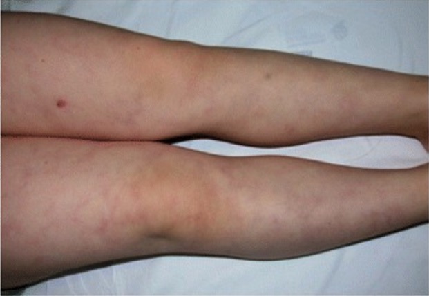
Livedo reticularisLivedo reticularisA condition characterized by a reticular or fishnet pattern on the skin of lower extremities and other parts of the body. This red and blue pattern is due to deoxygenated blood in unstable dermal blood vessels. The condition is intensified by cold exposure and relieved by rewarming.Chronic Kidney Disease: a red-blue netlike, reticulated pattern occurring due to compromised blood flowBlood flowBlood flow refers to the movement of a certain volume of blood through the vasculature over a given unit of time (e.g., mL per minute).Vascular Resistance, Flow, and Mean Arterial Pressure in medium-sized vessels
Rheumatologic findings: consistent with an associated rheumatologic disorder
RashesRashesRashes are a group of diseases that cause abnormal coloration and texture to the skin. The etiologies are numerous but can include irritation, allergens, infections, or inflammatory conditions. Rashes that present in only 1 area of the body are called localized rashes. Generalized rashes occur diffusely throughout the body. Generalized and Localized Rashes
Arthralgias
FatigueFatigueThe state of weariness following a period of exertion, mental or physical, characterized by a decreased capacity for work and reduced efficiency to respond to stimuli.Fibromyalgia
Livedo reticularis
Image: “Livedo reticularis in a patient with DADA2” by Roberta Caorsi et al. License: CC BY 4.0
Diagnosis
Diagnostic criteria[1–3]
To meet the diagnostic criteria for APS, a patient must meet both clinical andlaboratory criteria.
Table: Clinical and laboratory criteria for antiphospholipid syndromeAntiphospholipid syndromeAntiphospholipid syndrome (APLS) is an acquired autoimmune disorder characterized by the persistent presence of antiphospholipid antibodies, which create a hypercoagulable state. These antibodies are most commonly discovered during a workup for a thrombotic event or recurrent pregnancy loss, which are the 2 most common clinical manifestations.Antiphospholipid Syndrome[1-3]
Criterion
Events that satisfy the criterion
Clinical
Need to have a history of ≥ 1 of the following criteria (could be from either category):
Vascular thrombosisThrombosisFormation and development of a thrombus or blood clot in the blood vessel.Epidemic Typhus
≥ 1 deep vein, arterial, or small-vessel thrombosisThrombosisFormation and development of a thrombus or blood clot in the blood vessel.Epidemic Typhus in any organ or tissue confirmed by validated methods (e.g., unequivocal imaging findings, histopathology)
Superficial venous thrombosisVenous thrombosisThe formation or presence of a blood clot (thrombus) within a vein.Budd-Chiari Syndrome does not satisfy this criterion.
PregnancyPregnancyThe status during which female mammals carry their developing young (embryos or fetuses) in utero before birth, beginning from fertilization to birth.Pregnancy: Diagnosis, Physiology, and CaremorbidityMorbidityThe proportion of patients with a particular disease during a given year per given unit of population.Measures of Health Status
≥ 3 consecutive, spontaneous pregnancyPregnancyThe status during which female mammals carry their developing young (embryos or fetuses) in utero before birth, beginning from fertilization to birth.Pregnancy: Diagnosis, Physiology, and Care losses at < 10 WGA when the following have been excluded:
Maternal anatomic or hormonal abnormalities
Maternal or paternal chromosomal abnormalities
≥ 1 pregnancyPregnancyThe status during which female mammals carry their developing young (embryos or fetuses) in utero before birth, beginning from fertilization to birth.Pregnancy: Diagnosis, Physiology, and Care loss of a morphologically normal fetus at ≥ 10 WGA
≥ 1 prematurePrematureChildbirth before 37 weeks of pregnancy (259 days from the first day of the mother’s last menstrual period, or 245 days after fertilization).Necrotizing Enterocolitis delivery at < 34 WGA due to:
Severe preeclampsiaPreeclampsiaA complication of pregnancy, characterized by a complex of symptoms including maternal hypertension and proteinuria with or without pathological edema. Symptoms may range between mild and severe. Pre-eclampsia usually occurs after the 20th week of gestation, but may develop before this time in the presence of trophoblastic disease.Hypertensive Pregnancy Disorders/eclampsiaEclampsiaOnset of hyperreflexia; seizures; or coma in a previously diagnosed pre-eclamptic patient (pre-eclampsia).Hypertensive Pregnancy Disorders
Recognized signs of placental insufficiencyPlacental InsufficiencyFailure of the placenta to deliver an adequate supply of nutrients and oxygen to the fetus.Neonatal Polycythemia, which include:
OligohydramniosOligohydramniosOligohydramnios refers to amniotic fluid volume less than expected for the current gestational age. Oligohydramnios is diagnosed by ultrasound and defined as an amniotic fluid index (AFI) of ‰¤ 5 cm or a single deep pocket (SDP) of < 2 cm in the 2nd or 3rd trimester. Oligohydramnios (typically defined as an amniotic fluidAmniotic fluidA clear, yellowish liquid that envelopes the fetus inside the sac of amnion. In the first trimester, it is likely a transudate of maternal or fetal plasma. In the second trimester, amniotic fluid derives primarily from fetal lung and kidney. Cells or substances in this fluid can be removed for prenatal diagnostic tests (amniocentesis).Placenta, Umbilical Cord, and Amniotic Cavity index ≤ 5 cm)
Abnormal DopplerDopplerUltrasonography applying the doppler effect, with frequency-shifted ultrasound reflections produced by moving targets (usually red blood cells) in the bloodstream along the ultrasound axis in direct proportion to the velocity of movement of the targets, to determine both direction and velocity of blood flow.Ultrasound (Sonography)flowFlowBlood flows through the heart, arteries, capillaries, and veins in a closed, continuous circuit. Flow is the movement of volume per unit of time. Flow is affected by the pressure gradient and the resistance fluid encounters between 2 points. Vascular resistance is the opposition to flow, which is caused primarily by blood friction against vessel walls.Vascular Resistance, Flow, and Mean Arterial Pressure velocimetry of the umbilical arteryUmbilical arterySpecialized arterial vessels in the umbilical cord. They carry waste and deoxygenated blood from the fetus to the mother via the placenta. In humans, there are usually two umbilical arteries but sometimes one.Prenatal and Postnatal Physiology of the Neonate
Abnormal or non-reassuring fetal testing
Fetal growth restrictionFetal growth restrictionFetal growth restriction (FGR), also known as intrauterine fetal growth restriction (IUGR), is an estimated fetal weight (EFW) or abdominal circumference < 10th percentile for gestational age. The term small for gestational age (SGA) is sometimes erroneously used interchangeably with FGR. Fetal Growth Restriction (typically defined as an estimated fetal weightEstimated Fetal WeightObstetric Imaging < 10th percentile)
Laboratory
Need ≥ 1 positive antibody finding on ≥ 2 separate occasions ≥ 12 weeks apart:
Lupus anticoagulantLupus anticoagulantAn antiphospholipid antibody found in association with systemic lupus erythematosus, antiphospholipid syndrome; and in a variety of other diseases as well as in healthy individuals. In vitro, the antibody interferes with the conversion of prothrombin to thrombin and prolongs the partial thromboplastin time. In vivo, it exerts a procoagulant effect resulting in thrombosis mainly in the larger veins and arteries. It further causes obstetrical complications, including fetal death and spontaneous abortion, as well as a variety of hematologic and neurologic complications.Antiphospholipid Syndrome (positive = present)
Anti-cardiolipin antibodiesAntibodiesImmunoglobulins (Igs), also known as antibodies, are glycoprotein molecules produced by plasma cells that act in immune responses by recognizing and binding particular antigens. The various Ig classes are IgG (the most abundant), IgM, IgE, IgD, and IgA, which differ in their biologic features, structure, target specificity, and distribution.Immunoglobulins: Types and Functions (positive = ↑ IgGIgGThe major immunoglobulin isotype class in normal human serum. There are several isotype subclasses of igg, for example, igg1, igg2a, and igg2b.Hypersensitivity Pneumonitis and/or IgMIgMA class of immunoglobulin bearing mu chains (immunoglobulin mu-chains). Igm can fix complement. The name comes from its high molecular weight and originally being called a macroglobulin.Immunoglobulins: Types and Functions titers)
Β2-glycoprotein I antibodiesAntibodiesImmunoglobulins (Igs), also known as antibodies, are glycoprotein molecules produced by plasma cells that act in immune responses by recognizing and binding particular antigens. The various Ig classes are IgG (the most abundant), IgM, IgE, IgD, and IgA, which differ in their biologic features, structure, target specificity, and distribution.Immunoglobulins: Types and Functions (positive = ↑ IgGIgGThe major immunoglobulin isotype class in normal human serum. There are several isotype subclasses of igg, for example, igg1, igg2a, and igg2b.Hypersensitivity Pneumonitis and/or IgMIgMA class of immunoglobulin bearing mu chains (immunoglobulin mu-chains). Igm can fix complement. The name comes from its high molecular weight and originally being called a macroglobulin.Immunoglobulins: Types and Functions titers)
WGA: weeks of gestational age
aPLAPLAn acute myeloid leukemia in which abnormal promyelocytes predominate. It is frequently associated with disseminated intravascular coagulation.Acute Myeloid Leukemia testing
Indications:[1,4,5]
Any patient meeting the clinical diagnostic criteriafor APS-associated pregnancy-morbidity
Any of the following thrombotic events in patientsPatientsIndividuals participating in the health care system for the purpose of receiving therapeutic, diagnostic, or preventive procedures.Clinician–Patient Relationship < 50 years of age:
Unprovoked venous thromboembolismThromboembolismObstruction of a blood vessel (embolism) by a blood clot (thrombus) in the blood stream.Systemic Lupus Erythematosus (VTEVTEObstruction of a vein or veins (embolism) by a blood clot (thrombus) in the bloodstream.Hypercoagulable States)
Ischemic strokeIschemic StrokeAn ischemic stroke (also known as cerebrovascular accident) is an acute neurologic injury that occurs as a result of brain ischemia; this condition may be due to cerebral blood vessel occlusion by thrombosis or embolism, or rarely due to systemic hypoperfusion. Ischemic Stroke, TIATIATransient ischemic attack (TIA) is a temporary episode of neurologic dysfunction caused by ischemia without infarction that resolves completely when blood supply is restored. Transient ischemic attack is a neurologic emergency that warrants urgent medical attention. Transient Ischemic Attack (TIA), or other evidence of brain ischemiaBrain IschemiaLocalized reduction of blood flow to brain tissue due to arterial obstruction or systemic hypoperfusion. This frequently occurs in conjunction with brain hypoxia. Prolonged ischemia is associated with brain infarction.Ischemic Stroke
Any of the following thrombotic events in patientsPatientsIndividuals participating in the health care system for the purpose of receiving therapeutic, diagnostic, or preventive procedures.Clinician–Patient Relationship at any age:
VTEVTEObstruction of a vein or veins (embolism) by a blood clot (thrombus) in the bloodstream.Hypercoagulable States at unusual sites
Microvascular thrombosisThrombosisFormation and development of a thrombus or blood clot in the blood vessel.Epidemic Typhus
SLESLESystemic lupus erythematosus (SLE) is a chronic autoimmune, inflammatory condition that causes immune-complex deposition in organs, resulting in systemic manifestations. Women, particularly those of African American descent, are more commonly affected.Systemic Lupus Erythematosus
Consider testing in:
Immune thrombocytopeniaThrombocytopeniaThrombocytopenia occurs when the platelet count is < 150,000 per microliter. The normal range for platelets is usually 150,000-450,000/µL of whole blood. Thrombocytopenia can be a result of decreased production, increased destruction, or splenic sequestration of platelets. Patients are often asymptomatic until platelet counts are < 50,000/µL. Thrombocytopenia
Livedo reticularisLivedo reticularisA condition characterized by a reticular or fishnet pattern on the skin of lower extremities and other parts of the body. This red and blue pattern is due to deoxygenated blood in unstable dermal blood vessels. The condition is intensified by cold exposure and relieved by rewarming.Chronic Kidney Disease
PatientsPatientsIndividuals participating in the health care system for the purpose of receiving therapeutic, diagnostic, or preventive procedures.Clinician–Patient Relationship < 50 years of age with valvular heart disease and evidence of other autoimmune diseasesAutoimmune diseasesDisorders that are characterized by the production of antibodies that react with host tissues or immune effector cells that are autoreactive to endogenous peptides.Selective IgA Deficiency
Unexplained prolonged aPTT (incidental finding)
Serologies and titer levels:[1,2,4–6]
Lupus anticoagulantLupus anticoagulantAn antiphospholipid antibody found in association with systemic lupus erythematosus, antiphospholipid syndrome; and in a variety of other diseases as well as in healthy individuals. In vitro, the antibody interferes with the conversion of prothrombin to thrombin and prolongs the partial thromboplastin time. In vivo, it exerts a procoagulant effect resulting in thrombosis mainly in the larger veins and arteries. It further causes obstetrical complications, including fetal death and spontaneous abortion, as well as a variety of hematologic and neurologic complications.Antiphospholipid Syndrome:
Note: Despite being called an “anticoagulant,” this factor is actually prothrombotic.
Its name comes from the fact that it functions as an anticoagulant in some lab tests.
Acute-phase reactantsAcute-Phase ReactantsInflammation (including factor VIIIFactor VIIIFactor VIII of blood coagulation. Antihemophilic factor that is part of the factor viii/von Willebrand factor complex. Factor VIII is produced in the liver and acts in the intrinsic pathway of blood coagulation. It serves as a cofactor in factor X activation and this action is markedly enhanced by small amounts of thrombin.Hemostasis and C-reactive protein (CRP), which may be present after acute VTEVTEObstruction of a vein or veins (embolism) by a blood clot (thrombus) in the bloodstream.Hypercoagulable States)
PregnancyPregnancyThe status during which female mammals carry their developing young (embryos or fetuses) in utero before birth, beginning from fertilization to birth.Pregnancy: Diagnosis, Physiology, and Care
Anticardiolipin antibodiesAnticardiolipin antibodiesAntiphospholipid antibodies found in association with systemic lupus erythematosus, antiphospholipid syndrome; and in a variety of other diseases as well as in healthy individuals. The antibodies are detected by solid-phase immunoassay employing the purified phospholipid antigen cardiolipin.Antiphospholipid Syndrome:
Detected by ELISAELISAAn immunoassay utilizing an antibody labeled with an enzyme marker such as horseradish peroxidase. While either the enzyme or the antibody is bound to an immunosorbent substrate, they both retain their biologic activity; the change in enzyme activity as a result of the enzyme-antibody-antigen reaction is proportional to the concentration of the antigen and can be measured spectrophotometrically or with the naked eye. Many variations of the method have been developed.St. Louis Encephalitis Virus or automated solid-phase assay systems
Considered elevated if > 99th percentile of normal controls
Note: Anticardiolipin antibodiesAnticardiolipin antibodiesAntiphospholipid antibodies found in association with systemic lupus erythematosus, antiphospholipid syndrome; and in a variety of other diseases as well as in healthy individuals. The antibodies are detected by solid-phase immunoassay employing the purified phospholipid antigen cardiolipin.Antiphospholipid Syndrome can result in a false positiveFalse positiveAn FP test result indicates that a person has the disease when they do not.Epidemiological Values of Diagnostic Tests test for syphilisSyphilisSyphilis is a bacterial infection caused by the spirochete Treponema pallidum pallidum (T. p. pallidum), which is usually spread through sexual contact. Syphilis has 4 clinical stages: primary, secondary, latent, and tertiary. Syphilis because they react with the cardiolipin reagent in the rapid plasma reagin testRapid plasma reagin testTreponema.
Anti-β2-glycoprotein I antibodiesAntibodiesImmunoglobulins (Igs), also known as antibodies, are glycoprotein molecules produced by plasma cells that act in immune responses by recognizing and binding particular antigens. The various Ig classes are IgG (the most abundant), IgM, IgE, IgD, and IgA, which differ in their biologic features, structure, target specificity, and distribution.Immunoglobulins: Types and Functions
Detected by ELISAELISAAn immunoassay utilizing an antibody labeled with an enzyme marker such as horseradish peroxidase. While either the enzyme or the antibody is bound to an immunosorbent substrate, they both retain their biologic activity; the change in enzyme activity as a result of the enzyme-antibody-antigen reaction is proportional to the concentration of the antigen and can be measured spectrophotometrically or with the naked eye. Many variations of the method have been developed.St. Louis Encephalitis Virus or automated solid-phase assay systems
Considered elevated if > 99th percentile of normal controls
Note: All 3 primary aPLsAPLSAntiphospholipid syndrome (APLS) is an acquired autoimmune disorder characterized by the persistent presence of antiphospholipid antibodies, which create a hypercoagulable state. These antibodies are most commonly discovered during a workup for a thrombotic event or recurrent pregnancy loss, which are the 2 most common clinical manifestations.Antiphospholipid Syndrome should be tested for, since triple-positive patientsPatientsIndividuals participating in the health care system for the purpose of receiving therapeutic, diagnostic, or preventive procedures.Clinician–Patient Relationship are at increased risk for thrombosisThrombosisFormation and development of a thrombus or blood clot in the blood vessel.Epidemic Typhus and pregnancyPregnancyThe status during which female mammals carry their developing young (embryos or fetuses) in utero before birth, beginning from fertilization to birth.Pregnancy: Diagnosis, Physiology, and CaremorbidityMorbidityThe proportion of patients with a particular disease during a given year per given unit of population.Measures of Health Status.
Timing:[4,5]
Ideally, 2 tests ≥ 12 weeks apart (both should be positive to confirm the diagnosis)
In patientsPatientsIndividuals participating in the health care system for the purpose of receiving therapeutic, diagnostic, or preventive procedures.Clinician–Patient Relationship after a thrombotic event: no consensus on best timing
Direct oral anticoagulantsAnticoagulantsAnticoagulants are drugs that retard or interrupt the coagulation cascade. The primary classes of available anticoagulants include heparins, vitamin K-dependent antagonists (e.g., warfarin), direct thrombin inhibitors, and factor Xa inhibitors. Anticoagulants (DOAC):
48 hours after last dose
Simultaneously, check the DOAC level.
Vitamin KVitamin KA lipid cofactor that is required for normal blood clotting. Several forms of vitamin K have been identified: vitamin K 1 (phytomenadione) derived from plants, vitamin K 2 (menaquinone) from bacteria, and synthetic naphthoquinone provitamins, vitamin K 3 (menadione). Vitamin k 3 provitamins, after being alkylated in vivo, exhibit the antifibrinolytic activity of vitamin k. Green leafy vegetables, liver, cheese, butter, and egg yolk are good sources of vitamin k.Fat-soluble Vitamins and their Deficiencies antagonists (VKA): consider switching to LMWH and testing 1–2 weeks after last dose
In reproductive-aged women:
Best if performed when not pregnant
If tested during pregnancyPregnancyThe status during which female mammals carry their developing young (embryos or fetuses) in utero before birth, beginning from fertilization to birth.Pregnancy: Diagnosis, Physiology, and Care, repeat testing postpartum (≥ 6 weeks, but ideally 3 months after delivery).
Other laboratory testing
Coagulation studiesCoagulation studiesCoagulation studies are a group of hematologic laboratory studies that reflect the function of blood vessels, platelets, and coagulation factors, which all interact with one another to achieve hemostasis. Coagulation studies are usually ordered to evaluate patients with bleeding or hypercoagulation disorders.Coagulation Studies:[2,5,12,15]
Lupus anticoagulantLupus anticoagulantAn antiphospholipid antibody found in association with systemic lupus erythematosus, antiphospholipid syndrome; and in a variety of other diseases as well as in healthy individuals. In vitro, the antibody interferes with the conversion of prothrombin to thrombin and prolongs the partial thromboplastin time. In vivo, it exerts a procoagulant effect resulting in thrombosis mainly in the larger veins and arteries. It further causes obstetrical complications, including fetal death and spontaneous abortion, as well as a variety of hematologic and neurologic complications.Antiphospholipid Syndrome may interact with the assay and artificially prolong the aPTT.
Importance of baseline coagulation studiesCoagulation studiesCoagulation studies are a group of hematologic laboratory studies that reflect the function of blood vessels, platelets, and coagulation factors, which all interact with one another to achieve hemostasis. Coagulation studies are usually ordered to evaluate patients with bleeding or hypercoagulation disorders.Coagulation Studies:
Would need to use alternative assays that are not affected by the presence of aPLsAPLSAntiphospholipid syndrome (APLS) is an acquired autoimmune disorder characterized by the persistent presence of antiphospholipid antibodies, which create a hypercoagulable state. These antibodies are most commonly discovered during a workup for a thrombotic event or recurrent pregnancy loss, which are the 2 most common clinical manifestations.Antiphospholipid Syndrome (e.g., check anti-factor Xa activity for heparin monitoring)
CBC may show:[2,15]
AnemiaAnemiaAnemia is a condition in which individuals have low Hb levels, which can arise from various causes. Anemia is accompanied by a reduced number of RBCs and may manifest with fatigue, shortness of breath, pallor, and weakness. Subtypes are classified by the size of RBCs, chronicity, and etiology. Anemia: Overview and Types (hemolytic, microangiopathic)
ThrombocytopeniaThrombocytopeniaThrombocytopenia occurs when the platelet count is < 150,000 per microliter. The normal range for platelets is usually 150,000-450,000/µL of whole blood. Thrombocytopenia can be a result of decreased production, increased destruction, or splenic sequestration of platelets. Patients are often asymptomatic until platelet counts are < 50,000/µL. Thrombocytopenia:
PlateletsPlateletsPlatelets are small cell fragments involved in hemostasis. Thrombopoiesis takes place primarily in the bone marrow through a series of cell differentiation and is influenced by several cytokines. Platelets are formed after fragmentation of the megakaryocyte cytoplasm. Platelets: Histology are consumed in procoagulant states
Can be difficult to differentiate from other causes of thrombocytopeniaThrombocytopeniaThrombocytopenia occurs when the platelet count is < 150,000 per microliter. The normal range for platelets is usually 150,000-450,000/µL of whole blood. Thrombocytopenia can be a result of decreased production, increased destruction, or splenic sequestration of platelets. Patients are often asymptomatic until platelet counts are < 50,000/µL. Thrombocytopenia (e.g., ITPITPImmune thrombocytopenic purpura (ITP), formerly known as idiopathic thrombocytopenic purpura, is a condition that develops secondary to immune-mediated destruction of platelets, resulting in thrombocytopenia (platelet count < 100,000/mm³). Immune thrombocytopenic purpura can be either primary or secondary due to drugs or underlying disease. Immune Thrombocytopenic Purpura, DICDICDisseminated intravascular coagulation (DIC) is a condition characterized by systemic bodywide activation of the coagulation cascade. This cascade results in both widespread microvascular thrombi contributing to multiple organ dysfunction and consumption of clotting factors and platelets, leading to hemorrhage. Disseminated Intravascular Coagulation)
Complement studies:[7,10]
May show ↓ complement levels (e.g., C3 or C4)
Can be observed in both primary and secondary APS
Other assessments[14]
Other testing may be clinically indicated based on presentation. For example:
Imaging:
DopplerDopplerUltrasonography applying the doppler effect, with frequency-shifted ultrasound reflections produced by moving targets (usually red blood cells) in the bloodstream along the ultrasound axis in direct proportion to the velocity of movement of the targets, to determine both direction and velocity of blood flow.Ultrasound (Sonography) studies for a suspected DVTDVTDeep vein thrombosis (DVT) usually occurs in the deep veins of the lower extremities. The affected veins include the femoral, popliteal, iliofemoral, and pelvic veins. Proximal DVT is more likely to cause a pulmonary embolism (PE) and is generally considered more serious. Deep Vein Thrombosis
CT for a suspected stroke
ECGECGAn electrocardiogram (ECG) is a graphic representation of the electrical activity of the heart plotted against time. Adhesive electrodes are affixed to the skin surface allowing measurement of cardiac impulses from many angles. The ECG provides 3-dimensional information about the conduction system of the heart, the myocardium, and other cardiac structures. Electrocardiogram (ECG) for suspected MIMIMI is ischemia and death of an area of myocardial tissue due to insufficient blood flow and oxygenation, usually from thrombus formation on a ruptured atherosclerotic plaque in the epicardial arteries. Clinical presentation is most commonly with chest pain, but women and patients with diabetes may have atypical symptoms.Myocardial Infarction
ThrombophiliaThrombophiliaA disorder of hemostasis in which there is a tendency for the occurrence of thrombosis.Hypercoagulable States workup (e.g., if only presenting symptom is an unprovoked DVTDVTDeep vein thrombosis (DVT) usually occurs in the deep veins of the lower extremities. The affected veins include the femoral, popliteal, iliofemoral, and pelvic veins. Proximal DVT is more likely to cause a pulmonary embolism (PE) and is generally considered more serious. Deep Vein Thrombosis)
Rheumatologic workup (e.g., if patient has findings consistent with SLESLESystemic lupus erythematosus (SLE) is a chronic autoimmune, inflammatory condition that causes immune-complex deposition in organs, resulting in systemic manifestations. Women, particularly those of African American descent, are more commonly affected.Systemic Lupus Erythematosus)
Management
Management of acute thrombosisThrombosisFormation and development of a thrombus or blood clot in the blood vessel.Epidemic Typhus in APS
Management may vary depending on practice location. The following information is from US, UK, and European literature and clinical guidelines. Expert consultation (e.g., hematology) is recommended to assist with management.
The mainstay of therapy for both primary and secondary APS is anticoagulationAnticoagulationPulmonary Hypertension Drugs. Overall management of acute thrombosisThrombosisFormation and development of a thrombus or blood clot in the blood vessel.Epidemic Typhus in APS is similar to management in patientsPatientsIndividuals participating in the health care system for the purpose of receiving therapeutic, diagnostic, or preventive procedures.Clinician–Patient Relationship without APS, though warfarinWarfarinAn anticoagulant that acts by inhibiting the synthesis of vitamin K-dependent coagulation factors. Warfarin is indicated for the prophylaxis and/or treatment of venous thrombosis and its extension, pulmonary embolism, and atrial fibrillation with embolization. It is also used as an adjunct in the prophylaxis of systemic embolism after myocardial infarction. Warfarin is also used as a rodenticide.Anticoagulants is typically preferred over direct oral anticoagulantsAnticoagulantsAnticoagulants are drugs that retard or interrupt the coagulation cascade. The primary classes of available anticoagulants include heparins, vitamin K-dependent antagonists (e.g., warfarin), direct thrombin inhibitors, and factor Xa inhibitors. Anticoagulants (DOACsDOACsAnticoagulants).
May require supplemental O2, mechanical ventilationVentilationThe total volume of gas inspired or expired per unit of time, usually measured in liters per minute.Ventilation: Mechanics of Breathing, etcETCThe electron transport chain (ETC) sends electrons through a series of proteins, which generate an electrochemical proton gradient that produces energy in the form of adenosine triphosphate (ATP).Electron Transport Chain (ETC).
CirculationCirculationThe movement of the blood as it is pumped through the cardiovascular system.ABCDE Assessment/hemodynamic support:
Ensure stable blood pressure and heart rateHeart rateThe number of times the heart ventricles contract per unit of time, usually per minute.Cardiac Physiology
May require IV fluidsIV fluidsIntravenous fluids are one of the most common interventions administered in medicine to approximate physiologic bodily fluids. Intravenous fluids are divided into 2 categories: crystalloid and colloid solutions. Intravenous fluids have a wide variety of indications, including intravascular volume expansion, electrolyte manipulation, and maintenance fluids. Intravenous Fluids, vasopressorsVasopressorsSepsis in Children, cardiac drugs, etcETCThe electron transport chain (ETC) sends electrons through a series of proteins, which generate an electrochemical proton gradient that produces energy in the form of adenosine triphosphate (ATP).Electron Transport Chain (ETC).
Location and extent of the thrombosisThrombosisFormation and development of a thrombus or blood clot in the blood vessel.Epidemic Typhus/embolism
Bleeding risk
Age and comorbiditiesComorbiditiesThe presence of co-existing or additional diseases with reference to an initial diagnosis or with reference to the index condition that is the subject of study. Comorbidity may affect the ability of affected individuals to function and also their survival; it may be used as a prognostic indicator for length of hospital stay, cost factors, and outcome or survival.St. Louis Encephalitis Virus
Whether or not they are currently (or actively trying to become) pregnant
Preferred option: heparin overlapped with warfarinWarfarinAn anticoagulant that acts by inhibiting the synthesis of vitamin K-dependent coagulation factors. Warfarin is indicated for the prophylaxis and/or treatment of venous thrombosis and its extension, pulmonary embolism, and atrial fibrillation with embolization. It is also used as an adjunct in the prophylaxis of systemic embolism after myocardial infarction. Warfarin is also used as a rodenticide.Anticoagulants[7,15,16]
LMWH is typically the medication of choice.
Heparin is continued until the INR is in the therapeutic range for 2 consecutive days.
Heparin can then be discontinued, while warfarinWarfarinAn anticoagulant that acts by inhibiting the synthesis of vitamin K-dependent coagulation factors. Warfarin is indicated for the prophylaxis and/or treatment of venous thrombosis and its extension, pulmonary embolism, and atrial fibrillation with embolization. It is also used as an adjunct in the prophylaxis of systemic embolism after myocardial infarction. Warfarin is also used as a rodenticide.Anticoagulants is continued indefinitely for ongoing prophylaxisProphylaxisCephalosporins.
Goal INR on warfarinWarfarinAn anticoagulant that acts by inhibiting the synthesis of vitamin K-dependent coagulation factors. Warfarin is indicated for the prophylaxis and/or treatment of venous thrombosis and its extension, pulmonary embolism, and atrial fibrillation with embolization. It is also used as an adjunct in the prophylaxis of systemic embolism after myocardial infarction. Warfarin is also used as a rodenticide.Anticoagulants: typically, 2–3
Consider adding low-dose aspirinAspirinThe prototypical analgesic used in the treatment of mild to moderate pain. It has anti-inflammatory and antipyretic properties and acts as an inhibitor of cyclooxygenase which results in the inhibition of the biosynthesis of prostaglandins. Aspirin also inhibits platelet aggregation and is used in the prevention of arterial and venous thrombosis.Nonsteroidal Antiinflammatory Drugs (NSAIDs) to standard warfarinWarfarinAn anticoagulant that acts by inhibiting the synthesis of vitamin K-dependent coagulation factors. Warfarin is indicated for the prophylaxis and/or treatment of venous thrombosis and its extension, pulmonary embolism, and atrial fibrillation with embolization. It is also used as an adjunct in the prophylaxis of systemic embolism after myocardial infarction. Warfarin is also used as a rodenticide.Anticoagulants therapy.
Alternative: higher-dose warfarinWarfarinAn anticoagulant that acts by inhibiting the synthesis of vitamin K-dependent coagulation factors. Warfarin is indicated for the prophylaxis and/or treatment of venous thrombosis and its extension, pulmonary embolism, and atrial fibrillation with embolization. It is also used as an adjunct in the prophylaxis of systemic embolism after myocardial infarction. Warfarin is also used as a rodenticide.Anticoagulants (goal INR 3–4)
Note: WarfarinWarfarinAn anticoagulant that acts by inhibiting the synthesis of vitamin K-dependent coagulation factors. Warfarin is indicated for the prophylaxis and/or treatment of venous thrombosis and its extension, pulmonary embolism, and atrial fibrillation with embolization. It is also used as an adjunct in the prophylaxis of systemic embolism after myocardial infarction. Warfarin is also used as a rodenticide.Anticoagulants is contraindicated in pregnancyPregnancyThe status during which female mammals carry their developing young (embryos or fetuses) in utero before birth, beginning from fertilization to birth.Pregnancy: Diagnosis, Physiology, and Care.
RivaroxabanRivaroxabanA morpholine and thiophene derivative that functions as a factor Xa inhibitor and is used in the treatment and prevention of deep-vein thrombosis and pulmonary embolism. It is also used for the prevention of stroke and systemic embolization in patients with non-valvular atrial fibrillation, and for the prevention of atherothrombotic events in patients after an acute coronary syndrome.Anticoagulants
DabigatranDabigatranA thrombin inhibitor which acts by binding and blocking thrombogenic activity and the prevention of thrombus formation. It is used to reduce the risk of stroke and systemic embolism in patients with nonvalvular atrial fibrillation.Anticoagulants
Indications: may be considered in patientsPatientsIndividuals participating in the health care system for the purpose of receiving therapeutic, diagnostic, or preventive procedures.Clinician–Patient Relationship with non–high-risk APS with single- or double-positive aPLsAPLSAntiphospholipid syndrome (APLS) is an acquired autoimmune disorder characterized by the persistent presence of antiphospholipid antibodies, which create a hypercoagulable state. These antibodies are most commonly discovered during a workup for a thrombotic event or recurrent pregnancy loss, which are the 2 most common clinical manifestations.Antiphospholipid Syndrome who have previously done well on DOACsDOACsAnticoagulants (with good adherence)[9]
ContraindicationsContraindicationsA condition or factor associated with a recipient that makes the use of a drug, procedure, or physical agent improper or inadvisable. Contraindications may be absolute (life threatening) or relative (higher risk of complications in which benefits may outweigh risks).Noninvasive Ventilation (WarfarinWarfarinAn anticoagulant that acts by inhibiting the synthesis of vitamin K-dependent coagulation factors. Warfarin is indicated for the prophylaxis and/or treatment of venous thrombosis and its extension, pulmonary embolism, and atrial fibrillation with embolization. It is also used as an adjunct in the prophylaxis of systemic embolism after myocardial infarction. Warfarin is also used as a rodenticide.Anticoagulants is preferred over DOACsDOACsAnticoagulants in APS, especially in “high-risk” patientsPatientsIndividuals participating in the health care system for the purpose of receiving therapeutic, diagnostic, or preventive procedures.Clinician–Patient Relationship.):[7,9,16]
Triple aPLAPLAn acute myeloid leukemia in which abnormal promyelocytes predominate. It is frequently associated with disseminated intravascular coagulation.Acute Myeloid Leukemia positivity
Small-vessel thrombosisThrombosisFormation and development of a thrombus or blood clot in the blood vessel.Epidemic Typhus
Organ involvement
Heart valve disease
Recurrent thrombosisThrombosisFormation and development of a thrombus or blood clot in the blood vessel.Epidemic Typhus
Note: The European Medicines Agency (EMA) states that DOACsDOACsAnticoagulants are not recommended in APS and the US FDA has endorsed these recommendations.[7]
WarfarinWarfarinAn anticoagulant that acts by inhibiting the synthesis of vitamin K-dependent coagulation factors. Warfarin is indicated for the prophylaxis and/or treatment of venous thrombosis and its extension, pulmonary embolism, and atrial fibrillation with embolization. It is also used as an adjunct in the prophylaxis of systemic embolism after myocardial infarction. Warfarin is also used as a rodenticide.Anticoagulants is contraindicated owing to its teratogenicity.
Antiplatelet therapy with aspirinAspirinThe prototypical analgesic used in the treatment of mild to moderate pain. It has anti-inflammatory and antipyretic properties and acts as an inhibitor of cyclooxygenase which results in the inhibition of the biosynthesis of prostaglandins. Aspirin also inhibits platelet aggregation and is used in the prevention of arterial and venous thrombosis.Nonsteroidal Antiinflammatory Drugs (NSAIDs) is recommended to reduce obstetric morbidityMorbidityThe proportion of patients with a particular disease during a given year per given unit of population.Measures of Health Status.
IV glucocorticoidsGlucocorticoidsGlucocorticoids are a class within the corticosteroid family. Glucocorticoids are chemically and functionally similar to endogenous cortisol. There are a wide array of indications, which primarily benefit from the antiinflammatory and immunosuppressive effects of this class of drugs.Glucocorticoids (e.g., IV methylprednisoloneMethylprednisoloneA prednisolone derivative with similar anti-inflammatory action.Immunosuppressants 250–1,000 mg for 3 days)
Plasma exchangePlasma exchangeRemoval of plasma and replacement with various fluids, e.g., fresh frozen plasma, plasma protein fractions (ppf), albumin preparations, dextran solutions, saline. Used in treatment of autoimmune diseases, immune complex diseases, diseases of excess plasma factors, and other conditions.Thrombotic Thrombocytopenic Purpura
IV immunoglobulinsIV immunoglobulinsImmunoglobulin preparations used in intravenous infusion, containing primarily immunoglobulin g. They are used to treat a variety of diseases associated with decreased or abnormal immunoglobulin levels including pediatric aids; primary hypergammaglobulinemia; scid; cytomegalovirus infections in transplant recipients, lymphocytic leukemia, chronic; kawasaki syndrome, infection in neonates, and idiopathic thrombocytopenic purpura.DiGeorge Syndrome (IVIGIVIGDermatomyositis)
Prevention of thrombosisThrombosisFormation and development of a thrombus or blood clot in the blood vessel.Epidemic Typhus[7,15,16]
SmokingSmokingWillful or deliberate act of inhaling and exhaling smoke from burning substances or agents held by hand.Interstitial Lung Diseases cessation
Managing hyperlipidemia/hypertensionHypertensionHypertension, or high blood pressure, is a common disease that manifests as elevated systemic arterial pressures. Hypertension is most often asymptomatic and is found incidentally as part of a routine physical examination or during triage for an unrelated medical encounter. Hypertension
For patientsPatientsIndividuals participating in the health care system for the purpose of receiving therapeutic, diagnostic, or preventive procedures.Clinician–Patient Relationship who have never had a thrombotic event (i.e., primary prevention):[7,16]
Consider low-dose aspirinAspirinThe prototypical analgesic used in the treatment of mild to moderate pain. It has anti-inflammatory and antipyretic properties and acts as an inhibitor of cyclooxygenase which results in the inhibition of the biosynthesis of prostaglandins. Aspirin also inhibits platelet aggregation and is used in the prevention of arterial and venous thrombosis.Nonsteroidal Antiinflammatory Drugs (NSAIDs) (evidence is limited; consider entire clinical picture).
Preferred agents: LMWH (prophylactic dose) plus aspirinAspirinThe prototypical analgesic used in the treatment of mild to moderate pain. It has anti-inflammatory and antipyretic properties and acts as an inhibitor of cyclooxygenase which results in the inhibition of the biosynthesis of prostaglandins. Aspirin also inhibits platelet aggregation and is used in the prevention of arterial and venous thrombosis.Nonsteroidal Antiinflammatory Drugs (NSAIDs)
For patientsPatientsIndividuals participating in the health care system for the purpose of receiving therapeutic, diagnostic, or preventive procedures.Clinician–Patient Relationshipwith a history of thrombosisThrombosisFormation and development of a thrombus or blood clot in the blood vessel.Epidemic Typhus:
In patientsPatientsIndividuals participating in the health care system for the purpose of receiving therapeutic, diagnostic, or preventive procedures.Clinician–Patient Relationship who do not desire pregnancyPregnancyThe status during which female mammals carry their developing young (embryos or fetuses) in utero before birth, beginning from fertilization to birth.Pregnancy: Diagnosis, Physiology, and Care: warfarinWarfarinAn anticoagulant that acts by inhibiting the synthesis of vitamin K-dependent coagulation factors. Warfarin is indicated for the prophylaxis and/or treatment of venous thrombosis and its extension, pulmonary embolism, and atrial fibrillation with embolization. It is also used as an adjunct in the prophylaxis of systemic embolism after myocardial infarction. Warfarin is also used as a rodenticide.Anticoagulantstherapy with target INR 2–3
In patientsPatientsIndividuals participating in the health care system for the purpose of receiving therapeutic, diagnostic, or preventive procedures.Clinician–Patient Relationship who do desire pregnancyPregnancyThe status during which female mammals carry their developing young (embryos or fetuses) in utero before birth, beginning from fertilization to birth.Pregnancy: Diagnosis, Physiology, and Care: LMWH and aspirinAspirinThe prototypical analgesic used in the treatment of mild to moderate pain. It has anti-inflammatory and antipyretic properties and acts as an inhibitor of cyclooxygenase which results in the inhibition of the biosynthesis of prostaglandins. Aspirin also inhibits platelet aggregation and is used in the prevention of arterial and venous thrombosis.Nonsteroidal Antiinflammatory Drugs (NSAIDs) (warfarinWarfarinAn anticoagulant that acts by inhibiting the synthesis of vitamin K-dependent coagulation factors. Warfarin is indicated for the prophylaxis and/or treatment of venous thrombosis and its extension, pulmonary embolism, and atrial fibrillation with embolization. It is also used as an adjunct in the prophylaxis of systemic embolism after myocardial infarction. Warfarin is also used as a rodenticide.Anticoagulants is teratogenic)
For patientsPatientsIndividuals participating in the health care system for the purpose of receiving therapeutic, diagnostic, or preventive procedures.Clinician–Patient Relationshipwith aPLAPLAn acute myeloid leukemia in which abnormal promyelocytes predominate. It is frequently associated with disseminated intravascular coagulation.Acute Myeloid Leukemias but who do not meet diagnostic criteria for APS, antithrombotic medication is not recommended (may be considered on case-by-case basis with risk factors, such as SLESLESystemic lupus erythematosus (SLE) is a chronic autoimmune, inflammatory condition that causes immune-complex deposition in organs, resulting in systemic manifestations. Women, particularly those of African American descent, are more commonly affected.Systemic Lupus Erythematosus).[7,16]
aPL-positive, but no obstetric or thrombosisThrombosisFormation and development of a thrombus or blood clot in the blood vessel.Epidemic Typhus history:
AspirinAspirinThe prototypical analgesic used in the treatment of mild to moderate pain. It has anti-inflammatory and antipyretic properties and acts as an inhibitor of cyclooxygenase which results in the inhibition of the biosynthesis of prostaglandins. Aspirin also inhibits platelet aggregation and is used in the prevention of arterial and venous thrombosis.Nonsteroidal Antiinflammatory Drugs (NSAIDs) (81–100 mg) is conditionally recommended.
History of thrombotic APS:
AspirinAspirinThe prototypical analgesic used in the treatment of mild to moderate pain. It has anti-inflammatory and antipyretic properties and acts as an inhibitor of cyclooxygenase which results in the inhibition of the biosynthesis of prostaglandins. Aspirin also inhibits platelet aggregation and is used in the prevention of arterial and venous thrombosis.Nonsteroidal Antiinflammatory Drugs (NSAIDs) plus therapeutic-dose LMWH
Transition to warfarinWarfarinAn anticoagulant that acts by inhibiting the synthesis of vitamin K-dependent coagulation factors. Warfarin is indicated for the prophylaxis and/or treatment of venous thrombosis and its extension, pulmonary embolism, and atrial fibrillation with embolization. It is also used as an adjunct in the prophylaxis of systemic embolism after myocardial infarction. Warfarin is also used as a rodenticide.Anticoagulants after 6 weeks postpartum.
History of obstetric APS:
AspirinAspirinThe prototypical analgesic used in the treatment of mild to moderate pain. It has anti-inflammatory and antipyretic properties and acts as an inhibitor of cyclooxygenase which results in the inhibition of the biosynthesis of prostaglandins. Aspirin also inhibits platelet aggregation and is used in the prevention of arterial and venous thrombosis.Nonsteroidal Antiinflammatory Drugs (NSAIDs) plus prophylactic-dose LMWH
Continue through 6–12 weeks postpartum
Indicated to reduce morbidityMorbidityThe proportion of patients with a particular disease during a given year per given unit of population.Measures of Health Status due to APS, including:
PregnancyPregnancyThe status during which female mammals carry their developing young (embryos or fetuses) in utero before birth, beginning from fertilization to birth.Pregnancy: Diagnosis, Physiology, and Care loss (at any point)
Placental insufficiencyPlacental InsufficiencyFailure of the placenta to deliver an adequate supply of nutrients and oxygen to the fetus.Neonatal Polycythemia, which may lead to:
Fetal growth restrictionFetal growth restrictionFetal growth restriction (FGR), also known as intrauterine fetal growth restriction (IUGR), is an estimated fetal weight (EFW) or abdominal circumference < 10th percentile for gestational age. The term small for gestational age (SGA) is sometimes erroneously used interchangeably with FGR. Fetal Growth Restriction
OligohydramniosOligohydramniosOligohydramnios refers to amniotic fluid volume less than expected for the current gestational age. Oligohydramnios is diagnosed by ultrasound and defined as an amniotic fluid index (AFI) of ‰¤ 5 cm or a single deep pocket (SDP) of < 2 cm in the 2nd or 3rd trimester. Oligohydramnios
PreeclampsiaPreeclampsiaA complication of pregnancy, characterized by a complex of symptoms including maternal hypertension and proteinuria with or without pathological edema. Symptoms may range between mild and severe. Pre-eclampsia usually occurs after the 20th week of gestation, but may develop before this time in the presence of trophoblastic disease.Hypertensive Pregnancy Disorders/eclampsiaEclampsiaOnset of hyperreflexia; seizures; or coma in a previously diagnosed pre-eclamptic patient (pre-eclampsia).Hypertensive Pregnancy Disorders
Preterm birthPreterm birthPreterm labor refers to regular uterine contractions leading to cervical change prior to 37 weeks of gestation; preterm birth refers to birth prior to 37 weeks of gestation. Preterm birth may be spontaneous due to preterm labor, preterm prelabor rupture of membranes (PPROM), or cervical insufficiency. Preterm Labor and Birth
Many experts recommend serial ultrasonography and antepartum testingAntepartum TestingThe primary goals of antepartum testing and monitoring are to assess fetal well-being, identify treatable situations that may cause complications, and evaluate for chromosomal abnormalities. These tests are divided into screening tests (which include cell-free DNA testing, serum analyte testing, and nuchal translucency measurements), and diagnostic tests, which provide a definitive diagnosis of aneuploidy and include chorionic villus sampling (CVS) and amniocentesis.Antepartum Testing and Monitoring in the 3rd trimester.
Adjunctive therapies whithatch may help improve obstetric outcomes (should be guided by specialist recommendations):
GlucocorticoidsGlucocorticoidsGlucocorticoids are a class within the corticosteroid family. Glucocorticoids are chemically and functionally similar to endogenous cortisol. There are a wide array of indications, which primarily benefit from the antiinflammatory and immunosuppressive effects of this class of drugs.Glucocorticoids
HydroxychloroquineHydroxychloroquineA chemotherapeutic agent that acts against erythrocytic forms of malarial parasites. Hydroxychloroquine appears to concentrate in food vacuoles of affected protozoa. It inhibits plasmodial heme polymerase.Immunosuppressants (in patientsPatientsIndividuals participating in the health care system for the purpose of receiving therapeutic, diagnostic, or preventive procedures.Clinician–Patient Relationship with refractory thrombosisThrombosisFormation and development of a thrombus or blood clot in the blood vessel.Epidemic Typhus)
Other management points
Immunomodulatory agents:[1,7,15]
May be considered in some patientsPatientsIndividuals participating in the health care system for the purpose of receiving therapeutic, diagnostic, or preventive procedures.Clinician–Patient Relationship with:
Hematologic manifestations (e.g., thrombocytopeniaThrombocytopeniaThrombocytopenia occurs when the platelet count is < 150,000 per microliter. The normal range for platelets is usually 150,000-450,000/µL of whole blood. Thrombocytopenia can be a result of decreased production, increased destruction, or splenic sequestration of platelets. Patients are often asymptomatic until platelet counts are < 50,000/µL. Thrombocytopenia or thrombotic microangiopathies)
SLESLESystemic lupus erythematosus (SLE) is a chronic autoimmune, inflammatory condition that causes immune-complex deposition in organs, resulting in systemic manifestations. Women, particularly those of African American descent, are more commonly affected.Systemic Lupus Erythematosus
Limited evidence to guide use, though theoretically helpful because APS is an autoimmune disease
Agents to consider:
HydroxychloroquineHydroxychloroquineA chemotherapeutic agent that acts against erythrocytic forms of malarial parasites. Hydroxychloroquine appears to concentrate in food vacuoles of affected protozoa. It inhibits plasmodial heme polymerase.Immunosuppressants (especially in SLESLESystemic lupus erythematosus (SLE) is a chronic autoimmune, inflammatory condition that causes immune-complex deposition in organs, resulting in systemic manifestations. Women, particularly those of African American descent, are more commonly affected.Systemic Lupus Erythematosus): reverses aPL-induced platelet activationPlatelet activationA series of progressive, overlapping events, triggered by exposure of the platelets to subendothelial tissue. These events include shape change, adhesiveness, aggregation, and release reactions. When carried through to completion, these events lead to the formation of a stable hemostatic plug.Hemostasis and has beneficial effects on cholesterolCholesterolThe principal sterol of all higher animals, distributed in body tissues, especially the brain and spinal cord, and in animal fats and oils.Cholesterol Metabolism
RituximabRituximabA murine-derived monoclonal antibody and antineoplastic agent that binds specifically to the cd20 antigen and is used in the treatment of leukemia; lymphoma and rheumatoid arthritis.Immunosuppressants: monoclonal antibody against many B-cell precursors
ThrombocytopeniaThrombocytopeniaThrombocytopenia occurs when the platelet count is < 150,000 per microliter. The normal range for platelets is usually 150,000-450,000/µL of whole blood. Thrombocytopenia can be a result of decreased production, increased destruction, or splenic sequestration of platelets. Patients are often asymptomatic until platelet counts are < 50,000/µL. Thrombocytopenia:[15]
Cause of the thrombocytopeniaThrombocytopeniaThrombocytopenia occurs when the platelet count is < 150,000 per microliter. The normal range for platelets is usually 150,000-450,000/µL of whole blood. Thrombocytopenia can be a result of decreased production, increased destruction, or splenic sequestration of platelets. Patients are often asymptomatic until platelet counts are < 50,000/µL. Thrombocytopenia should still be investigated.
Very difficult to distinguish from immune thrombocytopeniaThrombocytopeniaThrombocytopenia occurs when the platelet count is < 150,000 per microliter. The normal range for platelets is usually 150,000-450,000/µL of whole blood. Thrombocytopenia can be a result of decreased production, increased destruction, or splenic sequestration of platelets. Patients are often asymptomatic until platelet counts are < 50,000/µL. Thrombocytopenia
StatinsStatinsStatins are competitive inhibitors of HMG-CoA reductase in the liver. HMG-CoA reductase is the rate-limiting step in cholesterol synthesis. Inhibition results in lowered intrahepatocytic cholesterol formation, resulting in up-regulation of LDL receptors and, ultimately, lowering levels of serum LDL and triglycerides.Statins:[7,15]
Have antithrombotic and antiinflammatory properties on endothelial cells
Consider adding in patientsPatientsIndividuals participating in the health care system for the purpose of receiving therapeutic, diagnostic, or preventive procedures.Clinician–Patient Relationship with:
aPLsAPLSAntiphospholipid syndrome (APLS) is an acquired autoimmune disorder characterized by the persistent presence of antiphospholipid antibodies, which create a hypercoagulable state. These antibodies are most commonly discovered during a workup for a thrombotic event or recurrent pregnancy loss, which are the 2 most common clinical manifestations.Antiphospholipid Syndrome and hyperlipidemia
Refractory thrombotic APS
PrognosisPrognosisA prediction of the probable outcome of a disease based on a individual’s condition and the usual course of the disease as seen in similar situations.Non-Hodgkin Lymphomas[1,15]
Risk of VTEVTEObstruction of a vein or veins (embolism) by a blood clot (thrombus) in the bloodstream.Hypercoagulable States in asymptomatic, nonpregnant patientsPatientsIndividuals participating in the health care system for the purpose of receiving therapeutic, diagnostic, or preventive procedures.Clinician–Patient Relationship with aPLsAPLSAntiphospholipid syndrome (APLS) is an acquired autoimmune disorder characterized by the persistent presence of antiphospholipid antibodies, which create a hypercoagulable state. These antibodies are most commonly discovered during a workup for a thrombotic event or recurrent pregnancy loss, which are the 2 most common clinical manifestations.Antiphospholipid Syndrome: < 1% per year
Risk of recurrent thrombosisThrombosisFormation and development of a thrombus or blood clot in the blood vessel.Epidemic Typhus in untreated patientsPatientsIndividuals participating in the health care system for the purpose of receiving therapeutic, diagnostic, or preventive procedures.Clinician–Patient Relationship with APS: 25% per year
Risk of thrombosisThrombosisFormation and development of a thrombus or blood clot in the blood vessel.Epidemic Typhus during pregnancyPregnancyThe status during which female mammals carry their developing young (embryos or fetuses) in utero before birth, beginning from fertilization to birth.Pregnancy: Diagnosis, Physiology, and Care/puerperium in patientsPatientsIndividuals participating in the health care system for the purpose of receiving therapeutic, diagnostic, or preventive procedures.Clinician–Patient Relationship with APS: 5%–12% per year
Clinical Relevance
Differential diagnosis for recurrent thrombosisThrombosisFormation and development of a thrombus or blood clot in the blood vessel.Epidemic Typhus
HypercoagulableHypercoagulableHypercoagulable states (also referred to as thrombophilias) are a group of hematologic diseases defined by an increased risk of clot formation (i.e., thrombosis) due to either an increase in procoagulants, a decrease in anticoagulants, or a decrease in fibrinolysis. Hypercoagulable States states: hypercoagulableHypercoagulableHypercoagulable states (also referred to as thrombophilias) are a group of hematologic diseases defined by an increased risk of clot formation (i.e., thrombosis) due to either an increase in procoagulants, a decrease in anticoagulants, or a decrease in fibrinolysis. Hypercoagulable States states or thrombophiliasThrombophiliasHypercoagulable states (also referred to as thrombophilias) are a group of hematologic diseases defined by an increased risk of clot formation (i.e., thrombosis) due to either an increase in procoagulants, a decrease in anticoagulants, or a decrease in fibrinolysis.Hypercoagulable States are defined by an increased risk of clot formation or thrombosisThrombosisFormation and development of a thrombus or blood clot in the blood vessel.Epidemic Typhus. The cause may be inherited or acquired; both lead to the production of clots that may cause occlusion of vessels in major organs, which can be fatal. Factor V LeidenFactor V LeidenHypercoagulable States is the most common inherited cause. Management usually involves the use of anticoagulantsAnticoagulantsAnticoagulants are drugs that retard or interrupt the coagulation cascade. The primary classes of available anticoagulants include heparins, vitamin K-dependent antagonists (e.g., warfarin), direct thrombin inhibitors, and factor Xa inhibitors. Anticoagulants.
Table: Causes of a hypercoagulableHypercoagulableHypercoagulable states (also referred to as thrombophilias) are a group of hematologic diseases defined by an increased risk of clot formation (i.e., thrombosis) due to either an increase in procoagulants, a decrease in anticoagulants, or a decrease in fibrinolysis. Hypercoagulable States state
Autosomal dominantAutosomal dominantAutosomal inheritance, both dominant and recessive, refers to the transmission of genes from the 22 autosomal chromosomes. Autosomal dominant diseases are expressed when only 1 copy of the dominant allele is inherited. Autosomal Recessive and Autosomal Dominant Inheritance with incomplete penetrancePenetranceThe percent frequency with which a dominant or homozygous recessive gene or gene combination manifests itself in the phenotype of the carriers.Familial Juvenile Polyposis
Caused by a point mutationPoint MutationA mutation caused by the substitution of one nucleotide for another. This results in the DNA molecule having a change in a single base pair.Types of Mutations substituting guanineGuanineNucleic Acids for adenineAdenineA purine base and a fundamental unit of adenine nucleotides.Nucleic Acids
Results in resistanceResistancePhysiologically, the opposition to flow of air caused by the forces of friction. As a part of pulmonary function testing, it is the ratio of driving pressure to the rate of air flow.Ventilation: Mechanics of Breathing to factor VFactor VHeat- and storage-labile plasma glycoprotein which accelerates the conversion of prothrombin to thrombin in blood coagulation. Factor V accomplishes this by forming a complex with factor Xa, phospholipid, and calcium (prothrombinase complex).Hemostasis degradation by protein C
ProthrombinProthrombinA plasma protein that is the inactive precursor of thrombin. It is converted to thrombin by a prothrombin activator complex consisting of factor Xa, factor V, phospholipid, and calcium ions.Hemostasis 20210A or factor II mutations
Autosomal dominantAutosomal dominantAutosomal inheritance, both dominant and recessive, refers to the transmission of genes from the 22 autosomal chromosomes. Autosomal dominant diseases are expressed when only 1 copy of the dominant allele is inherited. Autosomal Recessive and Autosomal Dominant Inheritance
Point mutationPoint MutationA mutation caused by the substitution of one nucleotide for another. This results in the DNA molecule having a change in a single base pair.Types of Mutations leads to ↑ prothrombinProthrombinA plasma protein that is the inactive precursor of thrombin. It is converted to thrombin by a prothrombin activator complex consisting of factor Xa, factor V, phospholipid, and calcium ions.Hemostasis function.
Autosomal dominantAutosomal dominantAutosomal inheritance, both dominant and recessive, refers to the transmission of genes from the 22 autosomal chromosomes. Autosomal dominant diseases are expressed when only 1 copy of the dominant allele is inherited. Autosomal Recessive and Autosomal Dominant Inheritance (or can be acquired)
AntithrombinAntithrombinEndogenous factors and drugs that directly inhibit the action of thrombin, usually by blocking its enzymatic activity. They are distinguished from indirect thrombin inhibitors, such as heparin, which act by enhancing the inhibitory effects of antithrombins.Anticoagulants normally inhibits thrombinThrombinAn enzyme formed from prothrombin that converts fibrinogen to fibrin.Hemostasis and factor Xa.
↑ ThrombinThrombinAn enzyme formed from prothrombin that converts fibrinogen to fibrin.Hemostasis and factor Xa
ResistanceResistancePhysiologically, the opposition to flow of air caused by the forces of friction. As a part of pulmonary function testing, it is the ratio of driving pressure to the rate of air flow.Ventilation: Mechanics of Breathing to LMWH
Protein C or S deficiency
ProteinsProteinsLinear polypeptides that are synthesized on ribosomes and may be further modified, crosslinked, cleaved, or assembled into complex proteins with several subunits. The specific sequence of amino acids determines the shape the polypeptide will take, during protein folding, and the function of the protein.Energy Homeostasis C and S are vitamin K-dependent anticoagulantsAnticoagulantsAnticoagulants are drugs that retard or interrupt the coagulation cascade. The primary classes of available anticoagulants include heparins, vitamin K-dependent antagonists (e.g., warfarin), direct thrombin inhibitors, and factor Xa inhibitors. Anticoagulants.
Deficiencies result in overactivity of factors V and VIII.
Secondary or acquired
Venous stasis
Immobility (cast, stroke patientsPatientsIndividuals participating in the health care system for the purpose of receiving therapeutic, diagnostic, or preventive procedures.Clinician–Patient Relationship, post-knee surgery, long flights)
VasculitisVasculitisInflammation of any one of the blood vessels, including the arteries; veins; and rest of the vasculature system in the body.Systemic Lupus Erythematosus
Oral contraceptiveOral contraceptiveCompounds, usually hormonal, taken orally in order to block ovulation and prevent the occurrence of pregnancy. The hormones are generally estrogen or progesterone or both.Benign Liver Tumors pills
Antiphospholipid syndromeAntiphospholipid syndromeAntiphospholipid syndrome (APLS) is an acquired autoimmune disorder characterized by the persistent presence of antiphospholipid antibodies, which create a hypercoagulable state. These antibodies are most commonly discovered during a workup for a thrombotic event or recurrent pregnancy loss, which are the 2 most common clinical manifestations.Antiphospholipid Syndrome
Nephrotic syndromeNephrotic syndromeNephrotic syndrome is characterized by severe proteinuria, hypoalbuminemia, and peripheral edema. In contrast, the nephritic syndromes present with hematuria, variable loss of renal function, and hypertension, although there is sometimes overlap of > 1 glomerular disease in the same individual. Nephrotic Syndrome
Paroxysmal nocturnal hemoglobinuriaHemoglobinuriaThe presence of free hemoglobin in the urine, indicating hemolysis of erythrocytes within the vascular system. After saturating the hemoglobin-binding proteins (haptoglobins), free hemoglobin begins to appear in the urine.Transfusion Reactions
Inflammatory bowel disease
Common presentations of recurrent thrombosisThrombosisFormation and development of a thrombus or blood clot in the blood vessel.Epidemic Typhus
Deep vein thrombosisThrombosisFormation and development of a thrombus or blood clot in the blood vessel.Epidemic Typhus (DVTDVTDeep vein thrombosis (DVT) usually occurs in the deep veins of the lower extremities. The affected veins include the femoral, popliteal, iliofemoral, and pelvic veins. Proximal DVT is more likely to cause a pulmonary embolism (PE) and is generally considered more serious. Deep Vein Thrombosis): thrombosisThrombosisFormation and development of a thrombus or blood clot in the blood vessel.Epidemic Typhus of the deep venous systems found in the lower limb. The condition is a clinical manifestation of hypercoagulableHypercoagulableHypercoagulable states (also referred to as thrombophilias) are a group of hematologic diseases defined by an increased risk of clot formation (i.e., thrombosis) due to either an increase in procoagulants, a decrease in anticoagulants, or a decrease in fibrinolysis. Hypercoagulable States states and requires management acutely to prevent the development of pulmonary emboli. Diagnosis is typically made using DopplerDopplerUltrasonography applying the doppler effect, with frequency-shifted ultrasound reflections produced by moving targets (usually red blood cells) in the bloodstream along the ultrasound axis in direct proportion to the velocity of movement of the targets, to determine both direction and velocity of blood flow.Ultrasound (Sonography) ultrasonography. The condition is treated with anticoagulantsAnticoagulantsAnticoagulants are drugs that retard or interrupt the coagulation cascade. The primary classes of available anticoagulants include heparins, vitamin K-dependent antagonists (e.g., warfarin), direct thrombin inhibitors, and factor Xa inhibitors. Anticoagulants.
Pulmonary embolismPulmonary EmbolismPulmonary embolism (PE) is a potentially fatal condition that occurs as a result of intraluminal obstruction of the main pulmonary artery or its branches. The causative factors include thrombi, air, amniotic fluid, and fat. In PE, gas exchange is impaired due to the decreased return of deoxygenated blood to the lungs. Pulmonary Embolism (PE): a medical emergency in which an embolus blockspart of the pulmonary arterial system, resulting in acute shortness of breathShortness of breathDyspnea is the subjective sensation of breathing discomfort. Dyspnea is a normal manifestation of heavy physical or psychological exertion, but also may be caused by underlying conditions (both pulmonary and extrapulmonary).Dyspnea and impaired ventilationVentilationThe total volume of gas inspired or expired per unit of time, usually measured in liters per minute.Ventilation: Mechanics of Breathing/perfusion. About 30% of these patientsPatientsIndividuals participating in the health care system for the purpose of receiving therapeutic, diagnostic, or preventive procedures.Clinician–Patient Relationship have a concurrent DVTDVTDeep vein thrombosis (DVT) usually occurs in the deep veins of the lower extremities. The affected veins include the femoral, popliteal, iliofemoral, and pelvic veins. Proximal DVT is more likely to cause a pulmonary embolism (PE) and is generally considered more serious. Deep Vein Thrombosis. Diagnosis can be made on a CT pulmonary angiographyAngiographyRadiography of blood vessels after injection of a contrast medium.Cardiac Surgery (CTPACTPAPulmonary Function Tests) scan and/or a V/Q. PatientsPatientsIndividuals participating in the health care system for the purpose of receiving therapeutic, diagnostic, or preventive procedures.Clinician–Patient Relationship are treated with anticoagulantsAnticoagulantsAnticoagulants are drugs that retard or interrupt the coagulation cascade. The primary classes of available anticoagulants include heparins, vitamin K-dependent antagonists (e.g., warfarin), direct thrombin inhibitors, and factor Xa inhibitors. Anticoagulants.
Billing and Coding
Diagnosis Codes:
This code is used to diagnose Antiphospholipid SyndromeAntiphospholipid syndromeAntiphospholipid syndrome (APLS) is an acquired autoimmune disorder characterized by the persistent presence of antiphospholipid antibodies, which create a hypercoagulable state. These antibodies are most commonly discovered during a workup for a thrombotic event or recurrent pregnancy loss, which are the 2 most common clinical manifestations.Antiphospholipid Syndrome (APS), an autoimmune disorderAutoimmune DisorderSeptic Arthritis characterized by recurrent arterial or venous thrombosisVenous thrombosisThe formation or presence of a blood clot (thrombus) within a vein.Budd-Chiari Syndrome and/or pregnancyPregnancyThe status during which female mammals carry their developing young (embryos or fetuses) in utero before birth, beginning from fertilization to birth.Pregnancy: Diagnosis, Physiology, and CaremorbidityMorbidityThe proportion of patients with a particular disease during a given year per given unit of population.Measures of Health Status in the presence of specific autoantibodiesAutoantibodiesAntibodies that react with self-antigens (autoantigens) of the organism that produced them.Blotting Techniques.
Coding System
Code
Description
ICD-10-CM
D68.61
Antiphospholipid syndromeAntiphospholipid syndromeAntiphospholipid syndrome (APLS) is an acquired autoimmune disorder characterized by the persistent presence of antiphospholipid antibodies, which create a hypercoagulable state. These antibodies are most commonly discovered during a workup for a thrombotic event or recurrent pregnancy loss, which are the 2 most common clinical manifestations.Antiphospholipid Syndrome
SNOMED CT
65344000
Antiphospholipid syndromeAntiphospholipid syndromeAntiphospholipid syndrome (APLS) is an acquired autoimmune disorder characterized by the persistent presence of antiphospholipid antibodies, which create a hypercoagulable state. These antibodies are most commonly discovered during a workup for a thrombotic event or recurrent pregnancy loss, which are the 2 most common clinical manifestations.Antiphospholipid Syndrome (disorder)
Evaluation & Workup:
These codes are for the laboratory tests required to diagnose APS, which include assays for lupus anticoagulantLupus anticoagulantAn antiphospholipid antibody found in association with systemic lupus erythematosus, antiphospholipid syndrome; and in a variety of other diseases as well as in healthy individuals. In vitro, the antibody interferes with the conversion of prothrombin to thrombin and prolongs the partial thromboplastin time. In vivo, it exerts a procoagulant effect resulting in thrombosis mainly in the larger veins and arteries. It further causes obstetrical complications, including fetal death and spontaneous abortion, as well as a variety of hematologic and neurologic complications.Antiphospholipid Syndrome, anticardiolipin antibodiesAnticardiolipin antibodiesAntiphospholipid antibodies found in association with systemic lupus erythematosus, antiphospholipid syndrome; and in a variety of other diseases as well as in healthy individuals. The antibodies are detected by solid-phase immunoassay employing the purified phospholipid antigen cardiolipin.Antiphospholipid Syndrome, and anti-beta 2 glycoprotein I antibodiesAntibodiesImmunoglobulins (Igs), also known as antibodies, are glycoprotein molecules produced by plasma cells that act in immune responses by recognizing and binding particular antigens. The various Ig classes are IgG (the most abundant), IgM, IgE, IgD, and IgA, which differ in their biologic features, structure, target specificity, and distribution.Immunoglobulins: Types and Functions.
This code is for warfarinWarfarinAn anticoagulant that acts by inhibiting the synthesis of vitamin K-dependent coagulation factors. Warfarin is indicated for the prophylaxis and/or treatment of venous thrombosis and its extension, pulmonary embolism, and atrial fibrillation with embolization. It is also used as an adjunct in the prophylaxis of systemic embolism after myocardial infarction. Warfarin is also used as a rodenticide.Anticoagulants, the primary oral anticoagulant used for long-term secondary prevention of thrombosisThrombosisFormation and development of a thrombus or blood clot in the blood vessel.Epidemic Typhus in patientsPatientsIndividuals participating in the health care system for the purpose of receiving therapeutic, diagnostic, or preventive procedures.Clinician–Patient Relationship with APS.
Coding System
Code
Description
RxNorm
11289
WarfarinWarfarinAn anticoagulant that acts by inhibiting the synthesis of vitamin K-dependent coagulation factors. Warfarin is indicated for the prophylaxis and/or treatment of venous thrombosis and its extension, pulmonary embolism, and atrial fibrillation with embolization. It is also used as an adjunct in the prophylaxis of systemic embolism after myocardial infarction. Warfarin is also used as a rodenticide.Anticoagulants (ingredient)
RxNorm
5224
Heparin (ingredient)
References
Committee on Practice Bulletins—Obstetrics, American College of Obstetricians and Gynecologists. (2012). Practice bulletin no. 132: antiphospholipid syndrome. Obstetrics and Gynecology, 120(6), 1514–1521. https://doi.org/10.1097/01.AOG.0000423816.39542.0f
Miyakis, S., Lockshin, M. D., Atsumi, T., et al. (2006). International consensus statement on an update of the classification criteria for definite antiphospholipid syndrome (APS). Journal of Thrombosis and Haemostasis, 4(2), 295–306. https://www.jthjournal.org/article/S1538-7836(22)12142-2/fulltext
The Practice Committee of the American Society for Reproductive Medicine. (2012). Evaluation and treatment of recurrent pregnancy loss: a committee opinion. Fertility and Sterility, 98(5), 1103‒1111. https://www.fertstert.org/article/S0015-0282(12)00701-7/fulltext
Devreese, K., de Groot, P. G., de Laat, B., et al. (2020). Guidance from the Scientific and Standardization Committee for Lupus Anticoagulant/Antiphospholipid Antibodies of the International Society on Thrombosis and Haemostasis: update of the guidelines for lupus anticoagulant detection and interpretation. Journal of Thrombosis and Haemostasis, 18(11), 2828–2839. https://www.jthjournal.org/article/S1538-7836(22)03725-4/fulltext
Devreese, K., Ortel, T. L., Pengo, V., de Laat, B., Subcommittee on Lupus Anticoagulant/Antiphospholipid Antibodies. (2018). Laboratory criteria for antiphospholipid syndrome: communication from the SSC of the ISTH. Journal of Thrombosis and Haemostasis, 16(4), 809–813. https://www.jthjournal.org/article/S1538-7836(22)00971-0/fulltext
Cohen, H., Cuadrado, M. J., Erkan, D., et al. (2020). 16th International Congress on Antiphospholipid Antibodies task force report on antiphospholipid syndrome treatment trends. Lupus, 29(12), 1571–1593. https://doi.org/10.1177/0961203320950461
Sammaritano, L. R., Bermas, B. L., Chakravarty, E. E., et al. (2020). 2020 American College of Rheumatology guideline for the management of reproductive health in rheumatic and musculoskeletal diseases. Arthritis & Rheumatology, 72(4), 529–556. https://doi.org/10.1002/art.41191
Zuily, S., Cohen, H., Isenberg, D., et al. (2020). Use of direct oral anticoagulants in patients with thrombotic antiphospholipid syndrome: guidance from the Scientific and Standardization Committee of the International Society on Thrombosis and Haemostasis. Journal of Thrombosis and Haemostasis, 18(9), 2126–2137. https://www.jthjournal.org/article/S1538-7836(22)01647-6/fulltext
Tektonidou, M. G., et al. (2019). EULAR recommendations for the management of antiphospholipid syndrome in adults. Annals of the Rheumatic Diseases, 78, 1296–1304. https://ard.bmj.com/content/78/10/1296
Create your free account or log in to continue reading!