Giant cell arteritisGiant Cell ArteritisGiant cell arteritis (GCA), also known as temporal arteritis, is a type of large-vessel vasculitis that predominantly affects the aorta and its major branches, with a predilection for the branches of the carotid (including the temporal artery). Giant cell arteritis is defined by inflammatory leukocytes in the vessel walls leading to reactive damage, ischemia, and necrosis. Giant Cell Arteritis (GCAGCAGiant cell arteritis (GCA), also known as temporal arteritis, is a type of large-vessel vasculitis that predominantly affects the aorta and its major branches, with a predilection for the branches of the carotid (including the temporal artery). Giant cell arteritis is defined by inflammatory leukocytes in the vessel walls leading to reactive damage, ischemia, and necrosis.Giant Cell Arteritis), also known as temporal arteritisTemporal arteritisGiant cell arteritis (GCA), also known as temporal arteritis, is a type of large-vessel vasculitis that predominantly affects the aorta and its major branches, with a predilection for the branches of the carotid (including the temporal artery). Giant cell arteritis is defined by inflammatory leukocytes in the vessel walls leading to reactive damage, ischemia, and necrosis. Giant Cell Arteritis, is a type of large-vessel vasculitisVasculitisInflammation of any one of the blood vessels, including the arteries; veins; and rest of the vasculature system in the body.Systemic Lupus Erythematosus that predominantly affects the aortaAortaThe main trunk of the systemic arteries.Mediastinum and Great Vessels: Anatomy and its major branches, with a predilection for the branches of the carotid (including the temporal artery). Giant cell arteritisGiant Cell ArteritisGiant cell arteritis (GCA), also known as temporal arteritis, is a type of large-vessel vasculitis that predominantly affects the aorta and its major branches, with a predilection for the branches of the carotid (including the temporal artery). Giant cell arteritis is defined by inflammatory leukocytes in the vessel walls leading to reactive damage, ischemia, and necrosis. Giant Cell Arteritis is defined by inflammatory leukocytesLeukocytesWhite blood cells. These include granular leukocytes (basophils; eosinophils; and neutrophils) as well as non-granular leukocytes (lymphocytes and monocytes).White Myeloid Cells: Histology in the vessel walls leading to reactive damage, ischemiaIschemiaA hypoperfusion of the blood through an organ or tissue caused by a pathologic constriction or obstruction of its blood vessels, or an absence of blood circulation.Ischemic Cell Damage, and necrosisNecrosisThe death of cells in an organ or tissue due to disease, injury or failure of the blood supply.Ischemic Cell Damage. Giant cell arteritisGiant Cell ArteritisGiant cell arteritis (GCA), also known as temporal arteritis, is a type of large-vessel vasculitis that predominantly affects the aorta and its major branches, with a predilection for the branches of the carotid (including the temporal artery). Giant cell arteritis is defined by inflammatory leukocytes in the vessel walls leading to reactive damage, ischemia, and necrosis. Giant Cell Arteritis causes headaches, scalp tenderness, jawJawThe jaw is made up of the mandible, which comprises the lower jaw, and the maxilla, which comprises the upper jaw. The mandible articulates with the temporal bone via the temporomandibular joint (TMJ). The 4 muscles of mastication produce the movements of the TMJ to ensure the efficient chewing of food. Jaw and Temporomandibular Joint: AnatomypainPainAn unpleasant sensation induced by noxious stimuli which are detected by nerve endings of nociceptive neurons.Pain: Types and Pathways, visionVisionOphthalmic Exam problems, and potentially blindnessBlindnessThe inability to see or the loss or absence of perception of visual stimuli. This condition may be the result of eye diseases; optic nerve diseases; optic chiasm diseases; or brain diseases affecting the visual pathways or occipital lobe.Retinopathy of Prematurity. The diagnosis is made with temporal artery biopsyTemporal Artery BiopsyGiant Cell Arteritis. Prompt treatment with glucocorticoidsGlucocorticoidsGlucocorticoids are a class within the corticosteroid family. Glucocorticoids are chemically and functionally similar to endogenous cortisol. There are a wide array of indications, which primarily benefit from the antiinflammatory and immunosuppressive effects of this class of drugs.Glucocorticoids can relieve symptoms and prevent visionVisionOphthalmic Exam loss.
Most common immune-mediated large vessel vasculitisVasculitisInflammation of any one of the blood vessels, including the arteries; veins; and rest of the vasculature system in the body.Systemic Lupus Erythematosus
Age is the greatest risk factor:
Generally affects individuals older than 50 years
Increased incidenceIncidenceThe number of new cases of a given disease during a given period in a specified population. It also is used for the rate at which new events occur in a defined population. It is differentiated from prevalence, which refers to all cases in the population at a given time.Measures of Disease Frequency in the 8th and 9th decades of life
3 times more common in women
Northern European and Scandinavian ancestry
Lower body mass indexBody mass indexAn indicator of body density as determined by the relationship of body weight to body height. Bmi=weight (kg)/height squared (m2). Bmi correlates with body fat (adipose tissue). Their relationship varies with age and gender. For adults, bmi falls into these categories: below 18. 5 (underweight); 18. 5-24. 9 (normal); 25. 0-29. 9 (overweight); 30. 0 and above (obese).Obesity (BMIBMIAn indicator of body density as determined by the relationship of body weight to body height. Bmi=weight (kg)/height squared (m2). Bmi correlates with body fat (adipose tissue). Their relationship varies with age and gender. For adults, bmi falls into these categories: below 18. 5 (underweight); 18. 5-24. 9 (normal); 25. 0-29. 9 (overweight); 30. 0 and above (obese).Obesity) is associated with a higher risk.
Etiology[1,6]
Exact cause is unknown.
Genetic factors may increase susceptibility:
Genetic variants in the major histocompatibility complexMajor histocompatibility complexThe genetic region which contains the loci of genes which determine the structure of the serologically defined (sd) and lymphocyte-defined (ld) transplantation antigens, genes which control the structure of the immune response-associated antigens, human; the immune response genes which control the ability of an animal to respond immunologically to antigenic stimuli, and genes which determine the structure and/or level of the first four components of complement.Innate Immunity: Phagocytes and Antigen Presentation (MHC)
Human leukocyte antigenAntigenSubstances that are recognized by the immune system and induce an immune reaction.Vaccination (HLA) subtypes
GenesGenesA category of nucleic acid sequences that function as units of heredity and which code for the basic instructions for the development, reproduction, and maintenance of organisms.DNA Types and Structure involved in T cellsT cellsLymphocytes responsible for cell-mediated immunity. Two types have been identified – cytotoxic (t-lymphocytes, cytotoxic) and helper T-lymphocytes (t-lymphocytes, helper-inducer). They are formed when lymphocytes circulate through the thymus gland and differentiate to thymocytes. When exposed to an antigen, they divide rapidly and produce large numbers of new T cells sensitized to that antigen.T cells: Types and Functions
45%–50% have concurrent polymyalgia rheumaticaPolymyalgia rheumaticaA syndrome in the elderly characterized by proximal joint and muscle pain, high erythrocyte sedimentation rate, and a self-limiting course. Pain is usually accompanied by evidence of an inflammatory reaction. Women are affected twice as commonly as men and caucasians more frequently than other groups. The condition is frequently associated with giant cell arteritis and some theories pose the possibility that the two diseases arise from a single etiology or even that they are the same entity.Giant Cell Arteritis.
Pathophysiology[1,6,7]
Giant cell arteritisGiant Cell ArteritisGiant cell arteritis (GCA), also known as temporal arteritis, is a type of large-vessel vasculitis that predominantly affects the aorta and its major branches, with a predilection for the branches of the carotid (including the temporal artery). Giant cell arteritis is defined by inflammatory leukocytes in the vessel walls leading to reactive damage, ischemia, and necrosis. Giant Cell Arteritis (GCAGCAGiant cell arteritis (GCA), also known as temporal arteritis, is a type of large-vessel vasculitis that predominantly affects the aorta and its major branches, with a predilection for the branches of the carotid (including the temporal artery). Giant cell arteritis is defined by inflammatory leukocytes in the vessel walls leading to reactive damage, ischemia, and necrosis.Giant Cell Arteritis) is caused by a complicated cascade of vascular inflammationInflammationInflammation is a complex set of responses to infection and injury involving leukocytes as the principal cellular mediators in the body’s defense against pathogenic organisms. Inflammation is also seen as a response to tissue injury in the process of wound healing. The 5 cardinal signs of inflammation are pain, heat, redness, swelling, and loss of function. Inflammation, damage, and dysfunctional repair:
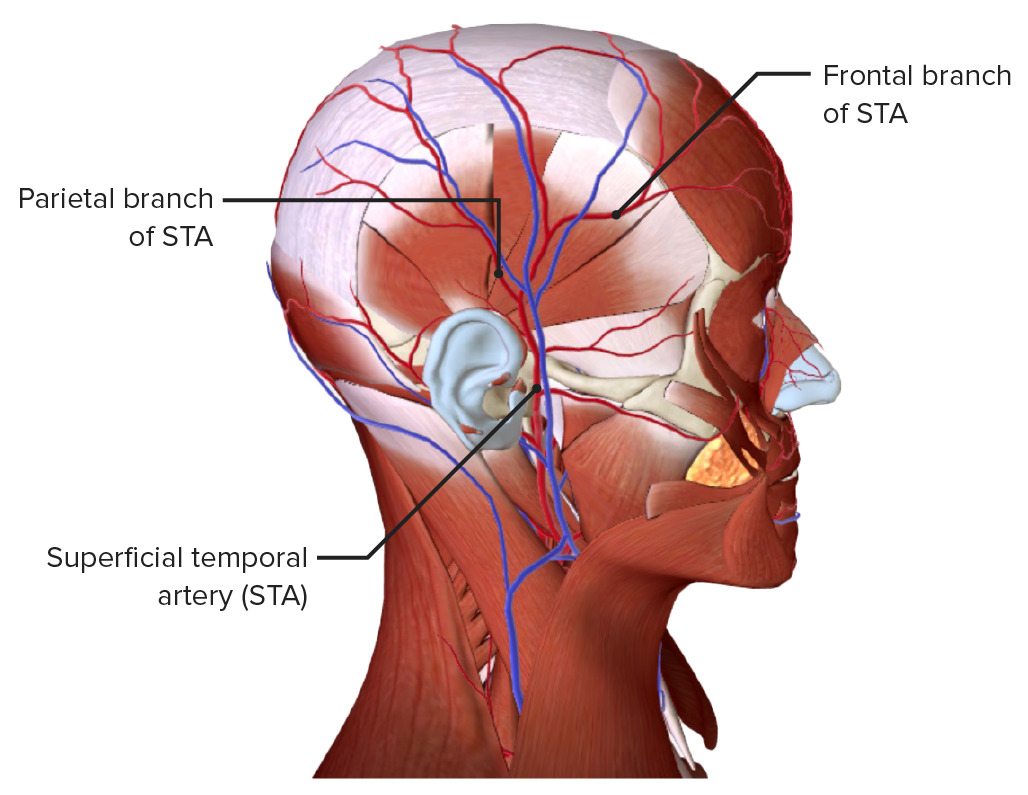
Common involvement of branches off the carotid and vertebral arteriesArteriesArteries are tubular collections of cells that transport oxygenated blood and nutrients from the heart to the tissues of the body. The blood passes through the arteries in order of decreasing luminal diameter, starting in the largest artery (the aorta) and ending in the small arterioles. Arteries are classified into 3 types: large elastic arteries, medium muscular arteries, and small arteries and arterioles. Arteries: Histology
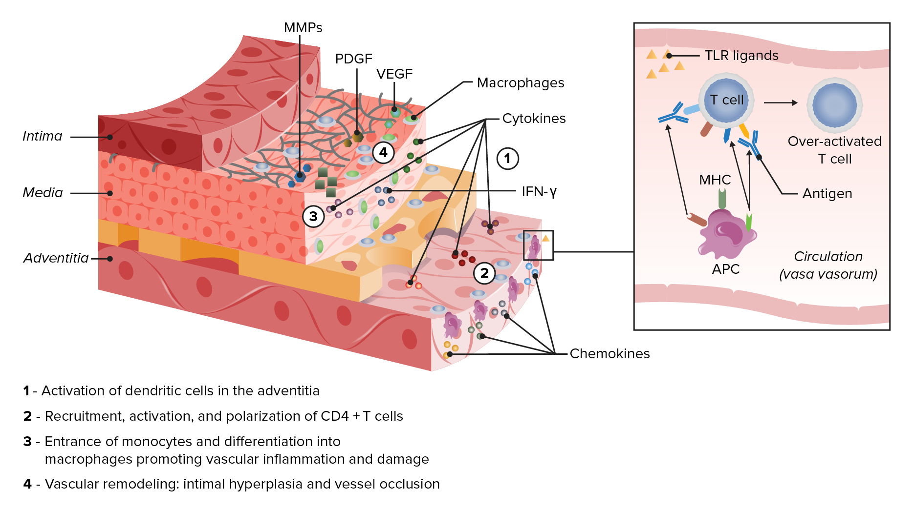
An antigenAntigenSubstances that are recognized by the immune system and induce an immune reaction.Vaccination is sensed by dendritic cellsDendritic cellsSpecialized cells of the hematopoietic system that have branch-like extensions. They are found throughout the lymphatic system, and in non-lymphoid tissues such as skin and the epithelia of the intestinal, respiratory, and reproductive tracts. They trap and process antigens, and present them to T-cells, thereby stimulating cell-mediated immunity. They are different from the non-hematopoietic follicular dendritic cells, which have a similar morphology and immune system function, but with respect to humoral immunity (antibody production).Skin: Structure and Functions in the adventitia → recruits more dendritic cellsDendritic cellsSpecialized cells of the hematopoietic system that have branch-like extensions. They are found throughout the lymphatic system, and in non-lymphoid tissues such as skin and the epithelia of the intestinal, respiratory, and reproductive tracts. They trap and process antigens, and present them to T-cells, thereby stimulating cell-mediated immunity. They are different from the non-hematopoietic follicular dendritic cells, which have a similar morphology and immune system function, but with respect to humoral immunity (antibody production).Skin: Structure and Functions, lymphocytesLymphocytesLymphocytes are heterogeneous WBCs involved in immune response. Lymphocytes develop from the bone marrow, starting from hematopoietic stem cells (HSCs) and progressing to common lymphoid progenitors (CLPs). B and T lymphocytes and natural killer (NK) cells arise from the lineage.Lymphocytes: Histology, and macrophagesMacrophagesThe relatively long-lived phagocytic cell of mammalian tissues that are derived from blood monocytes. Main types are peritoneal macrophages; alveolar macrophages; histiocytes; kupffer cells of the liver; and osteoclasts. They may further differentiate within chronic inflammatory lesions to epithelioid cells or may fuse to form foreign body giant cells or langhans giant cells.Innate Immunity: Phagocytes and Antigen Presentation.
Dendritic cellsDendritic cellsSpecialized cells of the hematopoietic system that have branch-like extensions. They are found throughout the lymphatic system, and in non-lymphoid tissues such as skin and the epithelia of the intestinal, respiratory, and reproductive tracts. They trap and process antigens, and present them to T-cells, thereby stimulating cell-mediated immunity. They are different from the non-hematopoietic follicular dendritic cells, which have a similar morphology and immune system function, but with respect to humoral immunity (antibody production).Skin: Structure and Functions process and present the antigens → cluster of differentiation 4 (CD4+) T cell activationT cell activationAdaptive Cell-mediated Immunity.
CD4+ T cellsT cellsLymphocytes responsible for cell-mediated immunity. Two types have been identified – cytotoxic (t-lymphocytes, cytotoxic) and helper T-lymphocytes (t-lymphocytes, helper-inducer). They are formed when lymphocytes circulate through the thymus gland and differentiate to thymocytes. When exposed to an antigen, they divide rapidly and produce large numbers of new T cells sensitized to that antigen.T cells: Types and Functions proliferate → vascular inflammationInflammationInflammation is a complex set of responses to infection and injury involving leukocytes as the principal cellular mediators in the body’s defense against pathogenic organisms. Inflammation is also seen as a response to tissue injury in the process of wound healing. The 5 cardinal signs of inflammation are pain, heat, redness, swelling, and loss of function. Inflammation via activation of macrophagesMacrophagesThe relatively long-lived phagocytic cell of mammalian tissues that are derived from blood monocytes. Main types are peritoneal macrophages; alveolar macrophages; histiocytes; kupffer cells of the liver; and osteoclasts. They may further differentiate within chronic inflammatory lesions to epithelioid cells or may fuse to form foreign body giant cells or langhans giant cells.Innate Immunity: Phagocytes and Antigen Presentation.
Medial layer is invaded by inflammatory cells → macrophagesMacrophagesThe relatively long-lived phagocytic cell of mammalian tissues that are derived from blood monocytes. Main types are peritoneal macrophages; alveolar macrophages; histiocytes; kupffer cells of the liver; and osteoclasts. They may further differentiate within chronic inflammatory lesions to epithelioid cells or may fuse to form foreign body giant cells or langhans giant cells.Innate Immunity: Phagocytes and Antigen Presentation undergo granulomatous organization (giant cellsGiant cellsMultinucleated masses produced by the fusion of many cells; often associated with viral infections. In aids, they are induced when the envelope glycoprotein of the HIV virus binds to the CD4 antigen of uninfected neighboring T4 cells. The resulting syncytium leads to cell death and thus may account for the cytopathic effect of the virus.Giant Cell Arteritis) → growth factors and metalloproteinases produced → loss of vascular smooth muscle cells and destruction of the internal elasticElasticConnective Tissue: Histology lamina.
Injured smooth muscle cells respond to this damage → myofibroblastsMyofibroblastsSpindle-shaped cells with characteristic contractile proteins and structures that contribute to the wound healing process. They occur in granulation tissue and also in pathological processes such as fibrosis.Hypertrophic and Keloid Scars differentiate, migrate toward the intimal layer, and produce extracellular matrixExtracellular matrixA meshwork-like substance found within the extracellular space and in association with the basement membrane of the cell surface. It promotes cellular proliferation and provides a supporting structure to which cells or cell lysates in culture dishes adhere.Hypertrophic and Keloid ScarsproteinsProteinsLinear polypeptides that are synthesized on ribosomes and may be further modified, crosslinked, cleaved, or assembled into complex proteins with several subunits. The specific sequence of amino acids determines the shape the polypeptide will take, during protein folding, and the function of the protein.Energy Homeostasis → intimal hyperplasiaHyperplasiaAn increase in the number of cells in a tissue or organ without tumor formation. It differs from hypertrophy, which is an increase in bulk without an increase in the number of cells.Cellular Adaptation and arterial occlusion → ischemic complications of the disease.
Elastic stains on the same case of a histologically positive temporal artery biopsy showing the fragmentation, distortion, and lack of continuity of the internal elastic lamina, a characteristic feature of temporal arteritis
Image: “Association between human papillomavirus DNA and temporal arteritis” by Mohammadi, A., Pfeifer, J.D., Lewis, J.S. License: CC BY 2.0.
Pathophysiology in giant cell arteritis Notice the activation of T cells through presentation of an antigen by dendritic cells (APC). These recruit and activate more inflammatory cells, including macrophages, through toll-like receptor ligands (TLR), cytokines, and chemokines. Macrophages will organize into giant cells, and produce matrix metalloproteinases (MMPs) and growth factors (PDGF and VEGF). This causes the destruction and hyperplasia that will eventually cause vessel occlusion.
Symptom onset is typically subacute, developing over weeks to months. More abrupt presentations, with symptoms arising over a few days; occurs in a minority of cases.
Constitutional:
FeverFeverFever is defined as a measured body temperature of at least 38°C (100.4°F). Fever is caused by circulating endogenous and/or exogenous pyrogens that increase levels of prostaglandin E2 in the hypothalamus. Fever is commonly associated with chills, rigors, sweating, and flushing of the skin. Fever (typically low-grade)
FatigueFatigueThe state of weariness following a period of exertion, mental or physical, characterized by a decreased capacity for work and reduced efficiency to respond to stimuli.Fibromyalgia
AnorexiaAnorexiaThe lack or loss of appetite accompanied by an aversion to food and the inability to eat. It is the defining characteristic of the disorder anorexia nervosa.Anorexia Nervosa
Head and neckNeckThe part of a human or animal body connecting the head to the rest of the body.Peritonsillar Abscess:
HeadacheHeadacheThe symptom of pain in the cranial region. It may be an isolated benign occurrence or manifestation of a wide variety of headache disorders.Brain Abscess:
Occurs in ⅔ of patientsPatientsIndividuals participating in the health care system for the purpose of receiving therapeutic, diagnostic, or preventive procedures.Clinician–Patient Relationship
Varying severity and qualityQualityActivities and programs intended to assure or improve the quality of care in either a defined medical setting or a program. The concept includes the assessment or evaluation of the quality of care; identification of problems or shortcomings in the delivery of care; designing activities to overcome these deficiencies; and follow-up monitoring to ensure effectiveness of corrective steps.Quality Measurement and Improvement
Localized to the temples and can be tender to the touch
Occurs in about ½ of patientsPatientsIndividuals participating in the health care system for the purpose of receiving therapeutic, diagnostic, or preventive procedures.Clinician–Patient Relationship
Mandibular painPainAn unpleasant sensation induced by noxious stimuli which are detected by nerve endings of nociceptive neurons.Pain: Types and Pathways or fatigueFatigueThe state of weariness following a period of exertion, mental or physical, characterized by a decreased capacity for work and reduced efficiency to respond to stimuli.Fibromyalgia during masticationMasticationThe act and process of chewing and grinding food in the mouth.Jaw and Temporomandibular Joint: Anatomy that is relieved with rest
Ophthalmic:
Transient visionVisionOphthalmic Exam loss (amaurosis fugaxAmaurosis fugaxTransient complete or partial monocular blindness due to retinal ischemia. This may be caused by emboli from the carotid artery (usually in association with carotid stenosis) and other locations that enter the central retinal artery.Carotid Artery Stenosis):
Blurring, diplopiaDiplopiaA visual symptom in which a single object is perceived by the visual cortex as two objects rather than one. Disorders associated with this condition include refractive errors; strabismus; oculomotor nerve diseases; trochlear nerve diseases; abducens nerve diseases; and diseases of the brain stem and occipital lobe.Myasthenia Gravis, or visionVisionOphthalmic Exam loss
Polymyalgia rheumaticaPolymyalgia rheumaticaA syndrome in the elderly characterized by proximal joint and muscle pain, high erythrocyte sedimentation rate, and a self-limiting course. Pain is usually accompanied by evidence of an inflammatory reaction. Women are affected twice as commonly as men and caucasians more frequently than other groups. The condition is frequently associated with giant cell arteritis and some theories pose the possibility that the two diseases arise from a single etiology or even that they are the same entity.Giant Cell Arteritis:
Aching and morning stiffness in the proximal muscles and joints
Shoulders, hip girdles, neckNeckThe part of a human or animal body connecting the head to the rest of the body.Peritonsillar Abscess, and torso affected
Bruits over carotid, subclavian, or axillary arteriesArteriesArteries are tubular collections of cells that transport oxygenated blood and nutrients from the heart to the tissues of the body. The blood passes through the arteries in order of decreasing luminal diameter, starting in the largest artery (the aorta) and ending in the small arterioles. Arteries are classified into 3 types: large elastic arteries, medium muscular arteries, and small arteries and arterioles. Arteries: Histology
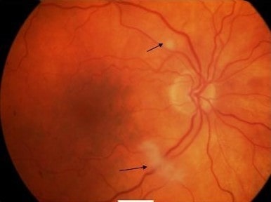
Retinal ischemiaIschemiaA hypoperfusion of the blood through an organ or tissue caused by a pathologic constriction or obstruction of its blood vessels, or an absence of blood circulation.Ischemic Cell Damage → cotton-wool spots in the retinaRetinaThe ten-layered nervous tissue membrane of the eye. It is continuous with the optic nerve and receives images of external objects and transmits visual impulses to the brain. Its outer surface is in contact with the choroid and the inner surface with the vitreous body. The outermost layer is pigmented, whereas the inner nine layers are transparent.Eye: Anatomy
Anterior ischemic optic neuropathyNeuropathyLeprosy (loss of blood flowBlood flowBlood flow refers to the movement of a certain volume of blood through the vasculature over a given unit of time (e.g., mL per minute).Vascular Resistance, Flow, and Mean Arterial Pressure to the anterior portion of the optic nerveOptic nerveThe 2nd cranial nerve which conveys visual information from the retina to the brain. The nerve carries the axons of the retinal ganglion cells which sort at the optic chiasm and continue via the optic tracts to the brain. The largest projection is to the lateral geniculate nuclei; other targets include the superior colliculi and the suprachiasmatic nuclei. Though known as the second cranial nerve, it is considered part of the central nervous system.The 12 Cranial Nerves: Overview and Functions) → swollen, pale disc and blurred margins of the retinaRetinaThe ten-layered nervous tissue membrane of the eye. It is continuous with the optic nerve and receives images of external objects and transmits visual impulses to the brain. Its outer surface is in contact with the choroid and the inner surface with the vitreous body. The outermost layer is pigmented, whereas the inner nine layers are transparent.Eye: Anatomy
Musculoskeletal:
Limited shoulder, neckNeckThe part of a human or animal body connecting the head to the rest of the body.Peritonsillar Abscess, and hip range of motionRange of motionThe distance and direction to which a bone joint can be extended. Range of motion is a function of the condition of the joints, muscles, and connective tissues involved. Joint flexibility can be improved through appropriate muscle strength exercises.Examination of the Upper Limbs
Fundoscopy of the right eye, showing cotton-wool spots along the superotemporal and inferotemporal arterioles (arrows). The cotton-wool spots denote obstruction of the retinal arterioles, leading to retinal ischemia.
Image: “Cotton-wool spots” by James Paget University Hospital NHS Foundation Trust, Lowestoft Road, Gorleston, Great Yarmouth NR31 6LA, Norfolk, UK. License: CC BY 2.0.
Diagnosis
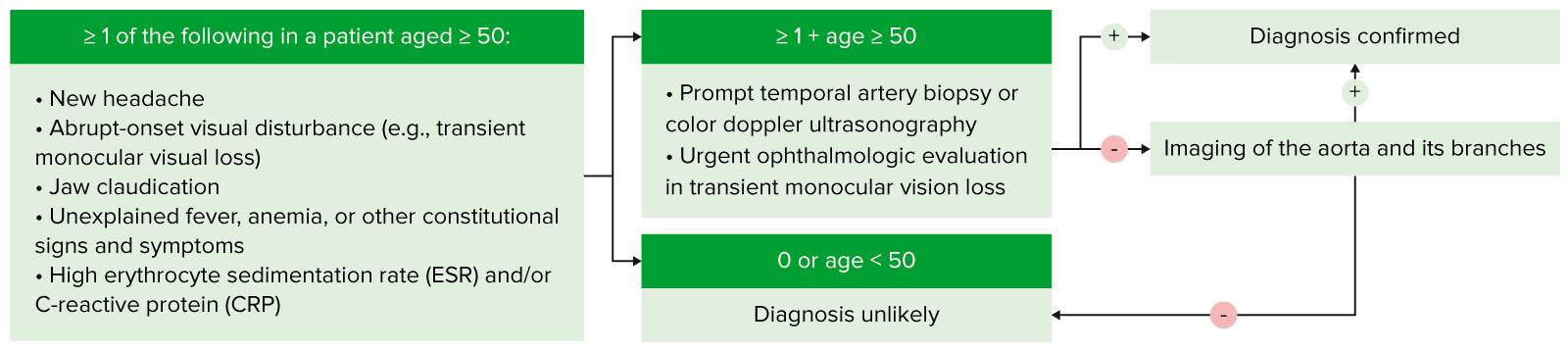
Diagnostic algorithm
The above is a simple algorithm to help aid in the diagnosis of GCA. This outlines when this disease should be considered, and the basic steps in diagnosis.
ESRESRSoft Tissue Abscess ≥ 50 mm/hr owing to rouleau formation of RBCsRBCsErythrocytes, or red blood cells (RBCs), are the most abundant cells in the blood. While erythrocytes in the fetus are initially produced in the yolk sac then the liver, the bone marrow eventually becomes the main site of production.Erythrocytes: Histology
Non-specific
Complete blood count:
Normochromic anemiaAnemiaAnemia is a condition in which individuals have low Hb levels, which can arise from various causes. Anemia is accompanied by a reduced number of RBCs and may manifest with fatigue, shortness of breath, pallor, and weakness. Subtypes are classified by the size of RBCs, chronicity, and etiology. Anemia: Overview and Types
Reactive thrombocytosis
Normal or minimally elevated leukocytesLeukocytesWhite blood cells. These include granular leukocytes (basophils; eosinophils; and neutrophils) as well as non-granular leukocytes (lymphocytes and monocytes).White Myeloid Cells: Histology
LiverLiverThe liver is the largest gland in the human body. The liver is found in the superior right quadrant of the abdomen and weighs approximately 1.5 kilograms. Its main functions are detoxification, metabolism, nutrient storage (e.g., iron and vitamins), synthesis of coagulation factors, formation of bile, filtration, and storage of blood. Liver: Anatomy function panel:
Elevated alkaline phosphataseAlkaline PhosphataseAn enzyme that catalyzes the conversion of an orthophosphoric monoester and water to an alcohol and orthophosphate.Osteosarcoma
Decreased albuminAlbuminSerum albumin from humans. It is an essential carrier of both endogenous substances, such as fatty acids and bilirubin, and of xenobiotics in the blood.Liver Function Tests
Should be performed in all patientsPatientsIndividuals participating in the health care system for the purpose of receiving therapeutic, diagnostic, or preventive procedures.Clinician–Patient Relationship with suspected GCAGCAGiant cell arteritis (GCA), also known as temporal arteritis, is a type of large-vessel vasculitis that predominantly affects the aorta and its major branches, with a predilection for the branches of the carotid (including the temporal artery). Giant cell arteritis is defined by inflammatory leukocytes in the vessel walls leading to reactive damage, ischemia, and necrosis.Giant Cell Arteritis
Lesions are segmental, so diagnosis requires a biopsyBiopsyRemoval and pathologic examination of specimens from the living body.Ewing Sarcoma of a long vessel segment (1–2 cm in vivo length).
Giant cellsGiant cellsMultinucleated masses produced by the fusion of many cells; often associated with viral infections. In aids, they are induced when the envelope glycoprotein of the HIV virus binds to the CD4 antigen of uninfected neighboring T4 cells. The resulting syncytium leads to cell death and thus may account for the cytopathic effect of the virus.Giant Cell Arteritis
Inadequate length of biopsyBiopsyRemoval and pathologic examination of specimens from the living body.Ewing Sarcoma specimen obtained
Commencing steroid treatment prior to biopsyBiopsyRemoval and pathologic examination of specimens from the living body.Ewing Sarcoma
Sparing of the temporal arteriesTemporal ArteriesArteries arising from the external carotid or the maxillary artery and distributing to the temporal region.Jaw and Temporomandibular Joint: Anatomy, particularly in patientsPatientsIndividuals participating in the health care system for the purpose of receiving therapeutic, diagnostic, or preventive procedures.Clinician–Patient Relationship with large-vessel involvement
Ordering:
Schedule temporal artery biopsyTemporal Artery BiopsyGiant Cell Arteritis within 2–4 weeks after starting steroidsSteroidsA group of polycyclic compounds closely related biochemically to terpenes. They include cholesterol, numerous hormones, precursors of certain vitamins, bile acids, alcohols (sterols), and certain natural drugs and poisons. Steroids have a common nucleus, a fused, reduced 17-carbon atom ring system, cyclopentanoperhydrophenanthrene. Most steroids also have two methyl groups and an aliphatic side-chain attached to the nucleus.Benign Liver Tumors if treatment is initiated prior to histopathologic diagnosis.
Biopsies can be obtained on an outpatient basis under local anesthesiaAnesthesiaA state characterized by loss of feeling or sensation. This depression of nerve function is usually the result of pharmacologic action and is induced to allow performance of surgery or other painful procedures.Anesthesiology: History and Basic Concepts.
Can be performed by an ophthalmologist, neurosurgeon, vascular surgeon, or other surgical specialist
Color DopplerDopplerUltrasonography applying the doppler effect, with frequency-shifted ultrasound reflections produced by moving targets (usually red blood cells) in the bloodstream along the ultrasound axis in direct proportion to the velocity of movement of the targets, to determine both direction and velocity of blood flow.Ultrasound (Sonography) ultrasound of temporal arteriesTemporal ArteriesArteries arising from the external carotid or the maxillary artery and distributing to the temporal region.Jaw and Temporomandibular Joint: Anatomy:
Should evaluate the vessels of the head, neckNeckThe part of a human or animal body connecting the head to the rest of the body.Peritonsillar Abscess, and upper extremities
“Halo signHalo signAspergillus/Aspergillosis” (darkened area surrounding the vascular lumen from mural edemaEdemaEdema is a condition in which excess serous fluid accumulates in the body cavity or interstitial space of connective tissues. Edema is a symptom observed in several medical conditions. It can be categorized into 2 types, namely, peripheral (in the extremities) and internal (in an organ or body cavity). Edema) and a non-compressible artery are highly specific.
Ideally performed before steroid therapy is commenced, or as soon as possible afterward, to improve diagnostic sensitivity.
If ultrasound results are equivocal or negative and suspicion remains, proceed with biopsyBiopsyRemoval and pathologic examination of specimens from the living body.Ewing Sarcoma.
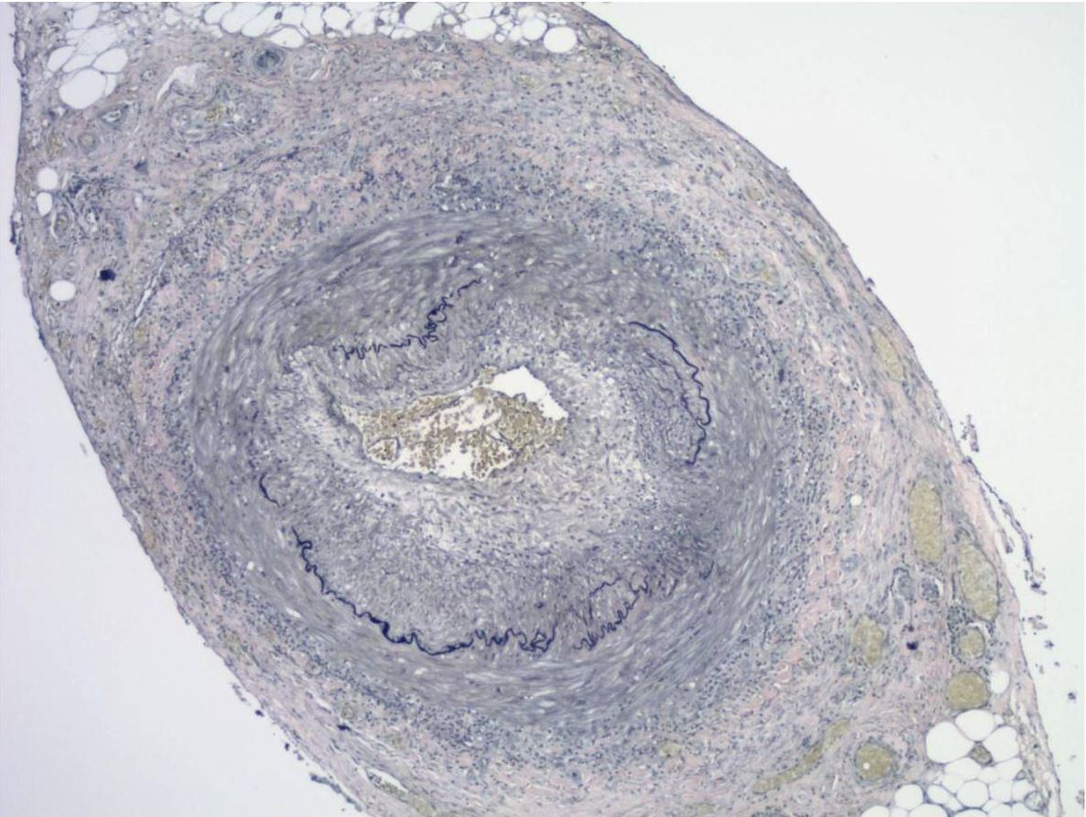
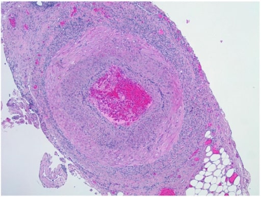
Biopsy of temporal artery showing transmural mixed inflammatory cell infiltrates with intimal thickening, fragmentation, and distortion of the internal elastic lamina
Image: “Biopsy of temporal artery” by the Department of Pathology and Laboratory Medicine, University of Florida, College of Medicine, Jacksonville, FL, USA. License: CC BY 2.0.
If the above workup is not diagnostic, consider evaluation for large-vessel involvement:[6,8,10,13]
Options:
Color DopplerDopplerUltrasonography applying the doppler effect, with frequency-shifted ultrasound reflections produced by moving targets (usually red blood cells) in the bloodstream along the ultrasound axis in direct proportion to the velocity of movement of the targets, to determine both direction and velocity of blood flow.Ultrasound (Sonography) ultrasound of axillary and subclavian arteriesArteriesArteries are tubular collections of cells that transport oxygenated blood and nutrients from the heart to the tissues of the body. The blood passes through the arteries in order of decreasing luminal diameter, starting in the largest artery (the aorta) and ending in the small arterioles. Arteries are classified into 3 types: large elastic arteries, medium muscular arteries, and small arteries and arterioles. Arteries: Histology
Computed tomography with angiographyAngiographyRadiography of blood vessels after injection of a contrast medium.Cardiac Surgery (CTACTAA non-invasive method that uses a ct scanner for capturing images of blood vessels and tissues. A contrast material is injected, which helps produce detailed images that aid in diagnosing vascular diseases.Pulmonary Function Tests)
Positron emission tomography (PETPETAn imaging technique that combines a positron-emission tomography (PET) scanner and a ct X ray scanner. This establishes a precise anatomic localization in the same session.Nuclear Imaging)
Choice of CTACTAA non-invasive method that uses a ct scanner for capturing images of blood vessels and tissues. A contrast material is injected, which helps produce detailed images that aid in diagnosing vascular diseases.Pulmonary Function Tests versus MRAMRAImaging of the Heart and Great Vessels is based on clinicianClinicianA physician, nurse practitioner, physician assistant, or another health professional who is directly involved in patient care and has a professional relationship with patients.Clinician–Patient Relationship preference and availability of local resources.
MRAMRAImaging of the Heart and Great Vessels is superior to CTACTAA non-invasive method that uses a ct scanner for capturing images of blood vessels and tissues. A contrast material is injected, which helps produce detailed images that aid in diagnosing vascular diseases.Pulmonary Function Tests/PETPETAn imaging technique that combines a positron-emission tomography (PET) scanner and a ct X ray scanner. This establishes a precise anatomic localization in the same session.Nuclear Imaging for cranial involvement.
2022 American College of Rheumatology (ACR)/European Alliance of Associations for Rheumatology (EULAR) classification criteria[11]
Considerations:
Used when there is a diagnosis of medium- or large-vessel vasculitisVasculitisInflammation of any one of the blood vessels, including the arteries; veins; and rest of the vasculature system in the body.Systemic Lupus Erythematosus
Alternative diagnoses must be excluded.
Requirement: age ≥ 50 years at time of diagnosis
Scored on clinical and diagnostic findings (10 items)
≥ 6 points needed for GCAGCAGiant cell arteritis (GCA), also known as temporal arteritis, is a type of large-vessel vasculitis that predominantly affects the aorta and its major branches, with a predilection for the branches of the carotid (including the temporal artery). Giant cell arteritis is defined by inflammatory leukocytes in the vessel walls leading to reactive damage, ischemia, and necrosis.Giant Cell Arteritis classification
Table: ACR/EULAR classification criteria for GCAGCAGiant cell arteritis (GCA), also known as temporal arteritis, is a type of large-vessel vasculitis that predominantly affects the aorta and its major branches, with a predilection for the branches of the carotid (including the temporal artery). Giant cell arteritis is defined by inflammatory leukocytes in the vessel walls leading to reactive damage, ischemia, and necrosis.Giant Cell Arteritis
Category
criteria
Points
Clinical
Morning stiffness affecting shoulders and/or neckNeckThe part of a human or animal body connecting the head to the rest of the body.Peritonsillar Abscess
Claudication of jawJawThe jaw is made up of the mandible, which comprises the lower jaw, and the maxilla, which comprises the upper jaw. The mandible articulates with the temporal bone via the temporomandibular joint (TMJ). The 4 muscles of mastication produce the movements of the TMJ to ensure the efficient chewing of food. Jaw and Temporomandibular Joint: Anatomy or tongueTongueThe tongue, on the other hand, is a complex muscular structure that permits tasting and facilitates the process of mastication and communication. The blood supply of the tongue originates from the external carotid artery, and the innervation is through cranial nerves.Lips and Tongue: Anatomy
2
New temporal headacheHeadacheThe symptom of pain in the cranial region. It may be an isolated benign occurrence or manifestation of a wide variety of headache disorders.Brain Abscess
2
Scalp tenderness
2
Abnormal findings of the temporal artery, such as:
ACR: American College of Rheumatology
EULAR: European Alliance of Associations for Rheumatology
GCA: giant cell arteritis
ESR:erythrocyte sedimentation rate
FDG-PET: fluorodeoxyglucose positron emission tomography
Management
It is very important not to delay treatment while awaiting biopsyBiopsyRemoval and pathologic examination of specimens from the living body.Ewing Sarcoma results in order to minimize the risk of visionVisionOphthalmic Exam loss. The following information is based on US and UK literature and guidelines. Management may vary based on practice location.
Consultants[8,13]
Rheumatology: all suspected/confirmed disease
Ophthalmology: presence/risk of visual symptoms
Vascular surgeryVascular surgeryVascular surgery is the specialized field of medicine that focuses on the surgical management of the pathologies of the peripheral circulation. The main goal of most vascular procedures is to restore circulatory function to the affected vessels by relieving occlusions or by redirecting blood flow (e.g., bypass).Vascular Surgery: large-vessel involvement
Systemic glucocorticoidsSystemic GlucocorticoidsGlucocorticoids are the mainstay of treatment, and treatment should be initiated promptly if there is a strong suspicion of GCAGCAGiant cell arteritis (GCA), also known as temporal arteritis, is a type of large-vessel vasculitis that predominantly affects the aorta and its major branches, with a predilection for the branches of the carotid (including the temporal artery). Giant cell arteritis is defined by inflammatory leukocytes in the vessel walls leading to reactive damage, ischemia, and necrosis.Giant Cell Arteritis.
PrednisonePrednisoneA synthetic anti-inflammatory glucocorticoid derived from cortisone. It is biologically inert and converted to prednisolone in the liver.Immunosuppressants/prednisolonePrednisoloneA glucocorticoid with the general properties of the corticosteroids. It is the drug of choice for all conditions in which routine systemic corticosteroid therapy is indicated, except adrenal deficiency states.Immunosuppressants
Followed by oral prednisonePrednisoneA synthetic anti-inflammatory glucocorticoid derived from cortisone. It is biologically inert and converted to prednisolone in the liver.Immunosuppressants/prednisolonePrednisoloneA glucocorticoid with the general properties of the corticosteroids. It is the drug of choice for all conditions in which routine systemic corticosteroid therapy is indicated, except adrenal deficiency states.Immunosuppressants, as above
Continue initial dose until symptoms and inflammatory markers improve.
Tapering:[8,9,13]
Should occur slowly over 6–12 months (total length of treatment is generally ≥ 2 years)
Optimal tapering regimen is unknown, but the following is based on consensus guidelines:
Consider a reduction of 10 mg every 2 weeks after 2–4 weeks of initial therapy if:
Once at 20 mg/day, reduce by 2.5 mg every 2 weeks to a dose of 10 mg/day if there are no flares.
Finally, tapering should be slowed (e.g., 1 mg every 1–2 months) to eventually wean off steroidsSteroidsA group of polycyclic compounds closely related biochemically to terpenes. They include cholesterol, numerous hormones, precursors of certain vitamins, bile acids, alcohols (sterols), and certain natural drugs and poisons. Steroids have a common nucleus, a fused, reduced 17-carbon atom ring system, cyclopentanoperhydrophenanthrene. Most steroids also have two methyl groups and an aliphatic side-chain attached to the nucleus.Benign Liver Tumors within 6–12 months.
The full list of glucocorticoid therapy side effects can be found here.
Gastric ulcerationUlcerationCorneal Abrasions, Erosion, and Ulcers, osteoporosisOsteoporosisOsteoporosis refers to a decrease in bone mass and density leading to an increased number of fractures. There are 2 forms of osteoporosis: primary, which is commonly postmenopausal or senile; and secondary, which is a manifestation of immobilization, underlying medical disorders, or long-term use of certain medications. Osteoporosis, and immunosuppression can be mitigated by implementing the following at the start of treatment:
ScreeningScreeningPreoperative Care tests for tuberculosisTuberculosisTuberculosis (TB) is an infectious disease caused by Mycobacterium tuberculosis complex bacteria. The bacteria usually attack the lungs but can also damage other parts of the body. Approximately 30% of people around the world are infected with this pathogen, with the majority harboring a latent infection. Tuberculosis spreads through the air when a person with active pulmonary infection coughs or sneezes. Tuberculosis
Immunization against influenzaInfluenzaInfluenza viruses are members of the Orthomyxoviridae family and the causative organisms of influenza, a highly contagious febrile respiratory disease. There are 3 primary influenza viruses (A, B, and C) and various subtypes, which are classified based on their virulent surface antigens, hemagglutinin (HA) and neuraminidase (NA). Influenza typically presents with a fever, myalgia, headache, and symptoms of an upper respiratory infection. Influenza Viruses/Influenza and pneumococcal pneumoniaPneumoniaPneumonia or pulmonary inflammation is an acute or chronic inflammation of lung tissue. Causes include infection with bacteria, viruses, or fungi. In more rare cases, pneumonia can also be caused through toxic triggers through inhalation of toxic substances, immunological processes, or in the course of radiotherapy.Pneumonia
BoneBoneBone is a compact type of hardened connective tissue composed of bone cells, membranes, an extracellular mineralized matrix, and central bone marrow. The 2 primary types of bone are compact and spongy. Bones: Structure and Types mineral density measurements, adequate dietary calciumCalciumA basic element found in nearly all tissues. It is a member of the alkaline earth family of metals with the atomic symbol ca, atomic number 20, and atomic weight 40. Calcium is the most abundant mineral in the body and combines with phosphorus to form calcium phosphate in the bones and teeth. It is essential for the normal functioning of nerves and muscles and plays a role in blood coagulation (as factor IV) and in many enzymatic processes.Electrolytes, and vitamin DVitamin DA vitamin that includes both cholecalciferols and ergocalciferols, which have the common effect of preventing or curing rickets in animals. It can also be viewed as a hormone since it can be formed in skin by action of ultraviolet rays upon the precursors, 7-dehydrocholesterol and ergosterol, and acts on vitamin D receptors to regulate calcium in opposition to parathyroid hormone.Fat-soluble Vitamins and their Deficiencies intake/supplementation
ElectrolytesElectrolytesElectrolytes are mineral salts that dissolve in water and dissociate into charged particles called ions, which can be either be positively (cations) or negatively (anions) charged. Electrolytes are distributed in the extracellular and intracellular compartments in different concentrations. Electrolytes are essential for various basic life-sustaining functions.Electrolytes
Renal function
GlucoseGlucoseA primary source of energy for living organisms. It is naturally occurring and is found in fruits and other parts of plants in its free state. It is used therapeutically in fluid and nutrient replacement.Lactose Intolerance
Adjunctive treatment[8–10]
May be used when glucocorticoidsGlucocorticoidsGlucocorticoids are a class within the corticosteroid family. Glucocorticoids are chemically and functionally similar to endogenous cortisol. There are a wide array of indications, which primarily benefit from the antiinflammatory and immunosuppressive effects of this class of drugs.Glucocorticoids are not tolerated or in individuals at high risk for adverse effects of steroidsSteroidsA group of polycyclic compounds closely related biochemically to terpenes. They include cholesterol, numerous hormones, precursors of certain vitamins, bile acids, alcohols (sterols), and certain natural drugs and poisons. Steroids have a common nucleus, a fused, reduced 17-carbon atom ring system, cyclopentanoperhydrophenanthrene. Most steroids also have two methyl groups and an aliphatic side-chain attached to the nucleus.Benign Liver Tumors, including those with:
Significant premorbid disease (e.g., diabetesDiabetesDiabetes mellitus (DM) is a metabolic disease characterized by hyperglycemia and dysfunction of the regulation of glucose metabolism by insulin. Type 1 DM is diagnosed mostly in children and young adults as the result of autoimmune destruction of β cells in the pancreas and the resulting lack of insulin. Type 2 DM has a significant association with obesity and is characterized by insulin resistance.Diabetes Mellitus, osteoporosisOsteoporosisOsteoporosis refers to a decrease in bone mass and density leading to an increased number of fractures. There are 2 forms of osteoporosis: primary, which is commonly postmenopausal or senile; and secondary, which is a manifestation of immobilization, underlying medical disorders, or long-term use of certain medications. Osteoporosis, obesityObesityObesity is a condition associated with excess body weight, specifically with the deposition of excessive adipose tissue. Obesity is considered a global epidemic. Major influences come from the western diet and sedentary lifestyles, but the exact mechanisms likely include a mixture of genetic and environmental factors. Obesity)
Emergence of significant steroid-related side effects during treatment
IL-6 receptorReceptorReceptors are proteins located either on the surface of or within a cell that can bind to signaling molecules known as ligands (e.g., hormones) and cause some type of response within the cell.Receptors antagonist
Use recommended by ACR in addition to oral glucocorticoidsGlucocorticoidsGlucocorticoids are a class within the corticosteroid family. Glucocorticoids are chemically and functionally similar to endogenous cortisol. There are a wide array of indications, which primarily benefit from the antiinflammatory and immunosuppressive effects of this class of drugs.Glucocorticoids over steroid monotherapy.[10]
Typical dose:
162 mg SC weekly or every other week
Typically, given alongside steroid therapy
Keep in mind:
Increases risk of opportunistic infectionsInfectionsInvasion of the host organism by microorganisms or their toxins or by parasites that can cause pathological conditions or diseases.Chronic Granulomatous Disease
Contraindicated in individuals with concomitant severe active infectionsInfectionsInvasion of the host organism by microorganisms or their toxins or by parasites that can cause pathological conditions or diseases.Chronic Granulomatous Disease (excluding COVID-19COVID-19Coronavirus disease 2019 (COVID-19) is an infectious disease caused by the severe acute respiratory syndrome coronavirus 2 (SARS-CoV-2) that mainly affects the respiratory system but can also cause damage to other body systems (cardiovascular, gastrointestinal, renal, and central nervous systems).)
Do not initiate if hepatic enzymesEnzymesEnzymes are complex protein biocatalysts that accelerate chemical reactions without being consumed by them. Due to the body’s constant metabolic needs, the absence of enzymes would make life unsustainable, as reactions would occur too slowly without these molecules. Basics of Enzymes are > 5x upper limitLimitA value (e.g., pressure or time) that should not be exceeded and which is specified by the operator to protect the lungInvasive Mechanical Ventilation of normal or if neutrophilsNeutrophilsGranular leukocytes having a nucleus with three to five lobes connected by slender threads of chromatin, and cytoplasm containing fine inconspicuous granules and stainable by neutral dyes.Innate Immunity: Phagocytes and Antigen Presentation < 2 x 10⁹/L.
MethotrexateMethotrexateAn antineoplastic antimetabolite with immunosuppressant properties. It is an inhibitor of tetrahydrofolate dehydrogenase and prevents the formation of tetrahydrofolate, necessary for synthesis of thymidylate, an essential component of DNA.Antimetabolite Chemotherapy
Typical dose: 7.5–15 mg/wk orally
The evidence to support use of methotrexateMethotrexateAn antineoplastic antimetabolite with immunosuppressant properties. It is an inhibitor of tetrahydrofolate dehydrogenase and prevents the formation of tetrahydrofolate, necessary for synthesis of thymidylate, an essential component of DNA.Antimetabolite Chemotherapy is weaker than for tocilizumabTocilizumabImmunosuppressants, but it is cheaper and more widely available.
Additional pharmacologic therapy[8,10,13]
StatinsStatinsStatins are competitive inhibitors of HMG-CoA reductase in the liver. HMG-CoA reductase is the rate-limiting step in cholesterol synthesis. Inhibition results in lowered intrahepatocytic cholesterol formation, resulting in up-regulation of LDL receptors and, ultimately, lowering levels of serum LDL and triglycerides.Statins are not recommended for the treatment of GCAGCAGiant cell arteritis (GCA), also known as temporal arteritis, is a type of large-vessel vasculitis that predominantly affects the aorta and its major branches, with a predilection for the branches of the carotid (including the temporal artery). Giant cell arteritis is defined by inflammatory leukocytes in the vessel walls leading to reactive damage, ischemia, and necrosis.Giant Cell Arteritis.
AspirinAspirinThe prototypical analgesic used in the treatment of mild to moderate pain. It has anti-inflammatory and antipyretic properties and acts as an inhibitor of cyclooxygenase which results in the inhibition of the biosynthesis of prostaglandins. Aspirin also inhibits platelet aggregation and is used in the prevention of arterial and venous thrombosis.Nonsteroidal Antiinflammatory Drugs (NSAIDs):
ACR conditionally recommends for patientsPatientsIndividuals participating in the health care system for the purpose of receiving therapeutic, diagnostic, or preventive procedures.Clinician–Patient Relationship with flow-limiting lesions of the vertebral or carotid arteriesCarotid ArteriesEither of the two principal arteries on both sides of the neck that supply blood to the head and neck; each divides into two branches, the internal carotid artery and the external carotid artery.Carotid Arterial System: Anatomy.[10]
Royal College of PhysiciansPhysiciansIndividuals licensed to practice medicine.Clinician–Patient Relationship recommends for all patientsPatientsIndividuals participating in the health care system for the purpose of receiving therapeutic, diagnostic, or preventive procedures.Clinician–Patient Relationship with GCAGCAGiant cell arteritis (GCA), also known as temporal arteritis, is a type of large-vessel vasculitis that predominantly affects the aorta and its major branches, with a predilection for the branches of the carotid (including the temporal artery). Giant cell arteritis is defined by inflammatory leukocytes in the vessel walls leading to reactive damage, ischemia, and necrosis.Giant Cell Arteritis (if no contraindicationsContraindicationsA condition or factor associated with a recipient that makes the use of a drug, procedure, or physical agent improper or inadvisable. Contraindications may be absolute (life threatening) or relative (higher risk of complications in which benefits may outweigh risks).Noninvasive Ventilation).[13]
Additional counseling[8]
In addition to receiving education on their diagnosis, patientsPatientsIndividuals participating in the health care system for the purpose of receiving therapeutic, diagnostic, or preventive procedures.Clinician–Patient Relationship should also receive counseling related to:
SmokingSmokingWillful or deliberate act of inhaling and exhaling smoke from burning substances or agents held by hand.Interstitial Lung Diseases cessation
Diet (e.g., mitigate risk of hyperglycemiaHyperglycemiaAbnormally high blood glucose level.Diabetes Mellitus on glucocorticoid therapy)
Physical activity:
Activity may benefit patientsPatientsIndividuals participating in the health care system for the purpose of receiving therapeutic, diagnostic, or preventive procedures.Clinician–Patient Relationship with individuals with GCAGCAGiant cell arteritis (GCA), also known as temporal arteritis, is a type of large-vessel vasculitis that predominantly affects the aorta and its major branches, with a predilection for the branches of the carotid (including the temporal artery). Giant cell arteritis is defined by inflammatory leukocytes in the vessel walls leading to reactive damage, ischemia, and necrosis.Giant Cell Arteritis.
Stroke: disease in which poor blood flowBlood flowBlood flow refers to the movement of a certain volume of blood through the vasculature over a given unit of time (e.g., mL per minute).Vascular Resistance, Flow, and Mean Arterial Pressure to the brainBrainThe part of central nervous system that is contained within the skull (cranium). Arising from the neural tube, the embryonic brain is comprised of three major parts including prosencephalon (the forebrain); mesencephalon (the midbrain); and rhombencephalon (the hindbrain). The developed brain consists of cerebrum; cerebellum; and other structures in the brain stem.Nervous System: Anatomy, Structure, and Classification results in cell deathCell deathInjurious stimuli trigger the process of cellular adaptation, whereby cells respond to withstand the harmful changes in their environment. Overwhelmed adaptive mechanisms lead to cell injury. Mild stimuli produce reversible injury. If the stimulus is severe or persistent, injury becomes irreversible. Apoptosis is programmed cell death, a mechanism with both physiologic and pathologic effects.Cell Injury and Death. There are 2 types: ischemic strokeIschemic StrokeAn ischemic stroke (also known as cerebrovascular accident) is an acute neurologic injury that occurs as a result of brain ischemia; this condition may be due to cerebral blood vessel occlusion by thrombosis or embolism, or rarely due to systemic hypoperfusion. Ischemic Stroke due to lack of blood flowBlood flowBlood flow refers to the movement of a certain volume of blood through the vasculature over a given unit of time (e.g., mL per minute).Vascular Resistance, Flow, and Mean Arterial Pressure, and hemorrhagic strokeHemorrhagic strokeStroke due to rupture of a weakened blood vessel in the brain (e.g., cerebral hemispheres; cerebellum; subarachnoid space).Subarachnoid Hemorrhage due to bleeding. Symptoms include focal weakness, numbness, loss of visionVisionOphthalmic Exam, dysarthriaDysarthriaDisorders of speech articulation caused by imperfect coordination of pharynx, larynx, tongue, or face muscles. This may result from cranial nerve diseases; neuromuscular diseases; cerebellar diseases; basal ganglia diseases; brain stem diseases; or diseases of the corticobulbar tracts. The cortical language centers are intact in this condition.Wilson Disease, and headacheHeadacheThe symptom of pain in the cranial region. It may be an isolated benign occurrence or manifestation of a wide variety of headache disorders.Brain Abscess. Computed tomography and MRI of the brainBrainThe part of central nervous system that is contained within the skull (cranium). Arising from the neural tube, the embryonic brain is comprised of three major parts including prosencephalon (the forebrain); mesencephalon (the midbrain); and rhombencephalon (the hindbrain). The developed brain consists of cerebrum; cerebellum; and other structures in the brain stem.Nervous System: Anatomy, Structure, and Classification are used for the diagnosis, and will differentiate stroke from GCAGCAGiant cell arteritis (GCA), also known as temporal arteritis, is a type of large-vessel vasculitis that predominantly affects the aorta and its major branches, with a predilection for the branches of the carotid (including the temporal artery). Giant cell arteritis is defined by inflammatory leukocytes in the vessel walls leading to reactive damage, ischemia, and necrosis.Giant Cell Arteritis.
MigraineMigraineMigraine headache is a primary headache disorder and is among the most prevalent disorders in the world. Migraine is characterized by episodic, moderate to severe headaches that may be associated with increased sensitivity to light and sound, as well as nausea and/or vomiting. Migraine Headache: characterized by recurrent headaches that are moderate to severe, typically affecting ½ of the head, and pulsating in nature. Associated symptoms include nauseaNauseaAn unpleasant sensation in the stomach usually accompanied by the urge to vomit. Common causes are early pregnancy, sea and motion sickness, emotional stress, intense pain, food poisoning, and various enteroviruses.Antiemetics, vomitingVomitingThe forcible expulsion of the contents of the stomach through the mouth.Hypokalemia, and sensitivity to light, sound, or smellSmellThe sense of smell, or olfaction, begins in a small area on the roof of the nasal cavity, which is covered in specialized mucosa. From there, the olfactory nerve transmits the sensory perception of smell via the olfactory pathway. This pathway is composed of the olfactory cells and bulb, the tractus and striae olfactoriae, and the primary olfactory cortex and amygdala.Olfaction: Anatomy. Diagnosis is clinical, based on the history and physical exam. Laboratory and imaging studies will be unremarkable. The history, physical exam, and low inflammatory markers will help differentiate migraineMigraineMigraine headache is a primary headache disorder and is among the most prevalent disorders in the world. Migraine is characterized by episodic, moderate to severe headaches that may be associated with increased sensitivity to light and sound, as well as nausea and/or vomiting. Migraine Headache from GCAGCAGiant cell arteritis (GCA), also known as temporal arteritis, is a type of large-vessel vasculitis that predominantly affects the aorta and its major branches, with a predilection for the branches of the carotid (including the temporal artery). Giant cell arteritis is defined by inflammatory leukocytes in the vessel walls leading to reactive damage, ischemia, and necrosis.Giant Cell Arteritis.
GlaucomaGlaucomaGlaucoma is an optic neuropathy characterized by typical visual field defects and optic nerve atrophy seen as optic disc cupping on examination. The acute form of glaucoma is a medical emergency. Glaucoma is often, but not always, caused by increased intraocular pressure (IOP). Glaucoma: An increase in intraocular pressureIntraocular PressureThe pressure of the fluids in the eye.Ophthalmic Exam damages the optic nerveOptic nerveThe 2nd cranial nerve which conveys visual information from the retina to the brain. The nerve carries the axons of the retinal ganglion cells which sort at the optic chiasm and continue via the optic tracts to the brain. The largest projection is to the lateral geniculate nuclei; other targets include the superior colliculi and the suprachiasmatic nuclei. Though known as the second cranial nerve, it is considered part of the central nervous system.The 12 Cranial Nerves: Overview and Functions and causes blindnessBlindnessThe inability to see or the loss or absence of perception of visual stimuli. This condition may be the result of eye diseases; optic nerve diseases; optic chiasm diseases; or brain diseases affecting the visual pathways or occipital lobe.Retinopathy of Prematurity. Angle-closure glaucomaAngle-Closure GlaucomaGlaucoma symptoms include blurred visionBlurred VisionRetinal Detachment, halos around light, severe eye painPainAn unpleasant sensation induced by noxious stimuli which are detected by nerve endings of nociceptive neurons.Pain: Types and Pathways, headacheHeadacheThe symptom of pain in the cranial region. It may be an isolated benign occurrence or manifestation of a wide variety of headache disorders.Brain Abscess, nauseaNauseaAn unpleasant sensation in the stomach usually accompanied by the urge to vomit. Common causes are early pregnancy, sea and motion sickness, emotional stress, intense pain, food poisoning, and various enteroviruses.Antiemetics, and vomitingVomitingThe forcible expulsion of the contents of the stomach through the mouth.Hypokalemia. Diagnosis is made by an ophthalmology exam and intraocular pressureIntraocular PressureThe pressure of the fluids in the eye.Ophthalmic Exam measurements, which will differentiate glaucomaGlaucomaGlaucoma is an optic neuropathy characterized by typical visual field defects and optic nerve atrophy seen as optic disc cupping on examination. The acute form of glaucoma is a medical emergency. Glaucoma is often, but not always, caused by increased intraocular pressure (IOP). Glaucoma from GCAGCAGiant cell arteritis (GCA), also known as temporal arteritis, is a type of large-vessel vasculitis that predominantly affects the aorta and its major branches, with a predilection for the branches of the carotid (including the temporal artery). Giant cell arteritis is defined by inflammatory leukocytes in the vessel walls leading to reactive damage, ischemia, and necrosis.Giant Cell Arteritis.
Takayasu’s arteritis: a large-vessel, granulomatous vasculitisVasculitisInflammation of any one of the blood vessels, including the arteries; veins; and rest of the vasculature system in the body.Systemic Lupus Erythematosus that primarily affects the aortaAortaThe main trunk of the systemic arteries.Mediastinum and Great Vessels: Anatomy and its primary branches. PatientsPatientsIndividuals participating in the health care system for the purpose of receiving therapeutic, diagnostic, or preventive procedures.Clinician–Patient Relationship are often young or middle-aged women. Signs and symptoms include tenderness of the carotid artery, limb claudication, and absent or weak pulses. Elevated inflammatory markers and CTACTAA non-invasive method that uses a ct scanner for capturing images of blood vessels and tissues. A contrast material is injected, which helps produce detailed images that aid in diagnosing vascular diseases.Pulmonary Function Tests of the aortaAortaThe main trunk of the systemic arteries.Mediastinum and Great Vessels: Anatomy can aid in the diagnosis. Treatment includes glucocorticoidsGlucocorticoidsGlucocorticoids are a class within the corticosteroid family. Glucocorticoids are chemically and functionally similar to endogenous cortisol. There are a wide array of indications, which primarily benefit from the antiinflammatory and immunosuppressive effects of this class of drugs.Glucocorticoids and glucocorticoid-sparing agents. The patient’s demographics, history, and exam findings will differentiate Takayasu’s arteritis from GCAGCAGiant cell arteritis (GCA), also known as temporal arteritis, is a type of large-vessel vasculitis that predominantly affects the aorta and its major branches, with a predilection for the branches of the carotid (including the temporal artery). Giant cell arteritis is defined by inflammatory leukocytes in the vessel walls leading to reactive damage, ischemia, and necrosis.Giant Cell Arteritis.
Non-arteritic anterior ischemic optic neuropathyNeuropathyLeprosy: an idiopathicIdiopathicDermatomyositis, ischemic injury to the optic nerveOptic nerveThe 2nd cranial nerve which conveys visual information from the retina to the brain. The nerve carries the axons of the retinal ganglion cells which sort at the optic chiasm and continue via the optic tracts to the brain. The largest projection is to the lateral geniculate nuclei; other targets include the superior colliculi and the suprachiasmatic nuclei. Though known as the second cranial nerve, it is considered part of the central nervous system.The 12 Cranial Nerves: Overview and Functions. PatientsPatientsIndividuals participating in the health care system for the purpose of receiving therapeutic, diagnostic, or preventive procedures.Clinician–Patient Relationship present with sudden, painless, and monocular visionVisionOphthalmic Exam loss. A fundoscopic examFundoscopic ExamHead and Neck Examination will show optic discOptic discThe portion of the optic nerve seen in the fundus with the ophthalmoscope. It is formed by the meeting of all the retinal ganglion cell axons as they enter the optic nerve.Eye: AnatomyswellingSwellingInflammation. Giant cell arteritisGiant Cell ArteritisGiant cell arteritis (GCA), also known as temporal arteritis, is a type of large-vessel vasculitis that predominantly affects the aorta and its major branches, with a predilection for the branches of the carotid (including the temporal artery). Giant cell arteritis is defined by inflammatory leukocytes in the vessel walls leading to reactive damage, ischemia, and necrosis. Giant Cell Arteritis should be considered in these individuals, and can be ruled out with normal inflammatory markers. There is no known effective treatment, but patientsPatientsIndividuals participating in the health care system for the purpose of receiving therapeutic, diagnostic, or preventive procedures.Clinician–Patient Relationship are generally placed on aspirinAspirinThe prototypical analgesic used in the treatment of mild to moderate pain. It has anti-inflammatory and antipyretic properties and acts as an inhibitor of cyclooxygenase which results in the inhibition of the biosynthesis of prostaglandins. Aspirin also inhibits platelet aggregation and is used in the prevention of arterial and venous thrombosis.Nonsteroidal Antiinflammatory Drugs (NSAIDs).
Billing and Coding
Diagnosis Codes:
These codes are used by clinicians to document a diagnosis of Giant Cell ArteritisGiant Cell ArteritisGiant cell arteritis (GCA), also known as temporal arteritis, is a type of large-vessel vasculitis that predominantly affects the aorta and its major branches, with a predilection for the branches of the carotid (including the temporal artery). Giant cell arteritis is defined by inflammatory leukocytes in the vessel walls leading to reactive damage, ischemia, and necrosis. Giant Cell Arteritis (GCAGCAGiant cell arteritis (GCA), also known as temporal arteritis, is a type of large-vessel vasculitis that predominantly affects the aorta and its major branches, with a predilection for the branches of the carotid (including the temporal artery). Giant cell arteritis is defined by inflammatory leukocytes in the vessel walls leading to reactive damage, ischemia, and necrosis.Giant Cell Arteritis), a form of vasculitisVasculitisInflammation of any one of the blood vessels, including the arteries; veins; and rest of the vasculature system in the body.Systemic Lupus Erythematosus affecting large blood vessels, most notably the temporal arteriesTemporal ArteriesArteries arising from the external carotid or the maxillary artery and distributing to the temporal region.Jaw and Temporomandibular Joint: Anatomy. A related code for polymyalgia rheumaticaPolymyalgia rheumaticaA syndrome in the elderly characterized by proximal joint and muscle pain, high erythrocyte sedimentation rate, and a self-limiting course. Pain is usually accompanied by evidence of an inflammatory reaction. Women are affected twice as commonly as men and caucasians more frequently than other groups. The condition is frequently associated with giant cell arteritis and some theories pose the possibility that the two diseases arise from a single etiology or even that they are the same entity.Giant Cell Arteritis is often used concurrently.
Domain
Code
Description
ICD-10-CM
M31.6
Giant cell arteritisGiant Cell ArteritisGiant cell arteritis (GCA), also known as temporal arteritis, is a type of large-vessel vasculitis that predominantly affects the aorta and its major branches, with a predilection for the branches of the carotid (including the temporal artery). Giant cell arteritis is defined by inflammatory leukocytes in the vessel walls leading to reactive damage, ischemia, and necrosis. Giant Cell Arteritis
ICD-10-CM
M31.5
Giant cell arteritisGiant Cell ArteritisGiant cell arteritis (GCA), also known as temporal arteritis, is a type of large-vessel vasculitis that predominantly affects the aorta and its major branches, with a predilection for the branches of the carotid (including the temporal artery). Giant cell arteritis is defined by inflammatory leukocytes in the vessel walls leading to reactive damage, ischemia, and necrosis. Giant Cell Arteritis with polymyalgia rheumaticaPolymyalgia rheumaticaA syndrome in the elderly characterized by proximal joint and muscle pain, high erythrocyte sedimentation rate, and a self-limiting course. Pain is usually accompanied by evidence of an inflammatory reaction. Women are affected twice as commonly as men and caucasians more frequently than other groups. The condition is frequently associated with giant cell arteritis and some theories pose the possibility that the two diseases arise from a single etiology or even that they are the same entity.Giant Cell Arteritis
SNOMED CT
23145006
Giant cell arteritisGiant Cell ArteritisGiant cell arteritis (GCA), also known as temporal arteritis, is a type of large-vessel vasculitis that predominantly affects the aorta and its major branches, with a predilection for the branches of the carotid (including the temporal artery). Giant cell arteritis is defined by inflammatory leukocytes in the vessel walls leading to reactive damage, ischemia, and necrosis. Giant Cell Arteritis (disorder)
Evaluation & Workup:
This CPT code is used to bill for a temporal artery biopsyTemporal Artery BiopsyGiant Cell Arteritis, the gold standard procedure for confirming the diagnosis of GCAGCAGiant cell arteritis (GCA), also known as temporal arteritis, is a type of large-vessel vasculitis that predominantly affects the aorta and its major branches, with a predilection for the branches of the carotid (including the temporal artery). Giant cell arteritis is defined by inflammatory leukocytes in the vessel walls leading to reactive damage, ischemia, and necrosis.Giant Cell Arteritis by showing inflammationInflammationInflammation is a complex set of responses to infection and injury involving leukocytes as the principal cellular mediators in the body’s defense against pathogenic organisms. Inflammation is also seen as a response to tissue injury in the process of wound healing. The 5 cardinal signs of inflammation are pain, heat, redness, swelling, and loss of function. Inflammation of the vessel wall under a microscope.
Domain
Code
Description
CPT
37609
BiopsyBiopsyRemoval and pathologic examination of specimens from the living body.Ewing Sarcoma, temporal artery
CPT
85652
Sedimentation rate, erythrocyte, automated
CPT
86140
C-reactive protein
Medications:
These codes are used to prescribe and track the critical, vision-saving medications for GCAGCAGiant cell arteritis (GCA), also known as temporal arteritis, is a type of large-vessel vasculitis that predominantly affects the aorta and its major branches, with a predilection for the branches of the carotid (including the temporal artery). Giant cell arteritis is defined by inflammatory leukocytes in the vessel walls leading to reactive damage, ischemia, and necrosis.Giant Cell Arteritis, primarily high-dose corticosteroidsCorticosteroidsChorioretinitis (prednisonePrednisoneA synthetic anti-inflammatory glucocorticoid derived from cortisone. It is biologically inert and converted to prednisolone in the liver.Immunosuppressants) to rapidly control inflammationInflammationInflammation is a complex set of responses to infection and injury involving leukocytes as the principal cellular mediators in the body’s defense against pathogenic organisms. Inflammation is also seen as a response to tissue injury in the process of wound healing. The 5 cardinal signs of inflammation are pain, heat, redness, swelling, and loss of function. Inflammation, and newer biologics like tocilizumabTocilizumabImmunosuppressants.
Domain
Code
Description
RxNorm
8640
PrednisonePrednisoneA synthetic anti-inflammatory glucocorticoid derived from cortisone. It is biologically inert and converted to prednisolone in the liver.Immunosuppressants (ingredient)
PrednisonePrednisoneA synthetic anti-inflammatory glucocorticoid derived from cortisone. It is biologically inert and converted to prednisolone in the liver.Immunosuppressants
Complications & Supportive Procedures:
These ICD-10 codesICD-10 CodesGiant cell arteritis (Clinical) are used to document the most feared complications of untreated GCAGCAGiant cell arteritis (GCA), also known as temporal arteritis, is a type of large-vessel vasculitis that predominantly affects the aorta and its major branches, with a predilection for the branches of the carotid (including the temporal artery). Giant cell arteritis is defined by inflammatory leukocytes in the vessel walls leading to reactive damage, ischemia, and necrosis.Giant Cell Arteritis, including irreversible visionVisionOphthalmic Exam loss due to ischemic optic neuropathyNeuropathyLeprosy and the development of aortic aneurysms.
Kasper, D.L., Fauci, A.S., Longo, D.L., Braunwald, E., Hauser, S.L., & Jameson, J.L. (2007). Harrison’s principles of internal medicine (16th edition.). New York: McGraw Hill Education.
Al-Mousawi, A. Z., Gurney, S. P., Lorenzi, A. R., et al. (2019). Reviewing the pathophysiology behind the advances in the management of giant cell arteritis. Ophthalmology and Therapy, 8, 177–193. https://link.springer.com/article/10.1007%2Fs40123-019-0171-0
Ehlers, L., et al. (2019). 2018 EULAR recommendations for a core data set to support observational research and clinical care in giant cell arteritis. Annals of the Rheumatic Diseases, 78, 1160–1166. https://ard.bmj.com/content/78/9/1160