Deep vein thrombosisThrombosisFormation and development of a thrombus or blood clot in the blood vessel.Epidemic Typhus (DVTDVTDeep vein thrombosis (DVT) usually occurs in the deep veins of the lower extremities. The affected veins include the femoral, popliteal, iliofemoral, and pelvic veins. Proximal DVT is more likely to cause a pulmonary embolism (PE) and is generally considered more serious. Deep Vein Thrombosis) is a blood clot in the deep veinsVeinsVeins are tubular collections of cells, which transport deoxygenated blood and waste from the capillary beds back to the heart. Veins are classified into 3 types: small veins/venules, medium veins, and large veins. Each type contains 3 primary layers: tunica intima, tunica media, and tunica adventitia. Veins: Histology, usually in the lower extremities (though they can occur in the upper extremities, mesenteric, and cerebral veinsVeinsVeins are tubular collections of cells, which transport deoxygenated blood and waste from the capillary beds back to the heart. Veins are classified into 3 types: small veins/venules, medium veins, and large veins. Each type contains 3 primary layers: tunica intima, tunica media, and tunica adventitia. Veins: Histology as well). The affected veinsVeinsVeins are tubular collections of cells, which transport deoxygenated blood and waste from the capillary beds back to the heart. Veins are classified into 3 types: small veins/venules, medium veins, and large veins. Each type contains 3 primary layers: tunica intima, tunica media, and tunica adventitia. Veins: Histology commonly include the popliteal, femoral, iliac, and pelvic veinsVeinsVeins are tubular collections of cells, which transport deoxygenated blood and waste from the capillary beds back to the heart. Veins are classified into 3 types: small veins/venules, medium veins, and large veins. Each type contains 3 primary layers: tunica intima, tunica media, and tunica adventitia. Veins: Histology. The 3 primary factors (known as the Virchow triad) that contribute to DVTDVTDeep vein thrombosis (DVT) usually occurs in the deep veins of the lower extremities. The affected veins include the femoral, popliteal, iliofemoral, and pelvic veins. Proximal DVT is more likely to cause a pulmonary embolism (PE) and is generally considered more serious. Deep Vein Thrombosis formation include: venous stasis, hypercoagulabilityHypercoagulabilityHypercoagulable States, and vascular endothelial damage. Any condition that worsens 1 (or more) of these 3 factors increases the risk of DVTDVTDeep vein thrombosis (DVT) usually occurs in the deep veins of the lower extremities. The affected veins include the femoral, popliteal, iliofemoral, and pelvic veins. Proximal DVT is more likely to cause a pulmonary embolism (PE) and is generally considered more serious. Deep Vein Thrombosis formation. Individuals can present with unilateral extremity painPainAn unpleasant sensation induced by noxious stimuli which are detected by nerve endings of nociceptive neurons.Pain: Types and Pathways, swellingSwellingInflammation, and/or rednessRednessInflammation around the DVTDVTDeep vein thrombosis (DVT) usually occurs in the deep veins of the lower extremities. The affected veins include the femoral, popliteal, iliofemoral, and pelvic veins. Proximal DVT is more likely to cause a pulmonary embolism (PE) and is generally considered more serious. Deep Vein Thrombosis; however, a majority of cases are asymptomatic. Ultrasound can visualize the thrombus. AnticoagulationAnticoagulationPulmonary Hypertension Drugs is the primary mode of treatment; the main objective is preventing a pulmonary embolismPulmonary EmbolismPulmonary embolism (PE) is a potentially fatal condition that occurs as a result of intraluminal obstruction of the main pulmonary artery or its branches. The causative factors include thrombi, air, amniotic fluid, and fat. In PE, gas exchange is impaired due to the decreased return of deoxygenated blood to the lungs. Pulmonary Embolism (PE).
PrevalencePrevalenceThe total number of cases of a given disease in a specified population at a designated time. It is differentiated from incidence, which refers to the number of new cases in the population at a given time.Measures of Disease Frequency of lower extremity DVTDVTDeep vein thrombosis (DVT) usually occurs in the deep veins of the lower extremities. The affected veins include the femoral, popliteal, iliofemoral, and pelvic veins. Proximal DVT is more likely to cause a pulmonary embolism (PE) and is generally considered more serious. Deep Vein Thrombosis: 1 per 1,000 population
IncidenceIncidenceThe number of new cases of a given disease during a given period in a specified population. It also is used for the rate at which new events occur in a defined population. It is differentiated from prevalence, which refers to all cases in the population at a given time.Measures of Disease Frequency is slightly higher in males and increases with age.
Proximal deep vein thrombosisThrombosisFormation and development of a thrombus or blood clot in the blood vessel.Epidemic Typhus (DVTs) are more likely to cause pulmonary embolismPulmonary EmbolismPulmonary embolism (PE) is a potentially fatal condition that occurs as a result of intraluminal obstruction of the main pulmonary artery or its branches. The causative factors include thrombi, air, amniotic fluid, and fat. In PE, gas exchange is impaired due to the decreased return of deoxygenated blood to the lungs. Pulmonary Embolism (PE).
10% of proximal legLegThe lower leg, or just “leg” in anatomical terms, is the part of the lower limb between the knee and the ankle joint. The bony structure is composed of the tibia and fibula bones, and the muscles of the leg are grouped into the anterior, lateral, and posterior compartments by extensions of fascia.Leg: Anatomy vein DVTs will lead to PE.
50% of untreated proximal DVTs will lead to PE within 3 months.
> 90% of PEsPESRemoval of plasma and replacement with various fluids, e.g., fresh frozen plasma, plasma protein fractions (ppf), albumin preparations, dextran solutions, saline. Used in treatment of autoimmune diseases, immune complex diseases, diseases of excess plasma factors, and other conditions.Thrombotic Thrombocytopenic Purpura are due to lower legLegThe lower leg, or just “leg” in anatomical terms, is the part of the lower limb between the knee and the ankle joint. The bony structure is composed of the tibia and fibula bones, and the muscles of the leg are grouped into the anterior, lateral, and posterior compartments by extensions of fascia.Leg: Anatomy DVTs.
Risk Factors[1,4,5]
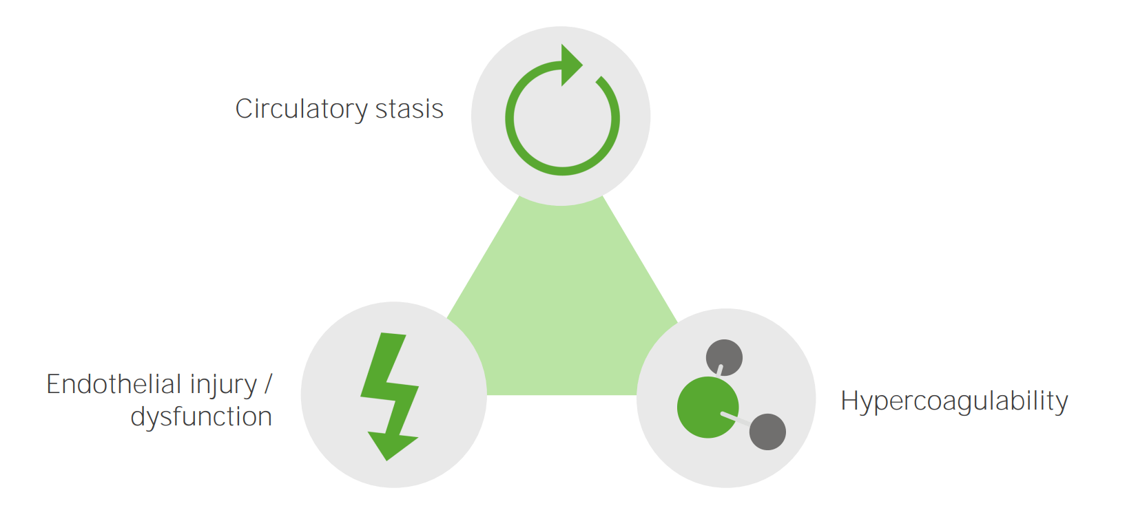
The 3 primary factors (known as the Virchow triad)that contribute to DVTDVTDeep vein thrombosis (DVT) usually occurs in the deep veins of the lower extremities. The affected veins include the femoral, popliteal, iliofemoral, and pelvic veins. Proximal DVT is more likely to cause a pulmonary embolism (PE) and is generally considered more serious. Deep Vein Thrombosis formation include: venous stasis, hypercoagulabilityHypercoagulabilityHypercoagulable States, and vascular endothelial damage. Any condition which worsens one (or more) of these three factors increases the risk of DVTDVTDeep vein thrombosis (DVT) usually occurs in the deep veins of the lower extremities. The affected veins include the femoral, popliteal, iliofemoral, and pelvic veins. Proximal DVT is more likely to cause a pulmonary embolism (PE) and is generally considered more serious. Deep Vein Thrombosis formation.
The primary etiologic factors which cause DVT: Circulatory stasis, endothelial injury or dysfunction, and hypercoagulability
Image by Lecturio.
Factors resulting in endothelial damage:
Hypertension: Increased shear stress leads to damage of the endothelium.
Surgery
Vascular catheter placement (e.g., hemodialysisHemodialysisProcedures which temporarily or permanently remedy insufficient cleansing of body fluids by the kidneys.Crush Syndrome catheters, peripherally inserted central catheters [PICC] lines): most common cause of upper extremity DVTDVTDeep vein thrombosis (DVT) usually occurs in the deep veins of the lower extremities. The affected veins include the femoral, popliteal, iliofemoral, and pelvic veins. Proximal DVT is more likely to cause a pulmonary embolism (PE) and is generally considered more serious. Deep Vein Thrombosis
Trauma and burnsBurnsA burn is a type of injury to the skin and deeper tissues caused by exposure to heat, electricity, chemicals, friction, or radiation. Burns are classified according to their depth as superficial (1st-degree), partial-thickness (2nd-degree), full-thickness (3rd-degree), and 4th-degree burns. Burns, especially involving the vasculature
Nephrotic syndromeNephrotic syndromeNephrotic syndrome is characterized by severe proteinuria, hypoalbuminemia, and peripheral edema. In contrast, the nephritic syndromes present with hematuria, variable loss of renal function, and hypertension, although there is sometimes overlap of > 1 glomerular disease in the same individual. Nephrotic Syndrome
Leads to loss of anticoagulant proteinsProteinsLinear polypeptides that are synthesized on ribosomes and may be further modified, crosslinked, cleaved, or assembled into complex proteins with several subunits. The specific sequence of amino acids determines the shape the polypeptide will take, during protein folding, and the function of the protein.Energy Homeostasis (e.g., antithrombinAntithrombinEndogenous factors and drugs that directly inhibit the action of thrombin, usually by blocking its enzymatic activity. They are distinguished from indirect thrombin inhibitors, such as heparin, which act by enhancing the inhibitory effects of antithrombins.Anticoagulants, proteinsProteinsLinear polypeptides that are synthesized on ribosomes and may be further modified, crosslinked, cleaved, or assembled into complex proteins with several subunits. The specific sequence of amino acids determines the shape the polypeptide will take, during protein folding, and the function of the protein.Energy Homeostasis C and S) via the urine due to damaged glomerular membranes
Leads to an increase in the production of fibrinogenFibrinogenPlasma glycoprotein clotted by thrombin, composed of a dimer of three non-identical pairs of polypeptide chains (alpha, beta, gamma) held together by disulfide bonds. Fibrinogen clotting is a sol-gel change involving complex molecular arrangements: whereas fibrinogen is cleaved by thrombin to form polypeptides a and b, the proteolytic action of other enzymes yields different fibrinogen degradation products.Hemostasis and other procoagulant proteinsProteinsLinear polypeptides that are synthesized on ribosomes and may be further modified, crosslinked, cleaved, or assembled into complex proteins with several subunits. The specific sequence of amino acids determines the shape the polypeptide will take, during protein folding, and the function of the protein.Energy Homeostasis in the liverLiverThe liver is the largest gland in the human body. The liver is found in the superior right quadrant of the abdomen and weighs approximately 1.5 kilograms. Its main functions are detoxification, metabolism, nutrient storage (e.g., iron and vitamins), synthesis of coagulation factors, formation of bile, filtration, and storage of blood. Liver: Anatomy due to protein loss and hypoalbuminemiaHypoalbuminemiaA condition in which albumin level in blood (serum albumin) is below the normal range. Hypoalbuminemia may be due to decreased hepatic albumin synthesis, increased albumin catabolism, altered albumin distribution, or albumin loss through the urine (albuminuria).Nephrotic Syndrome in Children
Factors resulting in venous stasis:
ImmobilizationImmobilizationDelirium (e.g., long air travel, after orthopedic surgery): 20 times increased risk of developing a DVTDVTDeep vein thrombosis (DVT) usually occurs in the deep veins of the lower extremities. The affected veins include the femoral, popliteal, iliofemoral, and pelvic veins. Proximal DVT is more likely to cause a pulmonary embolism (PE) and is generally considered more serious. Deep Vein Thrombosis
Heart failureHeart FailureA heterogeneous condition in which the heart is unable to pump out sufficient blood to meet the metabolic need of the body. Heart failure can be caused by structural defects, functional abnormalities (ventricular dysfunction), or a sudden overload beyond its capacity. Chronic heart failure is more common than acute heart failure which results from sudden insult to cardiac function, such as myocardial infarction.Total Anomalous Pulmonary Venous Return (TAPVR)
Hereditary thrombophiliaThrombophiliaA disorder of hemostasis in which there is a tendency for the occurrence of thrombosis.Hypercoagulable States, most often:
ProthrombinProthrombinA plasma protein that is the inactive precursor of thrombin. It is converted to thrombin by a prothrombin activator complex consisting of factor Xa, factor V, phospholipid, and calcium ions.Hemostasisgene mutationGene MutationMyotonic Dystrophies
PregnancyPregnancyThe status during which female mammals carry their developing young (embryos or fetuses) in utero before birth, beginning from fertilization to birth.Pregnancy: Diagnosis, Physiology, and Care/oral contraceptiveOral contraceptiveCompounds, usually hormonal, taken orally in order to block ovulation and prevent the occurrence of pregnancy. The hormones are generally estrogen or progesterone or both.Benign Liver Tumors pill (OCPOCPCompounds, usually hormonal, taken orally in order to block ovulation and prevent the occurrence of pregnancy. The hormones are generally estrogen or progesterone or both.Benign Liver Tumors)/hormone replacement therapyHormone Replacement TherapyHormone replacement therapy (HRT) is used to treat symptoms associated with female menopause and in combination to suppress ovulation. Risks and side effects include uterine bleeding, predisposition to cancer, breast tenderness, hyperpigmentation, migraine headaches, hypertension, bloating, and mood changes.Noncontraceptive Estrogen and Progestins use:EstrogenEstrogenCompounds that interact with estrogen receptors in target tissues to bring about the effects similar to those of estradiol. Estrogens stimulate the female reproductive organs, and the development of secondary female sex characteristics. Estrogenic chemicals include natural, synthetic, steroidal, or non-steroidal compounds.Ovaries: Anatomy increases the production of clotting factors in the liverLiverThe liver is the largest gland in the human body. The liver is found in the superior right quadrant of the abdomen and weighs approximately 1.5 kilograms. Its main functions are detoxification, metabolism, nutrient storage (e.g., iron and vitamins), synthesis of coagulation factors, formation of bile, filtration, and storage of blood. Liver: Anatomy.
Cancer (including myeloproliferative neoplasmsNeoplasmsNew abnormal growth of tissue. Malignant neoplasms show a greater degree of anaplasia and have the properties of invasion and metastasis, compared to benign neoplasms.Benign Bone Tumors)
SmokingSmokingWillful or deliberate act of inhaling and exhaling smoke from burning substances or agents held by hand.Interstitial Lung Diseases: Oxidant gases and other chemicals in cigarette smoke produce free radicalsFree radicalsHighly reactive molecules with an unsatisfied electron valence pair. Free radicals are produced in both normal and pathological processes. They are proven or suspected agents of tissue damage in a wide variety of circumstances including radiation, damage from environment chemicals, and aging. Natural and pharmacological prevention of free radical damage is being actively investigated.Ischemic Cell Damage which lead to platelet aggregationPlatelet aggregationThe attachment of platelets to one another. This clumping together can be induced by a number of agents (e.g., thrombin; collagen) and is part of the mechanism leading to the formation of a thrombus.Hemostasis and an increase in the production of procoagulant molecules.
Other risk factors and/or conditions affecting multiple components of the Virchow triad:
Prior DVTDVTDeep vein thrombosis (DVT) usually occurs in the deep veins of the lower extremities. The affected veins include the femoral, popliteal, iliofemoral, and pelvic veins. Proximal DVT is more likely to cause a pulmonary embolism (PE) and is generally considered more serious. Deep Vein Thrombosis/pulmonary embolismPulmonary EmbolismPulmonary embolism (PE) is a potentially fatal condition that occurs as a result of intraluminal obstruction of the main pulmonary artery or its branches. The causative factors include thrombi, air, amniotic fluid, and fat. In PE, gas exchange is impaired due to the decreased return of deoxygenated blood to the lungs. Pulmonary Embolism: 30 times increased risk of recurrent DVTDVTDeep vein thrombosis (DVT) usually occurs in the deep veins of the lower extremities. The affected veins include the femoral, popliteal, iliofemoral, and pelvic veins. Proximal DVT is more likely to cause a pulmonary embolism (PE) and is generally considered more serious. Deep Vein Thrombosis/PE
ObesityObesityObesity is a condition associated with excess body weight, specifically with the deposition of excessive adipose tissue. Obesity is considered a global epidemic. Major influences come from the western diet and sedentary lifestyles, but the exact mechanisms likely include a mixture of genetic and environmental factors. Obesity
Autoimmune diseasesAutoimmune diseasesDisorders that are characterized by the production of antibodies that react with host tissues or immune effector cells that are autoreactive to endogenous peptides.Selective IgA Deficiency:
Antiphospholipid syndromeAntiphospholipid syndromeAntiphospholipid syndrome (APLS) is an acquired autoimmune disorder characterized by the persistent presence of antiphospholipid antibodies, which create a hypercoagulable state. These antibodies are most commonly discovered during a workup for a thrombotic event or recurrent pregnancy loss, which are the 2 most common clinical manifestations.Antiphospholipid Syndrome
Systemic lupus erythematosusSystemic lupus erythematosusSystemic lupus erythematosus (SLE) is a chronic autoimmune, inflammatory condition that causes immune-complex deposition in organs, resulting in systemic manifestations. Women, particularly those of African American descent, are more commonly affected.Systemic Lupus Erythematosus (SLESLESystemic lupus erythematosus (SLE) is a chronic autoimmune, inflammatory condition that causes immune-complex deposition in organs, resulting in systemic manifestations. Women, particularly those of African American descent, are more commonly affected.Systemic Lupus Erythematosus)
COVID-19COVID-19Coronavirus disease 2019 (COVID-19) is an infectious disease caused by the severe acute respiratory syndrome coronavirus 2 (SARS-CoV-2) that mainly affects the respiratory system but can also cause damage to other body systems (cardiovascular, gastrointestinal, renal, and central nervous systems).
Mnemonic
To remember DVTDVTDeep vein thrombosis (DVT) usually occurs in the deep veins of the lower extremities. The affected veins include the femoral, popliteal, iliofemoral, and pelvic veins. Proximal DVT is more likely to cause a pulmonary embolism (PE) and is generally considered more serious. Deep Vein Thrombosis risk factors, think THROMBOSISThrombosisFormation and development of a thrombus or blood clot in the blood vessel.Epidemic Typhus.
Travel
Hypercoagulable/Hormone replacement therapy (HRTHRTHormone replacement therapy (HRT) is used to treat symptoms associated with female menopause and in combination to suppress ovulation. Risks and side effects include uterine bleeding, predisposition to cancer, breast tenderness, hyperpigmentation, migraine headaches, hypertension, bloating, and mood changes.Noncontraceptive Estrogen and Progestins)
Recreational drugs
Old (age > 60)
Malignancy
Blood disorders
Obesity/Obstetrics
Surgery/Smoking
Immobilization
Sickness (heart failureHeart FailureA heterogeneous condition in which the heart is unable to pump out sufficient blood to meet the metabolic need of the body. Heart failure can be caused by structural defects, functional abnormalities (ventricular dysfunction), or a sudden overload beyond its capacity. Chronic heart failure is more common than acute heart failure which results from sudden insult to cardiac function, such as myocardial infarction.Total Anomalous Pulmonary Venous Return (TAPVR) [HF]/myocardial infarctionMyocardial infarctionMI is ischemia and death of an area of myocardial tissue due to insufficient blood flow and oxygenation, usually from thrombus formation on a ruptured atherosclerotic plaque in the epicardial arteries. Clinical presentation is most commonly with chest pain, but women and patients with diabetes may have atypical symptoms.Myocardial Infarction [MIMIMI is ischemia and death of an area of myocardial tissue due to insufficient blood flow and oxygenation, usually from thrombus formation on a ruptured atherosclerotic plaque in the epicardial arteries. Clinical presentation is most commonly with chest pain, but women and patients with diabetes may have atypical symptoms.Myocardial Infarction], inflammatory bowel disease [IBD], nephrotic syndromeNephrotic syndromeNephrotic syndrome is characterized by severe proteinuria, hypoalbuminemia, and peripheral edema. In contrast, the nephritic syndromes present with hematuria, variable loss of renal function, and hypertension, although there is sometimes overlap of > 1 glomerular disease in the same individual. Nephrotic Syndrome, vasculitisVasculitisInflammation of any one of the blood vessels, including the arteries; veins; and rest of the vasculature system in the body.Systemic Lupus Erythematosus)
DVTDVTDeep vein thrombosis (DVT) usually occurs in the deep veins of the lower extremities. The affected veins include the femoral, popliteal, iliofemoral, and pelvic veins. Proximal DVT is more likely to cause a pulmonary embolism (PE) and is generally considered more serious. Deep Vein Thrombosis begins at the site of endothelial damage, often near the venous valves.
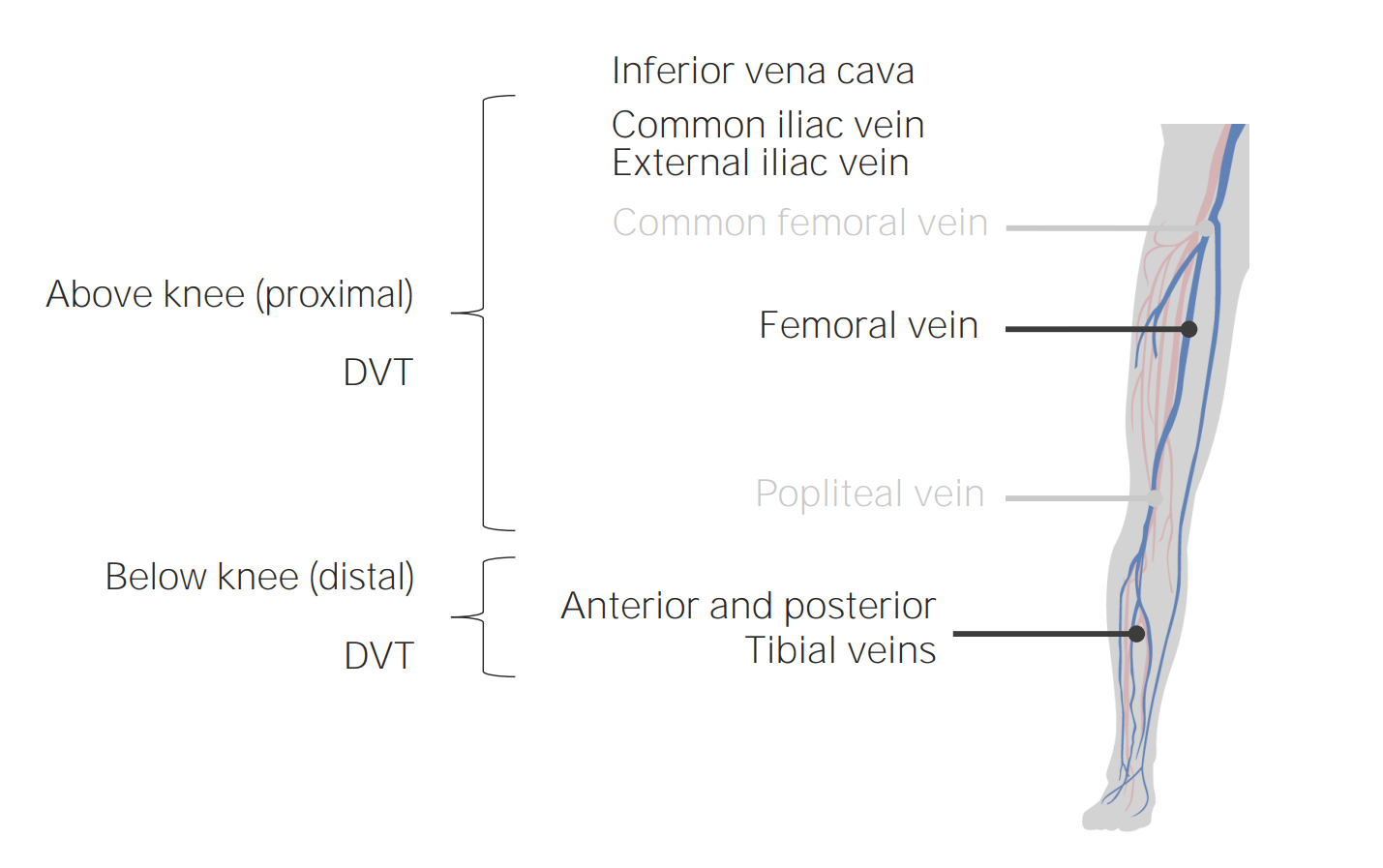
Clots usually begin in the deep vessels of the calf and extend proximally into the larger veinsVeinsVeins are tubular collections of cells, which transport deoxygenated blood and waste from the capillary beds back to the heart. Veins are classified into 3 types: small veins/venules, medium veins, and large veins. Each type contains 3 primary layers: tunica intima, tunica media, and tunica adventitia. Veins: Histology.
VeinsVeinsVeins are tubular collections of cells, which transport deoxygenated blood and waste from the capillary beds back to the heart. Veins are classified into 3 types: small veins/venules, medium veins, and large veins. Each type contains 3 primary layers: tunica intima, tunica media, and tunica adventitia. Veins: Histology affected the most:
Popliteal, femoral, and iliac veinsVeinsVeins are tubular collections of cells, which transport deoxygenated blood and waste from the capillary beds back to the heart. Veins are classified into 3 types: small veins/venules, medium veins, and large veins. Each type contains 3 primary layers: tunica intima, tunica media, and tunica adventitia. Veins: Histology
During pregnancyPregnancyThe status during which female mammals carry their developing young (embryos or fetuses) in utero before birth, beginning from fertilization to birth.Pregnancy: Diagnosis, Physiology, and Care:
Predominantly affects the lower left extremity veinsVeinsVeins are tubular collections of cells, which transport deoxygenated blood and waste from the capillary beds back to the heart. Veins are classified into 3 types: small veins/venules, medium veins, and large veins. Each type contains 3 primary layers: tunica intima, tunica media, and tunica adventitia. Veins: Histology (due to venous stasis produced by the right iliac artery compressing the left iliac vein, and by the uterusUterusThe uterus, cervix, and fallopian tubes are part of the internal female reproductive system. The uterus has a thick wall made of smooth muscle (the myometrium) and an inner mucosal layer (the endometrium). The most inferior portion of the uterus is the cervix, which connects the uterine cavity to the vagina.Uterus, Cervix, and Fallopian Tubes: Anatomy compressing the inferior vena cavaInferior vena cavaThe venous trunk which receives blood from the lower extremities and from the pelvic and abdominal organs.Mediastinum and Great Vessels: Anatomy)
ThrombosisThrombosisFormation and development of a thrombus or blood clot in the blood vessel.Epidemic Typhus of the pelvic veinsVeinsVeins are tubular collections of cells, which transport deoxygenated blood and waste from the capillary beds back to the heart. Veins are classified into 3 types: small veins/venules, medium veins, and large veins. Each type contains 3 primary layers: tunica intima, tunica media, and tunica adventitia. Veins: Histology are seen more in patientsPatientsIndividuals participating in the health care system for the purpose of receiving therapeutic, diagnostic, or preventive procedures.Clinician–Patient Relationship who are pregnant or postpartum.
Common locations of DVT
Image by Lecturio
Composition of thrombus[4,5]
Red blood cellsRed blood cellsErythrocytes, or red blood cells (RBCs), are the most abundant cells in the blood. While erythrocytes in the fetus are initially produced in the yolk sac then the liver, the bone marrow eventually becomes the main site of production.Erythrocytes: Histology
PlateletsPlateletsPlatelets are small cell fragments involved in hemostasis. Thrombopoiesis takes place primarily in the bone marrow through a series of cell differentiation and is influenced by several cytokines. Platelets are formed after fragmentation of the megakaryocyte cytoplasm. Platelets: Histology
Increased platelet activationPlatelet activationA series of progressive, overlapping events, triggered by exposure of the platelets to subendothelial tissue. These events include shape change, adhesiveness, aggregation, and release reactions. When carried through to completion, these events lead to the formation of a stable hemostatic plug.Hemostasis and adhesionAdhesionThe process whereby platelets adhere to something other than platelets, e.g., collagen; basement membrane; microfibrils; or other ‘foreign’ surfaces.Coagulation Studies
Decreased anticoagulantsAnticoagulantsAnticoagulants are drugs that retard or interrupt the coagulation cascade. The primary classes of available anticoagulants include heparins, vitamin K-dependent antagonists (e.g., warfarin), direct thrombin inhibitors, and factor Xa inhibitors. Anticoagulants
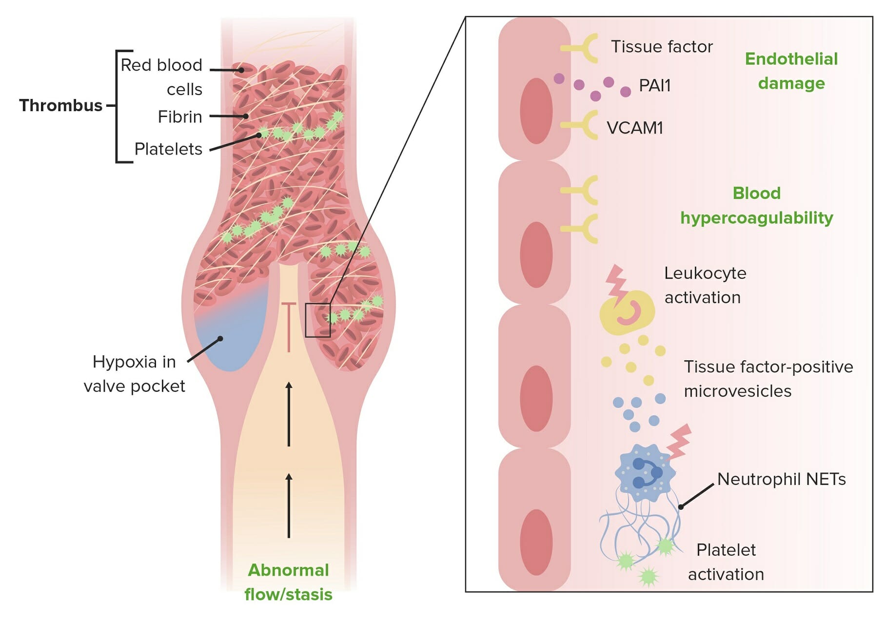
Endothelial damage: inflammationInflammationInflammation is a complex set of responses to infection and injury involving leukocytes as the principal cellular mediators in the body’s defense against pathogenic organisms. Inflammation is also seen as a response to tissue injury in the process of wound healing. The 5 cardinal signs of inflammation are pain, heat, redness, swelling, and loss of function. Inflammation/trauma →
Exposure of von Willebrand factorvon Willebrand factorA high-molecular-weight plasma protein, produced by endothelial cells and megakaryocytes, that is part of the factor VIII/von Willebrand factor complex. The von Willebrand factor has receptors for collagen, platelets, and ristocetin activity as well as the immunologically distinct antigenic determinants. It functions in adhesion of platelets to collagen and hemostatic plug formation. The prolonged bleeding time in von Willebrand diseases is due to the deficiency of this factor.Hemostasis (vWF) → results in platelet activationPlatelet activationA series of progressive, overlapping events, triggered by exposure of the platelets to subendothelial tissue. These events include shape change, adhesiveness, aggregation, and release reactions. When carried through to completion, these events lead to the formation of a stable hemostatic plug.Hemostasis and formation of the platelet plugPlatelet plugHemostasis
Exposure of tissue factor → activates the extrinsic pathwayExtrinsic pathwayThe extrinsic pathway is the primary physiological mechanism by which clotting is initiatedHemostasis in the coagulation cascadeCoagulation cascadeThe coagulation cascade is a series of reactions that ultimately generates a strong, cross-linked fibrin clot.Hemostasis → results in fibrinFibrinA protein derived from fibrinogen in the presence of thrombin, which forms part of the blood clot.Rapidly Progressive Glomerulonephritis clot formation
Abnormal flowFlowBlood flows through the heart, arteries, capillaries, and veins in a closed, continuous circuit. Flow is the movement of volume per unit of time. Flow is affected by the pressure gradient and the resistance fluid encounters between 2 points. Vascular resistance is the opposition to flow, which is caused primarily by blood friction against vessel walls.Vascular Resistance, Flow, and Mean Arterial Pressure/stasis: immobilizationImmobilizationDelirium, venous valve incompetence → stasis of blood → more time for clot formation
The main pathophysiologic mechanisms leading to deep vein thrombosis:
Endothelial damage, blood hypercoagulability, and blood stasis
Image by Lecturio.
Complications of DVTDVTDeep vein thrombosis (DVT) usually occurs in the deep veins of the lower extremities. The affected veins include the femoral, popliteal, iliofemoral, and pelvic veins. Proximal DVT is more likely to cause a pulmonary embolism (PE) and is generally considered more serious. Deep Vein Thrombosis[3,4]
Pulmonary embolismPulmonary EmbolismPulmonary embolism (PE) is a potentially fatal condition that occurs as a result of intraluminal obstruction of the main pulmonary artery or its branches. The causative factors include thrombi, air, amniotic fluid, and fat. In PE, gas exchange is impaired due to the decreased return of deoxygenated blood to the lungs. Pulmonary Embolism:
A condition in which a pulmonary arteryPulmonary arteryThe short wide vessel arising from the conus arteriosus of the right ventricle and conveying unaerated blood to the lungs.Lungs: Anatomy is obstructed by a portion of thrombus that has broken off of a DVTDVTDeep vein thrombosis (DVT) usually occurs in the deep veins of the lower extremities. The affected veins include the femoral, popliteal, iliofemoral, and pelvic veins. Proximal DVT is more likely to cause a pulmonary embolism (PE) and is generally considered more serious. Deep Vein Thrombosis and traveled to the lung
Potentially fatal
Has both cardiovascular and respiratory effects (hypotensionHypotensionHypotension is defined as low blood pressure, specifically < 90/60 mm Hg, and is most commonly a physiologic response. Hypotension may be mild, serious, or life threatening, depending on the cause. Hypotension and hypoxiaHypoxiaSub-optimal oxygen levels in the ambient air of living organisms.Ischemic Cell Damage)
Post-thrombotic syndromePost-thrombotic syndromeA condition caused by one or more episodes of deep vein thrombosis, usually the blood clots are lodged in the legs. Clinical features include edema; pain; aching; heaviness; and muscle cramp in the leg. When severe leg swelling leads to skin breakdown, it is called venous stasis ulcer.Deep Vein Thrombosis(most common complication of proximal DVTDVTDeep vein thrombosis (DVT) usually occurs in the deep veins of the lower extremities. The affected veins include the femoral, popliteal, iliofemoral, and pelvic veins. Proximal DVT is more likely to cause a pulmonary embolism (PE) and is generally considered more serious. Deep Vein Thrombosis)
Symptoms include painPainAn unpleasant sensation induced by noxious stimuli which are detected by nerve endings of nociceptive neurons.Pain: Types and Pathways and swellingSwellingInflammation.
Chronic venous insufficiencyChronic venous insufficiencyChronic venous disease is a spectrum of disorders characterized by venous dilation and/or abnormal vein function in the lower extremities resulting from venous hypertension. “Chronic venous insufficiency” refers to the more severe forms of chronic venous disease. Skin changes typically distinguish chronic venous insufficiency from milder forms of venous disease.Chronic Venous Insufficiency: Venous wall and valve dysfunction can occur as a result of fibrosisFibrosisAny pathological condition where fibrous connective tissue invades any organ, usually as a consequence of inflammation or other injury.Bronchiolitis Obliterans due to inflammationInflammationInflammation is a complex set of responses to infection and injury involving leukocytes as the principal cellular mediators in the body’s defense against pathogenic organisms. Inflammation is also seen as a response to tissue injury in the process of wound healing. The 5 cardinal signs of inflammation are pain, heat, redness, swelling, and loss of function. Inflammation around the thrombus.
Ulcers develop on lower extremities.
Mobility can be reduced.
Some patientsPatientsIndividuals participating in the health care system for the purpose of receiving therapeutic, diagnostic, or preventive procedures.Clinician–Patient Relationship experience paresthesiasParesthesiasSubjective cutaneous sensations (e.g., cold, warmth, tingling, pressure, etc.) that are experienced spontaneously in the absence of stimulation.Posterior Cord Syndrome.
Occurs in 25%–50% of all patientsPatientsIndividuals participating in the health care system for the purpose of receiving therapeutic, diagnostic, or preventive procedures.Clinician–Patient Relationship with DVTDVTDeep vein thrombosis (DVT) usually occurs in the deep veins of the lower extremities. The affected veins include the femoral, popliteal, iliofemoral, and pelvic veins. Proximal DVT is more likely to cause a pulmonary embolism (PE) and is generally considered more serious. Deep Vein Thrombosis
Many cases are asymptomatic, with up to 50% having nonspecific symptoms.[2,4,5]
Symptoms/manifestations are usually unilateral.
PainPainAn unpleasant sensation induced by noxious stimuli which are detected by nerve endings of nociceptive neurons.Pain: Types and Pathways
Warmth
EdemaEdemaEdema is a condition in which excess serous fluid accumulates in the body cavity or interstitial space of connective tissues. Edema is a symptom observed in several medical conditions. It can be categorized into 2 types, namely, peripheral (in the extremities) and internal (in an organ or body cavity). Edema
Intact distal pulses
FeverFeverFever is defined as a measured body temperature of at least 38°C (100.4°F). Fever is caused by circulating endogenous and/or exogenous pyrogens that increase levels of prostaglandin E2 in the hypothalamus. Fever is commonly associated with chills, rigors, sweating, and flushing of the skin. Fever (due to cytokine release)
Palpable cords representing the thrombotic vein (rare but significant finding)
Homan signHoman signDeep Vein Thrombosis: calf painPainAn unpleasant sensation induced by noxious stimuli which are detected by nerve endings of nociceptive neurons.Pain: Types and Pathways on dorsiflexion of the footFootThe foot is the terminal portion of the lower limb, whose primary function is to bear weight and facilitate locomotion. The foot comprises 26 bones, including the tarsal bones, metatarsal bones, and phalanges. The bones of the foot form longitudinal and transverse arches and are supported by various muscles, ligaments, and tendons.Foot: Anatomy (neither sensitive nor specific)
The 1st manifestation can be pulmonary embolismPulmonary EmbolismPulmonary embolism (PE) is a potentially fatal condition that occurs as a result of intraluminal obstruction of the main pulmonary artery or its branches. The causative factors include thrombi, air, amniotic fluid, and fat. In PE, gas exchange is impaired due to the decreased return of deoxygenated blood to the lungs. Pulmonary Embolism (which presents with chest painPainAn unpleasant sensation induced by noxious stimuli which are detected by nerve endings of nociceptive neurons.Pain: Types and Pathways, dyspneaDyspneaDyspnea is the subjective sensation of breathing discomfort. Dyspnea is a normal manifestation of heavy physical or psychological exertion, but also may be caused by underlying conditions (both pulmonary and extrapulmonary). Dyspnea).
Chronic DVTDVTDeep vein thrombosis (DVT) usually occurs in the deep veins of the lower extremities. The affected veins include the femoral, popliteal, iliofemoral, and pelvic veins. Proximal DVT is more likely to cause a pulmonary embolism (PE) and is generally considered more serious. Deep Vein Thrombosis can be asymptomatic and cause chronic venous insufficiencyChronic venous insufficiencyChronic venous disease is a spectrum of disorders characterized by venous dilation and/or abnormal vein function in the lower extremities resulting from venous hypertension. “Chronic venous insufficiency” refers to the more severe forms of chronic venous disease. Skin changes typically distinguish chronic venous insufficiency from milder forms of venous disease.Chronic Venous Insufficiency.
Phlegmasia alba dolens (“painful white inflammation”):
Occurs with a massive occlusion of the major deep vessels, but with patent collateral vessels and without ischemiaIschemiaA hypoperfusion of the blood through an organ or tissue caused by a pathologic constriction or obstruction of its blood vessels, or an absence of blood circulation.Ischemic Cell Damage
Presents with edemaEdemaEdema is a condition in which excess serous fluid accumulates in the body cavity or interstitial space of connective tissues. Edema is a symptom observed in several medical conditions. It can be categorized into 2 types, namely, peripheral (in the extremities) and internal (in an organ or body cavity). Edema, painPainAn unpleasant sensation induced by noxious stimuli which are detected by nerve endings of nociceptive neurons.Pain: Types and Pathways, and blanchingBlanchingDermatologic Examination without cyanosisCyanosisA bluish or purplish discoloration of the skin and mucous membranes due to an increase in the amount of deoxygenated hemoglobin in the blood or a structural defect in the hemoglobin molecule.Pulmonary Examination
Can progress to phlegmasia cerulea dolensPhlegmasia cerulea dolensNear-total occlusion of the deep venous system resulting in venous gangrene.Acute Limb Ischemia and compartment syndromeCompartment SyndromeCompartment syndrome is a surgical emergency usually occurring secondary to trauma. The condition is marked by increased pressure within a compartment that compromises the circulation and function of the tissues within that space.Compartment Syndrome
Obstruction of all veinsVeinsVeins are tubular collections of cells, which transport deoxygenated blood and waste from the capillary beds back to the heart. Veins are classified into 3 types: small veins/venules, medium veins, and large veins. Each type contains 3 primary layers: tunica intima, tunica media, and tunica adventitia. Veins: Histology of an extremity → severe venous congestion → limited arterial flowFlowBlood flows through the heart, arteries, capillaries, and veins in a closed, continuous circuit. Flow is the movement of volume per unit of time. Flow is affected by the pressure gradient and the resistance fluid encounters between 2 points. Vascular resistance is the opposition to flow, which is caused primarily by blood friction against vessel walls.Vascular Resistance, Flow, and Mean Arterial Pressure → ischemiaIschemiaA hypoperfusion of the blood through an organ or tissue caused by a pathologic constriction or obstruction of its blood vessels, or an absence of blood circulation.Ischemic Cell Damage → tissue death
Presents with edemaEdemaEdema is a condition in which excess serous fluid accumulates in the body cavity or interstitial space of connective tissues. Edema is a symptom observed in several medical conditions. It can be categorized into 2 types, namely, peripheral (in the extremities) and internal (in an organ or body cavity). Edema, painPainAn unpleasant sensation induced by noxious stimuli which are detected by nerve endings of nociceptive neurons.Pain: Types and Pathways, pulselessnessPulselessnessCardiac Arrest, and cyanosisCyanosisA bluish or purplish discoloration of the skin and mucous membranes due to an increase in the amount of deoxygenated hemoglobin in the blood or a structural defect in the hemoglobin molecule.Pulmonary Examination
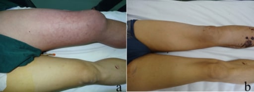
The image shows erythema, swelling, and cyanosis in a patient with phlegmasia cerulea dolens. a) Initial appearance of the left leg, showing significant swelling and cyanosis b) Appearance of the left leg 35 days after surgery, at the time of the patient’s discharge
Image: “Appearance of the left leg” by Division of Vascular and Thyroid Surgery, Department of Surgery, The First Affiliated Hospital, China Medical University, Shenyang 110001, China. License: CC BY 2.0
First test, depending on degree of suspicion[2,9‒11]
Wells criteria are used to assess risk of a DVTDVTDeep vein thrombosis (DVT) usually occurs in the deep veins of the lower extremities. The affected veins include the femoral, popliteal, iliofemoral, and pelvic veins. Proximal DVT is more likely to cause a pulmonary embolism (PE) and is generally considered more serious. Deep Vein Thrombosis based on the presentation of the individual (see table):
Low probabilityProbabilityProbability is a mathematical tool used to study randomness and provide predictions about the likelihood of something happening. There are several basic rules of probability that can be used to help determine the probability of multiple events happening together, separately, or sequentially.Basics of Probability: 0
Moderate probabilityProbabilityProbability is a mathematical tool used to study randomness and provide predictions about the likelihood of something happening. There are several basic rules of probability that can be used to help determine the probability of multiple events happening together, separately, or sequentially.Basics of Probability: 1 to 2 points
High probabilityProbabilityProbability is a mathematical tool used to study randomness and provide predictions about the likelihood of something happening. There are several basic rules of probability that can be used to help determine the probability of multiple events happening together, separately, or sequentially.Basics of Probability: 3 to 8 points
Modified Wells criteria:
Adds a clinical feature (previously documented DVTDVTDeep vein thrombosis (DVT) usually occurs in the deep veins of the lower extremities. The affected veins include the femoral, popliteal, iliofemoral, and pelvic veins. Proximal DVT is more likely to cause a pulmonary embolism (PE) and is generally considered more serious. Deep Vein Thrombosis) which is given a score of 1.
Given this modification, the DVTDVTDeep vein thrombosis (DVT) usually occurs in the deep veins of the lower extremities. The affected veins include the femoral, popliteal, iliofemoral, and pelvic veins. Proximal DVT is more likely to cause a pulmonary embolism (PE) and is generally considered more serious. Deep Vein Thrombosis is either likely or unlikely based on the scoring.
DVTDVTDeep vein thrombosis (DVT) usually occurs in the deep veins of the lower extremities. The affected veins include the femoral, popliteal, iliofemoral, and pelvic veins. Proximal DVT is more likely to cause a pulmonary embolism (PE) and is generally considered more serious. Deep Vein Thrombosis likely: ≥ 2
DVTDVTDeep vein thrombosis (DVT) usually occurs in the deep veins of the lower extremities. The affected veins include the femoral, popliteal, iliofemoral, and pelvic veins. Proximal DVT is more likely to cause a pulmonary embolism (PE) and is generally considered more serious. Deep Vein Thrombosis unlikely: < 2
If the suspicion of DVTDVTDeep vein thrombosis (DVT) usually occurs in the deep veins of the lower extremities. The affected veins include the femoral, popliteal, iliofemoral, and pelvic veins. Proximal DVT is more likely to cause a pulmonary embolism (PE) and is generally considered more serious. Deep Vein Thrombosis is high (Wells scoreWells scoreDeep Vein Thrombosis ≥ 2), the test of choice is ultrasonography with DopplerDopplerUltrasonography applying the doppler effect, with frequency-shifted ultrasound reflections produced by moving targets (usually red blood cells) in the bloodstream along the ultrasound axis in direct proportion to the velocity of movement of the targets, to determine both direction and velocity of blood flow.Ultrasound (Sonography). Diagnostic imaging findings include:
Decreased/absent flowFlowBlood flows through the heart, arteries, capillaries, and veins in a closed, continuous circuit. Flow is the movement of volume per unit of time. Flow is affected by the pressure gradient and the resistance fluid encounters between 2 points. Vascular resistance is the opposition to flow, which is caused primarily by blood friction against vessel walls.Vascular Resistance, Flow, and Mean Arterial Pressure
If the suspicion of DVTDVTDeep vein thrombosis (DVT) usually occurs in the deep veins of the lower extremities. The affected veins include the femoral, popliteal, iliofemoral, and pelvic veins. Proximal DVT is more likely to cause a pulmonary embolism (PE) and is generally considered more serious. Deep Vein Thrombosis is low (Wells scoreWells scoreDeep Vein Thrombosis < 2), the first test should be D-dimerD-dimerDeep Vein Thrombosis.
High sensitivity but low specificity
D-dimerD-dimerDeep Vein Thrombosis should not be obtained if it is expected to be positive (e.g., recent surgery).
Negative test (normal/low D-dimerD-dimerDeep Vein Thrombosis levels) rules out DVTDVTDeep vein thrombosis (DVT) usually occurs in the deep veins of the lower extremities. The affected veins include the femoral, popliteal, iliofemoral, and pelvic veins. Proximal DVT is more likely to cause a pulmonary embolism (PE) and is generally considered more serious. Deep Vein Thrombosis
Positive test (elevated D-dimerD-dimerDeep Vein Thrombosis levels) warrants ultrasonography for confirmation
Table: Modified Wells criteria for DVTDVTDeep vein thrombosis (DVT) usually occurs in the deep veins of the lower extremities. The affected veins include the femoral, popliteal, iliofemoral, and pelvic veins. Proximal DVT is more likely to cause a pulmonary embolism (PE) and is generally considered more serious. Deep Vein Thrombosis
Tenderness along the deep venous system
+1
Unilateral pitting edemaPitting edemaEdema caused by excess fluid without excess colloid. Leaves “pits” due to fluid displacement when pressure is applied to the areaEdema
+1
SwellingSwellingInflammation of the entire legLegThe lower leg, or just “leg” in anatomical terms, is the part of the lower limb between the knee and the ankle joint. The bony structure is composed of the tibia and fibula bones, and the muscles of the leg are grouped into the anterior, lateral, and posterior compartments by extensions of fascia.Leg: Anatomy
Collateral superficial non-varicose veinsVeinsVeins are tubular collections of cells, which transport deoxygenated blood and waste from the capillary beds back to the heart. Veins are classified into 3 types: small veins/venules, medium veins, and large veins. Each type contains 3 primary layers: tunica intima, tunica media, and tunica adventitia. Veins: Histology
+1
Active cancer
+1
Previous DVTDVTDeep vein thrombosis (DVT) usually occurs in the deep veins of the lower extremities. The affected veins include the femoral, popliteal, iliofemoral, and pelvic veins. Proximal DVT is more likely to cause a pulmonary embolism (PE) and is generally considered more serious. Deep Vein Thrombosis
Bedridden ≥ 3 days or major surgery within past 4 weeks
+1
Alternative diagnosis as likely/more likely than DVTDVTDeep vein thrombosis (DVT) usually occurs in the deep veins of the lower extremities. The affected veins include the femoral, popliteal, iliofemoral, and pelvic veins. Proximal DVT is more likely to cause a pulmonary embolism (PE) and is generally considered more serious. Deep Vein Thrombosis
-2
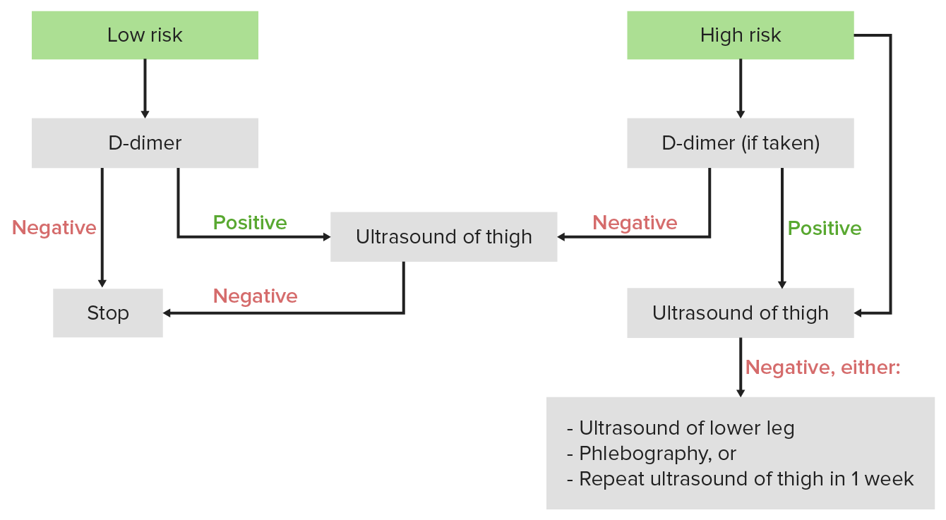
Diagnostic algorithm for DVT: If the Wells score is < 2, the individual is considered low risk, and the 1st test should be a D-dimer. If the Wells score is ≥ 2, the individual is considered high risk, and the 1st test should be an ultrasound.
Image by Lecturio.
Further tests[2,9,11]
Baseline laboratory studies to obtain:
CBC (assess hemoglobin and platelet count)
Chemistries (assess renal and liverLiverThe liver is the largest gland in the human body. The liver is found in the superior right quadrant of the abdomen and weighs approximately 1.5 kilograms. Its main functions are detoxification, metabolism, nutrient storage (e.g., iron and vitamins), synthesis of coagulation factors, formation of bile, filtration, and storage of blood. Liver: Anatomy function)
Coagulation panel (PT/INR, aPTT)
Age-appropriate cancer screeningScreeningPreoperative Care (e.g., digital rectal exam [DREDREA physical examination in which the qualified health care worker inserts a lubricated, gloved finger of one hand into the rectum and may use the other hand to press on the lower abdomen or pelvic area to palpate for abnormalities in the lower rectum, and nearby organs or tissues. The method is commonly used to check the lower rectum, the prostate gland in men, and the uterus and ovaries in women.Prostate Cancer Screening], mammographyMammographyRadiographic examination of the breast.Breast Cancer Screening, colonoscopyColonoscopyEndoscopic examination, therapy or surgery of the luminal surface of the colon.Colorectal Cancer Screening)
Hemodynamically stable individuals with a DVTDVTDeep vein thrombosis (DVT) usually occurs in the deep veins of the lower extremities. The affected veins include the femoral, popliteal, iliofemoral, and pelvic veins. Proximal DVT is more likely to cause a pulmonary embolism (PE) and is generally considered more serious. Deep Vein Thrombosis and signs of PE (e.g., chest painPainAn unpleasant sensation induced by noxious stimuli which are detected by nerve endings of nociceptive neurons.Pain: Types and Pathways, dyspneaDyspneaDyspnea is the subjective sensation of breathing discomfort. Dyspnea is a normal manifestation of heavy physical or psychological exertion, but also may be caused by underlying conditions (both pulmonary and extrapulmonary). Dyspnea): contrast-enhanced CT scan of the chest
Hemodynamically unstable patientsHemodynamically Unstable PatientsBlunt Chest Trauma with a DVTDVTDeep vein thrombosis (DVT) usually occurs in the deep veins of the lower extremities. The affected veins include the femoral, popliteal, iliofemoral, and pelvic veins. Proximal DVT is more likely to cause a pulmonary embolism (PE) and is generally considered more serious. Deep Vein Thrombosis and signs of PE: bedside echocardiogramEchocardiogramTransposition of the Great Arteries to visualize right ventricular dilation
Dye is injected into a dorsal footFootThe foot is the terminal portion of the lower limb, whose primary function is to bear weight and facilitate locomotion. The foot comprises 26 bones, including the tarsal bones, metatarsal bones, and phalanges. The bones of the foot form longitudinal and transverse arches and are supported by various muscles, ligaments, and tendons.Foot: Anatomy vein and images are taken to look for obstructing thrombi.
More costly with risk of contrast exposure and radiationRadiationEmission or propagation of acoustic waves (sound), electromagnetic energy waves (such as light; radio waves; gamma rays; or x-rays), or a stream of subatomic particles (such as electrons; neutrons; protons; or alpha particles).Osteosarcoma (in CTV)
Thrombus seen as an intraluminal filling defect
Management
AnticoagulationAnticoagulationPulmonary Hypertension Drugs is the mainstay of therapy for DVTs. Depending on the situation, some individuals may require thrombolysis, an invasive procedure, and/or lifelong anticoagulationAnticoagulationPulmonary Hypertension Drugs. The following is based on US and UK recommendations, but management may vary based on location.
Initial treatment considerations
Setting[6, 7]:
Outpatient treatment is appropriate for acute, uncomplicated DVTDVTDeep vein thrombosis (DVT) usually occurs in the deep veins of the lower extremities. The affected veins include the femoral, popliteal, iliofemoral, and pelvic veins. Proximal DVT is more likely to cause a pulmonary embolism (PE) and is generally considered more serious. Deep Vein Thrombosis.
HospitalizationHospitalizationThe confinement of a patient in a hospital.Delirium is indicated for:
Limb-threatening DVTDVTDeep vein thrombosis (DVT) usually occurs in the deep veins of the lower extremities. The affected veins include the femoral, popliteal, iliofemoral, and pelvic veins. Proximal DVT is more likely to cause a pulmonary embolism (PE) and is generally considered more serious. Deep Vein Thrombosis
Bleeding risk
Poor social support
Inability to afford or administer medications reliably
Inability to return to the hospital if clinical deterioration occurs
Proximal vs distal DVTDVTDeep vein thrombosis (DVT) usually occurs in the deep veins of the lower extremities. The affected veins include the femoral, popliteal, iliofemoral, and pelvic veins. Proximal DVT is more likely to cause a pulmonary embolism (PE) and is generally considered more serious. Deep Vein Thrombosis[8]:
Proximal DVTDVTDeep vein thrombosis (DVT) usually occurs in the deep veins of the lower extremities. The affected veins include the femoral, popliteal, iliofemoral, and pelvic veins. Proximal DVT is more likely to cause a pulmonary embolism (PE) and is generally considered more serious. Deep Vein Thrombosis:
Thrombus in the popliteal, femoral, or iliac veinsVeinsVeins are tubular collections of cells, which transport deoxygenated blood and waste from the capillary beds back to the heart. Veins are classified into 3 types: small veins/venules, medium veins, and large veins. Each type contains 3 primary layers: tunica intima, tunica media, and tunica adventitia. Veins: Histology
Distal DVTDVTDeep vein thrombosis (DVT) usually occurs in the deep veins of the lower extremities. The affected veins include the femoral, popliteal, iliofemoral, and pelvic veins. Proximal DVT is more likely to cause a pulmonary embolism (PE) and is generally considered more serious. Deep Vein Thrombosis:
Thrombus below the knee
Carries lower risk of embolizationEmbolizationA method of hemostasis utilizing various agents such as gelfoam, silastic, metal, glass, or plastic pellets, autologous clot, fat, and muscle as emboli. It has been used in the treatment of spinal cord and intracranial arteriovenous malformations, renal arteriovenous fistulas, gastrointestinal bleeding, epistaxis, hypersplenism, certain highly vascular tumors, traumatic rupture of blood vessels, and control of operative hemorrhage.Gastrointestinal Bleeding
AnticoagulationAnticoagulationPulmonary Hypertension Drugs is recommended for symptomatic distal DVTDVTDeep vein thrombosis (DVT) usually occurs in the deep veins of the lower extremities. The affected veins include the femoral, popliteal, iliofemoral, and pelvic veins. Proximal DVT is more likely to cause a pulmonary embolism (PE) and is generally considered more serious. Deep Vein Thrombosis.
AnticoagulationAnticoagulationPulmonary Hypertension Drugs is recommended for asymptomatic distal DVTDVTDeep vein thrombosis (DVT) usually occurs in the deep veins of the lower extremities. The affected veins include the femoral, popliteal, iliofemoral, and pelvic veins. Proximal DVT is more likely to cause a pulmonary embolism (PE) and is generally considered more serious. Deep Vein Thrombosis but with risk factors for extensionExtensionExamination of the Upper Limbs:
Unprovoked DVTDVTDeep vein thrombosis (DVT) usually occurs in the deep veins of the lower extremities. The affected veins include the femoral, popliteal, iliofemoral, and pelvic veins. Proximal DVT is more likely to cause a pulmonary embolism (PE) and is generally considered more serious. Deep Vein Thrombosis
Large thrombosisThrombosisFormation and development of a thrombus or blood clot in the blood vessel.Epidemic Typhus (> 5 cm length, > 7 mm in diameter)
Multiple veinsVeinsVeins are tubular collections of cells, which transport deoxygenated blood and waste from the capillary beds back to the heart. Veins are classified into 3 types: small veins/venules, medium veins, and large veins. Each type contains 3 primary layers: tunica intima, tunica media, and tunica adventitia. Veins: Histology involved
Persistent risk factors (e.g., cancer)
Prior history of DVTDVTDeep vein thrombosis (DVT) usually occurs in the deep veins of the lower extremities. The affected veins include the femoral, popliteal, iliofemoral, and pelvic veins. Proximal DVT is more likely to cause a pulmonary embolism (PE) and is generally considered more serious. Deep Vein Thrombosis or PE
Inpatient status
Limited mobility
COVID-19COVID-19Coronavirus disease 2019 (COVID-19) is an infectious disease caused by the severe acute respiratory syndrome coronavirus 2 (SARS-CoV-2) that mainly affects the respiratory system but can also cause damage to other body systems (cardiovascular, gastrointestinal, renal, and central nervous systems).
Serial US (once a week for 2 weeks) is an option for an isolated DVTDVTDeep vein thrombosis (DVT) usually occurs in the deep veins of the lower extremities. The affected veins include the femoral, popliteal, iliofemoral, and pelvic veins. Proximal DVT is more likely to cause a pulmonary embolism (PE) and is generally considered more serious. Deep Vein Thrombosis with low risk of extensionExtensionExamination of the Upper Limbs or with high risk of bleeding.
If clot extends towards the proximal veinsVeinsVeins are tubular collections of cells, which transport deoxygenated blood and waste from the capillary beds back to the heart. Veins are classified into 3 types: small veins/venules, medium veins, and large veins. Each type contains 3 primary layers: tunica intima, tunica media, and tunica adventitia. Veins: Histology, start anticoagulationAnticoagulationPulmonary Hypertension Drugs.
If clot is stable or extensionExtensionExamination of the Upper Limbs is limited to distal veinsVeinsVeins are tubular collections of cells, which transport deoxygenated blood and waste from the capillary beds back to the heart. Veins are classified into 3 types: small veins/venules, medium veins, and large veins. Each type contains 3 primary layers: tunica intima, tunica media, and tunica adventitia. Veins: Histology, consider anticoagulationAnticoagulationPulmonary Hypertension Drugs.
EnoxaparinEnoxaparinLow-molecular-weight fragment of heparin, having a 4-enopyranosuronate sodium structure at the non-reducing end of the chain. It is prepared by depolymerization of the benzylic ester of porcine mucosal heparin. Therapeutically, it is used as an antithrombotic agent.Anticoagulants: 1 mg/kg every 12 hours or 1.5 mg/kg daily
Unfractionated heparinUnfractionated heparinA highly acidic mucopolysaccharide formed of equal parts of sulfated d-glucosamine and d-glucuronic acid with sulfaminic bridges. The molecular weight ranges from six to twenty thousand. Heparin occurs in and is obtained from liver, lung, mast cells, etc. , of vertebrates. Its function is unknown, but it is used to prevent blood clotting in vivo and vitro, in the form of many different salts.Anticoagulants (UFHUFHA highly acidic mucopolysaccharide formed of equal parts of sulfated d-glucosamine and d-glucuronic acid with sulfaminic bridges. The molecular weight ranges from six to twenty thousand. Heparin occurs in and is obtained from liver, lung, mast cells, etc. , of vertebrates. Its function is unknown, but it is used to prevent blood clotting in vivo and vitro, in the form of many different salts.Anticoagulants)
Follow hospital protocol
Indirect factor Xa inhibitorIndirect Factor Xa InhibitorAnticoagulants: fondaparinuxFondaparinuxSynthetic pentasaccharide that mediates the interaction of heparin with antithrombins and inhibits factor Xa; it is used for prevention of venous thromboembolism after surgery.Anticoagulants
< 50 kg: 5 mg daily
50–100 kg: 7.5 mg daily
> 100 kg: 10 mg daily
Direct factor Xa inhibitors: rivaroxabanRivaroxabanA morpholine and thiophene derivative that functions as a factor Xa inhibitor and is used in the treatment and prevention of deep-vein thrombosis and pulmonary embolism. It is also used for the prevention of stroke and systemic embolization in patients with non-valvular atrial fibrillation, and for the prevention of atherothrombotic events in patients after an acute coronary syndrome.Anticoagulants, apixabanApixabanAnticoagulants
RivaroxabanRivaroxabanA morpholine and thiophene derivative that functions as a factor Xa inhibitor and is used in the treatment and prevention of deep-vein thrombosis and pulmonary embolism. It is also used for the prevention of stroke and systemic embolization in patients with non-valvular atrial fibrillation, and for the prevention of atherothrombotic events in patients after an acute coronary syndrome.Anticoagulants: 15 mg 2 times daily (with food) for 21 days, followed by 20 mg daily (with food)
ApixabanApixabanAnticoagulants: 10 mg 2 times daily for 7 days, followed by 5 mg 2 times daily
Direct thrombin inhibitorDirect Thrombin InhibitorAnticoagulants: dabigatranDabigatranA thrombin inhibitor which acts by binding and blocking thrombogenic activity and the prevention of thrombus formation. It is used to reduce the risk of stroke and systemic embolism in patients with nonvalvular atrial fibrillation.Anticoagulants
Can be administered after 5–10 days of initial treatment with LMWH or UFHUFHA highly acidic mucopolysaccharide formed of equal parts of sulfated d-glucosamine and d-glucuronic acid with sulfaminic bridges. The molecular weight ranges from six to twenty thousand. Heparin occurs in and is obtained from liver, lung, mast cells, etc. , of vertebrates. Its function is unknown, but it is used to prevent blood clotting in vivo and vitro, in the form of many different salts.Anticoagulants
150 mg 2 times daily
Medication is selected based on clinicianClinicianA physician, nurse practitioner, physician assistant, or another health professional who is directly involved in patient care and has a professional relationship with patients.Clinician–Patient Relationship experience, patient comorbiditiesComorbiditiesThe presence of co-existing or additional diseases with reference to an initial diagnosis or with reference to the index condition that is the subject of study. Comorbidity may affect the ability of affected individuals to function and also their survival; it may be used as a prognostic indicator for length of hospital stay, cost factors, and outcome or survival.St. Louis Encephalitis Virus, and patient preferences. DOACsDOACsAnticoagulants are currently preferred as first-line therapy for most patientsPatientsIndividuals participating in the health care system for the purpose of receiving therapeutic, diagnostic, or preventive procedures.Clinician–Patient Relationship, except:
Severe renal insufficiency
Moderate-severe liverLiverThe liver is the largest gland in the human body. The liver is found in the superior right quadrant of the abdomen and weighs approximately 1.5 kilograms. Its main functions are detoxification, metabolism, nutrient storage (e.g., iron and vitamins), synthesis of coagulation factors, formation of bile, filtration, and storage of blood. Liver: Anatomy disease
Antiphospholipid antibody syndrome
ContraindicationsContraindicationsA condition or factor associated with a recipient that makes the use of a drug, procedure, or physical agent improper or inadvisable. Contraindications may be absolute (life threatening) or relative (higher risk of complications in which benefits may outweigh risks).Noninvasive Ventilation:
Active bleeding
Acute intracranial hemorrhageIntracranial hemorrhageSubarachnoid hemorrhage (SAH) is a type of cerebrovascular accident (stroke) resulting from intracranial hemorrhage into the subarachnoid space between the arachnoid and the pia mater layers of the meninges surrounding the brain. Most sahs originate from a saccular aneurysm in the circle of willis but may also occur as a result of trauma, uncontrolled hypertension, vasculitis, anticoagulant use, or stimulant use.Subarachnoid Hemorrhage
Major trauma
Severe bleeding disordersBleeding disordersHypocoagulable conditions, also known as bleeding disorders or bleeding diathesis, are a diverse group of diseases that result in abnormal hemostasis. Physiologic hemostasis is dependent on the integrity of endothelial cells, subendothelial matrix, platelets, and coagulation factors. The hypocoagulable states result from abnormalities in one or more of these contributors, resulting in ineffective thrombosis and bleeding.Hypocoagulable Conditions
Notes:
WarfarinWarfarinAn anticoagulant that acts by inhibiting the synthesis of vitamin K-dependent coagulation factors. Warfarin is indicated for the prophylaxis and/or treatment of venous thrombosis and its extension, pulmonary embolism, and atrial fibrillation with embolization. It is also used as an adjunct in the prophylaxis of systemic embolism after myocardial infarction. Warfarin is also used as a rodenticide.Anticoagulants should not be used as initial treatment due to the brief period of hypercoagulabilityHypercoagulabilityHypercoagulable States that occurs when starting it.
UFHUFHA highly acidic mucopolysaccharide formed of equal parts of sulfated d-glucosamine and d-glucuronic acid with sulfaminic bridges. The molecular weight ranges from six to twenty thousand. Heparin occurs in and is obtained from liver, lung, mast cells, etc. , of vertebrates. Its function is unknown, but it is used to prevent blood clotting in vivo and vitro, in the form of many different salts.Anticoagulants is preferred in patientsPatientsIndividuals participating in the health care system for the purpose of receiving therapeutic, diagnostic, or preventive procedures.Clinician–Patient Relationship with renal failureRenal failureConditions in which the kidneys perform below the normal level in the ability to remove wastes, concentrate urine, and maintain electrolyte balance; blood pressure; and calcium metabolism. Renal insufficiency can be classified by the degree of kidney damage (as measured by the level of proteinuria) and reduction in glomerular filtration rate.Crush Syndrome.
> 3 months for unprovoked DVTDVTDeep vein thrombosis (DVT) usually occurs in the deep veins of the lower extremities. The affected veins include the femoral, popliteal, iliofemoral, and pelvic veins. Proximal DVT is more likely to cause a pulmonary embolism (PE) and is generally considered more serious. Deep Vein Thrombosis (3–6 months for active malignancyMalignancyHemothorax)
For individuals without malignancyMalignancyHemothorax, renal impairment, antiphospholipid syndromeAntiphospholipid syndromeAntiphospholipid syndrome (APLS) is an acquired autoimmune disorder characterized by the persistent presence of antiphospholipid antibodies, which create a hypercoagulable state. These antibodies are most commonly discovered during a workup for a thrombotic event or recurrent pregnancy loss, which are the 2 most common clinical manifestations.Antiphospholipid Syndrome, or hemodynamic instability:
RivaroxabanRivaroxabanA morpholine and thiophene derivative that functions as a factor Xa inhibitor and is used in the treatment and prevention of deep-vein thrombosis and pulmonary embolism. It is also used for the prevention of stroke and systemic embolization in patients with non-valvular atrial fibrillation, and for the prevention of atherothrombotic events in patients after an acute coronary syndrome.Anticoagulants
LMWH, either:
5-day course, followed by dabigatranDabigatranA thrombin inhibitor which acts by binding and blocking thrombogenic activity and the prevention of thrombus formation. It is used to reduce the risk of stroke and systemic embolism in patients with nonvalvular atrial fibrillation.Anticoagulants
5-day bridge with warfarinWarfarinAn anticoagulant that acts by inhibiting the synthesis of vitamin K-dependent coagulation factors. Warfarin is indicated for the prophylaxis and/or treatment of venous thrombosis and its extension, pulmonary embolism, and atrial fibrillation with embolization. It is also used as an adjunct in the prophylaxis of systemic embolism after myocardial infarction. Warfarin is also used as a rodenticide.Anticoagulants, followed by warfarinWarfarinAn anticoagulant that acts by inhibiting the synthesis of vitamin K-dependent coagulation factors. Warfarin is indicated for the prophylaxis and/or treatment of venous thrombosis and its extension, pulmonary embolism, and atrial fibrillation with embolization. It is also used as an adjunct in the prophylaxis of systemic embolism after myocardial infarction. Warfarin is also used as a rodenticide.Anticoagulants monotherapy to maintain INR ≥ 2.0
Direct factor Xa inhibitors or direct thrombinThrombinAn enzyme formed from prothrombin that converts fibrinogen to fibrin.Hemostasis inhibitors
LMWH
LMWH bridge to warfarinWarfarinAn anticoagulant that acts by inhibiting the synthesis of vitamin K-dependent coagulation factors. Warfarin is indicated for the prophylaxis and/or treatment of venous thrombosis and its extension, pulmonary embolism, and atrial fibrillation with embolization. It is also used as an adjunct in the prophylaxis of systemic embolism after myocardial infarction. Warfarin is also used as a rodenticide.Anticoagulants
ApixabanApixabanAnticoagulants or rivaroxabanRivaroxabanA morpholine and thiophene derivative that functions as a factor Xa inhibitor and is used in the treatment and prevention of deep-vein thrombosis and pulmonary embolism. It is also used for the prevention of stroke and systemic embolization in patients with non-valvular atrial fibrillation, and for the prevention of atherothrombotic events in patients after an acute coronary syndrome.Anticoagulants
LMWH for 5 days, followed by edoxabanEdoxabanAnticoagulants or dabigatranDabigatranA thrombin inhibitor which acts by binding and blocking thrombogenic activity and the prevention of thrombus formation. It is used to reduce the risk of stroke and systemic embolism in patients with nonvalvular atrial fibrillation.Anticoagulants (if CrCl ≥ 30 mL/min)
LMWH bridge to warfarinWarfarinAn anticoagulant that acts by inhibiting the synthesis of vitamin K-dependent coagulation factors. Warfarin is indicated for the prophylaxis and/or treatment of venous thrombosis and its extension, pulmonary embolism, and atrial fibrillation with embolization. It is also used as an adjunct in the prophylaxis of systemic embolism after myocardial infarction. Warfarin is also used as a rodenticide.Anticoagulants
UFHUFHA highly acidic mucopolysaccharide formed of equal parts of sulfated d-glucosamine and d-glucuronic acid with sulfaminic bridges. The molecular weight ranges from six to twenty thousand. Heparin occurs in and is obtained from liver, lung, mast cells, etc. , of vertebrates. Its function is unknown, but it is used to prevent blood clotting in vivo and vitro, in the form of many different salts.Anticoagulants
LMWH or UFHUFHA highly acidic mucopolysaccharide formed of equal parts of sulfated d-glucosamine and d-glucuronic acid with sulfaminic bridges. The molecular weight ranges from six to twenty thousand. Heparin occurs in and is obtained from liver, lung, mast cells, etc. , of vertebrates. Its function is unknown, but it is used to prevent blood clotting in vivo and vitro, in the form of many different salts.Anticoagulants concurrently with a vitamin KVitamin KA lipid cofactor that is required for normal blood clotting. Several forms of vitamin K have been identified: vitamin K 1 (phytomenadione) derived from plants, vitamin K 2 (menaquinone) from bacteria, and synthetic naphthoquinone provitamins, vitamin K 3 (menadione). Vitamin k 3 provitamins, after being alkylated in vivo, exhibit the antifibrinolytic activity of vitamin k. Green leafy vegetables, liver, cheese, butter, and egg yolk are good sources of vitamin k.Fat-soluble Vitamins and their Deficiencies antagonist (warfarinWarfarinAn anticoagulant that acts by inhibiting the synthesis of vitamin K-dependent coagulation factors. Warfarin is indicated for the prophylaxis and/or treatment of venous thrombosis and its extension, pulmonary embolism, and atrial fibrillation with embolization. It is also used as an adjunct in the prophylaxis of systemic embolism after myocardial infarction. Warfarin is also used as a rodenticide.Anticoagulants) for a minimum of 5 days, or when INR is therapeutic (at least INR 2) for 2 consecutive tests, followed by warfarinWarfarinAn anticoagulant that acts by inhibiting the synthesis of vitamin K-dependent coagulation factors. Warfarin is indicated for the prophylaxis and/or treatment of venous thrombosis and its extension, pulmonary embolism, and atrial fibrillation with embolization. It is also used as an adjunct in the prophylaxis of systemic embolism after myocardial infarction. Warfarin is also used as a rodenticide.Anticoagulants on its own
For individuals with antiphospholipid syndromeAntiphospholipid syndromeAntiphospholipid syndrome (APLS) is an acquired autoimmune disorder characterized by the persistent presence of antiphospholipid antibodies, which create a hypercoagulable state. These antibodies are most commonly discovered during a workup for a thrombotic event or recurrent pregnancy loss, which are the 2 most common clinical manifestations.Antiphospholipid Syndrome: LMWH bridge to warfarinWarfarinAn anticoagulant that acts by inhibiting the synthesis of vitamin K-dependent coagulation factors. Warfarin is indicated for the prophylaxis and/or treatment of venous thrombosis and its extension, pulmonary embolism, and atrial fibrillation with embolization. It is also used as an adjunct in the prophylaxis of systemic embolism after myocardial infarction. Warfarin is also used as a rodenticide.Anticoagulants
Secondary prevention of DVTDVTDeep vein thrombosis (DVT) usually occurs in the deep veins of the lower extremities. The affected veins include the femoral, popliteal, iliofemoral, and pelvic veins. Proximal DVT is more likely to cause a pulmonary embolism (PE) and is generally considered more serious. Deep Vein Thrombosis[6]
Administered for 3‒6 months to prevent recurrence (some individuals may require lifelong therapy)
Duration of therapy depends on risk factors for recurrence and bleeding (risk calculator).
Unprovoked DVTDVTDeep vein thrombosis (DVT) usually occurs in the deep veins of the lower extremities. The affected veins include the femoral, popliteal, iliofemoral, and pelvic veins. Proximal DVT is more likely to cause a pulmonary embolism (PE) and is generally considered more serious. Deep Vein Thrombosis
DVTDVTDeep vein thrombosis (DVT) usually occurs in the deep veins of the lower extremities. The affected veins include the femoral, popliteal, iliofemoral, and pelvic veins. Proximal DVT is more likely to cause a pulmonary embolism (PE) and is generally considered more serious. Deep Vein Thrombosis associated with a chronic or persistent risk factor
Options:
LMWH
Vitamin KVitamin KA lipid cofactor that is required for normal blood clotting. Several forms of vitamin K have been identified: vitamin K 1 (phytomenadione) derived from plants, vitamin K 2 (menaquinone) from bacteria, and synthetic naphthoquinone provitamins, vitamin K 3 (menadione). Vitamin k 3 provitamins, after being alkylated in vivo, exhibit the antifibrinolytic activity of vitamin k. Green leafy vegetables, liver, cheese, butter, and egg yolk are good sources of vitamin k.Fat-soluble Vitamins and their Deficiencies antagonists (warfarinWarfarinAn anticoagulant that acts by inhibiting the synthesis of vitamin K-dependent coagulation factors. Warfarin is indicated for the prophylaxis and/or treatment of venous thrombosis and its extension, pulmonary embolism, and atrial fibrillation with embolization. It is also used as an adjunct in the prophylaxis of systemic embolism after myocardial infarction. Warfarin is also used as a rodenticide.Anticoagulants):
Requires regularRegularInsulin monitoring of the prothrombin timeProthrombin timeClotting time of plasma recalcified in the presence of excess tissue thromboplastin. Factors measured are fibrinogen; prothrombin; factor V; factor VII; and factor X.Hemostasis (PT)
Numerous drug interactions
Contraindicated in pregnancyPregnancyThe status during which female mammals carry their developing young (embryos or fetuses) in utero before birth, beginning from fertilization to birth.Pregnancy: Diagnosis, Physiology, and Care (teratogenic)
Direct factor Xa inhibitors: rivaroxabanRivaroxabanA morpholine and thiophene derivative that functions as a factor Xa inhibitor and is used in the treatment and prevention of deep-vein thrombosis and pulmonary embolism. It is also used for the prevention of stroke and systemic embolization in patients with non-valvular atrial fibrillation, and for the prevention of atherothrombotic events in patients after an acute coronary syndrome.Anticoagulants, apixabanApixabanAnticoagulants, edoxabanEdoxabanAnticoagulants:
Monitoring generally not required
More expensive
ThrombinThrombinAn enzyme formed from prothrombin that converts fibrinogen to fibrin.Hemostasis inhibitors: dabigatranDabigatranA thrombin inhibitor which acts by binding and blocking thrombogenic activity and the prevention of thrombus formation. It is used to reduce the risk of stroke and systemic embolism in patients with nonvalvular atrial fibrillation.Anticoagulants:
Often used in individuals with a history of HIT
Depending on the initial and secondary therapies, “bridging” may be required to ensure full anticoagulationAnticoagulationPulmonary Hypertension Drugs during transition of therapy (e.g., continuing LMWH until warfarinWarfarinAn anticoagulant that acts by inhibiting the synthesis of vitamin K-dependent coagulation factors. Warfarin is indicated for the prophylaxis and/or treatment of venous thrombosis and its extension, pulmonary embolism, and atrial fibrillation with embolization. It is also used as an adjunct in the prophylaxis of systemic embolism after myocardial infarction. Warfarin is also used as a rodenticide.Anticoagulants is effective based on the PT).
Other management options[6, 8]
Thrombolysis:
Medications that cause thrombolysis (as opposed to simply preventing further coagulation and letting natural thrombolytic processes occur)
Primary drug: tissue plasminogen activatorTissue plasminogen activatorA proteolytic enzyme in the serine protease family found in many tissues which converts plasminogen to fibrinolysin. It has fibrin-binding activity and is immunologically different from urokinase-type plasminogen activator. The primary sequence, composed of 527 amino acids, is identical in both the naturally occurring and synthetic proteases.Hemostasis (tPAtPAIschemic Stroke)
Catheter-directed therapy is generally preferred over systemic thrombolysis.
Indications for thrombolysis:
Large proximal DVTDVTDeep vein thrombosis (DVT) usually occurs in the deep veins of the lower extremities. The affected veins include the femoral, popliteal, iliofemoral, and pelvic veins. Proximal DVT is more likely to cause a pulmonary embolism (PE) and is generally considered more serious. Deep Vein Thrombosis and/or phlegmasia cerulea dolensPhlegmasia cerulea dolensNear-total occlusion of the deep venous system resulting in venous gangrene.Acute Limb Ischemia (extensive DVTDVTDeep vein thrombosis (DVT) usually occurs in the deep veins of the lower extremities. The affected veins include the femoral, popliteal, iliofemoral, and pelvic veins. Proximal DVT is more likely to cause a pulmonary embolism (PE) and is generally considered more serious. Deep Vein Thrombosis causing occlusion of venous blood flowBlood flowBlood flow refers to the movement of a certain volume of blood through the vasculature over a given unit of time (e.g., mL per minute).Vascular Resistance, Flow, and Mean Arterial Pressure)
Pulmonary embolismPulmonary EmbolismPulmonary embolism (PE) is a potentially fatal condition that occurs as a result of intraluminal obstruction of the main pulmonary artery or its branches. The causative factors include thrombi, air, amniotic fluid, and fat. In PE, gas exchange is impaired due to the decreased return of deoxygenated blood to the lungs. Pulmonary Embolism with hemodynamic instability (systolic blood pressure < 90)
ThrombectomyThrombectomySurgical removal of an obstructing clot or foreign material from a blood vessel at the point of its formation. Removal of a clot arising from a distant site is called embolectomy.Vascular Surgery:
Surgical removal of a thrombus
Indications for thrombectomyThrombectomySurgical removal of an obstructing clot or foreign material from a blood vessel at the point of its formation. Removal of a clot arising from a distant site is called embolectomy.Vascular Surgery:
Large thrombus + contraindicationsContraindicationsA condition or factor associated with a recipient that makes the use of a drug, procedure, or physical agent improper or inadvisable. Contraindications may be absolute (life threatening) or relative (higher risk of complications in which benefits may outweigh risks).Noninvasive Ventilation to anticoagulationAnticoagulationPulmonary Hypertension Drugs/thrombolyticsThrombolyticsThrombolytics, also known as fibrinolytics, include recombinant tissue plasminogen activator (TPa) (i.e., alteplase, reteplase, and tenecteplase), urokinase, and streptokinase. The agents promote the breakdown of a blood clot by converting plasminogen to plasmin, which then degrades fibrin. Thrombolytics
Placement of a filter in the IVCIVCThe venous trunk which receives blood from the lower extremities and from the pelvic and abdominal organs.Mediastinum and Great Vessels: Anatomy to catch any thrombotic emboli that may break off from the DVTDVTDeep vein thrombosis (DVT) usually occurs in the deep veins of the lower extremities. The affected veins include the femoral, popliteal, iliofemoral, and pelvic veins. Proximal DVT is more likely to cause a pulmonary embolism (PE) and is generally considered more serious. Deep Vein Thrombosis in an effort to prevent PE
Indications for an IVC filterIVC filterMechanical devices inserted in the inferior vena cava that prevent the migration of blood clots from deep venous thrombosis of the leg.Hypercoagulable States
ContraindicationsContraindicationsA condition or factor associated with a recipient that makes the use of a drug, procedure, or physical agent improper or inadvisable. Contraindications may be absolute (life threatening) or relative (higher risk of complications in which benefits may outweigh risks).Noninvasive Ventilation to anticoagulationAnticoagulationPulmonary Hypertension Drugs/thrombolyticsThrombolyticsThrombolytics, also known as fibrinolytics, include recombinant tissue plasminogen activator (TPa) (i.e., alteplase, reteplase, and tenecteplase), urokinase, and streptokinase. The agents promote the breakdown of a blood clot by converting plasminogen to plasmin, which then degrades fibrin. Thrombolytics/thrombectomyThrombectomySurgical removal of an obstructing clot or foreign material from a blood vessel at the point of its formation. Removal of a clot arising from a distant site is called embolectomy.Vascular Surgery (e.g., major bleeding)
PatientsPatientsIndividuals participating in the health care system for the purpose of receiving therapeutic, diagnostic, or preventive procedures.Clinician–Patient Relationship who have DVTDVTDeep vein thrombosis (DVT) usually occurs in the deep veins of the lower extremities. The affected veins include the femoral, popliteal, iliofemoral, and pelvic veins. Proximal DVT is more likely to cause a pulmonary embolism (PE) and is generally considered more serious. Deep Vein Thrombosis or PE while on appropriate anticoagulationAnticoagulationPulmonary Hypertension Drugs
ThrombectomyThrombectomySurgical removal of an obstructing clot or foreign material from a blood vessel at the point of its formation. Removal of a clot arising from a distant site is called embolectomy.Vascular Surgery
FasciotomyFasciotomySurgical incision on the fascia. It is used to decompress compartment pressure (e.g. in compartment syndromes; circumferential burns and extremity injuries) or to release contractures (e.g. in dupuytren’s contracture).Compartment Syndrome if compartment syndromeCompartment SyndromeCompartment syndrome is a surgical emergency usually occurring secondary to trauma. The condition is marked by increased pressure within a compartment that compromises the circulation and function of the tissues within that space.Compartment Syndrome present
Fibrinolysis if thrombectomyThrombectomySurgical removal of an obstructing clot or foreign material from a blood vessel at the point of its formation. Removal of a clot arising from a distant site is called embolectomy.Vascular Surgery fails
AmputationAmputationAn amputation is the separation of a portion of the limb or the entire limb from the body, along with the bone. Amputations are generally indicated for conditions that compromise the viability of the limb or promote the spread of a local process that could manifest systemically. Amputation: if both thrombectomyThrombectomySurgical removal of an obstructing clot or foreign material from a blood vessel at the point of its formation. Removal of a clot arising from a distant site is called embolectomy.Vascular Surgery and fibrinolysis fail, leading to critical limb ischemiaIschemiaA hypoperfusion of the blood through an organ or tissue caused by a pathologic constriction or obstruction of its blood vessels, or an absence of blood circulation.Ischemic Cell Damage and limb loss
SmokingSmokingWillful or deliberate act of inhaling and exhaling smoke from burning substances or agents held by hand.Interstitial Lung Diseases cessation
Control of hypertensionHypertensionHypertension, or high blood pressure, is a common disease that manifests as elevated systemic arterial pressures. Hypertension is most often asymptomatic and is found incidentally as part of a routine physical examination or during triage for an unrelated medical encounter. Hypertension
Avoid estrogenEstrogenCompounds that interact with estrogen receptors in target tissues to bring about the effects similar to those of estradiol. Estrogens stimulate the female reproductive organs, and the development of secondary female sex characteristics. Estrogenic chemicals include natural, synthetic, steroidal, or non-steroidal compounds.Ovaries: Anatomy (e.g., combined oral contraceptive pillsCombined oral contraceptive pillsFixed drug combinations administered orally for contraceptive purposes.Hormonal Contraceptives (OCPs)) in high-risk patientsPatientsIndividuals participating in the health care system for the purpose of receiving therapeutic, diagnostic, or preventive procedures.Clinician–Patient Relationship
The following conditions can present similarly to DVTDVTDeep vein thrombosis (DVT) usually occurs in the deep veins of the lower extremities. The affected veins include the femoral, popliteal, iliofemoral, and pelvic veins. Proximal DVT is more likely to cause a pulmonary embolism (PE) and is generally considered more serious. Deep Vein Thrombosis:
Peripheral vascular disease: a chronic disease involving arteriesArteriesArteries are tubular collections of cells that transport oxygenated blood and nutrients from the heart to the tissues of the body. The blood passes through the arteries in order of decreasing luminal diameter, starting in the largest artery (the aorta) and ending in the small arterioles. Arteries are classified into 3 types: large elastic arteries, medium muscular arteries, and small arteries and arterioles. Arteries: Histology in the extremities, and the main cause of intermittent claudicationIntermittent claudicationA symptom complex characterized by pain and weakness in skeletal muscle group associated with exercise, such as leg pain and weakness brought on by walking. Such muscle limpness disappears after a brief rest and is often relates to arterial stenosis; muscle ischemia; and accumulation of lactate.Thromboangiitis Obliterans (Buerger Disease). The chronic atherosclerotic process leads to arterial stenosisStenosisHypoplastic Left Heart Syndrome (HLHS) and, at a later stage, to the complete occlusion of the arteriesArteriesArteries are tubular collections of cells that transport oxygenated blood and nutrients from the heart to the tissues of the body. The blood passes through the arteries in order of decreasing luminal diameter, starting in the largest artery (the aorta) and ending in the small arterioles. Arteries are classified into 3 types: large elastic arteries, medium muscular arteries, and small arteries and arterioles. Arteries: Histology (either from embolism or thrombosisThrombosisFormation and development of a thrombus or blood clot in the blood vessel.Epidemic Typhus).
Baker cyst: a synovial swellingSwellingInflammation in the popliteal space (space behind the knee). The painPainAn unpleasant sensation induced by noxious stimuli which are detected by nerve endings of nociceptive neurons.Pain: Types and Pathways worsens if the patient fully flexes or extends the knee. Baker cystsCystsAny fluid-filled closed cavity or sac that is lined by an epithelium. Cysts can be of normal, abnormal, non-neoplastic, or neoplastic tissues.Fibrocystic Change are commonly associated with rheumatoid or osteoarthritisOsteoarthritisOsteoarthritis (OA) is the most common form of arthritis, and is due to cartilage destruction and changes of the subchondral bone. The risk of developing this disorder increases with age, obesity, and repetitive joint use or trauma. Patients develop gradual joint pain, stiffness lasting < 30 minutes, and decreased range of motion. Osteoarthritis. A ruptured Baker cyst can mimic an acute DVTDVTDeep vein thrombosis (DVT) usually occurs in the deep veins of the lower extremities. The affected veins include the femoral, popliteal, iliofemoral, and pelvic veins. Proximal DVT is more likely to cause a pulmonary embolism (PE) and is generally considered more serious. Deep Vein Thrombosis.
LymphedemaLymphedemaEdema due to obstruction of lymph vessels or disorders of the lymph nodes.Lymphatic Filariasis (Elephantiasis): localized fluid retention and tissue swellingSwellingInflammation caused by a compromised lymphatic systemLymphatic systemA system of organs and tissues that process and transport immune cells and lymph.Primary Lymphatic Organs. LymphedemaLymphedemaEdema due to obstruction of lymph vessels or disorders of the lymph nodes.Lymphatic Filariasis (Elephantiasis) can be caused by surgery, parasitic infectionsInfectionsInvasion of the host organism by microorganisms or their toxins or by parasites that can cause pathological conditions or diseases.Chronic Granulomatous Disease, or hereditary conditions. The condition is often bilateral, unlike a DVTDVTDeep vein thrombosis (DVT) usually occurs in the deep veins of the lower extremities. The affected veins include the femoral, popliteal, iliofemoral, and pelvic veins. Proximal DVT is more likely to cause a pulmonary embolism (PE) and is generally considered more serious. Deep Vein Thrombosis, which is unilateral. Additionally, a D-dimerD-dimerDeep Vein Thrombosis may be negative with lymphedemaLymphedemaEdema due to obstruction of lymph vessels or disorders of the lymph nodes.Lymphatic Filariasis (Elephantiasis), but is almost always positive when a DVTDVTDeep vein thrombosis (DVT) usually occurs in the deep veins of the lower extremities. The affected veins include the femoral, popliteal, iliofemoral, and pelvic veins. Proximal DVT is more likely to cause a pulmonary embolism (PE) and is generally considered more serious. Deep Vein Thrombosis is present.
CellulitisCellulitisCellulitis is a common infection caused by bacteria that affects the dermis and subcutaneous tissue of the skin. It is frequently caused by Staphylococcus aureus and Streptococcus pyogenes. The skin infection presents as an erythematous and edematous area with warmth and tenderness. Cellulitis: an infection of the dermisDermisA layer of vascularized connective tissue underneath the epidermis. The surface of the dermis contains innervated papillae. Embedded in or beneath the dermis are sweat glands; hair follicles; and sebaceous glands.Skin: Structure and Functions and subcutaneous fatSubcutaneous fatFatty tissue under the skin throughout the body.Erythema Nodosum. PatientsPatientsIndividuals participating in the health care system for the purpose of receiving therapeutic, diagnostic, or preventive procedures.Clinician–Patient Relationship present with localized swellingSwellingInflammation, warmth, rednessRednessInflammation, and painPainAn unpleasant sensation induced by noxious stimuli which are detected by nerve endings of nociceptive neurons.Pain: Types and Pathways in an area. An abscessAbscessAccumulation of purulent material in tissues, organs, or circumscribed spaces, usually associated with signs of infection.Chronic Granulomatous Disease may form. A D-dimerD-dimerDeep Vein Thrombosis may be negative, and ultrasound will fail to show a DVTDVTDeep vein thrombosis (DVT) usually occurs in the deep veins of the lower extremities. The affected veins include the femoral, popliteal, iliofemoral, and pelvic veins. Proximal DVT is more likely to cause a pulmonary embolism (PE) and is generally considered more serious. Deep Vein Thrombosis.
Billing and Coding
Diagnosis Codes:
These codes are used to specify the location of a Deep Vein ThrombosisThrombosisFormation and development of a thrombus or blood clot in the blood vessel.Epidemic Typhus (DVTDVTDeep vein thrombosis (DVT) usually occurs in the deep veins of the lower extremities. The affected veins include the femoral, popliteal, iliofemoral, and pelvic veins. Proximal DVT is more likely to cause a pulmonary embolism (PE) and is generally considered more serious. Deep Vein Thrombosis), a blood clot in a deep vein, which is critical for assessing the risk of complications like pulmonary embolismPulmonary EmbolismPulmonary embolism (PE) is a potentially fatal condition that occurs as a result of intraluminal obstruction of the main pulmonary artery or its branches. The causative factors include thrombi, air, amniotic fluid, and fat. In PE, gas exchange is impaired due to the decreased return of deoxygenated blood to the lungs. Pulmonary Embolism.
Domain
Code
Description
ICD-10-CM
I82.411
Acute embolism and thrombosisThrombosisFormation and development of a thrombus or blood clot in the blood vessel.Epidemic Typhus of right femoral vein
ICD-10-CM
I82.432
Acute embolism and thrombosisThrombosisFormation and development of a thrombus or blood clot in the blood vessel.Epidemic Typhus of left poplital vein
ICD-10-CM
I82.4Z9
Acute embolism and thrombosisThrombosisFormation and development of a thrombus or blood clot in the blood vessel.Epidemic Typhus of unspecified deep veinsVeinsVeins are tubular collections of cells, which transport deoxygenated blood and waste from the capillary beds back to the heart. Veins are classified into 3 types: small veins/venules, medium veins, and large veins. Each type contains 3 primary layers: tunica intima, tunica media, and tunica adventitia. Veins: Histology of unspecified lower extremity
SNOMED CT
128053003
Deep vein thrombosisThrombosisFormation and development of a thrombus or blood clot in the blood vessel.Epidemic Typhus (disorder)
Evaluation & Workup:
These codes are used to order the diagnostic testsDiagnostic testsDiagnostic tests are important aspects in making a diagnosis. Some of the most important epidemiological values of diagnostic tests include sensitivity and specificity, false positives and false negatives, positive and negative predictive values, likelihood ratios, and pre-test and post-test probabilities. Epidemiological Values of Diagnostic Tests for a suspected DVTDVTDeep vein thrombosis (DVT) usually occurs in the deep veins of the lower extremities. The affected veins include the femoral, popliteal, iliofemoral, and pelvic veins. Proximal DVT is more likely to cause a pulmonary embolism (PE) and is generally considered more serious. Deep Vein Thrombosis. A venous duplex ultrasound is the primary imaging modality, while a D-dimerD-dimerDeep Vein Thrombosis blood test can help rule out a clot in low-risk patientsPatientsIndividuals participating in the health care system for the purpose of receiving therapeutic, diagnostic, or preventive procedures.Clinician–Patient Relationship.
Domain
Code
Description
CPT
93970
Duplex scan of extremity veinsVeinsVeins are tubular collections of cells, which transport deoxygenated blood and waste from the capillary beds back to the heart. Veins are classified into 3 types: small veins/venules, medium veins, and large veins. Each type contains 3 primary layers: tunica intima, tunica media, and tunica adventitia. Veins: Histology; complete bilateral study
These codes are used to prescribe anticoagulantsAnticoagulantsAnticoagulants are drugs that retard or interrupt the coagulation cascade. The primary classes of available anticoagulants include heparins, vitamin K-dependent antagonists (e.g., warfarin), direct thrombin inhibitors, and factor Xa inhibitors. Anticoagulants (blood thinners), the cornerstone of DVTDVTDeep vein thrombosis (DVT) usually occurs in the deep veins of the lower extremities. The affected veins include the femoral, popliteal, iliofemoral, and pelvic veins. Proximal DVT is more likely to cause a pulmonary embolism (PE) and is generally considered more serious. Deep Vein Thrombosis treatment, to prevent the clot from growing and breaking off. Options range from injectable heparin to oral agents like apixabanApixabanAnticoagulants.
RivaroxabanRivaroxabanA morpholine and thiophene derivative that functions as a factor Xa inhibitor and is used in the treatment and prevention of deep-vein thrombosis and pulmonary embolism. It is also used for the prevention of stroke and systemic embolization in patients with non-valvular atrial fibrillation, and for the prevention of atherothrombotic events in patients after an acute coronary syndrome.Anticoagulants (ingredient)
RxNorm
5224
Heparin (ingredient)
RxNorm
11289
WarfarinWarfarinAn anticoagulant that acts by inhibiting the synthesis of vitamin K-dependent coagulation factors. Warfarin is indicated for the prophylaxis and/or treatment of venous thrombosis and its extension, pulmonary embolism, and atrial fibrillation with embolization. It is also used as an adjunct in the prophylaxis of systemic embolism after myocardial infarction. Warfarin is also used as a rodenticide.Anticoagulants (ingredient)
These ICD-10 codesICD-10 CodesDeep Vein Thrombosis (Clinical) are used to document the most serious complication of a DVTDVTDeep vein thrombosis (DVT) usually occurs in the deep veins of the lower extremities. The affected veins include the femoral, popliteal, iliofemoral, and pelvic veins. Proximal DVT is more likely to cause a pulmonary embolism (PE) and is generally considered more serious. Deep Vein Thrombosis, a life-threatening pulmonary embolismPulmonary EmbolismPulmonary embolism (PE) is a potentially fatal condition that occurs as a result of intraluminal obstruction of the main pulmonary artery or its branches. The causative factors include thrombi, air, amniotic fluid, and fat. In PE, gas exchange is impaired due to the decreased return of deoxygenated blood to the lungs. Pulmonary Embolism (PE), as well as the chronic condition of postthrombotic syndrome.
Domain
Code
Description
ICD-10-CM
I26.99
Other pulmonary embolismPulmonary EmbolismPulmonary embolism (PE) is a potentially fatal condition that occurs as a result of intraluminal obstruction of the main pulmonary artery or its branches. The causative factors include thrombi, air, amniotic fluid, and fat. In PE, gas exchange is impaired due to the decreased return of deoxygenated blood to the lungs. Pulmonary Embolism without acute cor pulmonaleAcute cor pulmonaleCor Pulmonale
ICD-10-CM
I87.019
Postthrombotic syndrome with ulcer of unspecified lower extremity
O’Connor, C., and Saguil, A. (2018). Deep venous thrombosis: home vs. inpatient treatment. American Family Physician 98(11):642–644. https://www.aafp.org/afp/2018/1201/p642.html
National Institute for Health and Care Excellence (2020). Venous thromboembolic disease: Diagnosis, management and thrombophilia testing. https://www.nice.org.uk/guidance/ng158