Hydronephrosis is dilation of the renal collecting system as a result of the obstruction of urine outflow. Hydronephrosis can be unilateral or bilateral, acute or chronic, and partial or complete. Nephrolithiasis is the most common cause of hydronephrosis in young adults, while prostatic hyperplasia and neoplasm are seen in older patients. Hydronephrosis is considered to be physiologic in pregnant women. Clinical presentation depends on the acuity and extent of the obstruction. Patients can present with flank pain, dysuria, urgency, fever, a palpable abdominal mass, fever, and hypertension. Diagnosis includes imaging with ultrasonography, CT, or intravenous pyelography. Management is guided by the cause and degree of obstruction. Treatment includes pain control, fluid replacement, and relief of the obstruction, which may require surgery, depending on the cause.
HydronephrosisHydronephrosisHydronephrosis is dilation of the renal collecting system as a result of the obstruction of urine outflow. Hydronephrosis can be unilateral or bilateral. Nephrolithiasis is the most common cause of hydronephrosis in young adults, while prostatic hyperplasia and neoplasm are seen in older patients. Hydronephrosis is defined as the dilation of the renal pelvisRenal pelvisKidneys: Anatomy and calyces due to obstruction of urine outflow.
Epidemiology[3]
IncidenceIncidenceThe number of new cases of a given disease during a given period in a specified population. It also is used for the rate at which new events occur in a defined population. It is differentiated from prevalence, which refers to all cases in the population at a given time.Measures of Disease Frequency: about 3.1%
HydronephrosisHydronephrosisHydronephrosis is dilation of the renal collecting system as a result of the obstruction of urine outflow. Hydronephrosis can be unilateral or bilateral. Nephrolithiasis is the most common cause of hydronephrosis in young adults, while prostatic hyperplasia and neoplasm are seen in older patients. Hydronephrosis resolves spontaneously in 50% of cases.
Children:
Most often a transient and physiologic state
Most cases resolve spontaneously by 2 years of age.
HydronephrosisHydronephrosisHydronephrosis is dilation of the renal collecting system as a result of the obstruction of urine outflow. Hydronephrosis can be unilateral or bilateral. Nephrolithiasis is the most common cause of hydronephrosis in young adults, while prostatic hyperplasia and neoplasm are seen in older patients. Hydronephrosis is seen in 80% of pregnant women.
Men > women after 60 years of age, due to prostateProstateThe prostate is a gland in the male reproductive system. The gland surrounds the bladder neck and a portion of the urethra. The prostate is an exocrine gland that produces a weakly acidic secretion, which accounts for roughly 20% of the seminal fluid. disease
Vesicoureteral refluxVesicoureteral RefluxVesicoureteral reflux (VUR) is the retrograde flow of urine from the bladder into the upper urinary tract. Primary VUR often results from the incomplete closure of the ureterovesical junction, whereas secondary VUR is due to an anatomic or physiologic obstruction. Vesicoureteral Reflux
Multicystic dysplastic kidneyMulticystic dysplastic kidneyA nongenetic defect due to malformation of the kidney which appears as a bunch of grapes with multiple renal cysts but lacking the normal renal bean shape, and the collection drainage system. This condition can be detected in-utero with ultrasonography.Hydronephrosis
UreteroceleUreteroceleA cystic dilatation of the end of a ureter as it enters into the urinary bladder. It is characterized by the ballooning of the ureteral orifice into the lumen of the bladder and may obstruct urine flow.Urinary Tract Obstruction
NephrolithiasisNephrolithiasisNephrolithiasis is the formation of a stone, or calculus, anywhere along the urinary tract caused by precipitations of solutes in the urine. The most common type of kidney stone is the calcium oxalate stone, but other types include calcium phosphate, struvite (ammonium magnesium phosphate), uric acid, and cystine stones.Nephrolithiasis (most common cause in young adults)
IatrogenicIatrogenicAny adverse condition in a patient occurring as the result of treatment by a physician, surgeon, or other health professional, especially infections acquired by a patient during the course of treatment.Anterior Cord Syndrome injury to uretersUretersOne of a pair of thick-walled tubes that transports urine from the kidney pelvis to the urinary bladder.Urinary Tract: Anatomy
Neurogenic bladderBladderA musculomembranous sac along the urinary tract. Urine flows from the kidneys into the bladder via the ureters, and is held there until urination.Pyelonephritis and Perinephric Abscess
CystoceleCystoceleA hernia-like condition in which the weakened pelvic muscles cause the urinary bladder to drop from its normal position. Fallen urinary bladder is more common in females with the bladder dropping into the vagina and less common in males with the bladder dropping into the scrotum.Pelvic Organ Prolapse
MalignancyMalignancyHemothorax of the ureter, bladderBladderA musculomembranous sac along the urinary tract. Urine flows from the kidneys into the bladder via the ureters, and is held there until urination.Pyelonephritis and Perinephric Abscess, ovariesOvariesOvaries are the paired gonads of the female reproductive system that contain haploid gametes known as oocytes. The ovaries are located intraperitoneally in the pelvis, just posterior to the broad ligament, and are connected to the pelvic sidewall and to the uterus by ligaments. These organs function to secrete hormones (estrogen and progesterone) and to produce the female germ cells (oocytes).Ovaries: Anatomy, or prostateProstateThe prostate is a gland in the male reproductive system. The gland surrounds the bladder neck and a portion of the urethra. The prostate is an exocrine gland that produces a weakly acidic secretion, which accounts for roughly 20% of the seminal fluid.
Chronic granulomasGranulomasA relatively small nodular inflammatory lesion containing grouped mononuclear phagocytes, caused by infectious and noninfectious agents.Sarcoidosis at the lower ends of the uretersUretersOne of a pair of thick-walled tubes that transports urine from the kidney pelvis to the urinary bladder.Urinary Tract: Anatomy in schistosomiasisSchistosomiasisInfection with flukes (trematodes) of the genus schistosoma. Three species produce the most frequent clinical diseases: Schistosoma haematobium (endemic in Africa and the Middle East), Schistosoma Mansoni (in Egypt, northern and southern Africa, some West Indies islands, northern 2/3 of South america), and Schistosoma japonicum (in Japan, China, the Philippines, Celebes, Thailand, Laos). S. mansoni is often seen in Puerto Ricans living in the United States.Schistosoma/Schistosomiasis due to SchistosomaSchistosomaSchistosomiasis is an infection caused by Schistosoma, a trematode. Schistosomiasis occurs in developing countries with poor sanitation. Freshwater snails are the intermediate host and are transmitted to humans through skin contact with contaminated fresh water. The clinical presentation occurs as a result of the host’s immune response to antigens from the eggs. Schistosoma/Schistosomiasis haematobium[6]
Extrinsic causes:
PregnancyPregnancyThe status during which female mammals carry their developing young (embryos or fetuses) in utero before birth, beginning from fertilization to birth.Pregnancy: Diagnosis, Physiology, and Care
BenignBenignFibroadenoma prostatic hyperplasiaHyperplasiaAn increase in the number of cells in a tissue or organ without tumor formation. It differs from hypertrophy, which is an increase in bulk without an increase in the number of cells.Cellular Adaptation
Spinal cordSpinal cordThe spinal cord is the major conduction pathway connecting the brain to the body; it is part of the CNS. In cross section, the spinal cord is divided into an H-shaped area of gray matter (consisting of synapsing neuronal cell bodies) and a surrounding area of white matter (consisting of ascending and descending tracts of myelinated axons). Spinal Cord: Anatomy lesions above the sacral level can cause detrusor overactivity → severe outlet obstruction and hydronephrosisHydronephrosisHydronephrosis is dilation of the renal collecting system as a result of the obstruction of urine outflow. Hydronephrosis can be unilateral or bilateral. Nephrolithiasis is the most common cause of hydronephrosis in young adults, while prostatic hyperplasia and neoplasm are seen in older patients. Hydronephrosis
Pathophysiology[2,11]
Urinary tractUrinary tractThe urinary tract is located in the abdomen and pelvis and consists of the kidneys, ureters, urinary bladder, and urethra. The structures permit the excretion of urine from the body. Urine flows from the kidneys through the ureters to the urinary bladder and out through the urethra.Urinary Tract: Anatomy obstruction leading to hydronephrosisHydronephrosisHydronephrosis is dilation of the renal collecting system as a result of the obstruction of urine outflow. Hydronephrosis can be unilateral or bilateral. Nephrolithiasis is the most common cause of hydronephrosis in young adults, while prostatic hyperplasia and neoplasm are seen in older patients. Hydronephrosis is often associated with acute kidney injuryAcute Kidney InjuryAcute kidney injury refers to sudden and often reversible loss of renal function, which develops over days or weeks. Azotemia refers to elevated levels of nitrogen-containing substances in the blood that accompany AKI, which include BUN and creatinine. Acute Kidney Injury; a slower process may result in chronic kidney diseaseChronic Kidney DiseaseChronic kidney disease (CKD) is kidney impairment that lasts for ≥ 3 months, implying that it is irreversible. Hypertension and diabetes are the most common causes; however, there are a multitude of other etiologies. In the early to moderate stages, CKD is usually asymptomatic and is primarily diagnosed by laboratory abnormalities.Chronic Kidney Disease.
Obstructing the outward flowFlowBlood flows through the heart, arteries, capillaries, and veins in a closed, continuous circuit. Flow is the movement of volume per unit of time. Flow is affected by the pressure gradient and the resistance fluid encounters between 2 points. Vascular resistance is the opposition to flow, which is caused primarily by blood friction against vessel walls.Vascular Resistance, Flow, and Mean Arterial Pressure of urine → urine accumulation and ↑ pressure in the urinary tractUrinary tractThe urinary tract is located in the abdomen and pelvis and consists of the kidneys, ureters, urinary bladder, and urethra. The structures permit the excretion of urine from the body. Urine flows from the kidneys through the ureters to the urinary bladder and out through the urethra.Urinary Tract: Anatomy.
The GFRGFRThe volume of water filtered out of plasma through glomerular capillary walls into Bowman’s capsules per unit of time. It is considered to be equivalent to inulin clearance.Kidney Function Tests persists initially, contributing to ↑ pressure within the urinary tractUrinary tractThe urinary tract is located in the abdomen and pelvis and consists of the kidneys, ureters, urinary bladder, and urethra. The structures permit the excretion of urine from the body. Urine flows from the kidneys through the ureters to the urinary bladder and out through the urethra.Urinary Tract: Anatomy.
GFRGFRThe volume of water filtered out of plasma through glomerular capillary walls into Bowman’s capsules per unit of time. It is considered to be equivalent to inulin clearance.Kidney Function Tests ↓ significantly after several hours (which can lead to acute kidney injuryAcute Kidney InjuryAcute kidney injury refers to sudden and often reversible loss of renal function, which develops over days or weeks. Azotemia refers to elevated levels of nitrogen-containing substances in the blood that accompany AKI, which include BUN and creatinine. Acute Kidney Injury (AKIAKIAcute kidney injury refers to sudden and often reversible loss of renal function, which develops over days or weeks. Azotemia refers to elevated levels of nitrogen-containing substances in the blood that accompany AKI, which include BUN and creatinine. Acute Kidney Injury)).
High pressure is transmitted upward → dilation of the collecting tubulesCollecting tubulesStraight tubes commencing in the radiate part of the kidney cortex where they receive the curved ends of the distal convoluted tubules. In the medulla the collecting tubules of each pyramid converge to join a central tube (duct of Bellini) which opens on the summit of the papilla.Kidneys: Anatomy and renal calyces.
↑ Hydrostatic pressureHydrostatic pressureThe pressure due to the weight of fluid.Edema inside the collecting system → compressionCompressionBlunt Chest Trauma of the renal and especially inner medullary vasculature, which causes:
Impaired concentrating ability
Ischemic tubular atrophyAtrophyDecrease in the size of a cell, tissue, organ, or multiple organs, associated with a variety of pathological conditions such as abnormal cellular changes, ischemia, malnutrition, or hormonal changes.Cellular Adaptation
Interstitial fibrosisFibrosisAny pathological condition where fibrous connective tissue invades any organ, usually as a consequence of inflammation or other injury.Bronchiolitis Obliterans
Recovery of renal function depends on the duration and extent of the obstruction.
Clinical Presentation
Clinical manifestations vary depending on the acuity of symptom onset, the degree, and the site of the obstruction. Possible presentations include. Most patientsPatientsIndividuals participating in the health care system for the purpose of receiving therapeutic, diagnostic, or preventive procedures.Clinician–Patient Relationship without kidney stonesKidney stonesNephrolithiasis is the formation of a stone, or calculus, anywhere along the urinary tract caused by precipitations of solutes in the urine. The most common type of kidney stone is the calcium oxalate stone, but other types include calcium phosphate, struvite (ammonium magnesium phosphate), uric acid, and cystine stones.Nephrolithiasis as the cause of hydronephrosisHydronephrosisHydronephrosis is dilation of the renal collecting system as a result of the obstruction of urine outflow. Hydronephrosis can be unilateral or bilateral. Nephrolithiasis is the most common cause of hydronephrosis in young adults, while prostatic hyperplasia and neoplasm are seen in older patients. Hydronephrosis are asymptomatic.[2–4]
Asymptomatic (slowly developing or partial obstruction):
UretersUretersOne of a pair of thick-walled tubes that transports urine from the kidney pelvis to the urinary bladder.Urinary Tract: Anatomy
BladderBladderA musculomembranous sac along the urinary tract. Urine flows from the kidneys into the bladder via the ureters, and is held there until urination.Pyelonephritis and Perinephric Abscess
OvariesOvariesOvaries are the paired gonads of the female reproductive system that contain haploid gametes known as oocytes. The ovaries are located intraperitoneally in the pelvis, just posterior to the broad ligament, and are connected to the pelvic sidewall and to the uterus by ligaments. These organs function to secrete hormones (estrogen and progesterone) and to produce the female germ cells (oocytes).Ovaries: Anatomy
PregnancyPregnancyThe status during which female mammals carry their developing young (embryos or fetuses) in utero before birth, beginning from fertilization to birth.Pregnancy: Diagnosis, Physiology, and Care
PainPainAn unpleasant sensation induced by noxious stimuli which are detected by nerve endings of nociceptive neurons.Pain: Types and Pathways:
Caused by acute complete or incomplete obstruction (most often due to kidney stonesKidney stonesNephrolithiasis is the formation of a stone, or calculus, anywhere along the urinary tract caused by precipitations of solutes in the urine. The most common type of kidney stone is the calcium oxalate stone, but other types include calcium phosphate, struvite (ammonium magnesium phosphate), uric acid, and cystine stones.Nephrolithiasis or nephrolithiasisNephrolithiasisNephrolithiasis is the formation of a stone, or calculus, anywhere along the urinary tract caused by precipitations of solutes in the urine. The most common type of kidney stone is the calcium oxalate stone, but other types include calcium phosphate, struvite (ammonium magnesium phosphate), uric acid, and cystine stones.Nephrolithiasis)
Determined by site of obstruction
Upper urinary tractUrinary tractThe urinary tract is located in the abdomen and pelvis and consists of the kidneys, ureters, urinary bladder, and urethra. The structures permit the excretion of urine from the body. Urine flows from the kidneys through the ureters to the urinary bladder and out through the urethra.Urinary Tract: Anatomy obstruction causes flank painFlank painPain emanating from below the ribs and above the ilium.Renal Cell Carcinoma.
Lower urinary tractUrinary tractThe urinary tract is located in the abdomen and pelvis and consists of the kidneys, ureters, urinary bladder, and urethra. The structures permit the excretion of urine from the body. Urine flows from the kidneys through the ureters to the urinary bladder and out through the urethra.Urinary Tract: Anatomy obstruction results in suprapubic painPainAn unpleasant sensation induced by noxious stimuli which are detected by nerve endings of nociceptive neurons.Pain: Types and Pathways with radiationRadiationEmission or propagation of acoustic waves (sound), electromagnetic energy waves (such as light; radio waves; gamma rays; or x-rays), or a stream of subatomic particles (such as electrons; neutrons; protons; or alpha particles).Osteosarcoma to the labia or testicle.
Changes in urine volume:
AnuriaAnuriaAbsence of urine formation. It is usually associated with complete bilateral ureteral (ureter) obstruction, complete lower urinary tract obstruction, or unilateral ureteral obstruction when a solitary kidney is present.Acute Kidney Injury is seen with complete urethral (or bilateral ureteral) obstructions, which result in bilateral hydronephrosisHydronephrosisHydronephrosis is dilation of the renal collecting system as a result of the obstruction of urine outflow. Hydronephrosis can be unilateral or bilateral. Nephrolithiasis is the most common cause of hydronephrosis in young adults, while prostatic hyperplasia and neoplasm are seen in older patients. Hydronephrosis.
PolyuriaPolyuriaUrination of a large volume of urine with an increase in urinary frequency, commonly seen in diabetes.Renal Potassium Regulation:
Due to impaired concentrating ability
Seen with partial bilateral obstruction
HypertensionHypertensionHypertension, or high blood pressure, is a common disease that manifests as elevated systemic arterial pressures. Hypertension is most often asymptomatic and is found incidentally as part of a routine physical examination or during triage for an unrelated medical encounter. Hypertension:
Most common in acute unilateral hydronephrosisHydronephrosisHydronephrosis is dilation of the renal collecting system as a result of the obstruction of urine outflow. Hydronephrosis can be unilateral or bilateral. Nephrolithiasis is the most common cause of hydronephrosis in young adults, while prostatic hyperplasia and neoplasm are seen in older patients. Hydronephrosis
Occurs through activation of the renin-angiotensin system
Decreased GFRGFRThe volume of water filtered out of plasma through glomerular capillary walls into Bowman’s capsules per unit of time. It is considered to be equivalent to inulin clearance.Kidney Function Tests is seen in partial or complete bilateral hydronephrosisHydronephrosisHydronephrosis is dilation of the renal collecting system as a result of the obstruction of urine outflow. Hydronephrosis can be unilateral or bilateral. Nephrolithiasis is the most common cause of hydronephrosis in young adults, while prostatic hyperplasia and neoplasm are seen in older patients. Hydronephrosis.
FeverFeverFever is defined as a measured body temperature of at least 38°C (100.4°F). Fever is caused by circulating endogenous and/or exogenous pyrogens that increase levels of prostaglandin E2 in the hypothalamus. Fever is commonly associated with chills, rigors, sweating, and flushing of the skin. Fever: if urinary stasis causes infection
Urinary retentionUrinary retentionInability to empty the urinary bladder with voiding (urination).Delirium
Palpable bladderBladderA musculomembranous sac along the urinary tract. Urine flows from the kidneys into the bladder via the ureters, and is held there until urination.Pyelonephritis and Perinephric Abscess
The diagnosis of hydronephrosisHydronephrosisHydronephrosis is dilation of the renal collecting system as a result of the obstruction of urine outflow. Hydronephrosis can be unilateral or bilateral. Nephrolithiasis is the most common cause of hydronephrosis in young adults, while prostatic hyperplasia and neoplasm are seen in older patients. Hydronephrosis is made by imaging, which also often identifies the cause. History, exam, and lab findings can also help determine the underlying etiology.
History[2,4]
It is essential to take a thorough history to help identify possible hydronephrosisHydronephrosisHydronephrosis is dilation of the renal collecting system as a result of the obstruction of urine outflow. Hydronephrosis can be unilateral or bilateral. Nephrolithiasis is the most common cause of hydronephrosis in young adults, while prostatic hyperplasia and neoplasm are seen in older patients. Hydronephrosis and the need for imaging. Relevant areas of questioning include the following:
FeverFeverFever is defined as a measured body temperature of at least 38°C (100.4°F). Fever is caused by circulating endogenous and/or exogenous pyrogens that increase levels of prostaglandin E2 in the hypothalamus. Fever is commonly associated with chills, rigors, sweating, and flushing of the skin. Fever
Symptoms of lower urinary tractUrinary tractThe urinary tract is located in the abdomen and pelvis and consists of the kidneys, ureters, urinary bladder, and urethra. The structures permit the excretion of urine from the body. Urine flows from the kidneys through the ureters to the urinary bladder and out through the urethra.Urinary Tract: Anatomy obstruction:
Hesitancy
Dribbling
Prolonged micturition
Weak stream
PainPainAn unpleasant sensation induced by noxious stimuli which are detected by nerve endings of nociceptive neurons.Pain: Types and Pathways:
Location
QualityQualityActivities and programs intended to assure or improve the quality of care in either a defined medical setting or a program. The concept includes the assessment or evaluation of the quality of care; identification of problems or shortcomings in the delivery of care; designing activities to overcome these deficiencies; and follow-up monitoring to ensure effectiveness of corrective steps.Quality Measurement and Improvement
Duration
RadiationRadiationEmission or propagation of acoustic waves (sound), electromagnetic energy waves (such as light; radio waves; gamma rays; or x-rays), or a stream of subatomic particles (such as electrons; neutrons; protons; or alpha particles).Osteosarcoma
Symptoms of urinary tractUrinary tractThe urinary tract is located in the abdomen and pelvis and consists of the kidneys, ureters, urinary bladder, and urethra. The structures permit the excretion of urine from the body. Urine flows from the kidneys through the ureters to the urinary bladder and out through the urethra.Urinary Tract: Anatomy infection:
NauseaNauseaAn unpleasant sensation in the stomach usually accompanied by the urge to vomit. Common causes are early pregnancy, sea and motion sickness, emotional stress, intense pain, food poisoning, and various enteroviruses.Antiemetics and vomitingVomitingThe forcible expulsion of the contents of the stomach through the mouth.Hypokalemia
Physical exam findings[2,4]
The following exam findings, if present, can help identify the cause of hydronephrosisHydronephrosisHydronephrosis is dilation of the renal collecting system as a result of the obstruction of urine outflow. Hydronephrosis can be unilateral or bilateral. Nephrolithiasis is the most common cause of hydronephrosis in young adults, while prostatic hyperplasia and neoplasm are seen in older patients. Hydronephrosis:
Costovertebral angle tenderness (suggests nephrolithiasisNephrolithiasisNephrolithiasis is the formation of a stone, or calculus, anywhere along the urinary tract caused by precipitations of solutes in the urine. The most common type of kidney stone is the calcium oxalate stone, but other types include calcium phosphate, struvite (ammonium magnesium phosphate), uric acid, and cystine stones.Nephrolithiasis or upper tract infection)
Abdominal examinationAbdominal examinationThe abdominal examination is the portion of the physical exam evaluating the abdomen for signs of disease. The abdominal examination consists of inspection, auscultation, percussion, and palpation.Abdominal Examination:
Palpable kidney or bladderBladderA musculomembranous sac along the urinary tract. Urine flows from the kidneys into the bladder via the ureters, and is held there until urination.Pyelonephritis and Perinephric Abscess
Tenderness
Digital rectal examinationDigital Rectal ExaminationA physical examination in which the qualified health care worker inserts a lubricated, gloved finger of one hand into the rectum and may use the other hand to press on the lower abdomen or pelvic area to palpate for abnormalities in the lower rectum, and nearby organs or tissues. The method is commonly used to check the lower rectum, the prostate gland in men, and the uterus and ovaries in women.Prostate Cancer Screening:
Prostatic enlargement
Prostatic nodules
Sphincter tone
Pelvic exam (women):
PregnancyPregnancyThe status during which female mammals carry their developing young (embryos or fetuses) in utero before birth, beginning from fertilization to birth.Pregnancy: Diagnosis, Physiology, and Care
Pelvic massMassThree-dimensional lesion that occupies a space within the breastImaging of the Breast
Laboratory testing[3,5,10,13]
UrinalysisUrinalysisExamination of urine by chemical, physical, or microscopic means. Routine urinalysis usually includes performing chemical screening tests, determining specific gravity, observing any unusual color or odor, screening for bacteriuria, and examining the sediment microscopically.Urinary Tract Infections (UTIs) in Children:
PyuriaPyuriaThe presence of white blood cells (leukocytes) in the urine. It is often associated with bacterial infections of the urinary tract. Pyuria without bacteriuria can be caused by tuberculosis, stones, or cancer.Urinary Tract Infections (UTIs) suggests the presence of inflammationInflammationInflammation is a complex set of responses to infection and injury involving leukocytes as the principal cellular mediators in the body’s defense against pathogenic organisms. Inflammation is also seen as a response to tissue injury in the process of wound healing. The 5 cardinal signs of inflammation are pain, heat, redness, swelling, and loss of function. Inflammation.
Urine cultureUrine cultureUrinary Tract Infections (UTIs): obtain in those with suspected urinary tractUrinary tractThe urinary tract is located in the abdomen and pelvis and consists of the kidneys, ureters, urinary bladder, and urethra. The structures permit the excretion of urine from the body. Urine flows from the kidneys through the ureters to the urinary bladder and out through the urethra.Urinary Tract: Anatomy infection
↑ BUN and creatinine are seen in bilateral hydronephrosisHydronephrosisHydronephrosis is dilation of the renal collecting system as a result of the obstruction of urine outflow. Hydronephrosis can be unilateral or bilateral. Nephrolithiasis is the most common cause of hydronephrosis in young adults, while prostatic hyperplasia and neoplasm are seen in older patients. Hydronephrosis and indicate renal compromise; these are not usually seen with unilateral obstruction.
HyperkalemiaHyperkalemiaHyperkalemia is defined as a serum potassium (K+) concentration >5.2 mEq/L. Homeostatic mechanisms maintain the serum K+ concentration between 3.5 and 5.2 mEq/L, despite marked variation in dietary intake. Hyperkalemia can be due to a variety of causes, which include transcellular shifts, tissue breakdown, inadequate renal excretion, and drugs. Hyperkalemia and acidosisAcidosisA pathologic condition of acid accumulation or depletion of base in the body. The two main types are respiratory acidosis and metabolic acidosis, due to metabolic acid build up.Respiratory Acidosis indicate severe AKIAKIAcute kidney injury refers to sudden and often reversible loss of renal function, which develops over days or weeks. Azotemia refers to elevated levels of nitrogen-containing substances in the blood that accompany AKI, which include BUN and creatinine. Acute Kidney Injury.
Complete blood count:
↑ WBC indicates infectious process.
AnemiaAnemiaAnemia is a condition in which individuals have low Hb levels, which can arise from various causes. Anemia is accompanied by a reduced number of RBCs and may manifest with fatigue, shortness of breath, pallor, and weakness. Subtypes are classified by the size of RBCs, chronicity, and etiology. Anemia: Overview and Types suggests a chronic kidney diseaseChronic Kidney DiseaseChronic kidney disease (CKD) is kidney impairment that lasts for ≥ 3 months, implying that it is irreversible. Hypertension and diabetes are the most common causes; however, there are a multitude of other etiologies. In the early to moderate stages, CKD is usually asymptomatic and is primarily diagnosed by laboratory abnormalities.Chronic Kidney Disease or a chronic process such as malignancyMalignancyHemothorax.
Imaging[1,5]
HydronephrosisHydronephrosisHydronephrosis is dilation of the renal collecting system as a result of the obstruction of urine outflow. Hydronephrosis can be unilateral or bilateral. Nephrolithiasis is the most common cause of hydronephrosis in young adults, while prostatic hyperplasia and neoplasm are seen in older patients. Hydronephrosis due to urinary tractUrinary tractThe urinary tract is located in the abdomen and pelvis and consists of the kidneys, ureters, urinary bladder, and urethra. The structures permit the excretion of urine from the body. Urine flows from the kidneys through the ureters to the urinary bladder and out through the urethra.Urinary Tract: Anatomy obstruction is diagnosed most commonly using ultrasonography or CT.
Diagnostic findings:
Dilation of the collecting system in 1 or both kidneysKidneysThe kidneys are a pair of bean-shaped organs located retroperitoneally against the posterior wall of the abdomen on either side of the spine. As part of the urinary tract, the kidneys are responsible for blood filtration and excretion of water-soluble waste in the urine.Kidneys: Anatomy: the hallmark of obstruction
Imaging typically assesses:
KidneysKidneysThe kidneys are a pair of bean-shaped organs located retroperitoneally against the posterior wall of the abdomen on either side of the spine. As part of the urinary tract, the kidneys are responsible for blood filtration and excretion of water-soluble waste in the urine.Kidneys: Anatomy
UretersUretersOne of a pair of thick-walled tubes that transports urine from the kidney pelvis to the urinary bladder.Urinary Tract: Anatomy
BladderBladderA musculomembranous sac along the urinary tract. Urine flows from the kidneys into the bladder via the ureters, and is held there until urination.Pyelonephritis and Perinephric Abscess
Notation of dilated structures (and nondilated structures) can help identify the location of the obstruction (e.g., a dilated proximal right ureter with a normal bladderBladderA musculomembranous sac along the urinary tract. Urine flows from the kidneys into the bladder via the ureters, and is held there until urination.Pyelonephritis and Perinephric Abscess and contralateral structures = right ureteral obstructionUreteral obstructionBlockage in any part of the ureter causing obstruction of urine flow from the kidney to the urinary bladder. The obstruction may be congenital, acquired, unilateral, bilateral, complete, partial, acute, or chronic. Depending on the degree and duration of the obstruction, clinical features vary greatly such as hydronephrosis and obstructive nephropathy.Vesicoureteral Reflux)
Imaging in adults:
Ultrasonography:
Best 1st-line test in most individuals (90% sensitive; insufficient to exclude obstruction as early or mild obstruction may be missed)
Preferred over CT for pregnant women
Not as sensitive as CT in detecting ureteral stones
Primarily used to rule out obstruction and identify abnormal structures
Allows for simultaneous assessments of surrounding structures (e.g., prostateProstateThe prostate is a gland in the male reproductive system. The gland surrounds the bladder neck and a portion of the urethra. The prostate is an exocrine gland that produces a weakly acidic secretion, which accounts for roughly 20% of the seminal fluid. and gynecologic structures)
Findings:
Urine-filled dilated compartments are hypoechoicHypoechoicA structure that produces a low-amplitude echo (darker grays)Ultrasound (Sonography) (dark).
Calcified stones are hyperechoicHyperechoicA structure that produces a high-amplitude echo (lighter grays and white)Ultrasound (Sonography) (white) with posterior shadowing.
Can identify damage to the renal parenchyma (e.g., cortical thinning), cystsCystsAny fluid-filled closed cavity or sac that is lined by an epithelium. Cysts can be of normal, abnormal, non-neoplastic, or neoplastic tissues.Fibrocystic Change/masses, and urinary retentionUrinary retentionInability to empty the urinary bladder with voiding (urination).Delirium.
CT:
Noncontrast CT: Best imaging method to diagnose stones → 1st-line test when obstructing stones are suspected
Provides complementary information to ultrasounds, especially when evaluating cystsCystsAny fluid-filled closed cavity or sac that is lined by an epithelium. Cysts can be of normal, abnormal, non-neoplastic, or neoplastic tissues.Fibrocystic Change and masses
Contrast material is injected into the veinsVeinsVeins are tubular collections of cells, which transport deoxygenated blood and waste from the capillary beds back to the heart. Veins are classified into 3 types: small veins/venules, medium veins, and large veins. Each type contains 3 primary layers: tunica intima, tunica media, and tunica adventitia. Veins: Histology and a series of X-raysX-raysX-rays are high-energy particles of electromagnetic radiation used in the medical field for the generation of anatomical images. X-rays are projected through the body of a patient and onto a film, and this technique is called conventional or projectional radiography. X-rays are taken over the course of approximately 1 hour.
Assess how the contrast material is filtered into and moves through the urinary system.
Used infrequently for follow-up of stones or assessment of anatomy after stone removal
Generally replaced by CT urography
Imaging in children:[7–9,12]
Ultrasonography:
Test of choice in children, infants, and prenatally
GradingGradingMethods which attempt to express in replicable terms the level of cell differentiation in neoplasms as increasing anaplasia correlates with the aggressiveness of the neoplasm.Grading, Staging, and Metastasis of antenatal hydronephrosisHydronephrosisHydronephrosis is dilation of the renal collecting system as a result of the obstruction of urine outflow. Hydronephrosis can be unilateral or bilateral. Nephrolithiasis is the most common cause of hydronephrosis in young adults, while prostatic hyperplasia and neoplasm are seen in older patients. Hydronephrosis has been developed for prenatal and postnatal ureteropelvic-junction–type hydronephrosisHydronephrosisHydronephrosis is dilation of the renal collecting system as a result of the obstruction of urine outflow. Hydronephrosis can be unilateral or bilateral. Nephrolithiasis is the most common cause of hydronephrosis in young adults, while prostatic hyperplasia and neoplasm are seen in older patients. Hydronephrosis based on findings.
GradingGradingMethods which attempt to express in replicable terms the level of cell differentiation in neoplasms as increasing anaplasia correlates with the aggressiveness of the neoplasm.Grading, Staging, and Metastasis in infants differs between disciplines (e.g., pediatrics, radiology), and there is no consensus.
The Onen gradingGradingMethods which attempt to express in replicable terms the level of cell differentiation in neoplasms as increasing anaplasia correlates with the aggressiveness of the neoplasm.Grading, Staging, and Metastasis system (table and image below) provides details of renal damage.
This gradingGradingMethods which attempt to express in replicable terms the level of cell differentiation in neoplasms as increasing anaplasia correlates with the aggressiveness of the neoplasm.Grading, Staging, and Metastasis system helps distinguish cases that can be followed conservatively and those that require surgical intervention.
Voiding cystourethrography:
The bladderBladderA musculomembranous sac along the urinary tract. Urine flows from the kidneys into the bladder via the ureters, and is held there until urination.Pyelonephritis and Perinephric Abscess is filled with contrast material via a urethral catheter; fluoroscopic images are then taken while filling and voiding the contrast material.
Best imaging study to diagnose suspected vesicoureteral refluxVesicoureteral RefluxVesicoureteral reflux (VUR) is the retrograde flow of urine from the bladder into the upper urinary tract. Primary VUR often results from the incomplete closure of the ureterovesical junction, whereas secondary VUR is due to an anatomic or physiologic obstruction. Vesicoureteral Reflux
Diuretic renography:
A radionuclide and diuretic are injected intravenously and a series of images are taken as the marker moves through the kidney and urinary system.
Best for differentiating obstructive vs. nonobstructive hydronephrosisHydronephrosisHydronephrosis is dilation of the renal collecting system as a result of the obstruction of urine outflow. Hydronephrosis can be unilateral or bilateral. Nephrolithiasis is the most common cause of hydronephrosis in young adults, while prostatic hyperplasia and neoplasm are seen in older patients. Hydronephrosis
Assesses function of each individual kidney (i.e., how much of the “workload” is done by each kidney)
Magnetic resonance urography:
Better defines the anatomy
Used for surgery planning
Often requires general anesthesiaGeneral anesthesiaProcedure in which patients are induced into an unconscious state through use of various medications so that they do not feel pain during surgery.Anesthesiology: History and Basic Concepts or heavy sedation in children
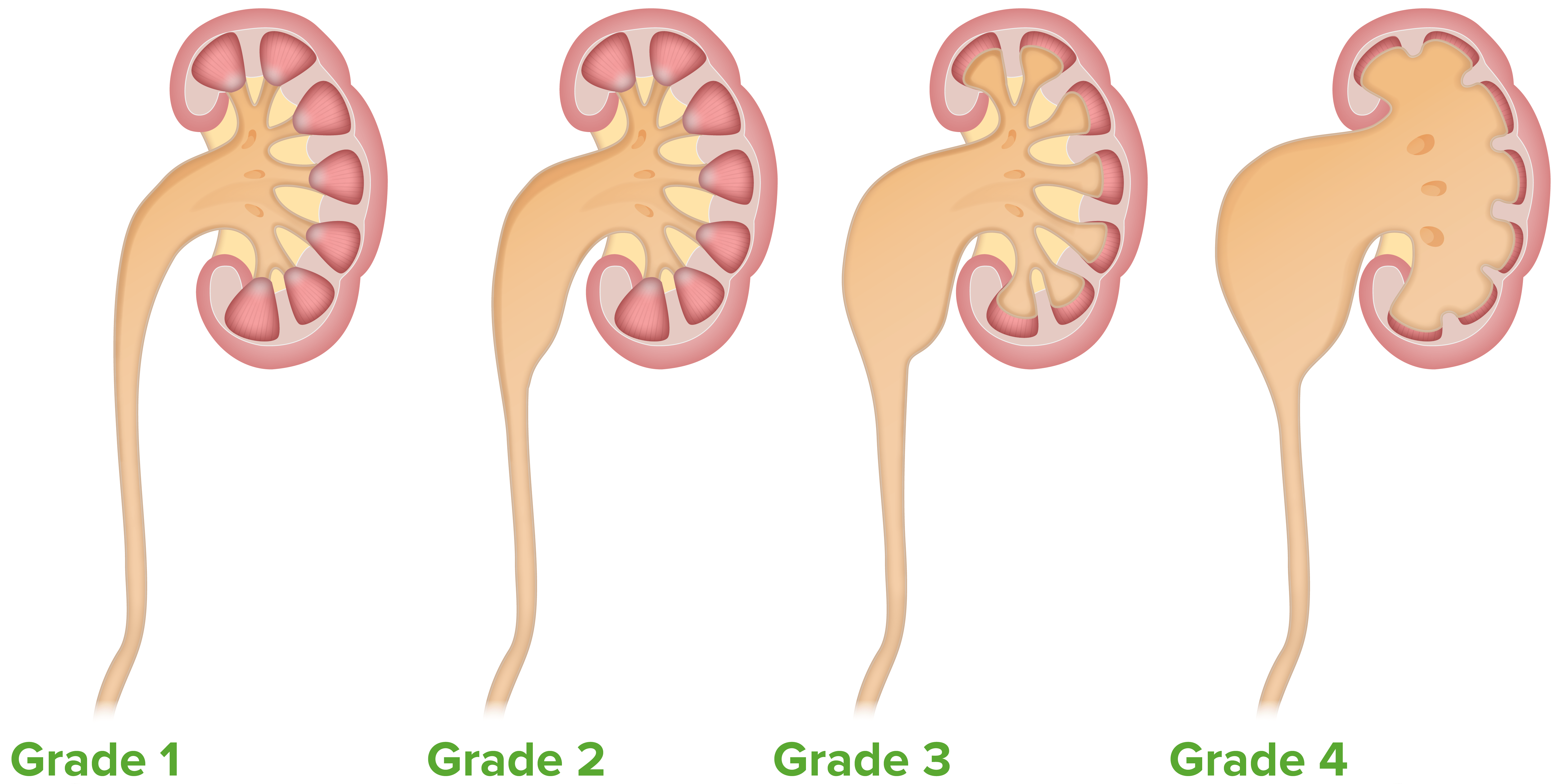
Table: The Onen GradingGradingMethods which attempt to express in replicable terms the level of cell differentiation in neoplasms as increasing anaplasia correlates with the aggressiveness of the neoplasm.Grading, Staging, and Metastasis System for hydronephrosisHydronephrosisHydronephrosis is dilation of the renal collecting system as a result of the obstruction of urine outflow. Hydronephrosis can be unilateral or bilateral. Nephrolithiasis is the most common cause of hydronephrosis in young adults, while prostatic hyperplasia and neoplasm are seen in older patients. Hydronephrosis[7]
Grade
Characteristics
I
Renal pelvic dilation
II
Grade I + calyceal dilation
III
Grade II + thinning of the medulla
IV
Grade III + cortical thinning + no corticomedullary differentiation
The Onen grading system of hydronephrosis
Image by Lecturio.
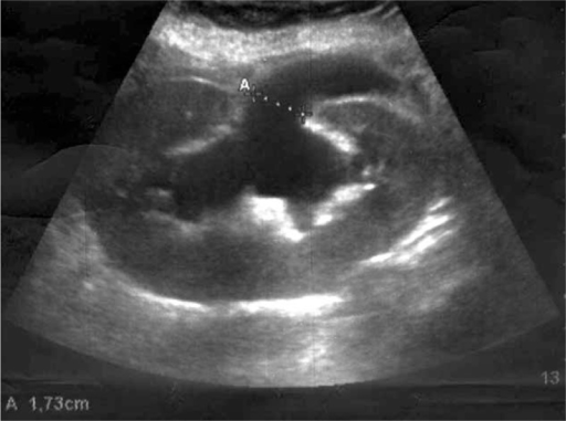
Renal ultrasound demonstrating severe hydronephrosis:
A: Calipers demonstrate dilation of the renal pelvis.
Image: “F1: Graft sonography performed 6 h after hernia repair demonstrates severe hydronephrosis.” by Massimiliano Veroux et al. License: CC BY 4.0
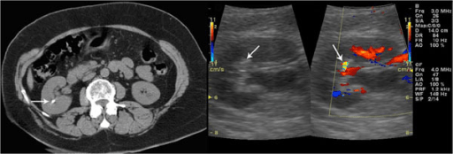
Unenhanced CT confirms the presence of a renal stone:
Left: a gray-scale sonogram showing a small hyperechoic spot without posterior acoustic shadowing
Right: a color Doppler sonogram showing a twinkling sign
Image: “F2: Unenhanced CT confirms the presence of renal stone. Gray-scale sonogram shows a small hyperechoic spot without posterior acoustic shadowing. Color Doppler sonogram shows a twinkling sign.” by Gianfranco Vallone et al. License: CC BY 2.0
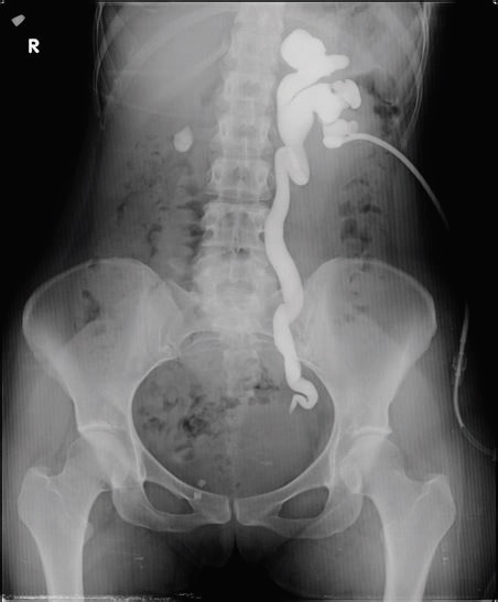
Anterograde pyelography demonstrates grade III left-sided hydronephrosis with obstruction at the ureterovesical junction.
Image: “Antegrade pyelogram of grade III hydronephrosis with obstruction at the ureterovesical junction” by Erkan Efe, Murat Bakacak, Salih Serin, Eyüp Kolus, Önder Ercan, and Sefa Resim. License: CC BY 4.0
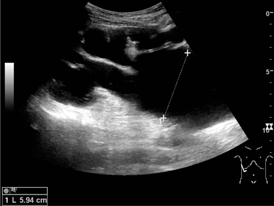
End-stage hydronephrosis with cortical thinning (grade IV):
Measurement of pelvic dilatation on the US image is illustrated by ‘+’ and a dashed line.
Image: “Ultrasonography of end-stage hydronephrosis with cortical thinning” by Kristoffer Lindskov Hansen, Michael Bachmann Nielsen and Caroline Ewertsen. License: CC BY 4.0
Management
Management depends on the cause of obstruction, the degree of metabolic abnormality, and the presence of infection.
AcetaminophenAcetaminophenAcetaminophen is an over-the-counter nonopioid analgesic and antipyretic medication and the most commonly used analgesic worldwide. Despite the widespread use of acetaminophen, its mechanism of action is not entirely understood.Acetaminophen
Antibiotics if infection present; duration of treatment depends on complicating factors:[13]
TrimethoprimTrimethoprimThe sulfonamides are a class of antimicrobial drugs inhibiting folic acid synthesize in pathogens. The prototypical drug in the class is sulfamethoxazole. Although not technically sulfonamides, trimethoprim, dapsone, and pyrimethamine are also important antimicrobial agents inhibiting folic acid synthesis. The agents are often combined with sulfonamides, resulting in a synergistic effect. Sulfonamides and Trimethoprim–sulfamethoxazoleSulfamethoxazoleA bacteriostatic antibacterial agent that interferes with folic acid synthesis in susceptible bacteria. Its broad spectrum of activity has been limited by the development of resistance.Sulfonamides and Trimethoprim: 160/800 mg (double-strength tablet) twice daily
Nitrofurantoin
CephalosporinsCephalosporinsCephalosporins are a group of bactericidal beta-lactam antibiotics (similar to penicillins) that exert their effects by preventing bacteria from producing their cell walls, ultimately leading to cell death. Cephalosporins are categorized by generation and all drug names begin with “cef-” or “ceph-.” Cephalosporins
NephrolithiasisNephrolithiasisNephrolithiasis is the formation of a stone, or calculus, anywhere along the urinary tract caused by precipitations of solutes in the urine. The most common type of kidney stone is the calcium oxalate stone, but other types include calcium phosphate, struvite (ammonium magnesium phosphate), uric acid, and cystine stones.Nephrolithiasis:
Medical expulsion therapy:
Alpha-blockersAlpha-blockersDrugs that bind to but do not activate alpha-adrenergic receptors thereby blocking the actions of endogenous or exogenous adrenergic agonists. Adrenergic alpha-antagonists are used in the treatment of hypertension, vasospasm, peripheral vascular disease, shock, and pheochromocytoma.Antiadrenergic Drugs if stone > 5 mm and ≤ 10 mm in diameter: tamsulosinTamsulosinA sulfonamide derivative and adrenergic alpha-1 receptor antagonist that is used to relieve symptoms of urinary obstruction caused by benign prostatic hyperplasia.Antiadrenergic Drugs 0.4 mg once daily (most common) until the stone passes, up to 4 weeks
CalciumCalciumA basic element found in nearly all tissues. It is a member of the alkaline earth family of metals with the atomic symbol ca, atomic number 20, and atomic weight 40. Calcium is the most abundant mineral in the body and combines with phosphorus to form calcium phosphate in the bones and teeth. It is essential for the normal functioning of nerves and muscles and plays a role in blood coagulation (as factor IV) and in many enzymatic processes.Electrolytes channel blocker (less effective than tamsulosinTamsulosinA sulfonamide derivative and adrenergic alpha-1 receptor antagonist that is used to relieve symptoms of urinary obstruction caused by benign prostatic hyperplasia.Antiadrenergic Drugs; used less frequently): nifedipineNifedipineA potent vasodilator agent with calcium antagonistic action. It is a useful anti-anginal agent that also lowers blood pressure.Class 4 Antiarrhythmic Drugs (Calcium Channel Blockers) extended-release 30 mg daily
Extracorporeal shockwave lithotripsy: High-energy shockwaves cause stones to fragment.
Percutaneous nephrolithotomy
Ureteroscopic or laparoscopic/open surgery
BenignBenignFibroadenoma prostatic hyperplasiaHyperplasiaAn increase in the number of cells in a tissue or organ without tumor formation. It differs from hypertrophy, which is an increase in bulk without an increase in the number of cells.Cellular Adaptation:
α1-Receptor blockers
TamsulosinTamsulosinA sulfonamide derivative and adrenergic alpha-1 receptor antagonist that is used to relieve symptoms of urinary obstruction caused by benign prostatic hyperplasia.Antiadrenergic Drugs 0.4 mg once daily; may increase to 0.8 mg daily after 2–4 weeks
DoxazosinDoxazosinA prazosin-related compound that is a selective alpha-1-adrenergic blocker.Antiadrenergic Drugs 4–8 mg once daily
5α-Reductase inhibitor: finasterideFinasterideAn orally active 3-oxo-5-alpha-steroid 4-dehydrogenase inhibitor. It is used as a surgical alternative for treatment of benign prostatic hyperplasia.Androgens and Antiandrogens 5 mg once daily, alone or with an alpha-blocker
Interventional radiologyInterventional radiologySubspecialty of radiology that combines organ system radiography, catheter techniques and sectional imaging.Penetrating Abdominal Injury: for percutaneous nephrostomy tube if needed
Urology: for hydronephrosisHydronephrosisHydronephrosis is dilation of the renal collecting system as a result of the obstruction of urine outflow. Hydronephrosis can be unilateral or bilateral. Nephrolithiasis is the most common cause of hydronephrosis in young adults, while prostatic hyperplasia and neoplasm are seen in older patients. Hydronephrosis due to prostatic enlargement or cancer, for ureteral repair, stents, or kidney surgery (pyeloplasty) if needed
Gynecology: hysterectomy consult for an individual with a large fibroid uterusUterusThe uterus, cervix, and fallopian tubes are part of the internal female reproductive system. The uterus has a thick wall made of smooth muscle (the myometrium) and an inner mucosal layer (the endometrium). The most inferior portion of the uterus is the cervix, which connects the uterine cavity to the vagina.Uterus, Cervix, and Fallopian Tubes: Anatomy causing obstruction
Oncology: chemotherapyChemotherapyOsteosarcoma and/or radiationRadiationEmission or propagation of acoustic waves (sound), electromagnetic energy waves (such as light; radio waves; gamma rays; or x-rays), or a stream of subatomic particles (such as electrons; neutrons; protons; or alpha particles).Osteosarcoma may be needed to reduce tumorTumorInflammation bulk in some malignancies (e.g., lymphomaLymphomaA general term for various neoplastic diseases of the lymphoid tissue.Imaging of the Mediastinum).
Relief of obstruction[5]
The goal is to decrease the pressure in the collecting system.
Indications:
Complete obstruction
Presence of infection
Evidence of compromised renal function
Lower urinary tractUrinary tractThe urinary tract is located in the abdomen and pelvis and consists of the kidneys, ureters, urinary bladder, and urethra. The structures permit the excretion of urine from the body. Urine flows from the kidneys through the ureters to the urinary bladder and out through the urethra.Urinary Tract: Anatomy obstruction:
Urethral catheterization
Suprapubic catheterization
Upper urinary tractUrinary tractThe urinary tract is located in the abdomen and pelvis and consists of the kidneys, ureters, urinary bladder, and urethra. The structures permit the excretion of urine from the body. Urine flows from the kidneys through the ureters to the urinary bladder and out through the urethra.Urinary Tract: Anatomy obstruction:
Placement of retrograde ureteric stents
Percutaneous nephrostomy (PCN):
Catheter is placed into the renal collecting system for decompression.
Helps facilitate endourologic surgery
Subsequent antegrade stenting can be done.
Potential complications[8]
Urinary tractUrinary tractThe urinary tract is located in the abdomen and pelvis and consists of the kidneys, ureters, urinary bladder, and urethra. The structures permit the excretion of urine from the body. Urine flows from the kidneys through the ureters to the urinary bladder and out through the urethra.Urinary Tract: Anatomy infection; pyelonephritisPyelonephritisPyelonephritis is infection affecting the renal pelvis and the renal parenchyma. This condition arises mostly as a complication of bladder infection that ascends to the upper urinary tract. Pyelonephritis can be acute or chronic (which results from persistent or chronic infections). Typical acute symptoms are flank pain, fever, and nausea with vomiting. TPyelonephritis and Perinephric Abscess
Postobstructive diuresis
Collecting-system rupture, presenting with an acute abdomenAcute AbdomenAcute abdomen, which is in many cases a surgical emergency, is the sudden onset of abdominal pain that may be caused by inflammation, infection, perforation, ischemia, or obstruction. The location of the pain, its characteristics, and associated symptoms (e.g., jaundice) are important tools that help narrow the differential diagnosis.Acute Abdomen (rare)
PrognosisPrognosisA prediction of the probable outcome of a disease based on a individual’s condition and the usual course of the disease as seen in similar situations.Non-Hodgkin Lymphomas[3,4]
Chronic hydronephrosisHydronephrosisHydronephrosis is dilation of the renal collecting system as a result of the obstruction of urine outflow. Hydronephrosis can be unilateral or bilateral. Nephrolithiasis is the most common cause of hydronephrosis in young adults, while prostatic hyperplasia and neoplasm are seen in older patients. Hydronephrosis causes ischemic injury, resulting in cortical and medullary atrophyAtrophyDecrease in the size of a cell, tissue, organ, or multiple organs, associated with a variety of pathological conditions such as abnormal cellular changes, ischemia, malnutrition, or hormonal changes.Cellular Adaptation, and permanent loss of renal function.
HydronephrosisHydronephrosisHydronephrosis is dilation of the renal collecting system as a result of the obstruction of urine outflow. Hydronephrosis can be unilateral or bilateral. Nephrolithiasis is the most common cause of hydronephrosis in young adults, while prostatic hyperplasia and neoplasm are seen in older patients. Hydronephrosis is reversible if the obstruction is promptly relieved.
Differential Diagnosis
Peripelvic cyst: cyst arising from renal hilusHilusLungs: Anatomy. A peripelvic cyst is contiguous to the renal pelvisRenal pelvisKidneys: Anatomy and calyces; its exact etiology is not known. These cystsCystsAny fluid-filled closed cavity or sac that is lined by an epithelium. Cysts can be of normal, abnormal, non-neoplastic, or neoplastic tissues.Fibrocystic Change are thought to be congenital or to arise from lymphatic obstruction. Peripelvic cystsCystsAny fluid-filled closed cavity or sac that is lined by an epithelium. Cysts can be of normal, abnormal, non-neoplastic, or neoplastic tissues.Fibrocystic Change are asymptomatic, but they can distort the renal pelvisRenal pelvisKidneys: Anatomy on imaging.. Diagnosis is with imaging, usually contrast-enhanced CT. Management includes percutaneous drainagePercutaneous DrainageEchinococcus/Echinococcosis or marsupialization.
PyelonephritisPyelonephritisPyelonephritis is infection affecting the renal pelvis and the renal parenchyma. This condition arises mostly as a complication of bladder infection that ascends to the upper urinary tract. Pyelonephritis can be acute or chronic (which results from persistent or chronic infections). Typical acute symptoms are flank pain, fever, and nausea with vomiting. TPyelonephritis and Perinephric Abscess: bacterial infection of the upper urinary tractUrinary tractThe urinary tract is located in the abdomen and pelvis and consists of the kidneys, ureters, urinary bladder, and urethra. The structures permit the excretion of urine from the body. Urine flows from the kidneys through the ureters to the urinary bladder and out through the urethra.Urinary Tract: Anatomy resulting in kidney inflammationInflammationInflammation is a complex set of responses to infection and injury involving leukocytes as the principal cellular mediators in the body’s defense against pathogenic organisms. Inflammation is also seen as a response to tissue injury in the process of wound healing. The 5 cardinal signs of inflammation are pain, heat, redness, swelling, and loss of function. Inflammation. PatientsPatientsIndividuals participating in the health care system for the purpose of receiving therapeutic, diagnostic, or preventive procedures.Clinician–Patient Relationship may present with feverFeverFever is defined as a measured body temperature of at least 38°C (100.4°F). Fever is caused by circulating endogenous and/or exogenous pyrogens that increase levels of prostaglandin E2 in the hypothalamus. Fever is commonly associated with chills, rigors, sweating, and flushing of the skin. Fever, flank painFlank painPain emanating from below the ribs and above the ilium.Renal Cell Carcinoma, nauseaNauseaAn unpleasant sensation in the stomach usually accompanied by the urge to vomit. Common causes are early pregnancy, sea and motion sickness, emotional stress, intense pain, food poisoning, and various enteroviruses.Antiemetics, vomitingVomitingThe forcible expulsion of the contents of the stomach through the mouth.Hypokalemia, and costovertebral angle tenderness. Diagnosis is based on history, physical examination findings, urinalysisUrinalysisExamination of urine by chemical, physical, or microscopic means. Routine urinalysis usually includes performing chemical screening tests, determining specific gravity, observing any unusual color or odor, screening for bacteriuria, and examining the sediment microscopically.Urinary Tract Infections (UTIs) in Children, and urine cultures. Other diagnostic workup includes CBC, imaging, and blood cultures. PyelonephritisPyelonephritisPyelonephritis is infection affecting the renal pelvis and the renal parenchyma. This condition arises mostly as a complication of bladder infection that ascends to the upper urinary tract. Pyelonephritis can be acute or chronic (which results from persistent or chronic infections). Typical acute symptoms are flank pain, fever, and nausea with vomiting. TPyelonephritis and Perinephric Abscess can be managed in the outpatient or inpatient setting depending on the presence of risk factors and the severity of the disease. Management includes the administration of analgesics, antibiotics, and antipyretics.
Congenital megacalyces: rare, usually unilateral, condition caused by the underdevelopment of medullary pyramids resulting in the dilation of the calyces without obstruction. PatientsPatientsIndividuals participating in the health care system for the purpose of receiving therapeutic, diagnostic, or preventive procedures.Clinician–Patient Relationship with congenital megacalyces are asymptomatic, but they can present with a urinary tractUrinary tractThe urinary tract is located in the abdomen and pelvis and consists of the kidneys, ureters, urinary bladder, and urethra. The structures permit the excretion of urine from the body. Urine flows from the kidneys through the ureters to the urinary bladder and out through the urethra.Urinary Tract: Anatomy infection due to stasis. Diagnosis is based on imaging with ultrasonography, IV pyelographyPyelographyHydronephrosis, or CT scan with contrast. Stones and infectionsInfectionsInvasion of the host organism by microorganisms or their toxins or by parasites that can cause pathological conditions or diseases.Chronic Granulomatous Disease are treated appropriately. Surgery is not required.
Billing and Coding
Diagnosis Codes:
These codes are used to document hydronephrosisHydronephrosisHydronephrosis is dilation of the renal collecting system as a result of the obstruction of urine outflow. Hydronephrosis can be unilateral or bilateral. Nephrolithiasis is the most common cause of hydronephrosis in young adults, while prostatic hyperplasia and neoplasm are seen in older patients. Hydronephrosis, the swellingSwellingInflammation of a kidney due to a build-up of urine, which is typically caused by a blockage in the ureter. The codes can specify the nature of the obstruction.
Coding System
Code
Description
ICD-10-CM
N13.30
Unspecified hydronephrosisHydronephrosisHydronephrosis is dilation of the renal collecting system as a result of the obstruction of urine outflow. Hydronephrosis can be unilateral or bilateral. Nephrolithiasis is the most common cause of hydronephrosis in young adults, while prostatic hyperplasia and neoplasm are seen in older patients. Hydronephrosis
ICD-10-CM
N13.0
HydronephrosisHydronephrosisHydronephrosis is dilation of the renal collecting system as a result of the obstruction of urine outflow. Hydronephrosis can be unilateral or bilateral. Nephrolithiasis is the most common cause of hydronephrosis in young adults, while prostatic hyperplasia and neoplasm are seen in older patients. Hydronephrosis with ureteropelvic junctionUreteropelvic junctionUrinary Tract: Anatomy obstruction
ICD-10-CM
N13.1
HydronephrosisHydronephrosisHydronephrosis is dilation of the renal collecting system as a result of the obstruction of urine outflow. Hydronephrosis can be unilateral or bilateral. Nephrolithiasis is the most common cause of hydronephrosis in young adults, while prostatic hyperplasia and neoplasm are seen in older patients. Hydronephrosis with ureteral strictureStricturePrimary Sclerosing Cholangitis, not elsewhere classified
Evaluation & Workup:
These CPT codes are for imaging studies used to diagnose hydronephrosisHydronephrosisHydronephrosis is dilation of the renal collecting system as a result of the obstruction of urine outflow. Hydronephrosis can be unilateral or bilateral. Nephrolithiasis is the most common cause of hydronephrosis in young adults, while prostatic hyperplasia and neoplasm are seen in older patients. Hydronephrosis and identify the location and cause of the obstruction. A renal ultrasound is often the initial test, while a CT urogram provides more detailed anatomical information.
Coding System
Code
Description
CPT
76770
Ultrasound, retroperitonealRetroperitonealPeritoneum: Anatomy (eg, renal, aortaAortaThe main trunk of the systemic arteries.Mediastinum and Great Vessels: Anatomy, nodes), real time with image documentationDocumentationSystematic organization, storage, retrieval, and dissemination of specialized information, especially of a scientific or technical nature. It often involves authenticating or validating information.Advance Directives; complete
CPT
74177
Computed tomography, abdomen and pelvisPelvisThe pelvis consists of the bony pelvic girdle, the muscular and ligamentous pelvic floor, and the pelvic cavity, which contains viscera, vessels, and multiple nerves and muscles. The pelvic girdle, composed of 2 “hip” bones and the sacrum, is a ring-like bony structure of the axial skeleton that links the vertebral column with the lower extremities.Pelvis: Anatomy; with contrast material(s)
Procedures/Interventions:
These codes represent procedures to relieve the urinary obstruction causing hydronephrosisHydronephrosisHydronephrosis is dilation of the renal collecting system as a result of the obstruction of urine outflow. Hydronephrosis can be unilateral or bilateral. Nephrolithiasis is the most common cause of hydronephrosis in young adults, while prostatic hyperplasia and neoplasm are seen in older patients. Hydronephrosis. A percutaneous nephrostomy tube drains urine directly from the kidney, while a ureteral stent bypasses the blockage internally.
Coding System
Code
Description
CPT
50432
Placement of nephrostomy catheter, percutaneous, including diagnostic nephrostogram and/or pyelogram when performed
CPT
52332
Cystourethroscopy, with insertion of indwelling ureteral stent
Complications:
These codes are used to document the consequences of prolonged or severe hydronephrosisHydronephrosisHydronephrosis is dilation of the renal collecting system as a result of the obstruction of urine outflow. Hydronephrosis can be unilateral or bilateral. Nephrolithiasis is the most common cause of hydronephrosis in young adults, while prostatic hyperplasia and neoplasm are seen in older patients. Hydronephrosis, including progressive kidney damage (obstructive uropathy) and acute renal failureRenal failureConditions in which the kidneys perform below the normal level in the ability to remove wastes, concentrate urine, and maintain electrolyte balance; blood pressure; and calcium metabolism. Renal insufficiency can be classified by the degree of kidney damage (as measured by the level of proteinuria) and reduction in glomerular filtration rate.Crush Syndrome.
Coding System
Code
Description
ICD-10-CM
N13.9
Obstructive and reflux uropathy, unspecified
ICD-10-CM
N17.9
Acute kidney failure, unspecified
References
Ilgi, M., et al. (2020). Rare causes of hydronephrosis in adults and diagnosis algorithm: analysis of 100 cases during 15 years. Cureus 12(5):e8226. https://doi.org/10.7759/cureus.8226
Expert Panel on Interventional Radiology, Scheidt, M. J., Hohenwalter, E. J., Pinchot, J. W., Ahmed, O., Bjurlin, M. A., Braun, A. R., Kim, C. Y., Knavel Koepsel, E. M., Schramm, K., Sella, D. M., Weiss, C. R., Lorenz, J. M. (2020). ACR Appropriateness Criteria® radiologic management of urinary tract obstruction. Journal of the American College of Radiology, 17(5S), S281–S292. https://doi.org/10.1016/j.jacr.2020.01.039
Carvalho, E. M., et al. (2019). Trematode infections. In Crow, M.K., et al. (Eds.), Goldman-Cecil Medicine (26th ed., vol. 2, pp. 2122–2124).
Alberter, A., & Sauer, J. (2022). Hydronephrosis and free fluid identified by point‐of‐care ultrasound leads to diagnosis. Journal of the American College of Emergency Physicians Open, 3(5). https://doi.org/10.1002/emp2.12824
Bayne, C. E., Majd, M., Rushton, H. G. (2019). Diuresis renography in the evaluation and management of pediatric hydronephrosis: What have we learned? Journal of Pediatric Urology, 15(2), 128–137. https://doi.org/10.1016/j.jpurol.2019.01.011
Caddeo, G., Williams, S. T., et al. (2013). Acute kidney injury in urology patients: incidence, causes and outcomes. Nephro-Urology Monthly, 5(5), 955–961. https://doi.org/10.5812/numonthly.12721
Meldrum, K. K. (2016). Pathophysiology of urinary tract obstruction. In Wein, A.J., et al. (Eds.), Campbell-Walsh Urology (11th ed., pp. 1089–1103).
de Bessa, J., Rodrigues, C. M., et al. (2018). Diagnostic accuracy of Onen’s Alternative Grading System combined with Doppler evaluation of ureteral jets as an alternative in the diagnosis of obstructive hydronephrosis in children. PeerJ, 6, e4791. https://doi.org/10.7717/peerj.4791
Herness, J., Buttolph, A., Hammer, N. C. (2020). Acute pyelonephritis in adults: rapid evidence review. American Family Physician, 102(3), 173–180.
Hitzeman, N., Williams, S. (2015). Alpha blockers to speed ureteral stone passage. American Family Physician, 91(3), 164–165.
Łoń, I., Wieliczko, M., Lewandowski, J., Małyszko, J. (2022). Retroperitoneal fibrosis is still an underdiagnosed entity with poor prognosis. Kidney & Blood Pressure Research, 47(3), 151–162. https://doi.org/10.1159/000521423