Hemolytic uremic syndrome (HUS) is a clinical phenomenon most commonly seen in children that consists of a classic triad of microangiopathic hemolytic anemia, thrombocytopenia, and acute kidney injury. Hemolytic uremic syndrome is a major cause of acute kidney injury in children and is most commonly associated with a prodrome of diarrheal illness caused by Shiga-like toxin-producing bacteria. Laboratory analysis confirms microangiopathic hemolytic anemia (hemoglobin < 8 g/dL, schistocytes, and negative direct Coombs), thrombocytopenia (platelet count < 140,000/mm³), and acute kidney injury (elevated creatinine and blood urea nitrogen (BUN)). The management of HUS is primarily through supportive care.
Hemolytic uremic syndromeHemolytic uremic syndromeA syndrome that is associated with microvascular diseases of the kidney, such as renal cortical necrosis. It is characterized by hemolytic anemia; thrombocytopenia; and acute renal failure.Hypocoagulable Conditions (HUSHUSHemolytic uremic syndrome (HUS) is a clinical phenomenon most commonly seen in children that consists of a classic triad of microangiopathic hemolytic anemia, thrombocytopenia, and acute kidney injury. Hemolytic uremic syndrome is a major cause of acute kidney injury in children and is most commonly associated with a prodrome of diarrheal illness caused by shiga-like toxin-producing bacteria.Hemolytic Uremic Syndrome) is a disease of the capillariesCapillariesCapillaries are the primary structures in the circulatory system that allow the exchange of gas, nutrients, and other materials between the blood and the extracellular fluid (ECF). Capillaries are the smallest of the blood vessels. Because a capillary diameter is so small, only 1 RBC may pass through at a time.Capillaries: Histology (microangiopathy) that causes the formation of blood clots, anemiaAnemiaAnemia is a condition in which individuals have low Hb levels, which can arise from various causes. Anemia is accompanied by a reduced number of RBCs and may manifest with fatigue, shortness of breath, pallor, and weakness. Subtypes are classified by the size of RBCs, chronicity, and etiology. Anemia: Overview and Types caused by the destruction of RBC in these clotted capillariesCapillariesCapillaries are the primary structures in the circulatory system that allow the exchange of gas, nutrients, and other materials between the blood and the extracellular fluid (ECF). Capillaries are the smallest of the blood vessels. Because a capillary diameter is so small, only 1 RBC may pass through at a time.Capillaries: Histology (hemolytic anemiaHemolytic AnemiaHemolytic anemia (HA) is the term given to a large group of anemias that are caused by the premature destruction/hemolysis of circulating red blood cells (RBCs). Hemolysis can occur within (intravascular hemolysis) or outside the blood vessels (extravascular hemolysis). Hemolytic Anemia), acute kidney injuryAcute Kidney InjuryAcute kidney injury refers to sudden and often reversible loss of renal function, which develops over days or weeks. Azotemia refers to elevated levels of nitrogen-containing substances in the blood that accompany AKI, which include BUN and creatinine. Acute Kidney Injury, and low plateletsPlateletsPlatelets are small cell fragments involved in hemostasis. Thrombopoiesis takes place primarily in the bone marrow through a series of cell differentiation and is influenced by several cytokines. Platelets are formed after fragmentation of the megakaryocyte cytoplasm. Platelets: Histology (thrombocytopeniaThrombocytopeniaThrombocytopenia occurs when the platelet count is < 150,000 per microliter. The normal range for platelets is usually 150,000-450,000/µL of whole blood. Thrombocytopenia can be a result of decreased production, increased destruction, or splenic sequestration of platelets. Patients are often asymptomatic until platelet counts are < 50,000/µL. Thrombocytopenia).
Epidemiology[1–4]
Majority of cases seen in children < 5 years of age
2–3 cases per 100,000 children in the United States
Major cause of acute kidney injuryAcute Kidney InjuryAcute kidney injury refers to sudden and often reversible loss of renal function, which develops over days or weeks. Azotemia refers to elevated levels of nitrogen-containing substances in the blood that accompany AKI, which include BUN and creatinine. Acute Kidney Injury in children (> 6 months to 5 years of age)
Previously classified as:
Typical HUSHUSHemolytic uremic syndrome (HUS) is a clinical phenomenon most commonly seen in children that consists of a classic triad of microangiopathic hemolytic anemia, thrombocytopenia, and acute kidney injury. Hemolytic uremic syndrome is a major cause of acute kidney injury in children and is most commonly associated with a prodrome of diarrheal illness caused by shiga-like toxin-producing bacteria.Hemolytic Uremic Syndrome:
Most commonly seen in the summer and in rural populations
90% of cases are associated with diarrheaDiarrheaDiarrhea is defined as ≥ 3 watery or loose stools in a 24-hour period. There are a multitude of etiologies, which can be classified based on the underlying mechanism of disease. The duration of symptoms (acute or chronic) and characteristics of the stools (e.g., watery, bloody, steatorrheic, mucoid) can help guide further diagnostic evaluation. Diarrhea caused by Shiga toxinShiga toxinA class of toxins that inhibit protein synthesis by blocking the interaction of ribosomal RNA; with peptide elongation factors. They include shiga toxin which is produced by Shigella dysenteriae and a variety of shiga-like toxins that are produced by pathologic strains of Escherichia coli such as Escherichia coli o157.Diarrheagenic E. coli–producing Escherichia coliEscherichia coliThe gram-negative bacterium Escherichia coli is a key component of the human gut microbiota. Most strains of E. coli are avirulent, but occasionally they escape the GI tract, infecting the urinary tract and other sites. Less common strains of E. coli are able to cause disease within the GI tract, most commonly presenting as abdominal pain and diarrhea. Escherichia coli(STEC).
Atypical HUSHUSHemolytic uremic syndrome (HUS) is a clinical phenomenon most commonly seen in children that consists of a classic triad of microangiopathic hemolytic anemia, thrombocytopenia, and acute kidney injury. Hemolytic uremic syndrome is a major cause of acute kidney injury in children and is most commonly associated with a prodrome of diarrheal illness caused by shiga-like toxin-producing bacteria.Hemolytic Uremic Syndrome: nondiarrheal
ResearchResearchCritical and exhaustive investigation or experimentation, having for its aim the discovery of new facts and their correct interpretation, the revision of accepted conclusions, theories, or laws in the light of newly discovered facts, or the practical application of such new or revised conclusions, theories, or laws.Conflict of Interest has shown that aside from infectionsInfectionsInvasion of the host organism by microorganisms or their toxins or by parasites that can cause pathological conditions or diseases.Chronic Granulomatous Disease, genetic mutationsGenetic MutationsCarcinogenesis and different triggering factors lead to HUSHUSHemolytic uremic syndrome (HUS) is a clinical phenomenon most commonly seen in children that consists of a classic triad of microangiopathic hemolytic anemia, thrombocytopenia, and acute kidney injury. Hemolytic uremic syndrome is a major cause of acute kidney injury in children and is most commonly associated with a prodrome of diarrheal illness caused by shiga-like toxin-producing bacteria.Hemolytic Uremic Syndrome (see Etiology).
Etiology is classified as acquired (infectious versus noninfectious) or hereditary.[2,5,6,13]
Acquired HUSHUSHemolytic uremic syndrome (HUS) is a clinical phenomenon most commonly seen in children that consists of a classic triad of microangiopathic hemolytic anemia, thrombocytopenia, and acute kidney injury. Hemolytic uremic syndrome is a major cause of acute kidney injury in children and is most commonly associated with a prodrome of diarrheal illness caused by shiga-like toxin-producing bacteria.Hemolytic Uremic Syndrome:
Infectious
Shiga toxinShiga toxinA class of toxins that inhibit protein synthesis by blocking the interaction of ribosomal RNA; with peptide elongation factors. They include shiga toxin which is produced by Shigella dysenteriae and a variety of shiga-like toxins that are produced by pathologic strains of Escherichia coli such as Escherichia coli o157.Diarrheagenic E. coli–producing E. coli (STEC): most common in children < 5 years of age
ShigellaShigellaShigella is a genus of gram-negative, non-lactose-fermenting facultative intracellular bacilli. Infection spreads most commonly via person-to-person contact or through contaminated food and water. Humans are the only known reservoir.Shigellaspp.
StreptococcusStreptococcusStreptococcus is one of the two medically important genera of gram-positive cocci, the other being Staphylococcus. Streptococci are identified as different species on blood agar on the basis of their hemolytic pattern and sensitivity to optochin and bacitracin. There are many pathogenic species of streptococci, including S. pyogenes, S. agalactiae, S. pneumoniae, and the viridans streptococci.Streptococcus pneumoniae
AutoantibodiesAutoantibodiesAntibodies that react with self-antigens (autoantigens) of the organism that produced them.Blotting Techniques to complement factors
Drugs associated with malignancyMalignancyHemothorax or organ transplant recipients (cyclosporineCyclosporineA cyclic undecapeptide from an extract of soil fungi. It is a powerful immunosupressant with a specific action on T-lymphocytes. It is used for the prophylaxis of graft rejection in organ and tissue transplantation.Immunosuppressants and tacrolimusTacrolimusA macrolide isolated from the culture broth of a strain of streptomyces tsukubaensis that has strong immunosuppressive activity in vivo and prevents the activation of T-lymphocytes in response to antigenic or mitogenic stimulation in vitro.Immunosuppressants)
Drugs of abuse (cocaineCocaineAn alkaloid ester extracted from the leaves of plants including coca. It is a local anesthetic and vasoconstrictor and is clinically used for that purpose, particularly in the eye, ear, nose, and throat. It also has powerful central nervous system effects similar to the amphetamines and is a drug of abuse. Cocaine, like amphetamines, acts by multiple mechanisms on brain catecholaminergic neurons; the mechanism of its reinforcing effects is thought to involve inhibition of dopamine uptake.Local Anesthetics)
Other:
Systemic lupus erythematosusSystemic lupus erythematosusSystemic lupus erythematosus (SLE) is a chronic autoimmune, inflammatory condition that causes immune-complex deposition in organs, resulting in systemic manifestations. Women, particularly those of African American descent, are more commonly affected.Systemic Lupus Erythematosus
Antiphospholipid syndromeAntiphospholipid syndromeAntiphospholipid syndrome (APLS) is an acquired autoimmune disorder characterized by the persistent presence of antiphospholipid antibodies, which create a hypercoagulable state. These antibodies are most commonly discovered during a workup for a thrombotic event or recurrent pregnancy loss, which are the 2 most common clinical manifestations.Antiphospholipid Syndrome
Hereditary HUSHUSHemolytic uremic syndrome (HUS) is a clinical phenomenon most commonly seen in children that consists of a classic triad of microangiopathic hemolytic anemia, thrombocytopenia, and acute kidney injury. Hemolytic uremic syndrome is a major cause of acute kidney injury in children and is most commonly associated with a prodrome of diarrheal illness caused by shiga-like toxin-producing bacteria.Hemolytic Uremic Syndrome:
Complement geneGeneA category of nucleic acid sequences that function as units of heredity and which code for the basic instructions for the development, reproduction, and maintenance of organisms.Basic Terms of Genetics mutations (involving complement proteinsProteinsLinear polypeptides that are synthesized on ribosomes and may be further modified, crosslinked, cleaved, or assembled into complex proteins with several subunits. The specific sequence of amino acids determines the shape the polypeptide will take, during protein folding, and the function of the protein.Energy Homeostasis C3 and CD46 and complement factors H, B, and I)
Inborn errors of metabolism (cobalaminCobalaminA cobalt-containing coordination compound produced by intestinal microorganisms and found also in soil and water. Higher plants do not concentrate vitamin B 12 from the soil and so are a poor source of the substance as compared with animal tissues. Intrinsic factor is important for the assimilation of vitamin B 12.Folate and Vitamin B12 C metabolism)
Pathophysiology
The pathophysiology for HUSHUSHemolytic uremic syndrome (HUS) is a clinical phenomenon most commonly seen in children that consists of a classic triad of microangiopathic hemolytic anemia, thrombocytopenia, and acute kidney injury. Hemolytic uremic syndrome is a major cause of acute kidney injury in children and is most commonly associated with a prodrome of diarrheal illness caused by shiga-like toxin-producing bacteria.Hemolytic Uremic Syndrome secondary to Shiga-like toxinShiga-Like ToxinHemolytic Uremic Syndrome has been well described.[7]
Infection, most commonly from E. colibacteriaBacteriaBacteria are prokaryotic single-celled microorganisms that are metabolically active and divide by binary fission. Some of these organisms play a significant role in the pathogenesis of diseases. Bacteriology, is contracted by ingestion or consumption of undercooked beef or unpasteurized milk.
Bacterial toxins are absorbed by the gut and enter the systemic circulationCirculationThe movement of the blood as it is pumped through the cardiovascular system.ABCDE Assessment.
Nitric oxideNitric OxideA free radical gas produced endogenously by a variety of mammalian cells, synthesized from arginine by nitric oxide synthase. Nitric oxide is one of the endothelium-dependent relaxing factors released by the vascular endothelium and mediates vasodilation. It also inhibits platelet aggregation, induces disaggregation of aggregated platelets, and inhibits platelet adhesion to the vascular endothelium. Nitric oxide activates cytosolic guanylate cyclase and thus elevates intracellular levels of cyclic gmp.Pulmonary Hypertension Drugs (NO) production
SecretionSecretionCoagulation Studies of interleukin-1Interleukin-1A soluble factor produced by monocytes; macrophages, and other cells which activates T-lymphocytes and potentiates their response to mitogens or antigens. Interleukin-1 is a general term refers to either of the two distinct proteins, interleukin-1alpha and interleukin-1beta. The biological effects of il-1 include the ability to replace macrophage requirements for t-cell activation.Interleukins (IL-1) and tumorTumorInflammationnecrosisNecrosisThe death of cells in an organ or tissue due to disease, injury or failure of the blood supply.Ischemic Cell Damage factor (TNFTNFTumor necrosis factor (TNF) is a major cytokine, released primarily by macrophages in response to stimuli. The presence of microbial products and dead cells and injury are among the stimulating factors. This protein belongs to the TNF superfamily, a group of ligands and receptors performing functions in inflammatory response, morphogenesis, and cell proliferation. Tumor Necrosis Factor (TNF))
Macrophage activationMacrophage activationThe process of altering the morphology and functional activity of macrophages so that they become avidly phagocytic. It is initiated by lymphokines, such as the macrophage activation factor (maf) and the macrophage migration-inhibitory factor (mmif), immune complexes, C3b, and various peptides, polysaccharides, and immunologic adjuvants.IL-12 Receptor Deficiency
Cause platelet aggregationPlatelet aggregationThe attachment of platelets to one another. This clumping together can be induced by a number of agents (e.g., thrombin; collagen) and is part of the mechanism leading to the formation of a thrombus.Hemostasis and localized thrombosisThrombosisFormation and development of a thrombus or blood clot in the blood vessel.Epidemic Typhus in capillariesCapillariesCapillaries are the primary structures in the circulatory system that allow the exchange of gas, nutrients, and other materials between the blood and the extracellular fluid (ECF). Capillaries are the smallest of the blood vessels. Because a capillary diameter is so small, only 1 RBC may pass through at a time.Capillaries: Histology and arteriolesArteriolesThe smallest divisions of the arteries located between the muscular arteries and the capillaries.Arteries: Histology:
Glomeruli vessels are especially affected → decreased glomerular filtration rateGlomerular filtration rateThe volume of water filtered out of plasma through glomerular capillary walls into Bowman’s capsules per unit of time. It is considered to be equivalent to inulin clearance.Kidney Function Tests (GFRGFRThe volume of water filtered out of plasma through glomerular capillary walls into Bowman’s capsules per unit of time. It is considered to be equivalent to inulin clearance.Kidney Function Tests)
Decreased renal blood flowRenal blood flowThe amount of the renal blood flow that is going to the functional renal tissue, i.e., parts of the kidney that are involved in production of urine.Glomerular Filtration → increased reninReninA highly specific (leu-leu) endopeptidase that generates angiotensin I from its precursor angiotensinogen, leading to a cascade of reactions which elevate blood pressure and increase sodium retention by the kidney in the renin-angiotensin system.Renal Sodium and Water Regulation
ErythrocytesErythrocytesErythrocytes, or red blood cells (RBCs), are the most abundant cells in the blood. While erythrocytes in the fetus are initially produced in the yolk sac then the liver, the bone marrow eventually becomes the main site of production. Erythrocytes: Histology are mechanically injured when going through the thrombotic microvasculature: causes microangiopathic hemolytic anemiaMicroangiopathic Hemolytic AnemiaHemolytic Uremic Syndrome
Other forms of HUSHUSHemolytic uremic syndrome (HUS) is a clinical phenomenon most commonly seen in children that consists of a classic triad of microangiopathic hemolytic anemia, thrombocytopenia, and acute kidney injury. Hemolytic uremic syndrome is a major cause of acute kidney injury in children and is most commonly associated with a prodrome of diarrheal illness caused by shiga-like toxin-producing bacteria.Hemolytic Uremic Syndrome:
Varied pathological mechanisms
All have endothelial damage in common.
Lead to thrombus production and hemolytic anemiaHemolytic AnemiaHemolytic anemia (HA) is the term given to a large group of anemias that are caused by the premature destruction/hemolysis of circulating red blood cells (RBCs). Hemolysis can occur within (intravascular hemolysis) or outside the blood vessels (extravascular hemolysis). Hemolytic Anemia
Clinical Presentation
History[2,8,9]
Typical HUSHUSHemolytic uremic syndrome (HUS) is a clinical phenomenon most commonly seen in children that consists of a classic triad of microangiopathic hemolytic anemia, thrombocytopenia, and acute kidney injury. Hemolytic uremic syndrome is a major cause of acute kidney injury in children and is most commonly associated with a prodrome of diarrheal illness caused by shiga-like toxin-producing bacteria.Hemolytic Uremic Syndrome: prodromal illness with abdominal painAbdominal PainAcute Abdomen, vomitingVomitingThe forcible expulsion of the contents of the stomach through the mouth.Hypokalemia, andbloody diarrheaBloody diarrheaDiarrhea
Pneumococcal HUSHUSHemolytic uremic syndrome (HUS) is a clinical phenomenon most commonly seen in children that consists of a classic triad of microangiopathic hemolytic anemia, thrombocytopenia, and acute kidney injury. Hemolytic uremic syndrome is a major cause of acute kidney injury in children and is most commonly associated with a prodrome of diarrheal illness caused by shiga-like toxin-producing bacteria.Hemolytic Uremic Syndrome is preceded by:
Severe pneumoniaPneumoniaPneumonia or pulmonary inflammation is an acute or chronic inflammation of lung tissue. Causes include infection with bacteria, viruses, or fungi. In more rare cases, pneumonia can also be caused through toxic triggers through inhalation of toxic substances, immunological processes, or in the course of radiotherapy.Pneumonia (usually with effusion) or
MeningitisMeningitisMeningitis is inflammation of the meninges, the protective membranes of the brain, and spinal cord. The causes of meningitis are varied, with the most common being bacterial or viral infection. The classic presentation of meningitis is a triad of fever, altered mental status, and nuchal rigidity. Meningitis or
Less commonly, sinus or ear infectionsInfectionsInvasion of the host organism by microorganisms or their toxins or by parasites that can cause pathological conditions or diseases.Chronic Granulomatous Disease
Other historical clues may include:
Concurrent known HUSHUSHemolytic uremic syndrome (HUS) is a clinical phenomenon most commonly seen in children that consists of a classic triad of microangiopathic hemolytic anemia, thrombocytopenia, and acute kidney injury. Hemolytic uremic syndrome is a major cause of acute kidney injury in children and is most commonly associated with a prodrome of diarrheal illness caused by shiga-like toxin-producing bacteria.Hemolytic Uremic Syndrome outbreak/family member with concurrent HUSHUSHemolytic uremic syndrome (HUS) is a clinical phenomenon most commonly seen in children that consists of a classic triad of microangiopathic hemolytic anemia, thrombocytopenia, and acute kidney injury. Hemolytic uremic syndrome is a major cause of acute kidney injury in children and is most commonly associated with a prodrome of diarrheal illness caused by shiga-like toxin-producing bacteria.Hemolytic Uremic Syndrome
Family member with history of HUSHUSHemolytic uremic syndrome (HUS) is a clinical phenomenon most commonly seen in children that consists of a classic triad of microangiopathic hemolytic anemia, thrombocytopenia, and acute kidney injury. Hemolytic uremic syndrome is a major cause of acute kidney injury in children and is most commonly associated with a prodrome of diarrheal illness caused by shiga-like toxin-producing bacteria.Hemolytic Uremic Syndrome (may be suggestive of a genetic cause)
Previous episode of HUSHUSHemolytic uremic syndrome (HUS) is a clinical phenomenon most commonly seen in children that consists of a classic triad of microangiopathic hemolytic anemia, thrombocytopenia, and acute kidney injury. Hemolytic uremic syndrome is a major cause of acute kidney injury in children and is most commonly associated with a prodrome of diarrheal illness caused by shiga-like toxin-producing bacteria.Hemolytic Uremic Syndrome (may be suggestive of a complement-mediated cause)
Physical examination[2,8,9]
Symptoms usually occur 5–7 days after diarrheaDiarrheaDiarrhea is defined as ≥ 3 watery or loose stools in a 24-hour period. There are a multitude of etiologies, which can be classified based on the underlying mechanism of disease. The duration of symptoms (acute or chronic) and characteristics of the stools (e.g., watery, bloody, steatorrheic, mucoid) can help guide further diagnostic evaluation. Diarrhea and may include:
Acute-onset lethargyLethargyA general state of sluggishness, listless, or uninterested, with being tired, and having difficulty concentrating and doing simple tasks. It may be related to depression or drug addiction.Hyponatremia or irritability
Pallor
Nephritic syndromeNephritic syndromeNephritic syndrome is a broad category of glomerular diseases characterized by glomerular hematuria, variable loss of renal function, and hypertension. These features are in contrast to those of nephrotic syndrome, which includes glomerular diseases characterized by severe proteinuria, although there is sometimes overlap of > 1 glomerular disease in the same individual. Nephritic Syndrome:
OliguriaOliguriaDecreased urine output that is below the normal range. Oliguria can be defined as urine output of less than or equal to 0. 5 or 1 ml/kg/hr depending on the age.Renal Potassium Regulation, volume overload, or dehydrationDehydrationThe condition that results from excessive loss of water from a living organism.Volume Depletion and Dehydration
SeizuresSeizuresA seizure is abnormal electrical activity of the neurons in the cerebral cortex that can manifest in numerous ways depending on the region of the brain affected. Seizures consist of a sudden imbalance that occurs between the excitatory and inhibitory signals in cortical neurons, creating a net excitation. The 2 major classes of seizures are focal and generalized. Seizures
Other organ involvement:
Cardiac: signs of fluid overload
LiverLiverThe liver is the largest gland in the human body. The liver is found in the superior right quadrant of the abdomen and weighs approximately 1.5 kilograms. Its main functions are detoxification, metabolism, nutrient storage (e.g., iron and vitamins), synthesis of coagulation factors, formation of bile, filtration, and storage of blood. Liver: Anatomy: hepatomegaly
Diagnosis
Clinical diagnosis[2,8,11,13]
Diagnosis per both US and UK criteriais clinical, based on the classic triad of:
↓ Haptoglobin, ↑ reticulocyteReticulocyteImmature erythrocytes. In humans, these are erythroid cells that have just undergone extrusion of their cell nucleus. They still contain some organelles that gradually decrease in number as the cells mature. Ribosomes are last to disappear. Certain staining techniques cause components of the ribosomes to precipitate into characteristic ‘reticulum’ (not the same as the endoplasmic reticulum), hence the name reticulocytes.Erythrocytes: Histology count, ↑ lactate dehydrogenaseLactate DehydrogenaseOsteosarcoma (LDHLDHOsteosarcoma), ↑ indirect bilirubinIndirect BilirubinLiver Function Tests
ThrombocytopeniaThrombocytopeniaThrombocytopenia occurs when the platelet count is < 150,000 per microliter. The normal range for platelets is usually 150,000-450,000/µL of whole blood. Thrombocytopenia can be a result of decreased production, increased destruction, or splenic sequestration of platelets. Patients are often asymptomatic until platelet counts are < 50,000/µL. Thrombocytopenia:
Platelet count < 140,000/mm³ (usually around 40,000/mm³)
Acute kidney injuryAcute Kidney InjuryAcute kidney injury refers to sudden and often reversible loss of renal function, which develops over days or weeks. Azotemia refers to elevated levels of nitrogen-containing substances in the blood that accompany AKI, which include BUN and creatinine. Acute Kidney Injury:
Elevated serum creatinine and blood ureaUreaA compound formed in the liver from ammonia produced by the deamination of amino acids. It is the principal end product of protein catabolism and constitutes about one half of the total urinary solids.Urea CyclenitrogenNitrogenAn element with the atomic symbol n, atomic number 7, and atomic weight [14. 00643; 14. 00728]. Nitrogen exists as a diatomic gas and makes up about 78% of the earth’s atmosphere by volume. It is a constituent of proteins and nucleic acids and found in all living cells.Urea Cycle (BUN)
May manifest as hematuriaHematuriaPresence of blood in the urine.Renal Cell Carcinoma, proteinuriaProteinuriaThe presence of proteins in the urine, an indicator of kidney diseases.Nephrotic Syndrome in Children, or oliguriaOliguriaDecreased urine output that is below the normal range. Oliguria can be defined as urine output of less than or equal to 0. 5 or 1 ml/kg/hr depending on the age.Renal Potassium Regulation
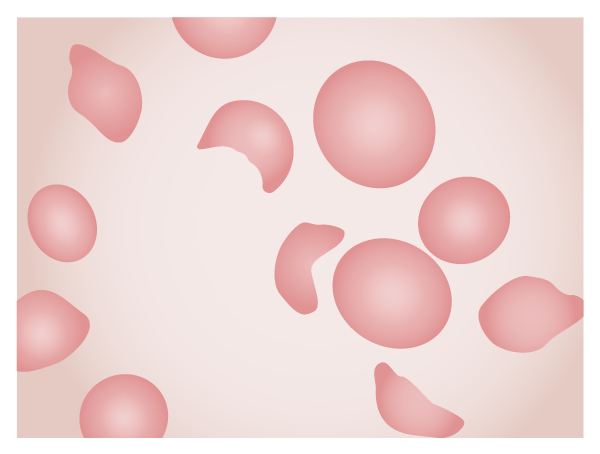
Blood slide of a patient with HUS. Note the schistocytes, fragments of erythrocytes left after mechanical injury of the cells in the microvasculature.
Image by Lecturio.
Additional tests[13,15,18,19]
Serum electrolytesElectrolytesElectrolytes are mineral salts that dissolve in water and dissociate into charged particles called ions, which can be either be positively (cations) or negatively (anions) charged. Electrolytes are distributed in the extracellular and intracellular compartments in different concentrations. Electrolytes are essential for various basic life-sustaining functions.Electrolytes, glucoseGlucoseA primary source of energy for living organisms. It is naturally occurring and is found in fruits and other parts of plants in its free state. It is used therapeutically in fluid and nutrient replacement.Lactose Intolerance, liver function testsLiver function testsLiver function tests, also known as hepatic function panels, are one of the most commonly performed screening blood tests. Such tests are also used to detect, evaluate, and monitor acute and chronic liver diseases.Liver Function Tests, amylaseAmylaseA group of amylolytic enzymes that cleave starch, glycogen, and related alpha-1, 4-glucans.Digestion and Absorption, lipaseLipaseAn enzyme of the hydrolase class that catalyzes the reaction of triacylglycerol and water to yield diacylglycerol and a fatty acid anion. It is produced by glands on the tongue and by the pancreas and initiates the digestion of dietary fats.Malabsorption and Maldigestion:
In severe renal injury, acidosisAcidosisA pathologic condition of acid accumulation or depletion of base in the body. The two main types are respiratory acidosis and metabolic acidosis, due to metabolic acid build up.Respiratory Acidosis with hyperkalemiaHyperkalemiaHyperkalemia is defined as a serum potassium (K+) concentration >5.2 mEq/L. Homeostatic mechanisms maintain the serum K+ concentration between 3.5 and 5.2 mEq/L, despite marked variation in dietary intake. Hyperkalemia can be due to a variety of causes, which include transcellular shifts, tissue breakdown, inadequate renal excretion, and drugs. Hyperkalemia can occur.
↑ TransaminaseTransaminaseA subclass of enzymes of the transferase class that catalyze the transfer of an amino group from a donor (generally an amino acid) to an acceptor (generally a 2-keto acid). Most of these enzymes are pyridoxyl phosphate proteins.Catabolism of Amino Acids levels can be seen.
PancreatitisPancreatitisInflammation of the pancreas. Pancreatitis is classified as acute unless there are computed tomographic or endoscopic retrograde cholangiopancreatographic findings of chronic pancreatitis. The two most common forms of acute pancreatitis are alcoholic pancreatitis and gallstone pancreatitis.Acute Pancreatitis and transient diabetesDiabetesDiabetes mellitus (DM) is a metabolic disease characterized by hyperglycemia and dysfunction of the regulation of glucose metabolism by insulin. Type 1 DM is diagnosed mostly in children and young adults as the result of autoimmune destruction of β cells in the pancreas and the resulting lack of insulin. Type 2 DM has a significant association with obesity and is characterized by insulin resistance.Diabetes Mellitus can develop from pancreatic involvement.
Prothrombin timeProthrombin timeClotting time of plasma recalcified in the presence of excess tissue thromboplastin. Factors measured are fibrinogen; prothrombin; factor V; factor VII; and factor X.Hemostasis (PT), partial thromboplastin timePartial thromboplastin timeThe time required for the appearance of fibrin strands following the mixing of plasma with phospholipid platelet substitute (e.g., crude cephalins, soybean phosphatides). It is a test of the intrinsic pathway (factors VIII, IX, XI, and XII) and the common pathway (fibrinogen, prothrombin, factors V and X) of blood coagulation.Hemostasis (PTT)
Stool culture and polymerase chain reactionPolymerase chain reactionPolymerase chain reaction (PCR) is a technique that amplifies DNA fragments exponentially for analysis. The process is highly specific, allowing for the targeting of specific genomic sequences, even with minuscule sample amounts. The PCR cycles multiple times through 3 phases: denaturation of the template DNA, annealing of a specific primer to the individual DNA strands, and synthesis/elongation of new DNA molecules.Polymerase Chain Reaction (PCR) (detecting Shiga toxinShiga toxinA class of toxins that inhibit protein synthesis by blocking the interaction of ribosomal RNA; with peptide elongation factors. They include shiga toxin which is produced by Shigella dysenteriae and a variety of shiga-like toxins that are produced by pathologic strains of Escherichia coli such as Escherichia coli o157.Diarrheagenic E. coli):
If atypical HUSHUSHemolytic uremic syndrome (HUS) is a clinical phenomenon most commonly seen in children that consists of a classic triad of microangiopathic hemolytic anemia, thrombocytopenia, and acute kidney injury. Hemolytic uremic syndrome is a major cause of acute kidney injury in children and is most commonly associated with a prodrome of diarrheal illness caused by shiga-like toxin-producing bacteria.Hemolytic Uremic Syndrome is suspected:
C3 and C4
Factor H and I concentration
Autoantibody screen
Genetic analysis
Other tests depend on the presentation (e.g., chest X-rayX-rayPenetrating electromagnetic radiation emitted when the inner orbital electrons of an atom are excited and release radiant energy. X-ray wavelengths range from 1 pm to 10 nm. Hard x-rays are the higher energy, shorter wavelength x-rays. Soft x-rays or grenz rays are less energetic and longer in wavelength. The short wavelength end of the x-ray spectrum overlaps the gamma rays wavelength range. The distinction between gamma rays and x-rays is based on their radiation source.Pulmonary Function Tests for patientsPatientsIndividuals participating in the health care system for the purpose of receiving therapeutic, diagnostic, or preventive procedures.Clinician–Patient Relationship with pneumoniaPneumoniaPneumonia or pulmonary inflammation is an acute or chronic inflammation of lung tissue. Causes include infection with bacteria, viruses, or fungi. In more rare cases, pneumonia can also be caused through toxic triggers through inhalation of toxic substances, immunological processes, or in the course of radiotherapy.Pneumonia, brainBrainThe part of central nervous system that is contained within the skull (cranium). Arising from the neural tube, the embryonic brain is comprised of three major parts including prosencephalon (the forebrain); mesencephalon (the midbrain); and rhombencephalon (the hindbrain). The developed brain consists of cerebrum; cerebellum; and other structures in the brain stem.Nervous System: Anatomy, Structure, and Classification MRI if CNS abnormalities are present)
BiopsyBiopsyRemoval and pathologic examination of specimens from the living body.Ewing Sarcoma is not required for the diagnosis of HUSHUSHemolytic uremic syndrome (HUS) is a clinical phenomenon most commonly seen in children that consists of a classic triad of microangiopathic hemolytic anemia, thrombocytopenia, and acute kidney injury. Hemolytic uremic syndrome is a major cause of acute kidney injury in children and is most commonly associated with a prodrome of diarrheal illness caused by shiga-like toxin-producing bacteria.Hemolytic Uremic Syndrome, but when performed for disambiguation, 3 patterns are usually seen:
Glomerular thrombotic microangiopathy:
CapillariesCapillariesCapillaries are the primary structures in the circulatory system that allow the exchange of gas, nutrients, and other materials between the blood and the extracellular fluid (ECF). Capillaries are the smallest of the blood vessels. Because a capillary diameter is so small, only 1 RBC may pass through at a time.Capillaries: Histology appear thicker and have a double “contour” due to an increase in thickness of the subendothelialSubendothelialMembranoproliferative Glomerulonephritis layer.
Glomeruli appear large and blocked with microclots
Patchy cortical necrosisNecrosisThe death of cells in an organ or tissue due to disease, injury or failure of the blood supply.Ischemic Cell Damage
Arterial thrombotic microangiopathy:
Primarily seen in non-STEC HUSHUSHemolytic uremic syndrome (HUS) is a clinical phenomenon most commonly seen in children that consists of a classic triad of microangiopathic hemolytic anemia, thrombocytopenia, and acute kidney injury. Hemolytic uremic syndrome is a major cause of acute kidney injury in children and is most commonly associated with a prodrome of diarrheal illness caused by shiga-like toxin-producing bacteria.Hemolytic Uremic Syndrome
Affects arteriolesArteriolesThe smallest divisions of the arteries located between the muscular arteries and the capillaries.Arteries: Histology, causing luminal wall necrosisNecrosisThe death of cells in an organ or tissue due to disease, injury or failure of the blood supply.Ischemic Cell Damage and clotting
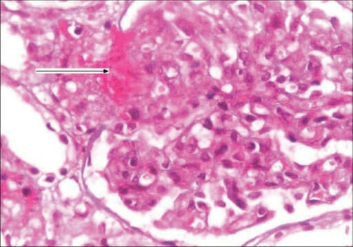
Kidney biopsy of hemolytic uremic syndrome (HUS):
Kidney biopsy showing the glomerulus with an increase in mesangial matrix, focal endocapillary cell swelling, arterioles with platelet-fibrin thrombi (arrow), and fibrinoid necrosis (H&E stain, 400x)
Image: “F0002: Kidney biopsy showing glomerulus with increase in mesangial matrix, focal endocapillary cell swelling and arterioles with platelet fibrin thrombi (arrow), and fibrinoid necrosis. (H&E stain, magnification ×400)” by G. Lakshminarayana, R. Rajesh, A. Jojo, G. Kurian, and V. N. Unni. License: CC BY 2.0
Diagnostic approach to a patient with thrombotic microangiopathy[11]
Thrombotic microangiopathy (TMA) is seen in multiple disease processes aside from HUSHUSHemolytic uremic syndrome (HUS) is a clinical phenomenon most commonly seen in children that consists of a classic triad of microangiopathic hemolytic anemia, thrombocytopenia, and acute kidney injury. Hemolytic uremic syndrome is a major cause of acute kidney injury in children and is most commonly associated with a prodrome of diarrheal illness caused by shiga-like toxin-producing bacteria.Hemolytic Uremic Syndrome. Among these, thrombotic thrombocytopenic purpuraThrombotic thrombocytopenic purpuraThrombotic thrombocytopenic purpura (TTP) is a life-threatening condition due to either a congenital or an acquired deficiency of ADAMTS-13, a metalloproteinase that cleaves multimers of von Willebrand factor (VWF). The large multimers then aggregate excessive platelets resulting in microvascular thrombosis and an increase in consumption of platelets. Thrombotic Thrombocytopenic Purpura (TTPTTPThrombotic thrombocytopenic purpura (TTP) is a life-threatening condition due to either a congenital or an acquired deficiency of adamts-13, a metalloproteinase that cleaves multimers of von Willebrand factor (vWF). The large multimers then aggregate excessive platelets resulting in microvascular thrombosis and an increase in consumption of platelets.Thrombotic Thrombocytopenic Purpura) is a medical emergency requiring urgent plasma exchangePlasma exchangeRemoval of plasma and replacement with various fluids, e.g., fresh frozen plasma, plasma protein fractions (ppf), albumin preparations, dextran solutions, saline. Used in treatment of autoimmune diseases, immune complex diseases, diseases of excess plasma factors, and other conditions.Thrombotic Thrombocytopenic Purpura. Because of this consideration, patientsPatientsIndividuals participating in the health care system for the purpose of receiving therapeutic, diagnostic, or preventive procedures.Clinician–Patient Relationship presenting with TMA should be evaluated as described below to determine the risk for TTPTTPThrombotic thrombocytopenic purpura (TTP) is a life-threatening condition due to either a congenital or an acquired deficiency of adamts-13, a metalloproteinase that cleaves multimers of von Willebrand factor (vWF). The large multimers then aggregate excessive platelets resulting in microvascular thrombosis and an increase in consumption of platelets.Thrombotic Thrombocytopenic Purpura and the necessity for treatment.
Includes patientsPatientsIndividuals participating in the health care system for the purpose of receiving therapeutic, diagnostic, or preventive procedures.Clinician–Patient Relationship with:
Clinical symptoms (neurologic, renal, etcETCThe electron transport chain (ETC) sends electrons through a series of proteins, which generate an electrochemical proton gradient that produces energy in the form of adenosine triphosphate (ATP).Electron Transport Chain (ETC).) suggestive of TMA with or without evidence of organ damage
Peripheral smear confirming microangiopathic hemolytic anemiaMicroangiopathic Hemolytic AnemiaHemolytic Uremic Syndrome (will show large plateletsPlateletsPlatelets are small cell fragments involved in hemostasis. Thrombopoiesis takes place primarily in the bone marrow through a series of cell differentiation and is influenced by several cytokines. Platelets are formed after fragmentation of the megakaryocyte cytoplasm. Platelets: Histology due to a strong bone marrowBone marrowThe soft tissue filling the cavities of bones. Bone marrow exists in two types, yellow and red. Yellow marrow is found in the large cavities of large bones and consists mostly of fat cells and a few primitive blood cells. Red marrow is a hematopoietic tissue and is the site of production of erythrocytes and granular leukocytes. Bone marrow is made up of a framework of connective tissue containing branching fibers with the frame being filled with marrow cells.Bone Marrow: Composition and Hematopoiesis response and fragments of red blood cellsRed blood cellsErythrocytes, or red blood cells (RBCs), are the most abundant cells in the blood. While erythrocytes in the fetus are initially produced in the yolk sac then the liver, the bone marrow eventually becomes the main site of production.Erythrocytes: Histology).
In these patientsPatientsIndividuals participating in the health care system for the purpose of receiving therapeutic, diagnostic, or preventive procedures.Clinician–Patient Relationship, rule out underlying conditions that can present with TMA:
Disseminated intravascular coagulationDisseminated intravascular coagulationDisseminated intravascular coagulation (DIC) is a condition characterized by systemic bodywide activation of the coagulation cascade. This cascade results in both widespread microvascular thrombi contributing to multiple organ dysfunction and consumption of clotting factors and platelets, leading to hemorrhage. Disseminated Intravascular Coagulation
PreeclampsiaPreeclampsiaA complication of pregnancy, characterized by a complex of symptoms including maternal hypertension and proteinuria with or without pathological edema. Symptoms may range between mild and severe. Pre-eclampsia usually occurs after the 20th week of gestation, but may develop before this time in the presence of trophoblastic disease.Hypertensive Pregnancy Disorders/HELLP syndromeHELLP syndromeA severe form of preeclampsia with Hemolysis (with: LDH > 600 IU/L, ↑ Bilirubin, Schistocytes on blood smear, Anemia), ↑ Liver enzymes (with AST and/or AST > 2 times upper limit of normal), ↓ Platelet count, and thrombocytopenia (< 100,000).Hypertensive Pregnancy Disorders
Organ transplant
Rheumatic disease
Important to note that:
Kidney injury is a prominent feature of HUSHUSHemolytic uremic syndrome (HUS) is a clinical phenomenon most commonly seen in children that consists of a classic triad of microangiopathic hemolytic anemia, thrombocytopenia, and acute kidney injury. Hemolytic uremic syndrome is a major cause of acute kidney injury in children and is most commonly associated with a prodrome of diarrheal illness caused by shiga-like toxin-producing bacteria.Hemolytic Uremic Syndrome (but is variableVariableVariables represent information about something that can change. The design of the measurement scales, or of the methods for obtaining information, will determine the data gathered and the characteristics of that data. As a result, a variable can be qualitative or quantitative, and may be further classified into subgroups.Types of Variables in TTPTTPThrombotic thrombocytopenic purpura (TTP) is a life-threatening condition due to either a congenital or an acquired deficiency of adamts-13, a metalloproteinase that cleaves multimers of von Willebrand factor (vWF). The large multimers then aggregate excessive platelets resulting in microvascular thrombosis and an increase in consumption of platelets.Thrombotic Thrombocytopenic Purpura).
Unlike HUSHUSHemolytic uremic syndrome (HUS) is a clinical phenomenon most commonly seen in children that consists of a classic triad of microangiopathic hemolytic anemia, thrombocytopenia, and acute kidney injury. Hemolytic uremic syndrome is a major cause of acute kidney injury in children and is most commonly associated with a prodrome of diarrheal illness caused by shiga-like toxin-producing bacteria.Hemolytic Uremic Syndrome, immune TTPTTPThrombotic thrombocytopenic purpura (TTP) is a life-threatening condition due to either a congenital or an acquired deficiency of adamts-13, a metalloproteinase that cleaves multimers of von Willebrand factor (vWF). The large multimers then aggregate excessive platelets resulting in microvascular thrombosis and an increase in consumption of platelets.Thrombotic Thrombocytopenic Purpura is characterized by severe ADAMTS13 (A Disintegrin And Metalloprotease with ThromboSpondin type 1Type 1Spinal Muscular Atrophy motif member 13) deficiency.
Approach those who present with TMA by applying the PLASMIC score:
Platelets, combined hemoLysis variableVariableVariables represent information about something that can change. The design of the measurement scales, or of the methods for obtaining information, will determine the data gathered and the characteristics of that data. As a result, a variable can be qualitative or quantitative, and may be further classified into subgroups.Types of Variables, absence of Active cancer, no Stem cell or solid organ transplant, MCVCVVasculitides, INR, Creatinine
1 point given for each of the findings below:
Platelet count < 30,000/µL
Hemolysis (defined by reticulocyteReticulocyteImmature erythrocytes. In humans, these are erythroid cells that have just undergone extrusion of their cell nucleus. They still contain some organelles that gradually decrease in number as the cells mature. Ribosomes are last to disappear. Certain staining techniques cause components of the ribosomes to precipitate into characteristic ‘reticulum’ (not the same as the endoplasmic reticulum), hence the name reticulocytes.Erythrocytes: Histology count > 2.5%, undetectable haptoglobin, or indirect bilirubinIndirect BilirubinLiver Function Tests > 2 mg/dL)
6–7 points: high likelihood of TTPTTPThrombotic thrombocytopenic purpura (TTP) is a life-threatening condition due to either a congenital or an acquired deficiency of adamts-13, a metalloproteinase that cleaves multimers of von Willebrand factor (vWF). The large multimers then aggregate excessive platelets resulting in microvascular thrombosis and an increase in consumption of platelets.Thrombotic Thrombocytopenic Purpura
5 points: intermediate likelihood of TTPTTPThrombotic thrombocytopenic purpura (TTP) is a life-threatening condition due to either a congenital or an acquired deficiency of adamts-13, a metalloproteinase that cleaves multimers of von Willebrand factor (vWF). The large multimers then aggregate excessive platelets resulting in microvascular thrombosis and an increase in consumption of platelets.Thrombotic Thrombocytopenic Purpura
0–4 points: low likelihood of TTPTTPThrombotic thrombocytopenic purpura (TTP) is a life-threatening condition due to either a congenital or an acquired deficiency of adamts-13, a metalloproteinase that cleaves multimers of von Willebrand factor (vWF). The large multimers then aggregate excessive platelets resulting in microvascular thrombosis and an increase in consumption of platelets.Thrombotic Thrombocytopenic Purpura
Next steps:
PatientsPatientsIndividuals participating in the health care system for the purpose of receiving therapeutic, diagnostic, or preventive procedures.Clinician–Patient Relationship with a PLASMIC scorePLASMIC scoreThe PLASMIC score predicts the probability of TTP in the presence of schistocytes on blood smear.Thrombotic Thrombocytopenic Purpura of ≥ 6 should be treated empirically for TTPTTPThrombotic thrombocytopenic purpura (TTP) is a life-threatening condition due to either a congenital or an acquired deficiency of adamts-13, a metalloproteinase that cleaves multimers of von Willebrand factor (vWF). The large multimers then aggregate excessive platelets resulting in microvascular thrombosis and an increase in consumption of platelets.Thrombotic Thrombocytopenic Purpura with therapeutic plasma exchangePlasma exchangeRemoval of plasma and replacement with various fluids, e.g., fresh frozen plasma, plasma protein fractions (ppf), albumin preparations, dextran solutions, saline. Used in treatment of autoimmune diseases, immune complex diseases, diseases of excess plasma factors, and other conditions.Thrombotic Thrombocytopenic Purpura.
PatientsPatientsIndividuals participating in the health care system for the purpose of receiving therapeutic, diagnostic, or preventive procedures.Clinician–Patient Relationship with a score of 5:
Send for ADAMTS13 activity testing.
Obtain expert consultation and consider plasma exchangePlasma exchangeRemoval of plasma and replacement with various fluids, e.g., fresh frozen plasma, plasma protein fractions (ppf), albumin preparations, dextran solutions, saline. Used in treatment of autoimmune diseases, immune complex diseases, diseases of excess plasma factors, and other conditions.Thrombotic Thrombocytopenic Purpura if no other etiology is identified.
PatientsPatientsIndividuals participating in the health care system for the purpose of receiving therapeutic, diagnostic, or preventive procedures.Clinician–Patient Relationship with a score of ≤ 4 should be risk-stratified (based on clinical suspicion).
Consider alternative conditions.
ADAMTS13 activity testing is done if no other etiology is identified.
Management
Practice guidelines may vary depending on location. The following information is based on US, European, and UK literature and guidelines.
Management of HUSHUSHemolytic uremic syndrome (HUS) is a clinical phenomenon most commonly seen in children that consists of a classic triad of microangiopathic hemolytic anemia, thrombocytopenia, and acute kidney injury. Hemolytic uremic syndrome is a major cause of acute kidney injury in children and is most commonly associated with a prodrome of diarrheal illness caused by shiga-like toxin-producing bacteria.Hemolytic Uremic Syndrome is primarily through supportive care.[2,12,13,15‒17,19]
AnemiaAnemiaAnemia is a condition in which individuals have low Hb levels, which can arise from various causes. Anemia is accompanied by a reduced number of RBCs and may manifest with fatigue, shortness of breath, pallor, and weakness. Subtypes are classified by the size of RBCs, chronicity, and etiology. Anemia: Overview and Types: RBC transfusions when indicated (hemoglobin < 6 or 7 g/dL)
ThrombocytopeniaThrombocytopeniaThrombocytopenia occurs when the platelet count is < 150,000 per microliter. The normal range for platelets is usually 150,000-450,000/µL of whole blood. Thrombocytopenia can be a result of decreased production, increased destruction, or splenic sequestration of platelets. Patients are often asymptomatic until platelet counts are < 50,000/µL. Thrombocytopenia: platelet transfusions only for significant clinical bleeding
Acute kidney injuryAcute Kidney InjuryAcute kidney injury refers to sudden and often reversible loss of renal function, which develops over days or weeks. Azotemia refers to elevated levels of nitrogen-containing substances in the blood that accompany AKI, which include BUN and creatinine. Acute Kidney Injury:
IV fluidsIV fluidsIntravenous fluids are one of the most common interventions administered in medicine to approximate physiologic bodily fluids. Intravenous fluids are divided into 2 categories: crystalloid and colloid solutions. Intravenous fluids have a wide variety of indications, including intravascular volume expansion, electrolyte manipulation, and maintenance fluids. Intravenous Fluids
Electrolyte management
Discontinuation of any nephrotoxic drugs
DialysisDialysisRenal replacement therapy refers to dialysis and/or kidney transplantation. Dialysis is a procedure by which toxins and excess water are removed from the circulation. Hemodialysis and peritoneal dialysis (PD) are the two types of dialysis, and their primary difference is the location of the filtration process (external to the body in hemodialysis versus inside the body for PD).Peritoneal Dialysis and Hemodialysis if severe:
UremiaUremiaA clinical syndrome associated with the retention of renal waste products or uremic toxins in the blood. It is usually the result of renal insufficiency. Most uremic toxins are end products of protein or nitrogen catabolism, such as urea or creatinine. Severe uremia can lead to multiple organ dysfunctions with a constellation of symptoms.Acute Kidney Injury present
Severe fluid overload refractory to medical therapy
Severe acidosisAcidosisA pathologic condition of acid accumulation or depletion of base in the body. The two main types are respiratory acidosis and metabolic acidosis, due to metabolic acid build up.Respiratory Acidosis and electrolyte refractory to medical therapy
AzotemiaAzotemiaA biochemical abnormality referring to an elevation of blood urea nitrogen and creatinine. Azotemia can be produced by kidney diseases or other extrarenal disorders. When azotemia becomes associated with a constellation of clinical signs, it is termed uremia.Acute Kidney Injury (BUN ≥ 80 mg/dL)
Nutritional support needed for a child with anuriaAnuriaAbsence of urine formation. It is usually associated with complete bilateral ureteral (ureter) obstruction, complete lower urinary tract obstruction, or unilateral ureteral obstruction when a solitary kidney is present.Acute Kidney Injury
HypertensionHypertensionHypertension, or high blood pressure, is a common disease that manifests as elevated systemic arterial pressures. Hypertension is most often asymptomatic and is found incidentally as part of a routine physical examination or during triage for an unrelated medical encounter. Hypertension (calciumCalciumA basic element found in nearly all tissues. It is a member of the alkaline earth family of metals with the atomic symbol ca, atomic number 20, and atomic weight 40. Calcium is the most abundant mineral in the body and combines with phosphorus to form calcium phosphate in the bones and teeth. It is essential for the normal functioning of nerves and muscles and plays a role in blood coagulation (as factor IV) and in many enzymatic processes.Electrolytes channel blockers (nifedipineNifedipineA potent vasodilator agent with calcium antagonistic action. It is a useful anti-anginal agent that also lowers blood pressure.Class 4 Antiarrhythmic Drugs (Calcium Channel Blockers) or nicardipineNicardipineA potent calcium channel blockader with marked vasodilator action. It has antihypertensive properties and is effective in the treatment of angina and coronary spasms without showing cardiodepressant effects. It has also been used in the treatment of asthma and enhances the action of specific antineoplastic agents.Class 4 Antiarrhythmic Drugs (Calcium Channel Blockers))):
Angiotensin-converting enzyme (ACE) inhibitors generally are not used during the acute phaseAcute phaseShort Bowel Syndrome (due to decreased renal perfusion).
Monoclonal antibody (binding C5) that blocks complement activationComplement ActivationThe sequential activation of serum complement proteins to create the complement membrane attack complex. Factors initiating complement activation include antigen-antibody complexes, microbial antigens, or cell surface polysaccharides.Systemic Lupus Erythematosus
Approved for atypical HUSHUSHemolytic uremic syndrome (HUS) is a clinical phenomenon most commonly seen in children that consists of a classic triad of microangiopathic hemolytic anemia, thrombocytopenia, and acute kidney injury. Hemolytic uremic syndrome is a major cause of acute kidney injury in children and is most commonly associated with a prodrome of diarrheal illness caused by shiga-like toxin-producing bacteria.Hemolytic Uremic Syndrome or complement-mediated HUSHUSHemolytic uremic syndrome (HUS) is a clinical phenomenon most commonly seen in children that consists of a classic triad of microangiopathic hemolytic anemia, thrombocytopenia, and acute kidney injury. Hemolytic uremic syndrome is a major cause of acute kidney injury in children and is most commonly associated with a prodrome of diarrheal illness caused by shiga-like toxin-producing bacteria.Hemolytic Uremic Syndrome
May be beneficial for patientsPatientsIndividuals participating in the health care system for the purpose of receiving therapeutic, diagnostic, or preventive procedures.Clinician–Patient Relationship presenting with severe neurologic involvement (e.g., comaComaComa is defined as a deep state of unarousable unresponsiveness, characterized by a score of 3 points on the GCS. A comatose state can be caused by a multitude of conditions, making the precise epidemiology and prognosis of coma difficult to determine. Coma, seizuresSeizuresA seizure is abnormal electrical activity of the neurons in the cerebral cortex that can manifest in numerous ways depending on the region of the brain affected. Seizures consist of a sudden imbalance that occurs between the excitatory and inhibitory signals in cortical neurons, creating a net excitation. The 2 major classes of seizures are focal and generalized. Seizures)
Plasma exchangePlasma exchangeRemoval of plasma and replacement with various fluids, e.g., fresh frozen plasma, plasma protein fractions (ppf), albumin preparations, dextran solutions, saline. Used in treatment of autoimmune diseases, immune complex diseases, diseases of excess plasma factors, and other conditions.Thrombotic Thrombocytopenic Purpura:
Uncertain benefits for STEC HUSHUSHemolytic uremic syndrome (HUS) is a clinical phenomenon most commonly seen in children that consists of a classic triad of microangiopathic hemolytic anemia, thrombocytopenia, and acute kidney injury. Hemolytic uremic syndrome is a major cause of acute kidney injury in children and is most commonly associated with a prodrome of diarrheal illness caused by shiga-like toxin-producing bacteria.Hemolytic Uremic Syndrome
Used for patientsPatientsIndividuals participating in the health care system for the purpose of receiving therapeutic, diagnostic, or preventive procedures.Clinician–Patient Relationship with suspected TTPTTPThrombotic thrombocytopenic purpura (TTP) is a life-threatening condition due to either a congenital or an acquired deficiency of adamts-13, a metalloproteinase that cleaves multimers of von Willebrand factor (vWF). The large multimers then aggregate excessive platelets resulting in microvascular thrombosis and an increase in consumption of platelets.Thrombotic Thrombocytopenic Purpura while awaiting genetic tests or with confirmed TTPTTPThrombotic thrombocytopenic purpura (TTP) is a life-threatening condition due to either a congenital or an acquired deficiency of adamts-13, a metalloproteinase that cleaves multimers of von Willebrand factor (vWF). The large multimers then aggregate excessive platelets resulting in microvascular thrombosis and an increase in consumption of platelets.Thrombotic Thrombocytopenic Purpura
Monitoring after the acute illness has passed:
Blood pressure
Renal function
UrinalysisUrinalysisExamination of urine by chemical, physical, or microscopic means. Routine urinalysis usually includes performing chemical screening tests, determining specific gravity, observing any unusual color or odor, screening for bacteriuria, and examining the sediment microscopically.Urinary Tract Infections (UTIs) in Children
Note that antibiotic treatment during bloody diarrheal illnesses caused by Shiga toxin-producing bacteriaBacteriaBacteria are prokaryotic single-celled microorganisms that are metabolically active and divide by binary fission. Some of these organisms play a significant role in the pathogenesis of diseases. Bacteriology is associated with an increased risk of developing HUSHUSHemolytic uremic syndrome (HUS) is a clinical phenomenon most commonly seen in children that consists of a classic triad of microangiopathic hemolytic anemia, thrombocytopenia, and acute kidney injury. Hemolytic uremic syndrome is a major cause of acute kidney injury in children and is most commonly associated with a prodrome of diarrheal illness caused by shiga-like toxin-producing bacteria.Hemolytic Uremic Syndrome.
Prognosis
Early diagnosis and treatment have a favorable outcome:mortalityMortalityAll deaths reported in a given population.Measures of Health Status: < 5%[1,3,4,10]
Most recover renal function completely; however:
30% are left with some degree of renal insufficiency.
5% are left dependent on dialysisDialysisRenal replacement therapy refers to dialysis and/or kidney transplantation. Dialysis is a procedure by which toxins and excess water are removed from the circulation. Hemodialysis and peritoneal dialysis (PD) are the two types of dialysis, and their primary difference is the location of the filtration process (external to the body in hemodialysis versus inside the body for PD).Peritoneal Dialysis and Hemodialysis.
When HUSHUSHemolytic uremic syndrome (HUS) is a clinical phenomenon most commonly seen in children that consists of a classic triad of microangiopathic hemolytic anemia, thrombocytopenia, and acute kidney injury. Hemolytic uremic syndrome is a major cause of acute kidney injury in children and is most commonly associated with a prodrome of diarrheal illness caused by shiga-like toxin-producing bacteria.Hemolytic Uremic Syndrome is not associated with diarrheaDiarrheaDiarrhea is defined as ≥ 3 watery or loose stools in a 24-hour period. There are a multitude of etiologies, which can be classified based on the underlying mechanism of disease. The duration of symptoms (acute or chronic) and characteristics of the stools (e.g., watery, bloody, steatorrheic, mucoid) can help guide further diagnostic evaluation. Diarrhea, the prognosisPrognosisA prediction of the probable outcome of a disease based on a individual’s condition and the usual course of the disease as seen in similar situations.Non-Hodgkin Lymphomas is more severe:
Pneumococci-related HUSHUSHemolytic uremic syndrome (HUS) is a clinical phenomenon most commonly seen in children that consists of a classic triad of microangiopathic hemolytic anemia, thrombocytopenia, and acute kidney injury. Hemolytic uremic syndrome is a major cause of acute kidney injury in children and is most commonly associated with a prodrome of diarrheal illness caused by shiga-like toxin-producing bacteria.Hemolytic Uremic Syndrome:
OliguriaOliguriaDecreased urine output that is below the normal range. Oliguria can be defined as urine output of less than or equal to 0. 5 or 1 ml/kg/hr depending on the age.Renal Potassium Regulation and thrombocytopeniaThrombocytopeniaThrombocytopenia occurs when the platelet count is < 150,000 per microliter. The normal range for platelets is usually 150,000-450,000/µL of whole blood. Thrombocytopenia can be a result of decreased production, increased destruction, or splenic sequestration of platelets. Patients are often asymptomatic until platelet counts are < 50,000/µL. Thrombocytopenia last longer.
Up to 80% of patientsPatientsIndividuals participating in the health care system for the purpose of receiving therapeutic, diagnostic, or preventive procedures.Clinician–Patient Relationship require dialysisDialysisRenal replacement therapy refers to dialysis and/or kidney transplantation. Dialysis is a procedure by which toxins and excess water are removed from the circulation. Hemodialysis and peritoneal dialysis (PD) are the two types of dialysis, and their primary difference is the location of the filtration process (external to the body in hemodialysis versus inside the body for PD).Peritoneal Dialysis and Hemodialysis.
Differential Diagnosis
Disseminated intravascular coagulationDisseminated intravascular coagulationDisseminated intravascular coagulation (DIC) is a condition characterized by systemic bodywide activation of the coagulation cascade. This cascade results in both widespread microvascular thrombi contributing to multiple organ dysfunction and consumption of clotting factors and platelets, leading to hemorrhage. Disseminated Intravascular Coagulation (DICDICDisseminated intravascular coagulation (DIC) is a condition characterized by systemic bodywide activation of the coagulation cascade. This cascade results in both widespread microvascular thrombi contributing to multiple organ dysfunction and consumption of clotting factors and platelets, leading to hemorrhage. Disseminated Intravascular Coagulation): associated with children with serious illnesses, such as septic shockSeptic shockSepsis associated with hypotension or hypoperfusion despite adequate fluid resuscitation. Perfusion abnormalities may include, but are not limited to lactic acidosis; oliguria; or acute alteration in mental status.Sepsis and Septic Shock. Unlike HUSHUSHemolytic uremic syndrome (HUS) is a clinical phenomenon most commonly seen in children that consists of a classic triad of microangiopathic hemolytic anemia, thrombocytopenia, and acute kidney injury. Hemolytic uremic syndrome is a major cause of acute kidney injury in children and is most commonly associated with a prodrome of diarrheal illness caused by shiga-like toxin-producing bacteria.Hemolytic Uremic Syndrome, DICDICDisseminated intravascular coagulation (DIC) is a condition characterized by systemic bodywide activation of the coagulation cascade. This cascade results in both widespread microvascular thrombi contributing to multiple organ dysfunction and consumption of clotting factors and platelets, leading to hemorrhage. Disseminated Intravascular Coagulation presents with abnormal coagulation studiesCoagulation studiesCoagulation studies are a group of hematologic laboratory studies that reflect the function of blood vessels, platelets, and coagulation factors, which all interact with one another to achieve hemostasis. Coagulation studies are usually ordered to evaluate patients with bleeding or hypercoagulation disorders.Coagulation Studies, including prolonged prothrombin timeProthrombin timeClotting time of plasma recalcified in the presence of excess tissue thromboplastin. Factors measured are fibrinogen; prothrombin; factor V; factor VII; and factor X.Hemostasis (PT) and partial thromboplastin timePartial thromboplastin timeThe time required for the appearance of fibrin strands following the mixing of plasma with phospholipid platelet substitute (e.g., crude cephalins, soybean phosphatides). It is a test of the intrinsic pathway (factors VIII, IX, XI, and XII) and the common pathway (fibrinogen, prothrombin, factors V and X) of blood coagulation.Hemostasis (PTT), decreased fibrinogenFibrinogenPlasma glycoprotein clotted by thrombin, composed of a dimer of three non-identical pairs of polypeptide chains (alpha, beta, gamma) held together by disulfide bonds. Fibrinogen clotting is a sol-gel change involving complex molecular arrangements: whereas fibrinogen is cleaved by thrombin to form polypeptides a and b, the proteolytic action of other enzymes yields different fibrinogen degradation products.Hemostasis, and increased D-dimerD-dimerDeep Vein Thrombosis.
Thrombotic thrombocytopenic purpuraThrombotic thrombocytopenic purpuraThrombotic thrombocytopenic purpura (TTP) is a life-threatening condition due to either a congenital or an acquired deficiency of ADAMTS-13, a metalloproteinase that cleaves multimers of von Willebrand factor (VWF). The large multimers then aggregate excessive platelets resulting in microvascular thrombosis and an increase in consumption of platelets. Thrombotic Thrombocytopenic Purpura (TTPTTPThrombotic thrombocytopenic purpura (TTP) is a life-threatening condition due to either a congenital or an acquired deficiency of adamts-13, a metalloproteinase that cleaves multimers of von Willebrand factor (vWF). The large multimers then aggregate excessive platelets resulting in microvascular thrombosis and an increase in consumption of platelets.Thrombotic Thrombocytopenic Purpura): occurs due to inhibition or deficiency of ADAMTS13, which causes decreased degradation of von Willebrand factorvon Willebrand factorA high-molecular-weight plasma protein, produced by endothelial cells and megakaryocytes, that is part of the factor VIII/von Willebrand factor complex. The von Willebrand factor has receptors for collagen, platelets, and ristocetin activity as well as the immunologically distinct antigenic determinants. It functions in adhesion of platelets to collagen and hemostatic plug formation. The prolonged bleeding time in von Willebrand diseases is due to the deficiency of this factor.Hemostasis (VWF) multimers, and increased platelet aggregationPlatelet aggregationThe attachment of platelets to one another. This clumping together can be induced by a number of agents (e.g., thrombin; collagen) and is part of the mechanism leading to the formation of a thrombus.Hemostasis and thrombosisThrombosisFormation and development of a thrombus or blood clot in the blood vessel.Epidemic Typhus. Laboratory analysis shows schistocytesSchistocytesHemolytic Uremic Syndrome, increased lactate dehydrogenaseLactate DehydrogenaseOsteosarcoma (LDHLDHOsteosarcoma), and normal coagulation parameters. Symptoms include a classic pentad of neurologic and renal symptoms, feverFeverFever is defined as a measured body temperature of at least 38°C (100.4°F). Fever is caused by circulating endogenous and/or exogenous pyrogens that increase levels of prostaglandin E2 in the hypothalamus. Fever is commonly associated with chills, rigors, sweating, and flushing of the skin. Fever, thrombocytopeniaThrombocytopeniaThrombocytopenia occurs when the platelet count is < 150,000 per microliter. The normal range for platelets is usually 150,000-450,000/µL of whole blood. Thrombocytopenia can be a result of decreased production, increased destruction, or splenic sequestration of platelets. Patients are often asymptomatic until platelet counts are < 50,000/µL. Thrombocytopenia, and microangiopathic hemolytic anemiaMicroangiopathic Hemolytic AnemiaHemolytic Uremic Syndrome.
Systemic vasculitisVasculitisInflammation of any one of the blood vessels, including the arteries; veins; and rest of the vasculature system in the body.Systemic Lupus Erythematosus: PatientsPatientsIndividuals participating in the health care system for the purpose of receiving therapeutic, diagnostic, or preventive procedures.Clinician–Patient Relationship with systemic vasculitisVasculitisInflammation of any one of the blood vessels, including the arteries; veins; and rest of the vasculature system in the body.Systemic Lupus Erythematosus would not have a prodromeProdromeSymptoms that appear 24–48 hours prior to migraine onset.Migraine Headache of diarrheaDiarrheaDiarrhea is defined as ≥ 3 watery or loose stools in a 24-hour period. There are a multitude of etiologies, which can be classified based on the underlying mechanism of disease. The duration of symptoms (acute or chronic) and characteristics of the stools (e.g., watery, bloody, steatorrheic, mucoid) can help guide further diagnostic evaluation. Diarrhea associated with illness. They would also likely have systemic symptoms, such as arthralgias and/or rashRashRocky Mountain Spotted Fever.
Billing and Coding
Diagnosis Codes:
This code is used to diagnose Hemolytic Uremic SyndromeHemolytic uremic syndromeA syndrome that is associated with microvascular diseases of the kidney, such as renal cortical necrosis. It is characterized by hemolytic anemia; thrombocytopenia; and acute renal failure.Hypocoagulable Conditions (HUSHUSHemolytic uremic syndrome (HUS) is a clinical phenomenon most commonly seen in children that consists of a classic triad of microangiopathic hemolytic anemia, thrombocytopenia, and acute kidney injury. Hemolytic uremic syndrome is a major cause of acute kidney injury in children and is most commonly associated with a prodrome of diarrheal illness caused by shiga-like toxin-producing bacteria.Hemolytic Uremic Syndrome), a serious condition characterized by the triad of hemolytic anemiaHemolytic AnemiaHemolytic anemia (HA) is the term given to a large group of anemias that are caused by the premature destruction/hemolysis of circulating red blood cells (RBCs). Hemolysis can occur within (intravascular hemolysis) or outside the blood vessels (extravascular hemolysis). Hemolytic Anemia, acute kidney injuryAcute Kidney InjuryAcute kidney injury refers to sudden and often reversible loss of renal function, which develops over days or weeks. Azotemia refers to elevated levels of nitrogen-containing substances in the blood that accompany AKI, which include BUN and creatinine. Acute Kidney Injury, and low platelet count, typically triggered by an E. coli infection.
Domain
Code
Description
ICD-10-CM
D59.31
Infection-associated hemolytic-uremic syndrome
SNOMED CT
23588008
Hemolytic-uremic syndrome (disorder)
Evaluation & Workup:
These codes are for the laboratory tests that define HUSHUSHemolytic uremic syndrome (HUS) is a clinical phenomenon most commonly seen in children that consists of a classic triad of microangiopathic hemolytic anemia, thrombocytopenia, and acute kidney injury. Hemolytic uremic syndrome is a major cause of acute kidney injury in children and is most commonly associated with a prodrome of diarrheal illness caused by shiga-like toxin-producing bacteria.Hemolytic Uremic Syndrome, including a CBC to show anemiaAnemiaAnemia is a condition in which individuals have low Hb levels, which can arise from various causes. Anemia is accompanied by a reduced number of RBCs and may manifest with fatigue, shortness of breath, pallor, and weakness. Subtypes are classified by the size of RBCs, chronicity, and etiology. Anemia: Overview and Types and thrombocytopeniaThrombocytopeniaThrombocytopenia occurs when the platelet count is < 150,000 per microliter. The normal range for platelets is usually 150,000-450,000/µL of whole blood. Thrombocytopenia can be a result of decreased production, increased destruction, or splenic sequestration of platelets. Patients are often asymptomatic until platelet counts are < 50,000/µL. Thrombocytopenia, a peripheral smear to identify schistocytesSchistocytesHemolytic Uremic Syndrome, and a metabolic panel to assess for acute kidney failure.
Domain
Code
Description
CPT
85027
Complete (CBC), automated (Hgb, Hct, RBC, WBC and platelet count)
This CPT code is for hemodialysisHemodialysisProcedures which temporarily or permanently remedy insufficient cleansing of body fluids by the kidneys.Crush Syndrome, a life-saving supportive procedure required for patientsPatientsIndividuals participating in the health care system for the purpose of receiving therapeutic, diagnostic, or preventive procedures.Clinician–Patient Relationship with HUSHUSHemolytic uremic syndrome (HUS) is a clinical phenomenon most commonly seen in children that consists of a classic triad of microangiopathic hemolytic anemia, thrombocytopenia, and acute kidney injury. Hemolytic uremic syndrome is a major cause of acute kidney injury in children and is most commonly associated with a prodrome of diarrheal illness caused by shiga-like toxin-producing bacteria.Hemolytic Uremic Syndrome who develop severe acute kidney injuryAcute Kidney InjuryAcute kidney injury refers to sudden and often reversible loss of renal function, which develops over days or weeks. Azotemia refers to elevated levels of nitrogen-containing substances in the blood that accompany AKI, which include BUN and creatinine. Acute Kidney Injury and are unable to filter waste products from their blood.
Domain
Code
Description
CPT
90935
HemodialysisHemodialysisProcedures which temporarily or permanently remedy insufficient cleansing of body fluids by the kidneys.Crush Syndrome procedure with single evaluation by a physician or other qualified health care professional
Complications & Supportive Procedures:
These codes are used to document the long-term consequences of HUSHUSHemolytic uremic syndrome (HUS) is a clinical phenomenon most commonly seen in children that consists of a classic triad of microangiopathic hemolytic anemia, thrombocytopenia, and acute kidney injury. Hemolytic uremic syndrome is a major cause of acute kidney injury in children and is most commonly associated with a prodrome of diarrheal illness caused by shiga-like toxin-producing bacteria.Hemolytic Uremic Syndrome, which can include the development of chronic kidney diseaseChronic Kidney DiseaseChronic kidney disease (CKD) is kidney impairment that lasts for ≥ 3 months, implying that it is irreversible. Hypertension and diabetes are the most common causes; however, there are a multitude of other etiologies. In the early to moderate stages, CKD is usually asymptomatic and is primarily diagnosed by laboratory abnormalities.Chronic Kidney Disease and hypertensionHypertensionHypertension, or high blood pressure, is a common disease that manifests as elevated systemic arterial pressures. Hypertension is most often asymptomatic and is found incidentally as part of a routine physical examination or during triage for an unrelated medical encounter. Hypertension due to permanent kidney damage.
Domain
Code
Description
ICD-10-CM
N18.9
Chronic kidney diseaseChronic Kidney DiseaseChronic kidney disease (CKD) is kidney impairment that lasts for ≥ 3 months, implying that it is irreversible. Hypertension and diabetes are the most common causes; however, there are a multitude of other etiologies. In the early to moderate stages, CKD is usually asymptomatic and is primarily diagnosed by laboratory abnormalities.Chronic Kidney Disease, unspecified
ICD-10-CM
I10
Essential (primary) hypertensionHypertensionHypertension, or high blood pressure, is a common disease that manifests as elevated systemic arterial pressures. Hypertension is most often asymptomatic and is found incidentally as part of a routine physical examination or during triage for an unrelated medical encounter. Hypertension
References
Ardissino, G., Salardi, S., Colombo, E., Testa, S., Borsa-Ghiringhelli, N., Paglialonga, F., Paracchini, V., Tel, F., Possenti, I., Belingheri, M., Civitillo, C. F., Sardini, S., Ceruti, R., Baldioli, C., Tommasi, P., Parola, L., Russo, F., Tedeschi, S. (2016). Epidemiology of haemolytic uremic syndrome in children. Data from the North Italian HUS network. European Journal of Pediatrics, 175, 465–473. https://doi.org/10.1007/s00431-015-2642-1
Noris, M., Remuzzi, G. (2005). Hemolytic uremic syndrome. Journal of the American Society of Nephrology, 16, 1035–1050. doi: 10.1681/ASN.2004100861. PMID: 15728781. https://pubmed.ncbi.nlm.nih.gov/15728781/
Constantinescu, A. R., Bitzan, M., Weiss, L. S., Christen, E., Kaplan, B. S., Cnaan, A., Trachtman, H. (2004). Non-enteropathic hemolytic uremic syndrome: causes and short-term course. American Journal of Kidney Diseases, 43, 976–982. https://doi.org/10.1053/j.ajkd.2004.02.010
Ito, K., Komatsu, Y. (1993). [Drug induced hemolytic uremic syndrome]. Nihon Rinsho, 51, 204–209. In Japanese. PMID: 7679450. https://pubmed.ncbi.nlm.nih.gov/7679450/
Geerdink, L. M., Westra, D., van Wijk, J. A., Dorresteijn, E. M., Lilien, M. R., Davin, J. C., Kömhoff, M., Van Hoeck, K., van der Vlugt, A., van den Heuvel, L. P., van de Kar, N. C. (2012). Atypical hemolytic uremic syndrome in children: complement mutations and clinical characteristics. Pediatric Nephrology, 27, 1283–1291. https://doi.org/10.1007/s00467-012-2131-y
Banerjee, R., Hersh, A. L., Newland, J., Beekmann, S. E., Polgreen, P. M., Bender, J., Shaw, J., Copelovitch, L., Kaplan, B. S., Shah, S. S., Emerging Infections Network Hemolytic-Uremic Syndrome Study Group. (2011). Streptococcus pneumoniae-associated hemolytic uremic syndrome among children in North America. Pediatric Infectious Disease Journal, 30, 736–739. https://doi.org/10.1097/INF.0b013e3182191c58
Habib R, Levy M, Gagnadoux M, et al. (1982). Prognosis of the hemolytic uremic syndrome in children. Advances in Nephrology of the Necker Hospital, 11:99.
Tarr, P. I., Gordon, C. A., Chandler, W. L. (2005). Shiga-toxin-producing Escherichia coli and haemolytic uraemic syndrome. Lancet, 365, 1073–1086. https://doi.org/10.1016/S0140-6736(05)71144-2
Percheron, L., Gramada, R., Tellier, S., Salomon, R., Harambat, J., Llanas, B., Fila, M., Allain-Launay, E., Lapeyraque, A. L., Leroy, V., Adra, A. L., Bérard, E., Bourdat-Michel, G., Chehade, H., Eckart, P., Merieau, E., Piètrement, C., Sellier-Leclerc, A. L., Frémeaux-Bacchi, V., Dimeglio, C., … Garnier, A. (2018). Eculizumab treatment in severe pediatric STEC-HUS: a multicenter retrospective study. Pediatric Nephrology, 33, 1385–1394. https://doi.org/10.1007/s00467-018-3903-9
Centers for Disease Control and Prevention (2009). Recommendation for diagnosis of Shiga toxin-producting Escherichia coli infections by clinical laboratories. MMWR Recommendations and Reports, 58(RR12):1-14. https://www.cdc.gov/mmwr/preview/mmwrhtml/rr5812a1.htm