Overview
Definition
Tuberculosis (TB) is an infectious disease affecting the lungs and, sometimes, other organs. Tuberculosis is caused by Mycobacterium tuberculosis complex (MTBC) bacteria.
Epidemiology
- TB is the deadliest infectious disease in the world, with approximately 1.4 million deaths per year.
- 30% of the world’s population is infected with TB.
- 10% of infected people will develop the active form of the disease.
- In 2019, 10 million people contracted TB.
- TB is the leading cause of death in patients with human immunodeficiency virus (HIV).
- Incidence: 2.7 per 100,000 people in the United States (2019)
- Regions with the highest incidence of TB:
- Southeast Asia
- Africa
- The Western Pacific
- Risk factors:
- Institutionalized patients
- Intravenous drug use
- HIV or other immunodeficiency
- Travel to high-risk regions
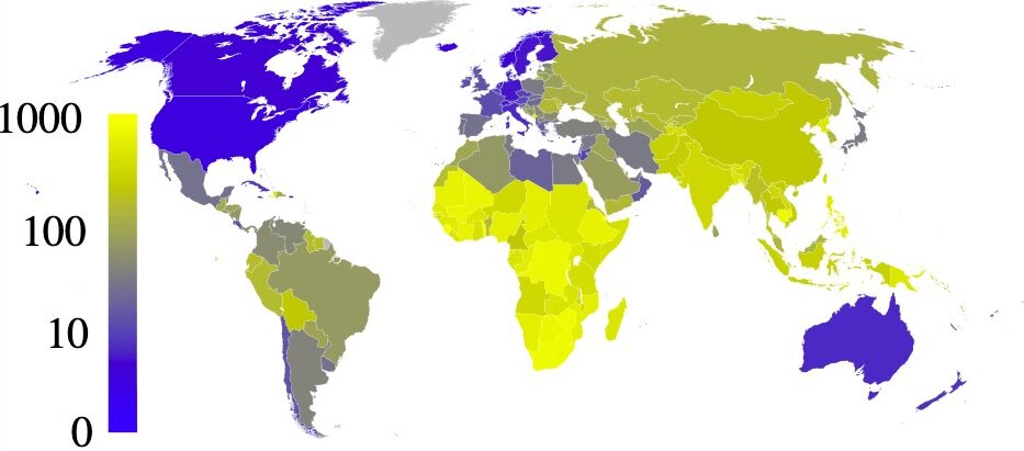
Estimated prevalence of tuberculosis per 100,000 people in 2007, per country
Image: “Estimated prevalence of tuberculosis” by Eubulides. License: Public DomainPathophysiology
Etiologic agent
The M. tuberculosis complex is a group of species that can cause TB in humans or other animals.
Key species:
- M. tuberculosis
- M. bovis
- M. africanum
- M. microti
- M. canetti
Characteristics:
- Acid fast:
- Property conferred by mycolic acid
- Do not destain by acid alcohol after being stained with aniline dyes
- Gram stain:
- Usually cannot penetrate MTBC waxy cell wall
- Most commonly produce no stain or variable results
- Slow growing
- Obligate aerobes
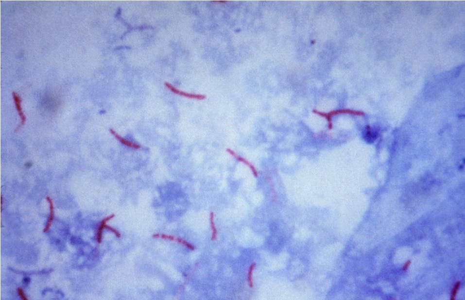
Acid-fast stain of M. tuberculosis
Image: “Mycobacterium tuberculosis bacteria” by CDC/Dr. George P. Kubica. License: Public DomainVirulence factors:
- Cell envelope:
- Major constituent: mycolic acid
- Mycolic acid is attached to glycolipids.
- Glycolipids are responsible for “cord formation” on microscopy (grossly corresponds to granuloma formation).
- Catalase-peroxidase: resists host cell oxidative response
- Sulfatides and trehalose dimycolate: triggers toxicity
- Lipoarabinomannan (LAM): induces cytokines
Transmission:
- Exclusively airborne
- From patients with active TB
Pathogenesis
- 1st step is inhalation of aerosol droplets.
- Droplets are deposited in the lungs.
- 3 possible outcomes:
- Clearance of bacteria
- Primary active disease
- Latent infection (clinical disease may occur many years later)
Primary active disease:
- Proliferation of bacteria within alveolar macrophages
- Cytokines produced by macrophages attract other phagocytic cells.
- A tubercle (granulomatous structure) forms.
- Tubercle expands into lung parenchyma → Ghon’s complex
- Bacteria then can spread to draining lymph nodes → lymphadenopathy
- Ghon’s complex + lymphadenopathy/calcification → Ranke complex
- If spread is not controlled by the immune cells, bacteremia with seeding of other organs may occur → miliary TB
- When bacteria erode into airways (caseating granulomas), the patient becomes contagious.
- Infection may progress to a chronic stage with episodes of healing and subsequent scarring of the lesions.
- Spontaneous eradication is rare.
Latent infection:
- Lifetime risk of reactivation is 5%–10%.
- Immunosuppression is a definite factor in reactivation.
- Risk factors:
- HIV
- Kidney disease
- Diabetes
- Steroids
- Lymphoma
- Advanced age
- Smoking
Clinical Presentation
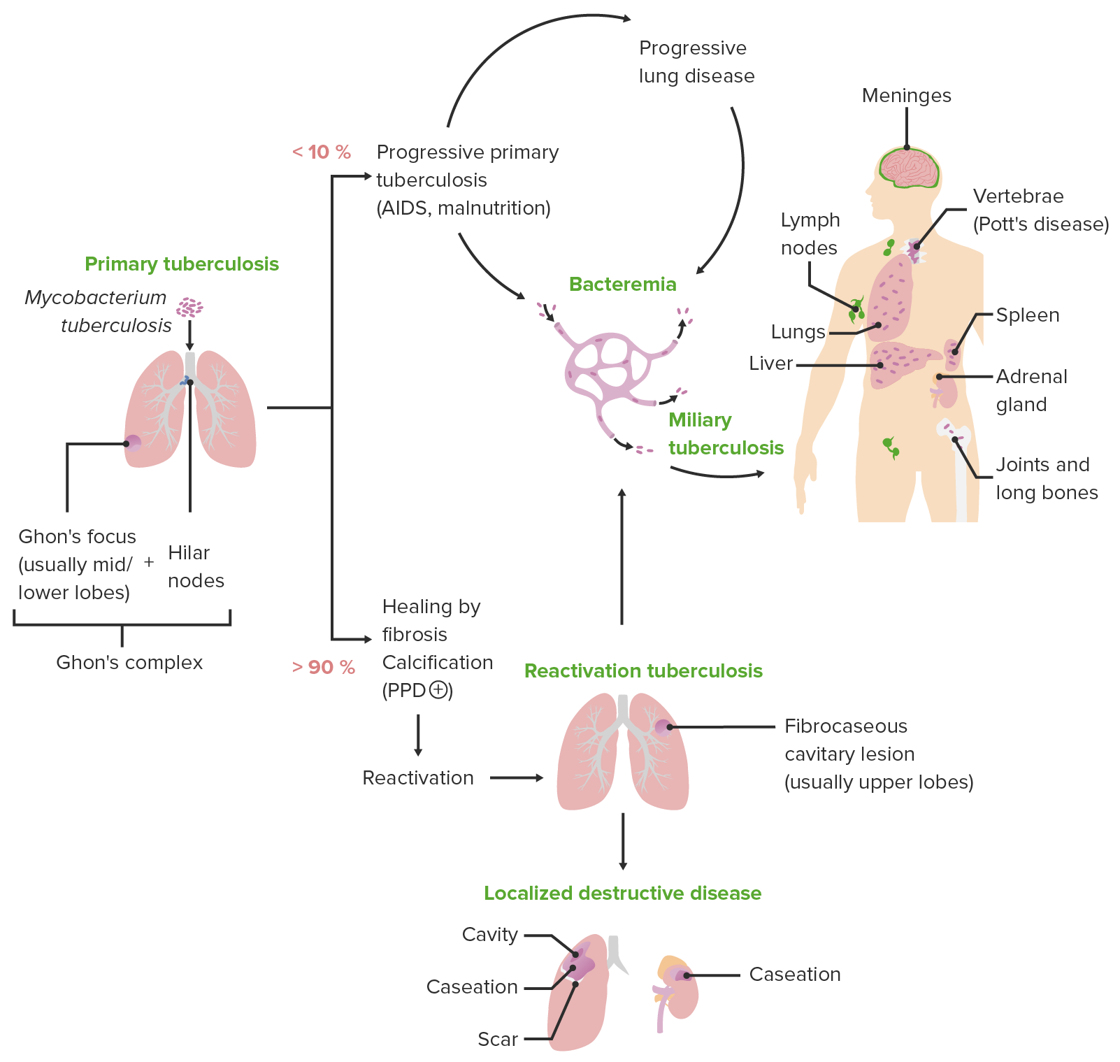
Schematic diagram depicting the various clinical presentations of tuberculosis along with the characteristic pathologic mechanisms of each presentation
Image by Lecturio.Primary TB
- Symptomatic primary disease develops in only about 10% of infected people.
- Fever:
- Most common symptom
- Mostly low grade, but may be up to 39°C (102.2°F)
- Lasts up to 10 weeks, but on average 14–21 days
- Pleuritic chest pain (may or may not be associated with effusion)
- Retrosternal/interscapular pain (due to bronchial lymphadenopathy)
- Cough
- Fatigue
- Arthralgia
- Pharyngitis
Reactivation TB
- Apical segments of upper lobes and superior segments of lower lobes are most commonly involved, likely because of:
- Increased oxygen tension
- Poor lymphatic drainage
- Onset of symptoms is gradual; may go undiagnosed for 2–3 years
- Fever:
- Low grade first, worsening with more advanced disease
- Classically diurnal: peaks in the afternoon, afebrile at night and in the early morning
- Night sweats
- Cough:
- Mild first, gradually worsening
- Initially only in the mornings
- Becomes more productive (greenish-yellow sputum) as disease progresses
- Nocturnal cough and hemoptysis: advanced disease
- Pneumothorax or effusions may present with dyspnea (rare).
- Anorexia, wasting, malaise
- Ulcers of mouth, tongue, larynx, and esophagus: due to infected expectorated secretions
- Fever:
Extrapulmonary and miliary TB
- Can develop in the course of primary or reactivation disease
- In up to 15%–20% of active cases
- Most common in children and immunocompromised individuals
- Extrapulmonary manifestations (can affect any organ system):
- Tuberculous pleurisy
- Adrenal gland insufficiency
- Meningitis
- Spondylitis tuberculosa (Pott’s disease, TB infection of > 1 vertebra)
- Constrictive pericarditis
- Lupus vulgaris (reddish-brown nodules that usually appear on the face around the nose, eyelids, lips, cheeks, ears, and neck)
- Scrofula (cervical lymphadenitis)
- Sterile pyuria
- Miliary TB:
- Massive spread of infection through blood and lymphatics
- Small granulomatous lesions through multiple organs
Related videos
Diagnosis
History
- Travel to endemic areas
- Exposure to individuals with known or suspected active infection
- HIV or other immune deficiencies
- Working in healthcare
- Living in a homeless shelter or correctional institution
Physical exam
- Findings are often non-specific.
- Pulmonary:
- Dullness to percussion (effusions)
- Crackles on auscultation
- Distant hollow breath sounds
- Extrapulmonary (depends on organ involvement):
- Cervical lymphadenopathy
- Hepatomegaly/splenomegaly
- Ascites, jaundice
- Meningismus, altered mental status
- Skin changes (lupus vulgaris)
Imaging
- Chest X-ray:
- Can be normal in primary TB
- Hilar lymphadenopathy
- Ghon’s complex: enlarged hilar lymph nodes + local shadowing
- Assmann’s focus: infraclavicular infiltration
- Pleural effusions
- Reactivation TB:
- Simon foci (calcified small scar from primary infection)
- Infiltrates in apical segments and upper segments of lower lobes
- Cavities with air-fluid levels
- Computed tomography (CT) scan:
- More sensitive than plain X-ray
- Used if chest X-ray is non-specific or alternative diagnosis is considered
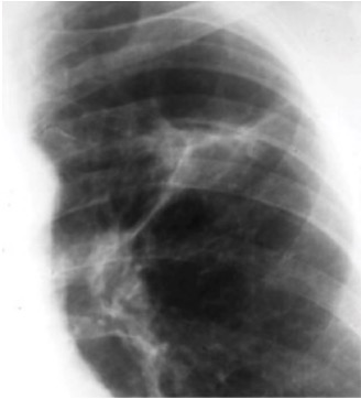
Chest X-ray of Ghon’s complex of active TB (left lung)
Image: “Ghon’s complex” by Basem Abbas Al Ubaidi. License: CC BY 4.0
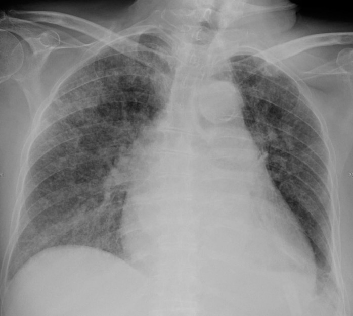
Chest X-ray of diffuse miliary infiltrates, characteristic of miliary TB
Image: “Chest radiograph of miliary tuberculosis” by Benjamín Herreros et al. License: CC BY 4.0
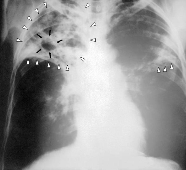
Chest X-ray of a patient with tuberculosis: bilateral reticular infiltrates (white triangles) and cavitary lesion (black arrows) in the right upper lobe
Image: “An anteroposterior X-ray” by Centers for Disease Control and Prevention. License: Public Domain
Laboratory identification
- Sputum:
- 3 specimens, at least 1 in the early morning
- Acid-fast bacillus (AFB) smear
- Mycobacterial culture
- Nuclear acid amplification (NAA) test
- Blood or urine mycobacterial culture: in HIV or immunocompromised patients
- Tuberculin skin test (TST; purified protein derivative (PPD) or Mantoux test):
- Intradermal injection of tuberculin antigen
- Can detect active or latent infection
- Measure induration area in 48–72 hours; positive if:
- > 5 mm in patients with HIV, immunosuppression, or recent contact with TB
- > 10 mm in patients from high-risk countries, IV drug users, medical and lab workers
- > 15 mm in patients with no known risk factors for TB
- lnterferon-y release assay (IGRA): no distinction between active and inactive TB
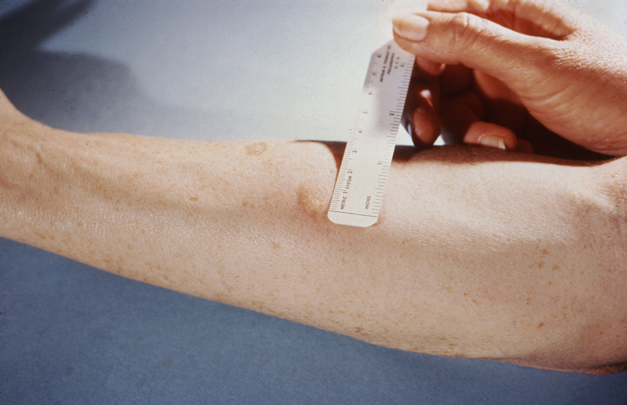
Measuring the reaction to a tuberculin skin test
Image: “Mendel-Mantoux-Test” by Public Health Image Library. License: Public Domain
Management
Pharmacologic management
- The mainstay of treatment is anti-mycobacterial drugs.
- Usually 2 treatment phases: intensive and stabilization
- Directly observed therapy (DOT) is preferred:
- Medications are administered to patients.
- Administration by healthcare providers ensures compliance and correct administration.
Summary of anti-tuberculosis regimens
Initial phase | Stabilization phase | |
---|---|---|
Active TB |
|
|
Latent TB | Treatment options:
|
Prevention
- TST screening for individuals at high risk
- Isolation of individuals with active pulmonary infection
- BCG (bacille Calmette-Guérin) vaccine:
- A nonvirulent form of M. bovis used as a live vaccine to provide active immunity against severe forms of TB
- Not recommended as a universal vaccine in countries with low TB burden
- May be considered for some individuals at high risk of exposure: infants and adolescents < 16 years of age in a high-incidence country
- 70%–80% effective against most severe forms of TB (miliary TB, tuberculous meningitis)
- Reduced effect against respiratory TB
- No proof of effectiveness in adults > 35
- Contraindicated in positive tuberculin reactions, AIDS, or immunosuppression
- Vaccine administration will result in a positive TST.
Prognosis
- Treatment with anti-mycobacterial drugs is 85% successful worldwide.
- 15% mortality rate worldwide
- 2.5%–5% rate of treatment failure or relapse in the United States
Differential Diagnosis
- Chronic bronchitis: a type of chronic obstructive pulmonary disease (COPD) involving inflammation and swelling of the airways, leading to productive cough for > 3 months in a year for > 2 consecutive years. Diagnosis is established by history, physical exam, and pulmonary function tests. Treatment involves bronchodilators and steroids.
- Atypical pneumonia: a form of pulmonary infection that typically has a slow onset and progression and presents with a non-productive, dry cough and extrapulmonary symptoms such as fatigue, malaise, and headaches. Diagnosis is made from history, physical exam, and chest imaging. Atypical pneumonia is usually treated with antibiotics.
- M. avium complex (MAC) infection: an AIDS-defining condition. Mycobacterium avium complex is an opportunistic infection caused by nontuberculous mycobacteria species that typically affects immunocompromised patients. Manifests with fever, night sweats, weight loss, abdominal pain, and diarrhea. Treatment includes antibiotics (macrolides and ethambutol).
- Lung mycoses: fungal diseases usually due to opportunistic pathogens that infect patients with immune deficiencies; includes candidiasis, cryptococcosis, and aspergillosis. Symptoms may include cough, hemoptysis, dyspnea, fevers, weight loss, and fatigue. Diagnosis is made by imaging and histologic examination. Treatment involves systemic antifungal medications.
- Bronchial carcinoma: a type of lung cancer that arises within the main airways. Frequently presents with cough, hemoptysis, and constitutional symptoms. Diagnosis is established by imaging and biopsy. Treatment depends on the stage and includes surgical resection, adjuvant radiation, and chemotherapy.
- Granulomatous diseases:
- Sarcoidosis: a chronic inflammatory disease characterized by the formation of noncaseating granulomas, typically in the lungs and, less commonly, in the liver, eyes, and skin. Pulmonary sarcoidosis presents with cough, dyspnea, chest pain, and constitutional symptoms. Definitive diagnosis is established by histology. Some cases are self-limited, while others are treated with steroids and immunosuppressive drugs.
- Pneumoconiosis: an occupational disease that consists of a group of restrictive interstitial lung diseases caused by inhalation of toxic dust. Presents with cough and progressive dyspnea. Diagnosed by imaging and histology. Management is largely supportive.
- Histoplasmosis: an infection caused by the fungus Histoplasma capsulatum. Presents with symptoms of pneumonia. Can also cause diffuse systemic infection in immunocompromised individuals. Diagnosis is established histologically. Treatment involves antifungal medications.
References
- Fordham von Reyn C. (2020). Vaccines for prevention of tuberculosis. Retrieved January 14, 2021, from https://www.uptodate.com/contents/vaccines-for-prevention-of-tuberculosis
- Pozniak A. (2019). Clinical manifestations and complications of pulmonary tuberculosis. Retrieved January 13, 2021, from https://www.uptodate.com/contents/clinical-manifestations-and-complications-of-pulmonary-tuberculosis
- Riley L.W. (2019). Tuberculosis: Natural history, microbiology, and pathogenesis. Retrieved January 13, 2021, from https://www.uptodate.com/contents/tuberculosis-natural-history-microbiology-and-pathogenesis
- Sterling, T. (2020). Treatment of drug-susceptible pulmonary tuberculosis in HIV-uninfected adults. Retrieved January 14, 2021, from https://www.uptodate.com/contents/treatment-of-drug-susceptible-pulmonary-tuberculosis-in-hiv-uninfected-adults
- Tuberculosis (TB). Centers for Disease Control and Prevention. Retrieved January 13, 2021, from https://www.cdc.gov/tb/default.htm