Complex regional pain syndrome (CRPS) is a chronic regional neuropathic pain condition characterized by excruciating pain (out of proportion to apparent tissue damage or inciting trauma), paresthesia, allodynia, temperature abnormalities, skin discoloration, edema, reduced range of motion, and bone demineralization. This syndrome is most often associated with an inciting traumatic event (e.g., fracture, surgery, burn) and predominantly affects the limb(s). Diagnosis is clinical, but it is supported by imaging and electrodiagnostic testing. Treatment centers around multidisciplinary pain management and maintenance of function.
EdemaEdemaEdema is a condition in which excess serous fluid accumulates in the body cavity or interstitial space of connective tissues. Edema is a symptom observed in several medical conditions. It can be categorized into 2 types, namely, peripheral (in the extremities) and internal (in an organ or body cavity). Edema
SkinSkinThe skin, also referred to as the integumentary system, is the largest organ of the body. The skin is primarily composed of the epidermis (outer layer) and dermis (deep layer). The epidermis is primarily composed of keratinocytes that undergo rapid turnover, while the dermis contains dense layers of connective tissue.Skin: Structure and Functions discoloration
Loss of range of motionRange of motionThe distance and direction to which a bone joint can be extended. Range of motion is a function of the condition of the joints, muscles, and connective tissues involved. Joint flexibility can be improved through appropriate muscle strength exercises.Examination of the Upper Limbs of local joint
PainPainAn unpleasant sensation induced by noxious stimuli which are detected by nerve endings of nociceptive neurons.Pain: Types and Pathways and disabilityDisabilityDetermination of the degree of a physical, mental, or emotional handicap. The diagnosis is applied to legal qualification for benefits and income under disability insurance and to eligibility for social security and workman’s compensation benefits.ABCDE Assessment greater than that expected from the original injury or apparent tissue damage
Epidemiology[1,3,6,8]
IncidenceIncidenceThe number of new cases of a given disease during a given period in a specified population. It also is used for the rate at which new events occur in a defined population. It is differentiated from prevalence, which refers to all cases in the population at a given time.Measures of Disease Frequency: 5–25 per 100,000 per year
Female-to-male ratio: 2–4 to 1
Highest in postmenopausal women
Most common in ages 50–70 years
Classification[1,8]
CRPS-1:
Previously known as reflex sympathetic dystrophyReflex sympathetic dystrophyA syndrome characterized by severe burning pain in an extremity accompanied by sudomotor, vasomotor, and trophic changes in bone without an associated specific nerve injury. This condition is most often precipitated by trauma to soft tissue or nerve complexes. The skin over the affected region is usually erythematous and demonstrates hypersensitivity to tactile stimuli and erythema.Complex Regional Pain Syndrome (CRPS) (RDSRDSRespiratory distress syndrome (RDS), also known as hyaline membrane disease, is caused by the lack of adequate pulmonary surfactant production in an immature lung. The syndrome is most commonly seen in preterm infants.Neonatal Respiratory Distress Syndrome)
No definitive nerve involvement
CRPS-2:
Previously known as causalgiaCausalgiaA complex regional pain syndrome characterized by burning pain and marked sensitivity to touch (hyperesthesia) in the distribution of an injured peripheral nerve. Autonomic dysfunction in the form of sudomotor (i.e., sympathetic innervation to sweat glands), vasomotor, and trophic skin changes may also occur.Complex Regional Pain Syndrome (CRPS)
FractureFractureA fracture is a disruption of the cortex of any bone and periosteum and is commonly due to mechanical stress after an injury or accident. Open fractures due to trauma can be a medical emergency. Fractures are frequently associated with automobile accidents, workplace injuries, and trauma.Overview of Bone Fractures (20%–40%)
Strain or sprain (10%–20%)
Postoperative (10%–20%)
Contusion or crush injuryCrush injuryExcessive compression of parts of the body that causes muscle swelling, fracture, and/or neurological disturbances in the affected areas. Crush injury with systemic manifestations is referred to as crush syndrome.Crush Syndrome (10%–20%)
The pathophysiology of CRPSCRPSComplex regional pain syndrome (CRPS) is a chronic regional neuropathic pain condition characterized by excruciating pain (out of proportion to apparent tissue damage or inciting trauma), paresthesia, allodynia, temperature abnormalities, skin discoloration, edema, reduced range of motion, and bone demineralization. Complex Regional Pain Syndrome (CRPS) is not completely understood, but there are multiple likely mechanisms.
InflammationInflammationInflammation is a complex set of responses to infection and injury involving leukocytes as the principal cellular mediators in the body’s defense against pathogenic organisms. Inflammation is also seen as a response to tissue injury in the process of wound healing. The 5 cardinal signs of inflammation are pain, heat, redness, swelling, and loss of function. Inflammation (cytokine-mediated):[1,3,6]
Abnormal local up-regulationUp-RegulationA positive regulatory effect on physiological processes at the molecular, cellular, or systemic level. At the molecular level, the major regulatory sites include membrane receptors, genes (gene expression regulation), mRNAs, and proteins.Pharmacokinetics and Pharmacodynamics of the immune systemImmune systemThe body’s defense mechanism against foreign organisms or substances and deviant native cells. It includes the humoral immune response and the cell-mediated response and consists of a complex of interrelated cellular, molecular, and genetic components.Primary Lymphatic Organs
Neurogenic inflammationInflammationInflammation is a complex set of responses to infection and injury involving leukocytes as the principal cellular mediators in the body’s defense against pathogenic organisms. Inflammation is also seen as a response to tissue injury in the process of wound healing. The 5 cardinal signs of inflammation are pain, heat, redness, swelling, and loss of function. Inflammation (neuropeptide-mediated):[1,3,6]
Local release of painPainAn unpleasant sensation induced by noxious stimuli which are detected by nerve endings of nociceptive neurons.Pain: Types and Pathways producing peptides by peripheral nervesPeripheral NervesThe nerves outside of the brain and spinal cord, including the autonomic, cranial, and spinal nerves. Peripheral nerves contain non-neuronal cells and connective tissue as well as axons. The connective tissue layers include, from the outside to the inside, the epineurium, the perineurium, and the endoneurium.Nervous System: Histology:
Calcitonin-gene–related peptide (CGRPCGRPA 37-amino acid peptide derived from the calcitonin gene. It occurs as a result of alternative processing of mRNA from the calcitonin gene. The neuropeptide is widely distributed in the brain, gut, perivascular nerves, and other tissue. The peptide produces multiple biological effects and has both circulatory and neurotransmitter modes of action. In particular, it is a potent endogenous vasodilator.Gastrointestinal Neural and Hormonal Signaling)
Abnormal signaling between afferentAfferentNeurons which conduct nerve impulses to the central nervous system.Nervous System: Histology and efferentEfferentNeurons which send impulses peripherally to activate muscles or secretory cells.Nervous System: Histology nerves
Central sensitizationCentral sensitizationIncreased responsiveness of nociceptive neurons in the central nervous system to normal or subthreshold input.Pain: Types and Pathways:[1,3,6,11]
Occurs in the setting of persistent nociceptive input to the dorsal hornDorsal hornOne of three central columns of the spinal cord. It is composed of gray matter spinal laminae i-vi.Brown-Séquard Syndrome of a given spinal segment or segments
Results in pathologic rearrangement of sensorySensoryNeurons which conduct nerve impulses to the central nervous system.Nervous System: Histology input territories in the dorsal hornDorsal hornOne of three central columns of the spinal cord. It is composed of gray matter spinal laminae i-vi.Brown-Séquard Syndrome of the spinal cordSpinal cordThe spinal cord is the major conduction pathway connecting the brain to the body; it is part of the CNS. In cross section, the spinal cord is divided into an H-shaped area of gray matter (consisting of synapsing neuronal cell bodies) and a surrounding area of white matter (consisting of ascending and descending tracts of myelinated axons). Spinal Cord: Anatomy (also known as Rexed laminaRexed LaminaComplex Regional Pain Syndrome (CRPS))
Results in abnormal cross talk at the level of the spinal interneurons
Sympathetic dysregulation:[1,3,6,11]
Local hyperactivityHyperactivityAttention Deficit Hyperactivity Disorder of the sympathetic nervous systemNervous systemThe nervous system is a small and complex system that consists of an intricate network of neural cells (or neurons) and even more glial cells (for support and insulation). It is divided according to its anatomical components as well as its functional characteristics. The brain and spinal cord are referred to as the central nervous system, and the branches of nerves from these structures are referred to as the peripheral nervous system.Nervous System: Anatomy, Structure, and Classification and/or abnormal local sensitivity to catecholaminesCatecholaminesA general class of ortho-dihydroxyphenylalkylamines derived from tyrosine.Adrenal Hormones
AfferentAfferentNeurons which conduct nerve impulses to the central nervous system.Nervous System: HistologyneuronsNeuronsThe basic cellular units of nervous tissue. Each neuron consists of a body, an axon, and dendrites. Their purpose is to receive, conduct, and transmit impulses in the nervous system.Nervous System: Histology carry nociceptive inputs to the CNS, triggering an abnormal reflex of vasoconstrictionVasoconstrictionThe physiological narrowing of blood vessels by contraction of the vascular smooth muscle.Vascular Resistance, Flow, and Mean Arterial Pressure in the corresponding area(s).
Up-regulationUp-RegulationA positive regulatory effect on physiological processes at the molecular, cellular, or systemic level. At the molecular level, the major regulatory sites include membrane receptors, genes (gene expression regulation), mRNAs, and proteins.Pharmacokinetics and Pharmacodynamics of adrenergic receptorsReceptorsReceptors are proteins located either on the surface of or within a cell that can bind to signaling molecules known as ligands (e.g., hormones) and cause some type of response within the cell.Receptors
Occurs in the setting of persistent nociceptive input to the cerebral cortexCerebral cortexThe cerebral cortex is the largest and most developed part of the human brain and CNS. Occupying the upper part of the cranial cavity, the cerebral cortex has 4 lobes and is divided into 2 hemispheres that are joined centrally by the corpus callosum. Cerebral Cortex: Anatomy of a given spinal segment or segments
Results in pathologic rearrangement of sensorySensoryNeurons which conduct nerve impulses to the central nervous system.Nervous System: Histology inputs in the cerebral cortexCerebral cortexThe cerebral cortex is the largest and most developed part of the human brain and CNS. Occupying the upper part of the cranial cavity, the cerebral cortex has 4 lobes and is divided into 2 hemispheres that are joined centrally by the corpus callosum. Cerebral Cortex: Anatomy areas corresponding to the affected area(s)
Genetic predisposition:[6]
CRPS-1 has been observed at a greater frequency in individuals with HLA-DQ1.
CRPSCRPSComplex regional pain syndrome (CRPS) is a chronic regional neuropathic pain condition characterized by excruciating pain (out of proportion to apparent tissue damage or inciting trauma), paresthesia, allodynia, temperature abnormalities, skin discoloration, edema, reduced range of motion, and bone demineralization. Complex Regional Pain Syndrome (CRPS) is more likely to become multifocalMultifocalRetinoblastoma in individuals with HLA-DR3.
History of trauma or other noxious event from which the individual has recovered:
An adequate amount of time for expected healing for a given injury(ies) has passed.
No residual wound or tissue damage that might otherwise produce painPainAn unpleasant sensation induced by noxious stimuli which are detected by nerve endings of nociceptive neurons.Pain: Types and Pathways is readily identifiable.
The painPainAn unpleasant sensation induced by noxious stimuli which are detected by nerve endings of nociceptive neurons.Pain: Types and Pathways is neuropathic in nature. Affected individuals describe the painPainAn unpleasant sensation induced by noxious stimuli which are detected by nerve endings of nociceptive neurons.Pain: Types and Pathways as:
Burning
Aching
Pricking
Shooting
Affected individuals may report painPainAn unpleasant sensation induced by noxious stimuli which are detected by nerve endings of nociceptive neurons.Pain: Types and Pathways with innocent activities:
Wearing socks or shoes (if footFootThe foot is the terminal portion of the lower limb, whose primary function is to bear weight and facilitate locomotion. The foot comprises 26 bones, including the tarsal bones, metatarsal bones, and phalanges. The bones of the foot form longitudinal and transverse arches and are supported by various muscles, ligaments, and tendons.Foot: Anatomy/feet affected)
Brushing hair (if scalp affected)
Individuals may report motorMotorNeurons which send impulses peripherally to activate muscles or secretory cells.Nervous System: Histology symptoms in the affected area(s):
Restless legsRestless legsA disorder characterized by aching or burning sensations in the lower and rarely the upper extremities that occur prior to sleep or may awaken the patient from sleep.Polyneuropathy
Individuals may report concomitant psychological symptoms:
Depression
AnxietyAnxietyFeelings or emotions of dread, apprehension, and impending disaster but not disabling as with anxiety disorders.Generalized Anxiety Disorder
Excruciating painPainAn unpleasant sensation induced by noxious stimuli which are detected by nerve endings of nociceptive neurons.Pain: Types and Pathways is the cardinal feature.
HyperalgesiaHyperalgesiaAn increased sensation of pain or discomfort produced by minimally noxious stimuli due to damage to soft tissue containing nociceptors or injury to a peripheral nerve.Pain: Types and Pathways:
Level of painPainAn unpleasant sensation induced by noxious stimuli which are detected by nerve endings of nociceptive neurons.Pain: Types and Pathways is disproportionate to the initial injury or apparent tissue damage.
PainPainAn unpleasant sensation induced by noxious stimuli which are detected by nerve endings of nociceptive neurons.Pain: Types and Pathways may persist longer than expected after noxious or nonnoxious stimulus is removed.
Pinprick test evokes excruciating painPainAn unpleasant sensation induced by noxious stimuli which are detected by nerve endings of nociceptive neurons.Pain: Types and Pathways.
AllodyniaAllodyniaPain due to a stimulus that does not typically provoke pain.Pain Management: Application of a normally nonnoxious mechanical stimulus is perceived as painful.
Light touch → mechanical allodyniaAllodyniaPain due to a stimulus that does not typically provoke pain.Pain Management
Cooling stimulus (e.g., drop of alcohol or water) → thermal allodyniaAllodyniaPain due to a stimulus that does not typically provoke pain.Pain Management
Greater receptive field:
HyperalgesiaHyperalgesiaAn increased sensation of pain or discomfort produced by minimally noxious stimuli due to damage to soft tissue containing nociceptors or injury to a peripheral nerve.Pain: Types and Pathways and allodyniaAllodyniaPain due to a stimulus that does not typically provoke pain.Pain Management beyond the area of initial injury
EdemaEdemaEdema is a condition in which excess serous fluid accumulates in the body cavity or interstitial space of connective tissues. Edema is a symptom observed in several medical conditions. It can be categorized into 2 types, namely, peripheral (in the extremities) and internal (in an organ or body cavity). Edema:
Mild to severe subcutaneous edemaEdemaEdema is a condition in which excess serous fluid accumulates in the body cavity or interstitial space of connective tissues. Edema is a symptom observed in several medical conditions. It can be categorized into 2 types, namely, peripheral (in the extremities) and internal (in an organ or body cavity). Edema is common in affected area(s).
Can mimic mild cellulitisCellulitisCellulitis is a common infection caused by bacteria that affects the dermis and subcutaneous tissue of the skin. It is frequently caused by Staphylococcus aureus and Streptococcus pyogenes. The skin infection presents as an erythematous and edematous area with warmth and tenderness. Cellulitis owing to accompanying erythemaErythemaRedness of the skin produced by congestion of the capillaries. This condition may result from a variety of disease processes.Chalazion and trophic changes of the skinSkinThe skin, also referred to as the integumentary system, is the largest organ of the body. The skin is primarily composed of the epidermis (outer layer) and dermis (deep layer). The epidermis is primarily composed of keratinocytes that undergo rapid turnover, while the dermis contains dense layers of connective tissue.Skin: Structure and Functions
ErythemaErythemaRedness of the skin produced by congestion of the capillaries. This condition may result from a variety of disease processes.Chalazion, pallor, and/or cyanosisCyanosisA bluish or purplish discoloration of the skin and mucous membranes due to an increase in the amount of deoxygenated hemoglobin in the blood or a structural defect in the hemoglobin molecule.Pulmonary Examination may manifest at any time in the same body area(s).
SkinSkinThe skin, also referred to as the integumentary system, is the largest organ of the body. The skin is primarily composed of the epidermis (outer layer) and dermis (deep layer). The epidermis is primarily composed of keratinocytes that undergo rapid turnover, while the dermis contains dense layers of connective tissue.Skin: Structure and Functions color can fluctuate in a matter of minutes to hours (e.g., reddish to pale or blue).
Livedo reticularisLivedo reticularisA condition characterized by a reticular or fishnet pattern on the skin of lower extremities and other parts of the body. This red and blue pattern is due to deoxygenated blood in unstable dermal blood vessels. The condition is intensified by cold exposure and relieved by rewarming.Chronic Kidney Disease is common.
Dystrophic manifestations:
Increased/decreased nail and hair growth in affected area(s)
HyperkeratosisHyperkeratosisIchthyosis Vulgaris/thin, glossy skinSkinThe skin, also referred to as the integumentary system, is the largest organ of the body. The skin is primarily composed of the epidermis (outer layer) and dermis (deep layer). The epidermis is primarily composed of keratinocytes that undergo rapid turnover, while the dermis contains dense layers of connective tissue.Skin: Structure and Functions
OsteoporosisOsteoporosisOsteoporosis refers to a decrease in bone mass and density leading to an increased number of fractures. There are 2 forms of osteoporosis: primary, which is commonly postmenopausal or senile; and secondary, which is a manifestation of immobilization, underlying medical disorders, or long-term use of certain medications. Osteoporosis of underlying bones
MotorMotorNeurons which send impulses peripherally to activate muscles or secretory cells.Nervous System: Histology dysfunction:
Mild weakness
Decreased range of motionRange of motionThe distance and direction to which a bone joint can be extended. Range of motion is a function of the condition of the joints, muscles, and connective tissues involved. Joint flexibility can be improved through appropriate muscle strength exercises.Examination of the Upper Limbs
DystoniaDystoniaDystonia is a hyperkinetic movement disorder characterized by the involuntary contraction of muscles, resulting in abnormal postures or twisting and repetitive movements. Dystonia can present in various ways as may affect many different skeletal muscle groups. Dystonia
TremorTremorCyclical movement of a body part that can represent either a physiologic process or a manifestation of disease. Intention or action tremor, a common manifestation of cerebellar diseases, is aggravated by movement. In contrast, resting tremor is maximal when there is no attempt at voluntary movement, and occurs as a relatively frequent manifestation of parkinson disease.Myotonic Dystrophies/myoclonusMyoclonusInvoluntary shock-like contractions, irregular in rhythm and amplitude, followed by relaxation, of a muscle or a group of muscles. This condition may be a feature of some central nervous system diseases; (e.g., epilepsy-myoclonic). Nocturnal myoclonus is the principal feature of the nocturnal myoclonus syndrome.Neurological Examination
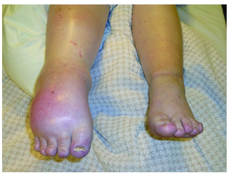
Complex regional pain syndrome type 1 (CRPS-1) The individual presented dystonic equinus of the right ankle and a swollen foot and calf with tightened, pale, gleaming, cold skin.
Image: “CRPS-1 (Complex regional pain syndrome)” by Voet C, le Polain de Waroux B, Forget P, Deumens R, Masquelier E. License: CC BY 4.0
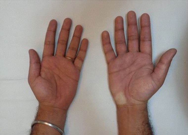
Individual with complex regional pain syndrome, presenting with a swollen right hand, hyperemia and a pale spot on the hypothenar region: The individual sustained an electrical shock injury to the right hand while repairing a domestic appliance.
Image: “Swollen right hand showing hyperemia” by Ahmad F, Kumar KH. License: CC BY 4.0
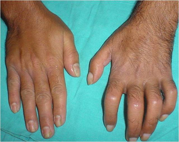
Typical features of complex regional pain syndrome: On the left hand, note the hypertrichosis, edema, joint stiffness, nail changes, and skin atrophy as compared with the opposite hand.
Image: “Typical features of CRPS” by Sunder RA, Toshniwal G, Dureja GP. License: CC BY 2.0
There were formerly thought to be 3 stages of CRPSCRPSComplex regional pain syndrome (CRPS) is a chronic regional neuropathic pain condition characterized by excruciating pain (out of proportion to apparent tissue damage or inciting trauma), paresthesia, allodynia, temperature abnormalities, skin discoloration, edema, reduced range of motion, and bone demineralization. Complex Regional Pain Syndrome (CRPS). However, these stages are no longer widely used owing to a lack of supporting evidence.
PainPainAn unpleasant sensation induced by noxious stimuli which are detected by nerve endings of nociceptive neurons.Pain: Types and Pathways/hypersensitivity/allodyniaAllodyniaPain due to a stimulus that does not typically provoke pain.Pain Management in primary affected area
Mild edemaEdemaEdema is a condition in which excess serous fluid accumulates in the body cavity or interstitial space of connective tissues. Edema is a symptom observed in several medical conditions. It can be categorized into 2 types, namely, peripheral (in the extremities) and internal (in an organ or body cavity). Edema
SkinSkinThe skin, also referred to as the integumentary system, is the largest organ of the body. The skin is primarily composed of the epidermis (outer layer) and dermis (deep layer). The epidermis is primarily composed of keratinocytes that undergo rapid turnover, while the dermis contains dense layers of connective tissue.Skin: Structure and Functions temperature increased to the touch
Minimal localized boneBoneBone is a compact type of hardened connective tissue composed of bone cells, membranes, an extracellular mineralized matrix, and central bone marrow. The 2 primary types of bone are compact and spongy. Bones: Structure and Types demineralization on x-rayX-rayPenetrating electromagnetic radiation emitted when the inner orbital electrons of an atom are excited and release radiant energy. X-ray wavelengths range from 1 pm to 10 nm. Hard x-rays are the higher energy, shorter wavelength x-rays. Soft x-rays or grenz rays are less energetic and longer in wavelength. The short wavelength end of the x-ray spectrum overlaps the gamma rays wavelength range. The distinction between gamma rays and x-rays is based on their radiation source.Pulmonary Function Tests
Stage 2:
Worsening of painPainAn unpleasant sensation induced by noxious stimuli which are detected by nerve endings of nociceptive neurons.Pain: Types and Pathways
Expansion of affected area(s)
Worsening soft tissueSoft TissueSoft Tissue AbscessedemaEdemaEdema is a condition in which excess serous fluid accumulates in the body cavity or interstitial space of connective tissues. Edema is a symptom observed in several medical conditions. It can be categorized into 2 types, namely, peripheral (in the extremities) and internal (in an organ or body cavity). Edema
Muscle atrophyAtrophyDecrease in the size of a cell, tissue, organ, or multiple organs, associated with a variety of pathological conditions such as abnormal cellular changes, ischemia, malnutrition, or hormonal changes.Cellular Adaptation
Onset of atrophic skinSkinThe skin, also referred to as the integumentary system, is the largest organ of the body. The skin is primarily composed of the epidermis (outer layer) and dermis (deep layer). The epidermis is primarily composed of keratinocytes that undergo rapid turnover, while the dermis contains dense layers of connective tissue.Skin: Structure and Functions and soft tissueSoft TissueSoft Tissue Abscess changes
Stage 3:
Severe painPainAn unpleasant sensation induced by noxious stimuli which are detected by nerve endings of nociceptive neurons.Pain: Types and Pathways
Spread of painPainAn unpleasant sensation induced by noxious stimuli which are detected by nerve endings of nociceptive neurons.Pain: Types and Pathways to contralateral (unaffected) limb, distant body region(s) or even entire body
Cold and cyanotic extremities
Significant atrophic skinSkinThe skin, also referred to as the integumentary system, is the largest organ of the body. The skin is primarily composed of the epidermis (outer layer) and dermis (deep layer). The epidermis is primarily composed of keratinocytes that undergo rapid turnover, while the dermis contains dense layers of connective tissue.Skin: Structure and Functions changes
Joint contracturesContracturesProlonged shortening of the muscle or other soft tissue around a joint, preventing movement of the joint.Wound Healing
Muscular and boneBoneBone is a compact type of hardened connective tissue composed of bone cells, membranes, an extracellular mineralized matrix, and central bone marrow. The 2 primary types of bone are compact and spongy. Bones: Structure and TypesatrophyAtrophyDecrease in the size of a cell, tissue, organ, or multiple organs, associated with a variety of pathological conditions such as abnormal cellular changes, ischemia, malnutrition, or hormonal changes.Cellular Adaptation
Suspect CRPSCRPSComplex regional pain syndrome (CRPS) is a chronic regional neuropathic pain condition characterized by excruciating pain (out of proportion to apparent tissue damage or inciting trauma), paresthesia, allodynia, temperature abnormalities, skin discoloration, edema, reduced range of motion, and bone demineralization. Complex Regional Pain Syndrome (CRPS) when the following historical features are present:
Inciting trauma
Persistent symptoms after 4–6 weeks or expected course of healing:
PainPainAn unpleasant sensation induced by noxious stimuli which are detected by nerve endings of nociceptive neurons.Pain: Types and Pathways/sensorySensoryNeurons which conduct nerve impulses to the central nervous system.Nervous System: Histology changes
The following Budapest criteriaBudapest CriteriaComplex Regional Pain Syndrome (CRPS) were adopted as a more sensitive and specific clinical tool for screeningScreeningPreoperative Care for CRPSCRPSComplex regional pain syndrome (CRPS) is a chronic regional neuropathic pain condition characterized by excruciating pain (out of proportion to apparent tissue damage or inciting trauma), paresthesia, allodynia, temperature abnormalities, skin discoloration, edema, reduced range of motion, and bone demineralization. Complex Regional Pain Syndrome (CRPS) than previous consensus criteria. Keep in mind that the presentation can vary among individuals and over time.[10]
To make a clinical diagnosis, all of the following must be metMETPreoperative Care:[2,3,8]
Continuing painPainAn unpleasant sensation induced by noxious stimuli which are detected by nerve endings of nociceptive neurons.Pain: Types and Pathways (disproportionate to any triggers or inciting event)
SkinSkinThe skin, also referred to as the integumentary system, is the largest organ of the body. The skin is primarily composed of the epidermis (outer layer) and dermis (deep layer). The epidermis is primarily composed of keratinocytes that undergo rapid turnover, while the dermis contains dense layers of connective tissue.Skin: Structure and Functions color changes
SkinSkinThe skin, also referred to as the integumentary system, is the largest organ of the body. The skin is primarily composed of the epidermis (outer layer) and dermis (deep layer). The epidermis is primarily composed of keratinocytes that undergo rapid turnover, while the dermis contains dense layers of connective tissue.Skin: Structure and Functions color asymmetryAsymmetryExamination of the Upper Limbs
Sudomotor/edemaEdemaEdema is a condition in which excess serous fluid accumulates in the body cavity or interstitial space of connective tissues. Edema is a symptom observed in several medical conditions. It can be categorized into 2 types, namely, peripheral (in the extremities) and internal (in an organ or body cavity). Edema:
Asymmetrical edemaEdemaEdema is a condition in which excess serous fluid accumulates in the body cavity or interstitial space of connective tissues. Edema is a symptom observed in several medical conditions. It can be categorized into 2 types, namely, peripheral (in the extremities) and internal (in an organ or body cavity). Edema
MotorMotorNeurons which send impulses peripherally to activate muscles or secretory cells.Nervous System: Histology/trophic:
↓ Range of motionRange of motionThe distance and direction to which a bone joint can be extended. Range of motion is a function of the condition of the joints, muscles, and connective tissues involved. Joint flexibility can be improved through appropriate muscle strength exercises.Examination of the Upper Limbs
MotorMotorNeurons which send impulses peripherally to activate muscles or secretory cells.Nervous System: Histology dysfunction (e.g., weakness, tremorTremorCyclical movement of a body part that can represent either a physiologic process or a manifestation of disease. Intention or action tremor, a common manifestation of cerebellar diseases, is aggravated by movement. In contrast, resting tremor is maximal when there is no attempt at voluntary movement, and occurs as a relatively frequent manifestation of parkinson disease.Myotonic Dystrophies, dystoniaDystoniaDystonia is a hyperkinetic movement disorder characterized by the involuntary contraction of muscles, resulting in abnormal postures or twisting and repetitive movements. Dystonia can present in various ways as may affect many different skeletal muscle groups. Dystonia)
Dystrophic changes in the hair, nails, and/or skinSkinThe skin, also referred to as the integumentary system, is the largest organ of the body. The skin is primarily composed of the epidermis (outer layer) and dermis (deep layer). The epidermis is primarily composed of keratinocytes that undergo rapid turnover, while the dermis contains dense layers of connective tissue.Skin: Structure and Functions
≥ 1 sign in ≥ 2 of the following categories at the time of evaluation:
HyperalgesiaHyperalgesiaAn increased sensation of pain or discomfort produced by minimally noxious stimuli due to damage to soft tissue containing nociceptors or injury to a peripheral nerve.Pain: Types and Pathways (to pinprick)
AllodyniaAllodyniaPain due to a stimulus that does not typically provoke pain.Pain Management (to light touch and/or temperature sensation and/or deep somatic pressure and/or joint movement)
SkinSkinThe skin, also referred to as the integumentary system, is the largest organ of the body. The skin is primarily composed of the epidermis (outer layer) and dermis (deep layer). The epidermis is primarily composed of keratinocytes that undergo rapid turnover, while the dermis contains dense layers of connective tissue.Skin: Structure and Functions color changes
SkinSkinThe skin, also referred to as the integumentary system, is the largest organ of the body. The skin is primarily composed of the epidermis (outer layer) and dermis (deep layer). The epidermis is primarily composed of keratinocytes that undergo rapid turnover, while the dermis contains dense layers of connective tissue.Skin: Structure and Functions color asymmetryAsymmetryExamination of the Upper Limbs
Sudomotor/edemaEdemaEdema is a condition in which excess serous fluid accumulates in the body cavity or interstitial space of connective tissues. Edema is a symptom observed in several medical conditions. It can be categorized into 2 types, namely, peripheral (in the extremities) and internal (in an organ or body cavity). Edema:
Asymmetrical edemaEdemaEdema is a condition in which excess serous fluid accumulates in the body cavity or interstitial space of connective tissues. Edema is a symptom observed in several medical conditions. It can be categorized into 2 types, namely, peripheral (in the extremities) and internal (in an organ or body cavity). Edema
MotorMotorNeurons which send impulses peripherally to activate muscles or secretory cells.Nervous System: Histology/trophic:
↓ Range of motionRange of motionThe distance and direction to which a bone joint can be extended. Range of motion is a function of the condition of the joints, muscles, and connective tissues involved. Joint flexibility can be improved through appropriate muscle strength exercises.Examination of the Upper Limbs
MotorMotorNeurons which send impulses peripherally to activate muscles or secretory cells.Nervous System: Histology dysfunction (e.g., weakness, tremorTremorCyclical movement of a body part that can represent either a physiologic process or a manifestation of disease. Intention or action tremor, a common manifestation of cerebellar diseases, is aggravated by movement. In contrast, resting tremor is maximal when there is no attempt at voluntary movement, and occurs as a relatively frequent manifestation of parkinson disease.Myotonic Dystrophies, dystoniaDystoniaDystonia is a hyperkinetic movement disorder characterized by the involuntary contraction of muscles, resulting in abnormal postures or twisting and repetitive movements. Dystonia can present in various ways as may affect many different skeletal muscle groups. Dystonia)
Dystrophic changes in the hair, nails, and/or skinSkinThe skin, also referred to as the integumentary system, is the largest organ of the body. The skin is primarily composed of the epidermis (outer layer) and dermis (deep layer). The epidermis is primarily composed of keratinocytes that undergo rapid turnover, while the dermis contains dense layers of connective tissue.Skin: Structure and Functions
No other diagnosis to better explain the signs and symptoms
Imaging[6,8]
CRPSCRPSComplex regional pain syndrome (CRPS) is a chronic regional neuropathic pain condition characterized by excruciating pain (out of proportion to apparent tissue damage or inciting trauma), paresthesia, allodynia, temperature abnormalities, skin discoloration, edema, reduced range of motion, and bone demineralization. Complex Regional Pain Syndrome (CRPS) cannot be diagnosed with imaging, but the following tests may be ordered to exclude other causes for the patient’s signs and symptoms.
Plain films (x-rayX-rayPenetrating electromagnetic radiation emitted when the inner orbital electrons of an atom are excited and release radiant energy. X-ray wavelengths range from 1 pm to 10 nm. Hard x-rays are the higher energy, shorter wavelength x-rays. Soft x-rays or grenz rays are less energetic and longer in wavelength. The short wavelength end of the x-ray spectrum overlaps the gamma rays wavelength range. The distinction between gamma rays and x-rays is based on their radiation source.Pulmonary Function Tests):
May show evidence of fractureFractureA fracture is a disruption of the cortex of any bone and periosteum and is commonly due to mechanical stress after an injury or accident. Open fractures due to trauma can be a medical emergency. Fractures are frequently associated with automobile accidents, workplace injuries, and trauma.Overview of Bone Fractures or bony abnormality related to inciting trauma
May show patchy osteoporosisOsteoporosisOsteoporosis refers to a decrease in bone mass and density leading to an increased number of fractures. There are 2 forms of osteoporosis: primary, which is commonly postmenopausal or senile; and secondary, which is a manifestation of immobilization, underlying medical disorders, or long-term use of certain medications. Osteoporosis as compared with the contralateral (unaffected) limb or affected area
MRI:
Not useful for the diagnosis of CRPSCRPSComplex regional pain syndrome (CRPS) is a chronic regional neuropathic pain condition characterized by excruciating pain (out of proportion to apparent tissue damage or inciting trauma), paresthesia, allodynia, temperature abnormalities, skin discoloration, edema, reduced range of motion, and bone demineralization. Complex Regional Pain Syndrome (CRPS), but may be useful in excluding other diagnoses
May show patchy areas of osteoporosisOsteoporosisOsteoporosis refers to a decrease in bone mass and density leading to an increased number of fractures. There are 2 forms of osteoporosis: primary, which is commonly postmenopausal or senile; and secondary, which is a manifestation of immobilization, underlying medical disorders, or long-term use of certain medications. Osteoporosis, but not recommended for screeningScreeningPreoperative Care of bony lesions
Bone scanBone ScanOsteosarcoma (also known as boneBoneBone is a compact type of hardened connective tissue composed of bone cells, membranes, an extracellular mineralized matrix, and central bone marrow. The 2 primary types of bone are compact and spongy. Bones: Structure and TypesscintigraphyScintigraphySjögren Syndrome):
Used to detect a large variety of boneBoneBone is a compact type of hardened connective tissue composed of bone cells, membranes, an extracellular mineralized matrix, and central bone marrow. The 2 primary types of bone are compact and spongy. Bones: Structure and Types and joint disorders:
FractureFractureA fracture is a disruption of the cortex of any bone and periosteum and is commonly due to mechanical stress after an injury or accident. Open fractures due to trauma can be a medical emergency. Fractures are frequently associated with automobile accidents, workplace injuries, and trauma.Overview of Bone Fractures
Metabolic boneBoneBone is a compact type of hardened connective tissue composed of bone cells, membranes, an extracellular mineralized matrix, and central bone marrow. The 2 primary types of bone are compact and spongy. Bones: Structure and Types disease
Useful for detecting and monitoring bony demineralization in CRPSCRPSComplex regional pain syndrome (CRPS) is a chronic regional neuropathic pain condition characterized by excruciating pain (out of proportion to apparent tissue damage or inciting trauma), paresthesia, allodynia, temperature abnormalities, skin discoloration, edema, reduced range of motion, and bone demineralization. Complex Regional Pain Syndrome (CRPS)
Presence of bony demineralization is not diagnostic of CRPSCRPSComplex regional pain syndrome (CRPS) is a chronic regional neuropathic pain condition characterized by excruciating pain (out of proportion to apparent tissue damage or inciting trauma), paresthesia, allodynia, temperature abnormalities, skin discoloration, edema, reduced range of motion, and bone demineralization. Complex Regional Pain Syndrome (CRPS), but it supports the diagnosis.
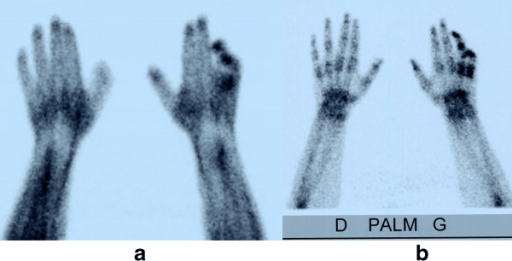
Three-phase bone scintigraphy:
Early and delayed hyperfixation on the 4th and 5th fingers suggest complex regional pain syndrome (CRPS).
a: early phase
b: delayed phase
Image: “F2: Three-phase bone scintigraphy: early phase (a) and delayed phase (b) Same patient as Figure 1. Staged early and delayed hyperfixation on 4th and 5th fingers suggesting CRPS stage 1.” by Michel Konzelmann, Olivier Deriaz, and François Luthi. License: CC BY 2.0
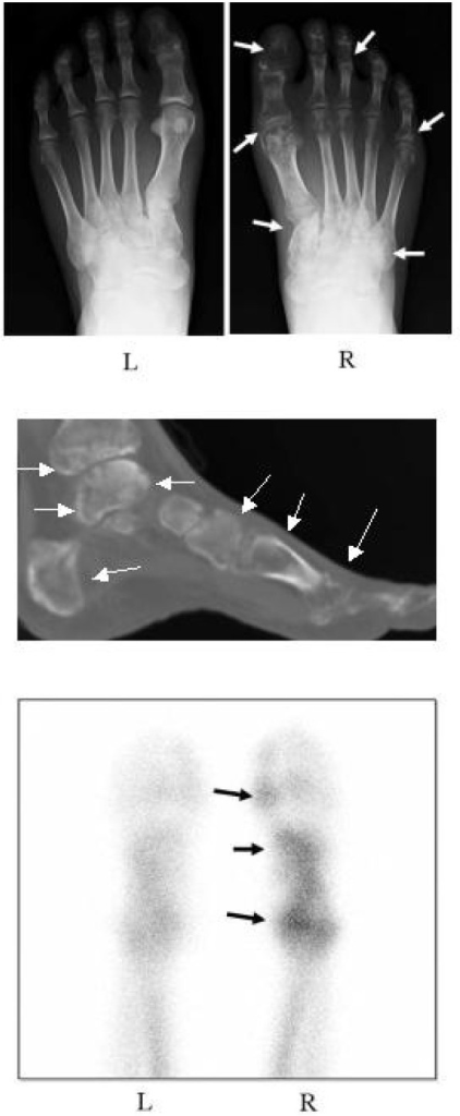
Images of an individual’s feet with complex regional pain syndrome (CRPS) before treatment with bisphosphonates:
Top picture: Radiograph shows regional osteoporotic changes in the phalanges, metatarsals, and tarsals of the right foot (white arrows). The left foot is normal.
Middle picture: A reconstitution CT shows extensive osteoporotic changes in the foot and ankle (white arrows).
Lower picture: A bone scintigraphy with 99mTc-methylene diphosphonate shows a marked increase in radioactivity in the right foot (black arrows).
Image: “F5: Radiograph (A), reconstitution computed tomography (CT) image (B), and scintigraph (C) of the both feet before treatment.” by Yasuhisa Abe, Kousuke Iba, Junichi Takada, Takuro Wada, Toshihiko Yamashita. License: CC BY 2.0
Autonomic testing[6,11]
The following are not required but have been used to evaluate cases of suspected CRPSCRPSComplex regional pain syndrome (CRPS) is a chronic regional neuropathic pain condition characterized by excruciating pain (out of proportion to apparent tissue damage or inciting trauma), paresthesia, allodynia, temperature abnormalities, skin discoloration, edema, reduced range of motion, and bone demineralization. Complex Regional Pain Syndrome (CRPS). Appropriate autonomic testing may require specialty assistance.
Resting sweat output (RSO)
Resting skinSkinThe skin, also referred to as the integumentary system, is the largest organ of the body. The skin is primarily composed of the epidermis (outer layer) and dermis (deep layer). The epidermis is primarily composed of keratinocytes that undergo rapid turnover, while the dermis contains dense layers of connective tissue.Skin: Structure and Functions temperature (RST)
Quantitative sudomotor axon reflex test (QSART):
Utilizes a cholinergic challenge
Looks at the difference in sweat production between unaffected and affected limbs
Diagnostic regional anesthesiaAnesthesiaA state characterized by loss of feeling or sensation. This depression of nerve function is usually the result of pharmacologic action and is induced to allow performance of surgery or other painful procedures.Anesthesiology: History and Basic Concepts procedures[10,11]
The following procedures were formerly used to establish the diagnosis of CRPSCRPSComplex regional pain syndrome (CRPS) is a chronic regional neuropathic pain condition characterized by excruciating pain (out of proportion to apparent tissue damage or inciting trauma), paresthesia, allodynia, temperature abnormalities, skin discoloration, edema, reduced range of motion, and bone demineralization. Complex Regional Pain Syndrome (CRPS). However, they have fallen out of favor but may be performed by an interventional painPainAn unpleasant sensation induced by noxious stimuli which are detected by nerve endings of nociceptive neurons.Pain: Types and Pathways specialist.
IV regional anesthesiaAnesthesiaA state characterized by loss of feeling or sensation. This depression of nerve function is usually the result of pharmacologic action and is induced to allow performance of surgery or other painful procedures.Anesthesiology: History and Basic Concepts technique:
An IV is placed in the affected limb (typically, the handHandThe hand constitutes the distal part of the upper limb and provides the fine, precise movements needed in activities of daily living. It consists of 5 metacarpal bones and 14 phalanges, as well as numerous muscles innervated by the median and ulnar nerves. Hand: Anatomy).
An Esmarch bandage is used to exsanguinate the limb.
Then, a tourniquet is inflated and IV lidocaineLidocaineA local anesthetic and cardiac depressant used as an antiarrhythmic agent. Its actions are more intense and its effects more prolonged than those of procaine but its duration of action is shorter than that of bupivacaine or prilocaine.Local Anesthetics is injected.
Accomplishes sympathetic and somatic peripheral nerve block regionally
A positive response leads to painPainAn unpleasant sensation induced by noxious stimuli which are detected by nerve endings of nociceptive neurons.Pain: Types and Pathways relief and improvement in autonomic manifestations.
A positive response is not diagnostic, but it is supportive of CRPSCRPSComplex regional pain syndrome (CRPS) is a chronic regional neuropathic pain condition characterized by excruciating pain (out of proportion to apparent tissue damage or inciting trauma), paresthesia, allodynia, temperature abnormalities, skin discoloration, edema, reduced range of motion, and bone demineralization. Complex Regional Pain Syndrome (CRPS) diagnosis.
Regional sympathetic nerve block:
Interventional injection technique performed under image guidance wherein a needle is advanced to the sympathetic chain ganglia corresponding to a given affected limb or body region (i.e., stellate ganglion for upper extremity, lumbar sympathetic ganglia for lower extremity).
A positive response leads to painPainAn unpleasant sensation induced by noxious stimuli which are detected by nerve endings of nociceptive neurons.Pain: Types and Pathways relief and improvement in autonomic manifestations.
A positive response is not diagnostic, but it is supportive of CRPSCRPSComplex regional pain syndrome (CRPS) is a chronic regional neuropathic pain condition characterized by excruciating pain (out of proportion to apparent tissue damage or inciting trauma), paresthesia, allodynia, temperature abnormalities, skin discoloration, edema, reduced range of motion, and bone demineralization. Complex Regional Pain Syndrome (CRPS) diagnosis.
Management
Management may vary based on practice location. The following information is based on US and UK guidelines.
General considerations[1,8]
The management of CRPSCRPSComplex regional pain syndrome (CRPS) is a chronic regional neuropathic pain condition characterized by excruciating pain (out of proportion to apparent tissue damage or inciting trauma), paresthesia, allodynia, temperature abnormalities, skin discoloration, edema, reduced range of motion, and bone demineralization. Complex Regional Pain Syndrome (CRPS) is difficult. Management requires a multidisciplinary approach to painPainAn unpleasant sensation induced by noxious stimuli which are detected by nerve endings of nociceptive neurons.Pain: Types and Pathways in order to be successful.
The members of the care team should include, but are not limited to:
Primary care physician
PainPainAn unpleasant sensation induced by noxious stimuli which are detected by nerve endings of nociceptive neurons.Pain: Types and Pathways management
Neurology
Rheumatology
Surgery
Psychiatry/psychology
Physical and/or occupational therapyOccupational TherapySkilled treatment that helps individuals achieve independence in all facets of their lives. It assists in the development of skills needed for independent living.Fetal Alcohol Spectrum Disorder
Goals of management:
Swift diagnosis and initiation of care
Patient education about CRPSCRPSComplex regional pain syndrome (CRPS) is a chronic regional neuropathic pain condition characterized by excruciating pain (out of proportion to apparent tissue damage or inciting trauma), paresthesia, allodynia, temperature abnormalities, skin discoloration, edema, reduced range of motion, and bone demineralization. Complex Regional Pain Syndrome (CRPS)
Physical and occupational rehabilitation
PainPainAn unpleasant sensation induced by noxious stimuli which are detected by nerve endings of nociceptive neurons.Pain: Types and Pathways management
Psychosocial support
Pharmacologic painPainAn unpleasant sensation induced by noxious stimuli which are detected by nerve endings of nociceptive neurons.Pain: Types and Pathways management
Control of painPainAn unpleasant sensation induced by noxious stimuli which are detected by nerve endings of nociceptive neurons.Pain: Types and Pathways is critical for full complianceComplianceDistensibility measure of a chamber such as the lungs (lung compliance) or bladder. Compliance is expressed as a change in volume per unit change in pressure.Veins: Histology with rehabilitation. Initial painPainAn unpleasant sensation induced by noxious stimuli which are detected by nerve endings of nociceptive neurons.Pain: Types and Pathways management may be started by a primary care physician, with an escalation of pharmacotherapy managed by a painPainAn unpleasant sensation induced by noxious stimuli which are detected by nerve endings of nociceptive neurons.Pain: Types and Pathways specialist. Duration of therapy will vary.
Used to suppress underlying inflammationInflammationInflammation is a complex set of responses to infection and injury involving leukocytes as the principal cellular mediators in the body’s defense against pathogenic organisms. Inflammation is also seen as a response to tissue injury in the process of wound healing. The 5 cardinal signs of inflammation are pain, heat, redness, swelling, and loss of function. Inflammation that contributes to CRPSCRPSComplex regional pain syndrome (CRPS) is a chronic regional neuropathic pain condition characterized by excruciating pain (out of proportion to apparent tissue damage or inciting trauma), paresthesia, allodynia, temperature abnormalities, skin discoloration, edema, reduced range of motion, and bone demineralization. Complex Regional Pain Syndrome (CRPS) pathogenesis and painPainAn unpleasant sensation induced by noxious stimuli which are detected by nerve endings of nociceptive neurons.Pain: Types and Pathways
Examples:
NaproxenNaproxenAn anti-inflammatory agent with analgesic and antipyretic properties. Both the acid and its sodium salt are used in the treatment of rheumatoid arthritis and other rheumatic or musculoskeletal disorders, dysmenorrhea, and acute gout.Nonsteroidal Antiinflammatory Drugs (NSAIDs)
May help with neuropathic painNeuropathic painCaused by lesion or disease affecting the nervous system (PNS or CNS).Pain: Types and Pathways, depression, and/or sleepSleepA readily reversible suspension of sensorimotor interaction with the environment, usually associated with recumbency and immobility.Physiology of Sleep disturbance
Tricyclic antidepressantsTricyclic antidepressantsTricyclic antidepressants (TCAs) are a class of medications used in the management of mood disorders, primarily depression. These agents, named after their 3-ring chemical structure, act via reuptake inhibition of neurotransmitters (particularly norepinephrine and serotonin) in the brain.Tricyclic Antidepressants (TCAsTCAsTricyclic antidepressants (TCAs) are a class of medications used in the management of mood disorders, primarily depression. These agents, named after their 3-ring chemical structure, act via reuptake inhibition of neurotransmitters (particularly norepinephrine and serotonin) in the brain.Tricyclic Antidepressants), such as:
AmitriptylineAmitriptylineTricyclic antidepressant with anticholinergic and sedative properties. It appears to prevent the reuptake of norepinephrine and serotonin at nerve terminals, thus potentiating the action of these neurotransmitters. Amitriptyline also appears to antagonize cholinergic and alpha-1 adrenergic responses to bioactive amines.Tricyclic Antidepressants
NortriptylineNortriptylineA metabolite of amitriptyline that is also used as an antidepressant agent. Nortriptyline is used in major depression, dysthymia, and atypical depressions.Tricyclic Antidepressants
SerotoninSerotoninA biochemical messenger and regulator, synthesized from the essential amino acid l-tryptophan. In humans it is found primarily in the central nervous system, gastrointestinal tract, and blood platelets. Serotonin mediates several important physiological functions including neurotransmission, gastrointestinal motility, hemostasis, and cardiovascular integrity.Receptors and Neurotransmitters of the CNS–norepinephrineNorepinephrinePrecursor of epinephrine that is secreted by the adrenal medulla and is a widespread central and autonomic neurotransmitter. Norepinephrine is the principal transmitter of most postganglionic sympathetic fibers, and of the diffuse projection system in the brain that arises from the locus ceruleus.Receptors and Neurotransmitters of the CNS reuptake inhibitors (SNRIsSNRIsSerotonin Reuptake Inhibitors and Similar Antidepressants), such as:
DuloxetineDuloxetineA thiophene derivative and selective neurotransmitter uptake inhibitor for serotonin and noradrenaline (SNRI). It is an antidepressant agent and anxiolytic, and is also used for the treatment of pain in patients with diabetes mellitus and fibromyalgia.Serotonin Reuptake Inhibitors and Similar Antidepressants
GabapentinGabapentinA cyclohexane-gamma-aminobutyric acid derivative that is used for the treatment of partial seizures; neuralgia; and restless legs syndrome.Second-Generation Anticonvulsant Drugs
PregabalinPregabalinA gamma-aminobutyric acid (gaba) derivative that functions as a calcium channel blocker and is used as an anticonvulsant as well as an anti-anxiety agent. It is also used as an analgesic in the treatment of neuropathic pain and fibromyalgia.Second-Generation Anticonvulsant Drugs
Other anticonvulsants may be tried as a 2nd-line adjunct, but they are generally not as effective:
CarbamazepineCarbamazepineA dibenzazepine that acts as a sodium channel blocker. It is used as an anticonvulsant for the treatment of grand mal and psychomotor or focal seizures. It may also be used in the management of bipolar disorder, and has analgesic properties.First-Generation Anticonvulsant Drugs
BisphosphonatesBisphosphonatesBisphosphonates are pyrophosphate analogs most well-known for treating osteoporosis by preventing bone loss. Bisphosphonates end in the suffix “-dronate” or “-dronic acid” (e.g., alendronate, risedronate, pamidronate) and bind to hydroxyapatite crystals in bone, inhibiting osteoclast-induced bone resorption.Bisphosphonates:
Potentially have analgesic properties
Should be tried in patientsPatientsIndividuals participating in the health care system for the purpose of receiving therapeutic, diagnostic, or preventive procedures.Clinician–Patient Relationship with:
Early (< 6 months) CRPSCRPSComplex regional pain syndrome (CRPS) is a chronic regional neuropathic pain condition characterized by excruciating pain (out of proportion to apparent tissue damage or inciting trauma), paresthesia, allodynia, temperature abnormalities, skin discoloration, edema, reduced range of motion, and bone demineralization. Complex Regional Pain Syndrome (CRPS)
AlendronateAlendronateA nonhormonal medication for the treatment of postmenopausal osteoporosis in women. This drug builds healthy bone, restoring some of the bone loss as a result of osteoporosis.Bisphosphonates
PamidronatePamidronateAn aminobisphosphonate that inhibits bone resorption and is used for the treatment of osteolytic lesions, bone pain, and severe hypercalcemia associated with malignancies.Bisphosphonates
CalcitoninCalcitoninA peptide hormone that lowers calcium concentration in the blood. In humans, it is released by thyroid cells and acts to decrease the formation and absorptive activity of osteoclasts. Its role in regulating plasma calcium is much greater in children and in certain diseases than in normal adults.Other Antiresorptive Drugs 100‒300 IU intranasally per day[10]
Adjunctive vitamin DVitamin DA vitamin that includes both cholecalciferols and ergocalciferols, which have the common effect of preventing or curing rickets in animals. It can also be viewed as a hormone since it can be formed in skin by action of ultraviolet rays upon the precursors, 7-dehydrocholesterol and ergosterol, and acts on vitamin D receptors to regulate calcium in opposition to parathyroid hormone.Fat-soluble Vitamins and their Deficiencies and/or calciumCalciumA basic element found in nearly all tissues. It is a member of the alkaline earth family of metals with the atomic symbol ca, atomic number 20, and atomic weight 40. Calcium is the most abundant mineral in the body and combines with phosphorus to form calcium phosphate in the bones and teeth. It is essential for the normal functioning of nerves and muscles and plays a role in blood coagulation (as factor IV) and in many enzymatic processes.Electrolytes supplementation may be considered as a prophylactic or therapeutic measure.
Transdermal analgesics:[1,10]
LidocaineLidocaineA local anesthetic and cardiac depressant used as an antiarrhythmic agent. Its actions are more intense and its effects more prolonged than those of procaine but its duration of action is shorter than that of bupivacaine or prilocaine.Local Anesthetics
Capsaicin
Adjuncts:[1,8,10]
Less effective
Weaker supporting evidence
Should be used sparingly
May include:
OpioidsOpioidsOpiates are drugs that are derived from the sap of the opium poppy. Opiates have been used since antiquity for the relief of acute severe pain. Opioids are synthetic opiates with properties that are substantially similar to those of opiates. Opioid Analgesics
Possibly beneficial early in the course of the disease
PrednisolonePrednisoloneA glucocorticoid with the general properties of the corticosteroids. It is the drug of choice for all conditions in which routine systemic corticosteroid therapy is indicated, except adrenal deficiency states.Immunosuppressants 30‒40 mg daily for 2 weeks followed by a taper has been used.[1]
Alpha-adrenergic agonists (e.g., clonidineClonidineAn imidazoline sympatholytic agent that stimulates alpha-2 adrenergic receptors and central imidazoline receptors. It is commonly used in the management of hypertension.Sympathomimetic Drugs)
Free radicalFree RadicalHighly reactive molecules with an unsatisfied electron valence pair. Free radicals are produced in both normal and pathological processes. They are proven or suspected agents of tissue damage in a wide variety of circumstances including radiation, damage from environment chemicals, and aging. Natural and pharmacological prevention of free radical damage is being actively investigated.Nitroimidazoles scavengers (e.g., dimethylsulfoxide (DMSO))
KetamineKetamineA cyclohexanone derivative used for induction of anesthesia. Its mechanism of action is not well understood, but ketamine can block NMDA receptors (n-methyl-d-aspartate receptors) and may interact with sigma receptors.Intravenous Anesthetics infusions
ImmunoglobulinsImmunoglobulinsImmunoglobulins (Igs), also known as antibodies, are glycoprotein molecules produced by plasma cells that act in immune responses by recognizing and binding particular antigens. The various Ig classes are IgG (the most abundant), IgM, IgE, IgD, and IgA, which differ in their biologic features, structure, target specificity, and distribution.Immunoglobulins: Types and Functions
Procedural therapy[1,10,11]
The following should be performed only by an interventional painPainAn unpleasant sensation induced by noxious stimuli which are detected by nerve endings of nociceptive neurons.Pain: Types and Pathways specialist:
Target nerves known or suspected to be involved in the receptive field of the affected area(s)
Injection performed proximal to the affected area(s)
Diagnostic blocks are performed by injecting a local anesthetic only to the target nerve(s) and noting response to somatic blockade.
Successful blockade may be followed by:
Repeat injection with added steroid for a longer duration of action
Chemical or thermal neurolysis for an even longer duration of action (an option only for pure sensorySensoryNeurons which conduct nerve impulses to the central nervous system.Nervous System: Histologyperipheral nervesPeripheral NervesThe nerves outside of the brain and spinal cord, including the autonomic, cranial, and spinal nerves. Peripheral nerves contain non-neuronal cells and connective tissue as well as axons. The connective tissue layers include, from the outside to the inside, the epineurium, the perineurium, and the endoneurium.Nervous System: Histology)
Sympathetic nerve blocks:
Targeting the sympathetic chain ganglia (preaortic and/or periaortic) at the spinal level corresponding with involved area(s)
Diagnostic blocks are performed by injecting a local anesthetic only to the target area and noting response to sympathetic blockade.
Successful blockade may be followed by:
Repeat injection with added steroid for a longer duration of action
Chemical or thermal neurolysis for an even longer duration of action (higher risk of complications)
Spinal cordSpinal cordThe spinal cord is the major conduction pathway connecting the brain to the body; it is part of the CNS. In cross section, the spinal cord is divided into an H-shaped area of gray matter (consisting of synapsing neuronal cell bodies) and a surrounding area of white matter (consisting of ascending and descending tracts of myelinated axons). Spinal Cord: Anatomy stimulation (SCS) or dorsal root ganglion (DRGDRGRespiratory Regulation) stimulation:
Involves the placement of conductive leads over the posterior columns (SCS) or DRGDRGRespiratory Regulation corresponding to the affected area(s)
Electrical stimulation delivered by an implantable pulse generator (IPG) “overrides” incoming nociceptive input from the affected area(s)
This is an example of the “gate theory” of afferentAfferentNeurons which conduct nerve impulses to the central nervous system.Nervous System: Histology nerve conduction.
Intrathecal drug delivery:
Implantable reservoirReservoirAnimate or inanimate sources which normally harbor disease-causing organisms and thus serve as potential sources of disease outbreaks. Reservoirs are distinguished from vectors (disease vectors) and carriers, which are agents of disease transmission rather than continuing sources of potential disease outbreaks. Humans may serve both as disease reservoirs and carriers.Escherichia coli of liquid drug agents delivered continuously to the intrathecal space via a tunneled catheter
MorphineMorphineThe principal alkaloid in opium and the prototype opiate analgesic and narcotic. Morphine has widespread effects in the central nervous system and on smooth muscle.Opioid Analgesics is the only drug approved by the FDA for intrathecal delivery for painPainAn unpleasant sensation induced by noxious stimuli which are detected by nerve endings of nociceptive neurons.Pain: Types and Pathways control.
KetamineKetamineA cyclohexanone derivative used for induction of anesthesia. Its mechanism of action is not well understood, but ketamine can block NMDA receptors (n-methyl-d-aspartate receptors) and may interact with sigma receptors.Intravenous Anesthetics, baclofenBaclofenA gamma-aminobutyric acid derivative that is a specific agonist of gaba-b receptors. It is used in the treatment of muscle spasticity, especially that due to spinal cord injuries. Its therapeutic effects result from actions at spinal and supraspinal sites, generally the reduction of excitatory transmission.Spasmolytics, bupivacaineBupivacaineA widely used local anesthetic agent.Local Anesthetics, ziconotide, and hydromorphoneHydromorphoneAn opioid analgesic made from morphine and used mainly as an analgesic. It has a shorter duration of action than morphine.Opioid Analgesics are often added “off-label” as adjuvants or may be used as monotherapy.
Physical and occupational therapyOccupational TherapySkilled treatment that helps individuals achieve independence in all facets of their lives. It assists in the development of skills needed for independent living.Fetal Alcohol Spectrum Disorder[8,10,11]
Individuals with CRPSCRPSComplex regional pain syndrome (CRPS) is a chronic regional neuropathic pain condition characterized by excruciating pain (out of proportion to apparent tissue damage or inciting trauma), paresthesia, allodynia, temperature abnormalities, skin discoloration, edema, reduced range of motion, and bone demineralization. Complex Regional Pain Syndrome (CRPS) should be referred promptly.
The main goal of treatment is to improve function of the affected limb.
Affected individuals must use the affected area regardless of painPainAn unpleasant sensation induced by noxious stimuli which are detected by nerve endings of nociceptive neurons.Pain: Types and Pathways level.
Irreversible atrophyAtrophyDecrease in the size of a cell, tissue, organ, or multiple organs, associated with a variety of pathological conditions such as abnormal cellular changes, ischemia, malnutrition, or hormonal changes.Cellular Adaptation and joint ankylosis may arise if an extremity is not used.
Using the extremity is important for desensitization and re-educating centrally mediated painPainAn unpleasant sensation induced by noxious stimuli which are detected by nerve endings of nociceptive neurons.Pain: Types and Pathways.
PsychotherapyPsychotherapyPsychotherapy is interpersonal treatment based on the understanding of psychological principles and mechanisms of mental disease. The treatment approach is often individualized, depending on the psychiatric condition(s) or circumstance. Psychotherapy[8,10,11]
Psychological support is often very important to encourage participation, motivate, and maintain complianceComplianceDistensibility measure of a chamber such as the lungs (lung compliance) or bladder. Compliance is expressed as a change in volume per unit change in pressure.Veins: Histology with these difficult treatment regimens.
Follow-up must be frequent owing to unpredictable prognosisPrognosisA prediction of the probable outcome of a disease based on a individual’s condition and the usual course of the disease as seen in similar situations.Non-Hodgkin Lymphomas and potential for sudden deterioration of condition.
Recurrence[7,8,11]
Occurs in about 10%–30% of cases (greater risk in younger individuals)
Can be triggered by new trauma, cold exposure, psychological stressPsychological stressStress wherein emotional factors predominate.Acute Stress Disorder, or new surgical procedures
Surgery/interventional procedures are avoided during exacerbations of symptoms.
Risk-reduction measures for individuals who require surgery:
Intensive rehabilitation
Sympathetic block before surgery
Regional anesthesiaAnesthesiaA state characterized by loss of feeling or sensation. This depression of nerve function is usually the result of pharmacologic action and is induced to allow performance of surgery or other painful procedures.Anesthesiology: History and Basic Concepts/analgesiaAnalgesiaMethods of pain relief that may be used with or in place of analgesics.Anesthesiology: History and Basic Concepts
Perioperative calcitoninCalcitoninA peptide hormone that lowers calcium concentration in the blood. In humans, it is released by thyroid cells and acts to decrease the formation and absorptive activity of osteoclasts. Its role in regulating plasma calcium is much greater in children and in certain diseases than in normal adults.Other Antiresorptive DrugsprophylaxisProphylaxisCephalosporins
Neuromodulation after surgery
PrognosisPrognosisA prediction of the probable outcome of a disease based on a individual’s condition and the usual course of the disease as seen in similar situations.Non-Hodgkin Lymphomas[7,8,11]
VariableVariableVariables represent information about something that can change. The design of the measurement scales, or of the methods for obtaining information, will determine the data gathered and the characteristics of that data. As a result, a variable can be qualitative or quantitative, and may be further classified into subgroups.Types of Variables
Majority of individuals have at least moderate persistent chronic painChronic painAching sensation that persists for more than a few months. It may or may not be associated with trauma or disease, and may persist after the initial injury has healed. Its localization, character, and timing are more vague than with acute pain.Pain Management and some level of disabilityDisabilityDetermination of the degree of a physical, mental, or emotional handicap. The diagnosis is applied to legal qualification for benefits and income under disability insurance and to eligibility for social security and workman’s compensation benefits.ABCDE Assessment.
Prompt treatment improves prognosisPrognosisA prediction of the probable outcome of a disease based on a individual’s condition and the usual course of the disease as seen in similar situations.Non-Hodgkin Lymphomas.
Multidisciplinary painPainAn unpleasant sensation induced by noxious stimuli which are detected by nerve endings of nociceptive neurons.Pain: Types and Pathways management (pharmacologic, procedural, physical therapyPhysical TherapyBecker Muscular Dystrophy, psychiatric) improves prognosisPrognosisA prediction of the probable outcome of a disease based on a individual’s condition and the usual course of the disease as seen in similar situations.Non-Hodgkin Lymphomas
Recurrence: 10%–30% of cases (greater risk in younger individuals)
Somatic symptom disorderSomatic symptom disorderSomatic symptom disorder (SSD) is a condition characterized by the presence of 1 or more physical symptoms associated with excessive thoughts and feelings about symptom severity. Symptoms are usually not dangerous, but the patient devotes excessive time and energy to figuring out their underlying cause and how to treat them. Somatic Symptom Disorder (SSDSSDSomatic symptom disorder (SSD) is a condition characterized by the presence of 1 or more physical symptoms associated with excessive thoughts and feelings about symptom severity. Symptoms are usually not dangerous, but the patient devotes excessive time and energy to figuring out their underlying cause and how to treat them.Somatic Symptom Disorder): condition characterized by the presence of ≥ 1 physical symptom associated with excessive thoughts and feelings about their severity. Symptoms are usually not dangerous, but the individual devotes excessive time and energy to figuring out their underlying cause and how to treat them. Management relies on a strong therapeutic alliance between the individual and the provider.
FibromyalgiaFibromyalgiaFibromyalgia is a chronic pain syndrome characterized by widespread body pain, chronic fatigue, mood disturbance, and cognitive disturbance. It also presents with other comorbid symptoms such as migraine headaches, depression, sleep disturbance, and irritable bowel syndrome. Fibromyalgia: chronic pain syndromeChronic Pain SyndromeFibromyalgia characterized by widespread body painPainAn unpleasant sensation induced by noxious stimuli which are detected by nerve endings of nociceptive neurons.Pain: Types and Pathways, chronic fatigueChronic FatigueFibromyalgia, mood disturbance, and cognitive disturbance. Diagnosis is clinical, with laboratory exams and imaging reserved to rule out other causes for the spectrum of symptoms. Management is centered around education and lifestyle modification, with both pharmacologic (e.g., antidepressants, anticonvulsants) and nonpharmacologic (low-impact exercise, CBT) therapy showing efficacy.
Deep vein thrombosisThrombosisFormation and development of a thrombus or blood clot in the blood vessel.Epidemic Typhus (DVTDVTDeep vein thrombosis (DVT) usually occurs in the deep veins of the lower extremities. The affected veins include the femoral, popliteal, iliofemoral, and pelvic veins. Proximal DVT is more likely to cause a pulmonary embolism (PE) and is generally considered more serious. Deep Vein Thrombosis): most common form of thrombosisThrombosisFormation and development of a thrombus or blood clot in the blood vessel.Epidemic Typhus in the deep veinsVeinsVeins are tubular collections of cells, which transport deoxygenated blood and waste from the capillary beds back to the heart. Veins are classified into 3 types: small veins/venules, medium veins, and large veins. Each type contains 3 primary layers: tunica intima, tunica media, and tunica adventitia. Veins: Histology of the calf. Deep vein thrombosisThrombosisFormation and development of a thrombus or blood clot in the blood vessel.Epidemic Typhus describes the occlusion of the lumen of the deep venous plexus of the lower limb, due to endothelial injury, hypercoagulabilityHypercoagulabilityHypercoagulable States, or venous stasis. The disorder can be distal or proximal, and the latter is more likely to cause pulmonary embolismPulmonary EmbolismPulmonary embolism (PE) is a potentially fatal condition that occurs as a result of intraluminal obstruction of the main pulmonary artery or its branches. The causative factors include thrombi, air, amniotic fluid, and fat. In PE, gas exchange is impaired due to the decreased return of deoxygenated blood to the lungs. Pulmonary Embolism (PE). One of the cardinal manifestations of DVTDVTDeep vein thrombosis (DVT) usually occurs in the deep veins of the lower extremities. The affected veins include the femoral, popliteal, iliofemoral, and pelvic veins. Proximal DVT is more likely to cause a pulmonary embolism (PE) and is generally considered more serious. Deep Vein Thrombosis is unilateral edemaEdemaEdema is a condition in which excess serous fluid accumulates in the body cavity or interstitial space of connective tissues. Edema is a symptom observed in several medical conditions. It can be categorized into 2 types, namely, peripheral (in the extremities) and internal (in an organ or body cavity). Edema. EdemaEdemaEdema is a condition in which excess serous fluid accumulates in the body cavity or interstitial space of connective tissues. Edema is a symptom observed in several medical conditions. It can be categorized into 2 types, namely, peripheral (in the extremities) and internal (in an organ or body cavity). Edema in CRPSCRPSComplex regional pain syndrome (CRPS) is a chronic regional neuropathic pain condition characterized by excruciating pain (out of proportion to apparent tissue damage or inciting trauma), paresthesia, allodynia, temperature abnormalities, skin discoloration, edema, reduced range of motion, and bone demineralization. Complex Regional Pain Syndrome (CRPS) can be so severe that DopplerDopplerUltrasonography applying the doppler effect, with frequency-shifted ultrasound reflections produced by moving targets (usually red blood cells) in the bloodstream along the ultrasound axis in direct proportion to the velocity of movement of the targets, to determine both direction and velocity of blood flow.Ultrasound (Sonography) ultrasonography is required to rule out DVTDVTDeep vein thrombosis (DVT) usually occurs in the deep veins of the lower extremities. The affected veins include the femoral, popliteal, iliofemoral, and pelvic veins. Proximal DVT is more likely to cause a pulmonary embolism (PE) and is generally considered more serious. Deep Vein Thrombosis.
Local infection:OsteomyelitisOsteomyelitisOsteomyelitis is an infection of the bone that results from the spread of microorganisms from the blood (hematogenous), nearby infected tissue, or open wounds (non-hematogenous). Infections are most commonly caused by Staphylococcus aureus.Osteomyelitis, cellulitisCellulitisCellulitis is a common infection caused by bacteria that affects the dermis and subcutaneous tissue of the skin. It is frequently caused by Staphylococcus aureus and Streptococcus pyogenes. The skin infection presents as an erythematous and edematous area with warmth and tenderness. Cellulitis, or an infected wound may present with painPainAn unpleasant sensation induced by noxious stimuli which are detected by nerve endings of nociceptive neurons.Pain: Types and Pathways, rednessRednessInflammation, and swellingSwellingInflammation of the affected limb or body area. Diagnosis is clinical but supported by imaging and cultures of the infected tissue. The mainstay of treatment is antimicrobial therapy.
Compartment syndromeCompartment SyndromeCompartment syndrome is a surgical emergency usually occurring secondary to trauma. The condition is marked by increased pressure within a compartment that compromises the circulation and function of the tissues within that space.Compartment Syndrome: typically preceded by trauma (e.g., fractureFractureA fracture is a disruption of the cortex of any bone and periosteum and is commonly due to mechanical stress after an injury or accident. Open fractures due to trauma can be a medical emergency. Fractures are frequently associated with automobile accidents, workplace injuries, and trauma.Overview of Bone Fractures, crush injuryCrush injuryExcessive compression of parts of the body that causes muscle swelling, fracture, and/or neurological disturbances in the affected areas. Crush injury with systemic manifestations is referred to as crush syndrome.Crush Syndrome). The associated edemaEdemaEdema is a condition in which excess serous fluid accumulates in the body cavity or interstitial space of connective tissues. Edema is a symptom observed in several medical conditions. It can be categorized into 2 types, namely, peripheral (in the extremities) and internal (in an organ or body cavity). Edema is confined to fascial compartments and increases pressure. The increased pressure may lead to vascular ischemiaIschemiaA hypoperfusion of the blood through an organ or tissue caused by a pathologic constriction or obstruction of its blood vessels, or an absence of blood circulation.Ischemic Cell Damage and nerve and muscular tissue damage. The diagnosis is clinical and characterized by painPainAn unpleasant sensation induced by noxious stimuli which are detected by nerve endings of nociceptive neurons.Pain: Types and Pathways, pallor, and pulselessnessPulselessnessCardiac Arrest. Compartment syndromeCompartment SyndromeCompartment syndrome is a surgical emergency usually occurring secondary to trauma. The condition is marked by increased pressure within a compartment that compromises the circulation and function of the tissues within that space.Compartment Syndrome is a surgical emergencySurgical EmergencyAcute Abdomen that requires prompt intervention to prevent imminent limb loss.
Billing and Coding
Diagnosis Codes:
These codes are used to diagnose Complex Regional PainRegional PainComplex Regional Pain Syndrome (CRPS) Syndrome (CRPSCRPSComplex regional pain syndrome (CRPS) is a chronic regional neuropathic pain condition characterized by excruciating pain (out of proportion to apparent tissue damage or inciting trauma), paresthesia, allodynia, temperature abnormalities, skin discoloration, edema, reduced range of motion, and bone demineralization. Complex Regional Pain Syndrome (CRPS)), a chronic painChronic painAching sensation that persists for more than a few months. It may or may not be associated with trauma or disease, and may persist after the initial injury has healed. Its localization, character, and timing are more vague than with acute pain.Pain Management condition that most often affects one limb after an injury. The codes distinguish between Type 1Type 1Spinal Muscular Atrophy (no confirmed nerve injuryNerve InjurySurgical Complications) and Type 2 (with confirmed nerve injuryNerve InjurySurgical Complications).
This code is for a sympathetic nerve block, an injection procedure used both diagnostically and therapeutically for CRPSCRPSComplex regional pain syndrome (CRPS) is a chronic regional neuropathic pain condition characterized by excruciating pain (out of proportion to apparent tissue damage or inciting trauma), paresthesia, allodynia, temperature abnormalities, skin discoloration, edema, reduced range of motion, and bone demineralization. Complex Regional Pain Syndrome (CRPS) to interrupt the sympathetic nerve signals that may be contributing to the painPainAn unpleasant sensation induced by noxious stimuli which are detected by nerve endings of nociceptive neurons.Pain: Types and Pathways.
Coding System
Code
Description
CPT
64520
Injection, anesthetic agent; lumbar or thoracic (paravertebral sympathetic)
Harden, R. N., Bruehl, S., Stanton-Hicks, M., Wilson, P. R. (2007). Proposed new diagnostic criteria for complex regional pain syndrome. Pain Medicine, 8, 326–331. https://doi.org/10.1111/j.1526-4637.2006.00169.x
Dinakar, P. (2021). Pain management. In Jankovic, J., et al. (Eds.), Bradley and Daroff’s Neurology in Clinical Practice (8th ed. pp. 753–775). Elsevier.