Overview
Epidemiology
- Breast cancer is the most common cancer in women.
- Accounts for 29% of all malignant diseases among women in the United States
- Incidence: 125 cases per 100,000 women
- Risk increases with age, with 90% of cases occurring in women > 40 years of age
- Male breast cancer accounts for < 1% of total cases.
- An important cause of death in women:
- United States: the 2nd leading cause of cancer-related deaths
- Developing countries: the leading cause of cancer-related deaths
- Early detection and improved treatments have reduced death rates.
Risk factors
Unmodifiable factors that increase the risk:
- Family history:
- Breast cancer in 1st- or 2nd-degree relatives (mother, grandmother, sister)
- Ashkenazi Jewish descent
- Hormonal influences: long hormone exposure due to early menarche and/or late menopause
- Genetic mutations (examples):
- BRCA1 (on chromosome 17q)
- BRCA2 (on chromosome 13q)
- p53 (on chromosome 17)
- Increasing age
- Breast cancer on the contralateral side
Modifiable risk factors:
- Lifestyle factors that increase the risk:
- High-fat diet
- Obesity (especially after menopause)
- Heavy alcohol use
- Tobacco
- Hormonal influences that increase the risk:
- Higher age at 1st delivery (> 30 years of age)
- Nulliparity
- Hormone replacement therapy after menopause (> 5 years)
- Hormonal influences that decrease risk: breastfeeding for at least 6 months
Mnemonics:
“BReast-CAncer 1 and 2” = Mutated genes are the BRCA1 and BRCA2 genes.
Histologic classification
Non-invasive:
- Ductal carcinoma in situ (DCIS)
- Lobular carcinoma in situ (LCIS)
Invasive:
- Infiltrating ductal (most common)
- Infiltrating lobular
- Ductal/lobular
- Mucinous/colloid
- Tubular
- Medullary
- Micropapillary
Other clinical forms:
- Paget’s disease of the breast
- Inflammatory carcinoma
Molecular classification
Based on expression of:
- Estrogen receptors (ERs)
- Progesterone receptors (PRs)
- Human epidermal growth factor receptor 2 (HER2)
- Cell proliferation regulator/protein (Ki-67)
Molecular types:
- Luminal A:
- ER positive, HER2 negative
- PR positive, low Ki-67 (low proliferation)
- Favorable prognosis
- Luminal B:
- ER positive, HER2 negative/positive
- PR positive, high Ki-67 (high proliferation)
- Worse prognosis than luminal A
- HER2 amplified:
- Overexpress HER2
- ER negative, PR negative
- Grows faster than luminal cancers
- Basal-like or triple-negative breast cancers (TNBCs):
- Negative for ER, PR, and HER2
- More common in African American women
Pathophysiology
Familial breast cancer
- 25%–30% of breast cancers are associated with susceptibility genes.
- Tumor suppressor genes (TSGs):
- Most significant susceptibility genes for familial breast cancer
- Autosomal dominant inheritance of risk, which occurs with loss-of-function mutations
- TSG mutations:
- BRCA1 (on chromosome 17q21) and BRCA2 (on chromosome 13q12.3) mutations (familial breast and ovarian cancer):
- 80%–90% of single gene familial breast cancers
- At-risk population: strong family history, certain ethnic groups (Ashkenazi Jewish descent)
- Associated with poorly differentiated cancer
- TP53 (Li-Fraumeni syndrome)
- PTEN (Cowden syndrome)
- STK11 (Peutz-Jeghers syndrome)
- CDH1 (hereditary diffuse gastric cancer syndrome)
- PALPB2 (hereditary breast cancer)
- BRCA1 (on chromosome 17q21) and BRCA2 (on chromosome 13q12.3) mutations (familial breast and ovarian cancer):
- Normally functioning TSGs induce cell cycle arrest and deoxyribonucleic acid (DNA) repair in the setting of DNA damage.
- Impaired function of TSGs → ↑ DNA damage → ↑ oncogenic mutations
Sporadic breast cancer
- Tumor progression:
- Breast cancer occurs from malignant transformation of secretory epithelial cells that are part of the normal breast epithelium (2 layers):
- Basal myo-epithelial layer
- Luminal epithelial layer
- Early genetic mutations cause proliferative epithelial replacement.
- Additional genetic events + hormone signaling → increased and abnormal overgrowth (hyperplasia, precursor lesion(s)) → carcinoma in situ → eventually invasive ductal carcinoma
- The morphology and clinical behavior depend on the molecular type.
- Breast cancer occurs from malignant transformation of secretory epithelial cells that are part of the normal breast epithelium (2 layers):
- In the luminal type (ER positive/HER2 negative):
- Up to 65% of breast malignancies
- Estrogen exposure (major factor):
- ↑ Growth factors (e.g., transforming growth factor ɑ)
- Stimulate epithelial cell proliferation (puberty, menses, pregnancy), increasing the risk of malignant transformation
- ↑ Age and ↑ estrogen exposure with more menstrual cycles → ↑ breast cancer risk
- ER-positive precursor lesions: flat atypia and atypical ductal and lobular hyperplasia (more associated with luminal A)
- In the HER2-positive/amplified type:
- Up to 20% of breast cancers
- Associated with amplification of the HER2 gene (chromosome 17q)
- HER2 (also called ERBB2): protein that promotes proliferation, and opposes apoptosis
- Used to have a poor outcome but with biologic therapy (trastuzumab), prognosis has improved
- No definite precursor lesion
- In TNBCs:
- 15% of breast cancers
- Basal-like: have markers characterizing the basal myoepithelial cells
- Associated with genomic instability from defects in DNA repair
- Possible precursor lesion: cells with TP53 mutations
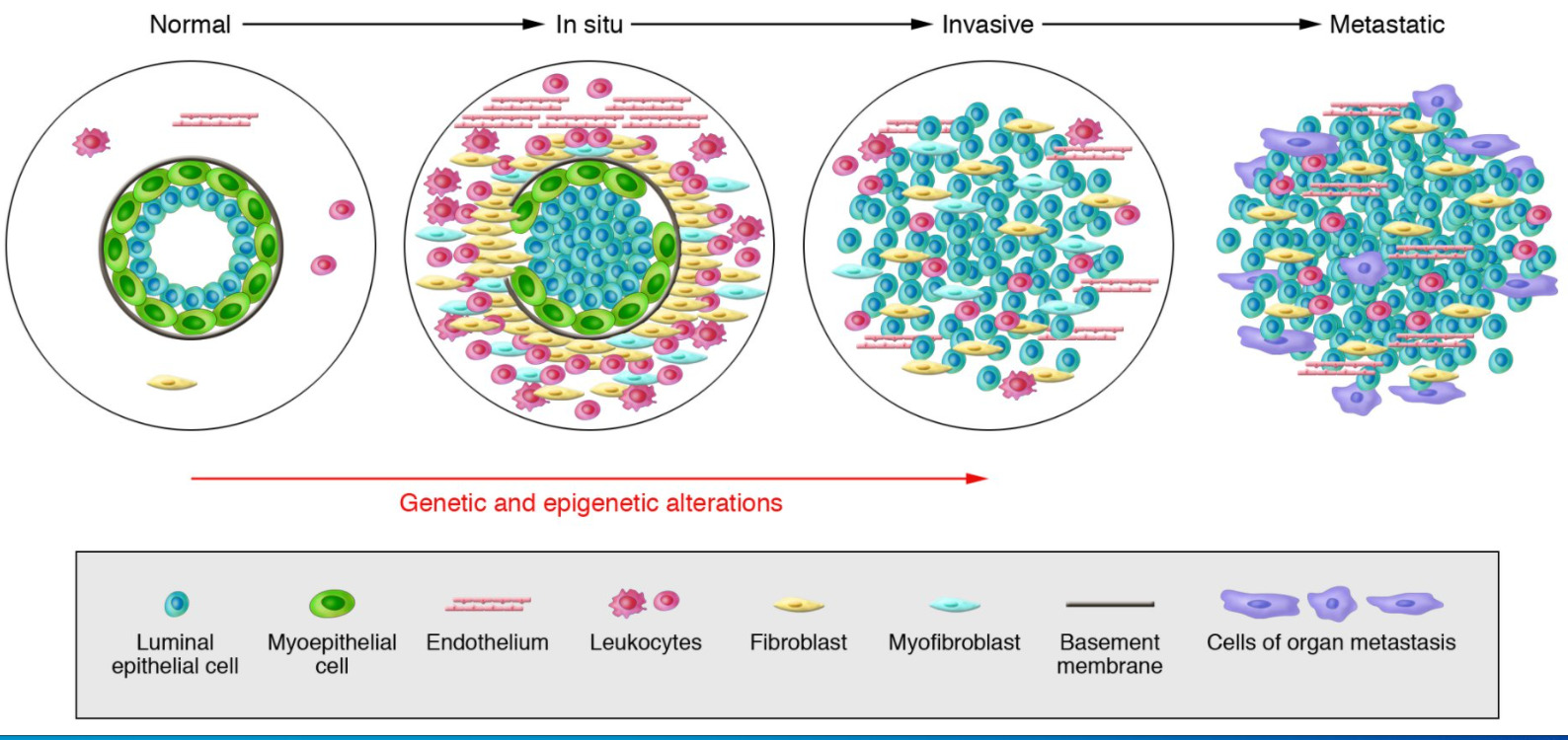
Model of breast tumor progression
From left to right: Normal breast ducts are composed of the basement membrane and a layer of luminal epithelial and myoepithelial cells. The stroma includes various leukocytes, fibroblasts, myofibroblasts, and endothelial cells. In in situ carcinomas: The myoepithelial cells are epigenetically and phenotypically altered and their number decreases, potentially due to degradation of the basement membrane. The stromal fibroblasts, myofibroblasts, lymphocytes, and endothelial cells increase. In invasive carcinomas, there is a loss of myoepithelial cells and basement membrane, in which tumor cells can invade surrounding tissues. The tumor cells migrate to distant organs, eventually leading to metastases.
Clinical Presentation
Clinical findings
In areas with established breast cancer screening: Most cases of cancer are diagnosed by having an abnormal mammogram.
Symptoms:
- Patient feels a lump/mass.
- Skin changes (dimpling, erythema, thickening)
- Nipple changes (appearance, discharge)
Signs:
- Firm or hard mass with poorly defined margins, fixed or immovable (on its own not enough to distinguish malignancy)
- Location:
- Highest frequency: upper outer quadrant (50% of cases)
- Lowest frequency: lower inner quadrant (5%)
- Possible to have multifocal/multiple foci in the same quadrant
- Possible to have different quadrants of the breasts affected (multicentric)
- Contralateral breast also affected by the tumor in 5%–10% of cases
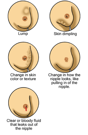
Possible signs of breast cancer
From right to left, top to bottom: a breast lump/mass, skin dimpling, change in skin color/texture, nipple changes including retraction (pulling in of the nipple), and nipple discharge
Metastasis
- Presentation depends on organ(s) involved
- Most common sites:
- Bone (back or leg pain)
- Liver (jaundice, abdominal pain, nausea, abnormal liver tests)
- Lungs (shortness of breath, cough, abnormal chest imaging)
Related videos
Non-invasive Breast Cancer
DCIS
- Proliferation of cytologically malignant cells within the mammary ductal system, with no invasion of the surrounding stroma
- ⅓ develop invasive cancer in 5 years
- Frequently detected by mammography
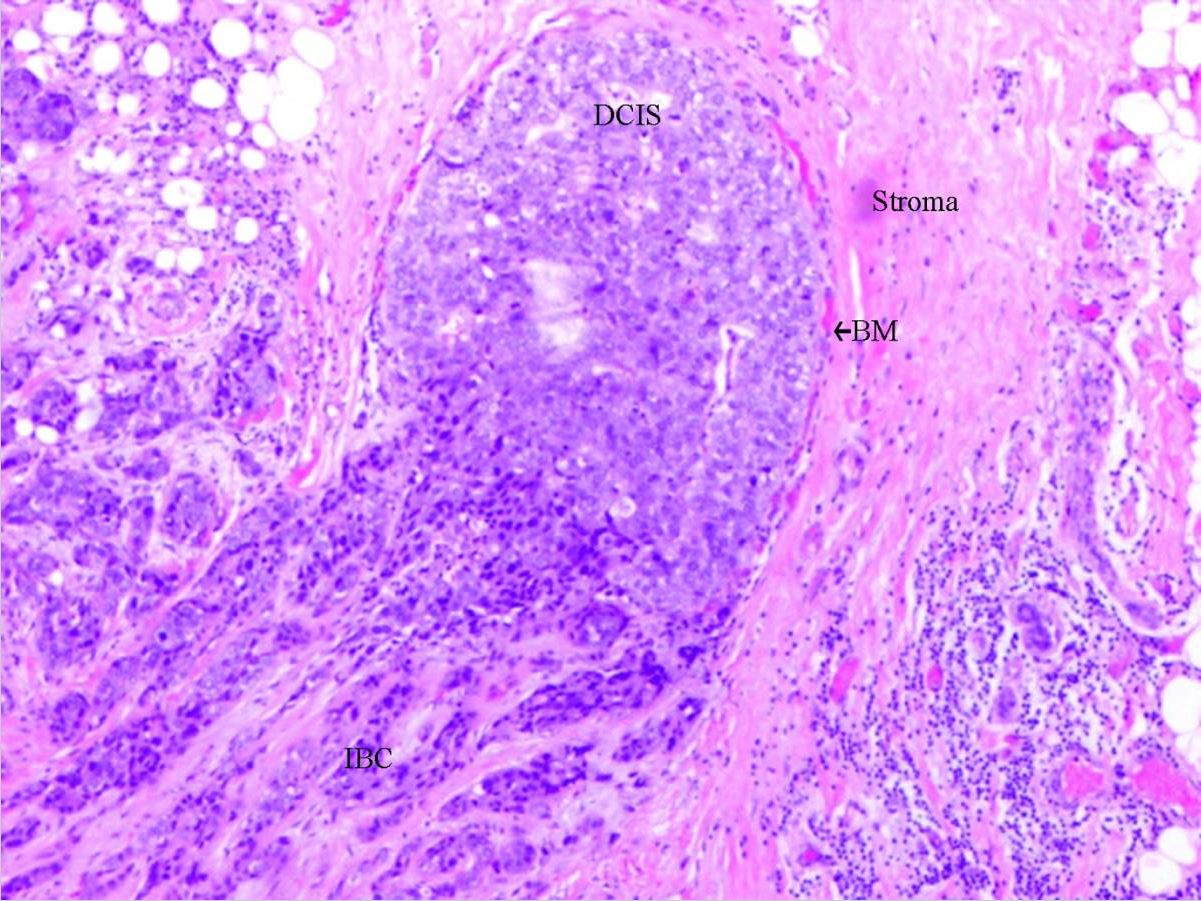
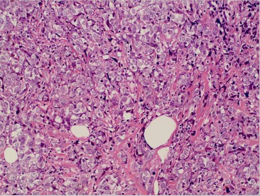
Ductal carcinoma in situ: DCIS with invasion
Image: “Ductal carcinoma in situ of the breast: Morphological and molecular features implicated in progression” by Bioscience Reports. License: CC BY 3.0.
Abbreviations used:
DCIS: ductal carcinoma in situ
BM: basal membrane
LCIS
- Proliferation of malignant cells within the lobules, growing in an incohesive manner
- Mucin-positive signet cell rings usually noted
- Loss of cellular adhesion also observed (from dysfunction of E-cadherin)
- Rarely with calcifications (incidence not changed by mammography)
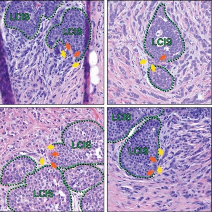
Lobular carcinoma in situ in association with invasive lobular carcinoma (ILC)
Image: “Molecular drivers of lobular carcinoma in situ” by Logan GJ, Dabbs DJ, Lucas PC, Jankowitz RC, Brown DD, Clark BZ, Oesterreich S, McAuliffe PF. License: CC BY 4.0.
Histologic sections from multiple patients capture areas of LCIS that appear to have focal myoepithelial layer disruption adjacent to ILC, suggesting possible progression of LCIS to ILC at such transitions. Green hatched lines mark the myoepithelial layer; orange arrows are possible foci of myoepithelial disruption; yellow arrows highlight invasive cells. In addition to the cells marked by yellow arrows, additional ILC cells are present in each image throughout the stroma, surrounding the areas of LCIS.
Comparison DCIS versus LCIS
DCIS | LCIS | |
---|---|---|
Presentation | Unifocal | Multifocal |
Patterns |
| Solid |
Calcification | Yes/no | Usually no |
Risk of cancer | Higher | Lower |
Location of cancer | Ipsilateral breast | Ipsilateral or contralateral |
LCIS: lobular carcinoma in situ
Related videos
Invasive Breast Cancer
Infiltrating ductal carcinoma
- Most common invasive breast cancer (76% of all breast carcinomas)
- Mostly unilateral
- Gross appearance:
- Firm, fibrous, “rock-hard” mass with irregular stellate shape
- Often 2–3 cm in size
- Microscopic findings (Nottingham Histologic Score):
- Well differentiated (grade 1): tubular/cribriform pattern, small uniform nuclei, low proliferative rate
- Moderately differentiated (grade 2): cells in clusters or single infiltrating cells, nuclear polymorphism, + mitotic figures
- Poorly differentiated (grade 3): ragged nest pattern, large irregular nuclei, high proliferative rate
Breast cancer: invasive ductal carcinoma in the left breast
Image: “Invasive ductal breast cancer metastatic to the sigmoid colon” by Zhou XC, Zhou H, Ye YH, Zhang XF, Jiang Y. License: CC BY 2.0.
Infiltrating lobular carcinoma
- 2nd most common invasive breast cancer (5%–10%)
- Gross appearance:
- May not have a mass lesion
- Difficult to palpate or detect by mammography
- Microscopic findings:
- Incohesive infiltrating tumor cells (often with signet ring cells)
- Staining with E-cadherin (negative in infiltrating lobular carcinoma): helps distinguish from infiltrating ductal carcinoma
- Metastatic pattern: involves the peritoneum, retroperitoneum, leptomeninges, ovaries/uterus, and gastrointestinal tract
- Heterozygous germline mutations in CDH1: increased risk of lobular carcinoma
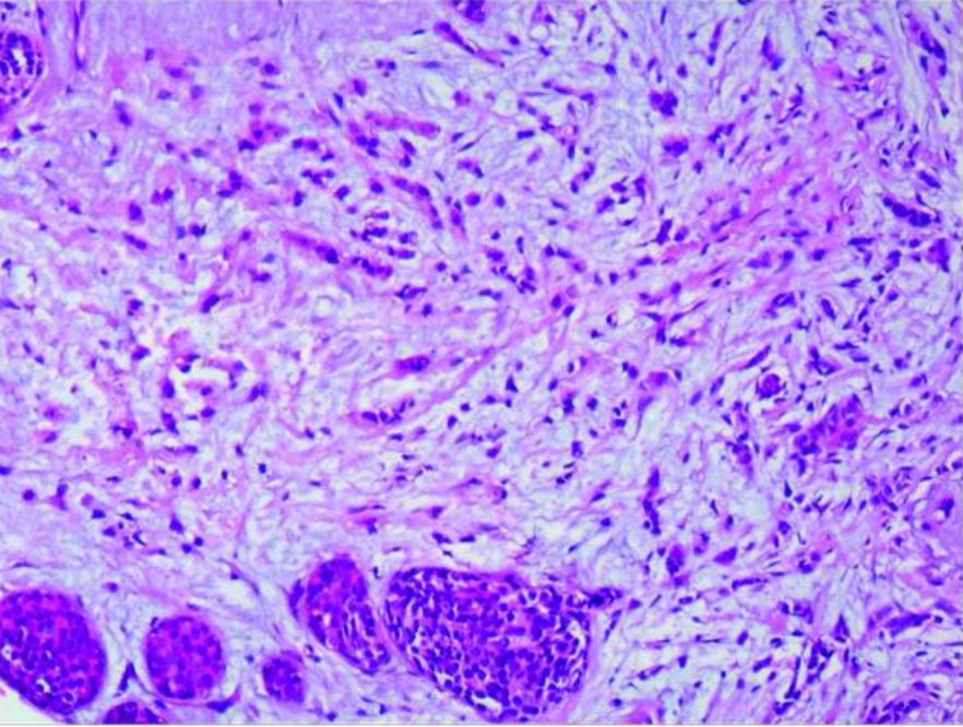
Lobular carcinoma: infiltrating lobular carcinoma of the right breast and LCIS (amplified 20×10)
Image: “Case report of small bowel obstruction caused by small intestinal metastasis of bilateral breast cancer” by Lv L, Zhao Y, Liu H, Peng Z. License: CC BY 3.0.
Tubular carcinoma
- ≤ 2% of invasive breast cancers
- Microscopic findings: well-formed tubules; associated with low-grade DCIS
- Good prognosis, metastasis rare
Mucinous/colloid carcinoma
- 1%–2% of invasive breast cancers
- More common in older patients
- Favorable prognosis
- Gross appearance: soft, circumscribed, pale gray-blue gelatin
- Microscopic findings: clusters of tumor cells within lakes of mucin
Medullary carcinoma
- More frequently found in BRCA1 carcinomas
- Occurs in younger patients
- Better prognosis than other poorly differentiated carcinomas
- Microscopic findings:
- Well circumscribed, with areas of necrosis and hemorrhage
- Sheets of large cells, pleomorphic nuclei with prominent nucleoli
- Marked lymphoplasmacytic infiltrate
Micropapillary carcinoma
- Rare but aggressive cancer
- High propensity for lymphovascular invasion and lymph node metastasis
- Microscopic findings: Clusters of cells float within an empty stromal space.
Paget’s disease of the breast
- 1%–4% of cases
- Appearance:
- Unilateral eczematous, erythematous patches on the nipple, and nipple retraction
- In > 50%, palpable mass present (often invasive carcinoma, ER negative and overexpress HER2)
- Those without a mass: Most have DCIS.
- Paget’s cells: large, round, malignant cells in the epidermis
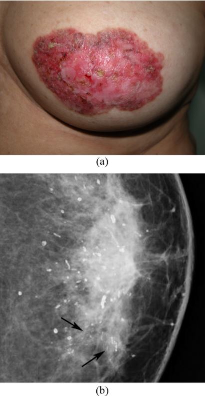
Paget’s disease of the breast: left nipple areolar changes
(a) Photograph of the left breast shows skin thickening, redness, erythema, erosion of the nipple, and scaling around the nipple-areola area.
(b) Mammogram shows scattered rod-like calcifications and groups of pleomorphic, fine, linear microcalcifications in the inner quadrant (arrows). Simple mastectomy revealed DCIS and secretory calcifications in the breast and Paget’s disease of the nipple.
Inflammatory carcinoma
- Characterized by dermal lymphovascular invasion of tumor cells
- Appearance:
- Breast skin erythema and thickening
- Breast skin resembles orange peel (peau d’orange).
- Poor prognosis
Related videos
Diagnosis
Clinical and diagnostic tools
- Breast exam
- Screening mammography:
- May offer screening starting at age 40 years.
- Signs of a malignant finding:
- Soft-tissue mass or density
- Clustered microcalcifications
- Spiculated high-density mass (most specific for invasive cancer)
- The presence of a breast lump with a negative mammogram still warrants further investigation.
- Ultrasonography:
- Complementary test to mammography
- Benefit:
- No radiation exposure
- Differentiates solid (such as a benign fibroadenoma or cancer) from fluid-filled cystic (such as a benign cyst) lesions
- Disadvantage: highly operator dependent, not suited for screening on its own
- Signs of malignant finding:
- Internal calcifications
- Hypoechoic
- Shadowing
- Spiculated margins
- Magnetic resonance imaging (MRI):
- Screening for women at high risk for breast cancer
- Benefit: high soft-tissue contrast, very high sensitivity
- Disadvantage: low specificity, no detection of microcalcifications
- Signs of malignant finding:
- Spiculated mass
- Enhancing internal septa
- Heterogenous internal enhancement
- Biopsy (confirms diagnosis):
- Fine-needle aspiration: small sample, with a false-negative rate of 10%
- Core needle biopsy (recommended): larger sample, and allows for immunohistochemical testing
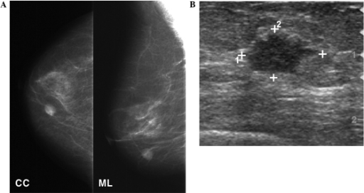
Mammography and breast ultrasound
Image: “Secretory breast carcinoma: A report of three cases and a review of the literature” by Lee SG, Jung SP, Lee HY, Kim S, Kim HY, Kim I, Bae JW. License: CC BY 3.0.
A: Mammography shows a round, high-density mass in the lower inner quadrant of the right breast.
B: Ultrasonography follow-up shows a 1.3-cm irregularly shaped, hypoechoic mass in the breast.
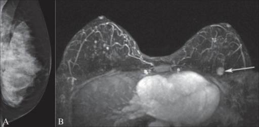
Mammography and breast MRI
Image: “MRI for breast cancer: Current indications” by Ojeda-Fournier H, Comstock CE. License: CC BY 2.0.
Image A shows mammography of the left breast in a BRCA1 gene mutation carrier. Note the extremely dense breast tissue.
MRI image B demonstrates an enhancing mass (arrow) in the upper left breast that was occult on the mammogram.
Post-diagnostic tools
- Breast cancer receptor testing:
- When cancer is confirmed, determine ER/PR receptor expression:
- If > 1% of the tumor cells stain positive (on immunohistochemistry or IHC) for ER/PR → hormone receptor positive
- ER/PR-positive carcinoma is suitable for endocrine therapy → better prognosis
- Overexpression of HER2 receptor:
- Detected by IHC or fluorescence in situ hybridization (FISH)
- HER2 positive → treatment with trastuzumab (HER2 antibody)
- When cancer is confirmed, determine ER/PR receptor expression:
- Additional imaging (for metastatic breast cancer):
- Bone scan: for patients with bone pain or elevated alkaline phosphatase
- Computed tomography (CT) of the chest: for patients with pulmonary symptoms
- CT of the abdomen and pelvis or MRI: for patients with abnormal pain and/or examination, elevated liver enzymes/alkaline phosphatase
- Positron emission tomography-CT (PET-CT): for whole-body screening for metastasis (stage III or higher)
- Tumor markers:
- CA 15-3, CA 27.29, and carcinoembryonic antigen (CEA)
- Biochemical markers are not specific for breast cancer relapse.
- Used to monitor treatment response of patients with metastatic disease
- Genetic testing:
- United States Preventive Services Task Force recommends the use of approved familial risk assessment tools for the following:
- A personal or family history of breast, ovarian, tubal, or peritoneal cancer
- Ancestry associated with BRCA1 or 2 mutations
- Positive assessment indicates the need for genetic counseling and testing.
- Other expert groups have varying recommendations.
- United States Preventive Services Task Force recommends the use of approved familial risk assessment tools for the following:
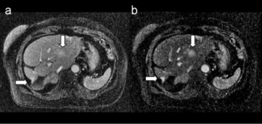
MRI of breast metastasis: images show metastatic breast lesions in the liver
A. T1-weighted 3D postcontrast image
B. corresponding 3D subtraction image
Arrows indicate the metastatic lesions.
Staging
Overview of breast cancer staging
- Based on the American Joint Committee on Cancer (AJCC) and Union for International Cancer Control (UICC) 8th edition
- Includes the tumor, node, metastasis (TNM) staging system and incorporates biologic factors for prognostic staging
- Information included in staging:
- Tumor
- Node
- Metastasis
- ER status
- PR status
- HER2 status
- Histologic grade
- Recurrence score (Oncotype Dx) considered
- Anatomic stage grouping relies on TNM.
- Prognostic stage grouping:
- Relies on TNM, ER/PR/HER2, grade
- Primary prognostic staging system for those who receive neoadjuvant treatment or for those who do not undergo surgery
TNM staging
Tumor stage | Description |
---|---|
Tx | Primary tumor unable to be assessed |
T0 | No evidence of primary tumor |
Tis |
|
T1 | ≤ 20-mm tumor in greatest dimension |
T2 | > 20-mm but ≥ 50-mm tumor in greatest dimension |
T3 | > 50-mm tumor in greatest dimension |
T4 | Tumor of any size, with direct extension to the chest wall and/or skin (ulceration or macroscopic nodules) |
Node stage | Description |
---|---|
cNX | Regional LNs cannot be assessed. |
cN0 | No regional LN metastases |
cN1 | Metastasis to movable ipsilateral level I, II axillary LNs |
cN2 |
|
cN3 |
|
Cn: clinical node
Note: Spread to other LN (cervical or contralateral axillary) is considered metastatic (M1).
Metastasis | Description |
---|---|
M0 | No evidence of distant metastasis (clinical or radiographic) |
M1 | Detectable metastasis |
Stage | Substages | Tumor | Node | Metastasis |
---|---|---|---|---|
0 | Tis | N0 | M0 | |
I |
| T0-T1 | N1 | M0 |
II |
| T0-T3 | N0-N1 | M0 |
III |
| T0-T3 | N1-N2 | M0 |
IV | Any T | Any N | M1 |
UICC: Union for International Cancer Control
T: tumor
N: node
M: metastasis
Categories
- Early-stage non-metastatic breast cancer: stage I, IIA, a subset of stage IIB (T2N1)
- Locally advanced non-metastatic breast cancer: subset of stage IIB (T3N0), stage IIIA–IIIC
- Metastatic breast cancer: stage IV
Management and Prognosis
DCIS
- Surgery: breast-conserving surgery (BCS) (e.g., lumpectomy) or mastectomy
- Radiation therapy (RT) considered in those with high risk of recurrence
- Endocrine therapy for ER-positive DCIS (for 5 years):
- Tamoxifen:
- ER antagonist
- Option for all women
- Aromatase inhibitors (e.g., anastrozole):
- Inactivates aromatase, reducing peripheral conversion of androgens to estrogens
- Alternative for post-menopausal women
- Tamoxifen:
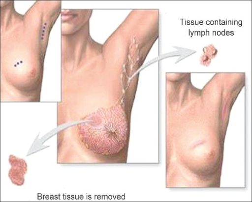
Breast-conserving surgery (lumpectomy): excision of the tumor to the negative margins and axillary lymph node evaluation
Image: “Phantom breast syndrome” by the Indian Journal of Palliative Care. License: CC BY 2.0.Early-stage cancer
- Surgery:
- BCS or mastectomy
- Axillary LN assessment performed with sentinel lymph node biopsy (if positive, axillary dissection is done)
- RT in most cases (if BCS is done and for those at risk for local recurrence)
- Adjuvant or systemic therapy given in addition to surgery:
- Endocrine therapy:
- For hormone receptor (ER/PR)–positive patients
- Aromatase inhibitors or tamoxifen
- HER2-targeted therapy:
- For HER2-positive cancers
- Trastuzumab +/- pertuzumab
- Chemotherapy (doxorubicin + cyclophosphamide, then paclitaxel):
- For TNBCs (≥ 0.5 cm)
- For HER2-positive cancers (> 1 cm)
- For hormone receptor–positive cancers (with high-risk features: ≥ 2-cm size, + LNs, high-grade lesions)
- Endocrine therapy:
Locally advanced breast cancer
- Neoadjuvant systemic therapy:
- HER2-directed therapy + chemotherapy: for HER2-positive cancer
- Chemotherapy given 1st for hormone receptor–positive cancer
- Chemotherapy given for TNBCs
- Surgery (BCS or mastectomy + LN assessment) follows neoadjuvant therapy.
- Adjuvant therapy:
- Given if there was no neoadjuvant therapy (similar principles as early-stage cancer)
- For those who had neoadjuvant therapy:
- Endocrine therapy or HER2-directed therapy continued as indicated
- Chemotherapy use is dictated by clinical status and tumor characteristics.
- RT
Metastatic breast cancer
- Systemic therapy:
- Aromatase inhibitors with/without chemotherapy for ER-positive cancers
- Trastuzumab with chemotherapy for HER2-positive cancers
- Chemotherapy for receptor-negative cancers
- Immunotherapy (immune checkpoint inhibitors)
- RT for palliation
Prognosis
- 10-year survival:
- Stage 0 (DCIS): 97%
- Stage I: 87%
- Stage II: 65%
- Stage III: 40%
- Stage IV: 5%
- With distant metastasis, there is no cure.
Differential Diagnosis
- Fibroadenoma: benign breast tumors that arise in young women. Diagnosis is based on the physical finding of a mobile, non-tender breast mass. Some fibroadenoma are asymptomatic, small in size, and should be treated conservatively. Other fibroadenoma grow rapidly in size, are symptomatic, and should be surgically excised.
- Fat necrosis of the breast: a benign condition caused by trauma to the breast, which may go unnoticed, leading to adipocyte necrosis and calcification. Patients may present with a solid, painless mass with or without skin changes. Fat necrosis of the breast can mimic malignancy and definitive diagnosis is by imaging and biopsy.
- Mastitis: inflammation of the mammary gland tissue that can be lactational or non-lactational. Mastitis is most common in women of childbearing age. Patients usually present with high fever, chills, fatigue, malaise, and myalgia. Analgesics and antibiotics are given as part of the management.
- Breast abscess: a breast mass associated with mastitis. Breast abscess presents as a unilateral and fluctuant mass, accompanied by fever, and a painful, erythematous, and edematous breast. History, physical findings, and needle aspiration (revealing purulent contents) help differentiate a breast abscess. Treatment includes incision and drainage, with antibiotics.
- Intraductal papillomas: abnormal growths with a papillary configuration of the breast stroma and epithelium within a breast duct. The most common presenting feature is bloody or serous nipple discharge. Core needle biopsy confirms the diagnosis.
- Simple breast cyst: round or oval fluid-filled masses from the terminal duct lobular unit. A cyst is felt as a palpable mass that can cause pain. Ultrasonography shows a cyst as a well-circumscribed, anechoic mass with no solid components.
References
- Bleiweiss, I., Chagpar, A., & Vora, S. (2020). Pathology of breast cancer. UpToDate. Retrieved Jan 15, 2021, from https://www.uptodate.com/contents/pathology-of-breast-cancer
- Hao, S., Zhao, YY., Peng, JJ., et al. (2019). Invasive micropapillary carcinoma of the breast had no difference in prognosis compared with invasive ductal carcinoma: a propensity-matched analysis. Sci Rep 9, 286. https://doi.org/10.1038/s41598-018-36362-8
- Hayes D.F., & Lippman M.E. (2018). Breast cancer. In Jameson JL, et al. (Ed.), Harrison’s Principles of Internal Medicine, 20th ed. McGraw-Hill.
- Lester, S. (2020). The breast. In Kumar, V., Abbas, A. K., Aster, J.C., (Eds.), Robbins & Cotran Pathologic Basis of Disease. (10th ed. pp. 1037–1064).
- Moasser M.M., & Ai W.Z. (2019). Neoplasia. In Hammer G.D., & McPhee S.J. (Eds.), Pathophysiology of Disease: An Introduction to Clinical Medicine, 8th ed. McGraw-Hill.
- Nascimento, R., & Otoni, K. (2020). Histological and molecular classification of breast cancer: What do we know? Mastology. https://www.mastology.org/wp-content/uploads/2020/09/MAS_2020024_AOP.pdf
- Polyak, K. (2007). Breast cancer: Origins and evolution. J Clin Invest. 117(11), 3155–63. https://doi.org/10.1172/JCI33295
- Taghian, A., Meravjer, S., Hayes, D., & Vora, S. (2020). Overview of the treatment of newly diagnosed, invasive, non-metastatic breast cancer. UpToDate. Retrieved Jan 16, 2021, from https://www.uptodate.com/contents/overview-of-the-treatment-of-newly-diagnosed-invasive-non-metastatic-breast-cancer