Physical examination of the breasts is important both in the evaluation of a breast complaint and screening for asymptomatic breast pathology such as a breast mass. The examination involves inspection of the breasts to look for asymmetry or skin/nipple changes, as well as palpation of the breasts, nipples, and axilla. Coupled with medical history, the outcome of a breast examination can be normal, lead to a clinical diagnosis (e.g., infection), or require additional diagnostic evaluation (e.g., the evaluation of a palpable breast mass or skin/nipple changes).
Last updated: Dec 15, 2025
The anatomy of the breast and axilla Axilla The axilla is a pyramid-shaped space located between the upper thorax and the arm. The axilla has a base, an apex, and 4 walls (anterior, medial, lateral, posterior). The base of the pyramid is made up of the axillary skin. The apex is the axillary inlet, located between the 1st rib, superior border of the scapula, and clavicle. Axilla and Brachial Plexus: Anatomy as well as the relationship to the thorax should be reviewed before starting. Knowledge of the underlying tissue anatomy makes interpreting and describing the findings easier.
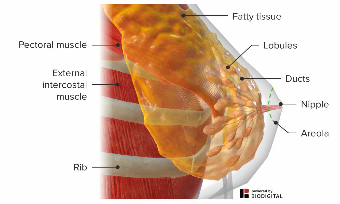
Anatomy of the breast
Image by BioDigital, edited by Lecturio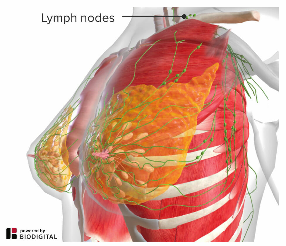
Tail of Spence
Image by BioDigital, edited by Lecturio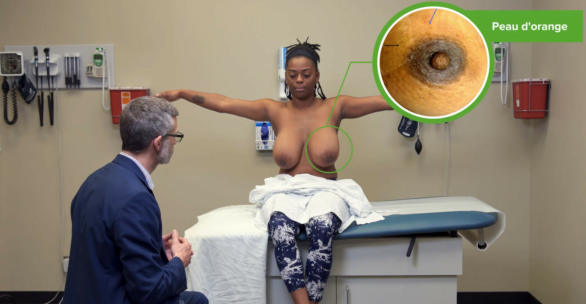
Inspection of the breasts highlighting skin dimpling (peau d’orange)
Image by Lecturio.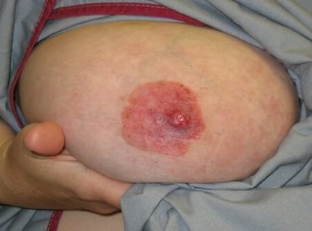
Paget disease:
Scaling, itching, ulceration, bleeding, and erythema of the nipple spreading to the areola and surrounding area may be suggestive of Paget disease, a rare malignancy of the breast tissue.
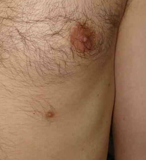
Accessory or supernumerary nipple:
Errors in migration during embryonic development may cause remnants of mammary tissue to form accessory or supernumerary nipples.
To preserve modesty, cover the individual with an extra gown/sheet and expose only the breast to be examined.

Include the tail of Spence in palpation of the breast.
Image by Lecturio.
Assess for discharge in palpation of the nipple.
Image by Lecturio.
Examination of the breast
Image by Lecturio.Describe findings accurately to allow the practitioner to monitor for changes and, when imaging is required, communicate findings to the radiologist.
| Fibroadenoma Fibroadenoma Fibroadenomas are the most common benign tumor of the female breast and the most common breast tumor in adolescent and young women. The tumors are well-circumscribed, mobile, and unencapsulated, with a rubbery or firm consistency. Fibroadenoma | Fibrocystic Fibrocystic Fibrocystic Change changes | Breast cancer Breast cancer Breast cancer is a disease characterized by malignant transformation of the epithelial cells of the breast. Breast cancer is the most common form of cancer and 2nd most common cause of cancer-related death among women. Breast Cancer | |
|---|---|---|---|
| Usual age | 15–35 years old | 30–50 years old with regression Regression Corneal Abrasions, Erosion, and Ulcers after menopause Menopause Menopause is a physiologic process in women characterized by the permanent cessation of menstruation that occurs after the loss of ovarian activity. Menopause can only be diagnosed retrospectively, after 12 months without menstrual bleeding. Menopause (except on estrogen replacement therapy Estrogen Replacement Therapy The use of hormonal agents with estrogen-like activity in postmenopausal or other estrogen-deficient women to alleviate effects of hormone deficiency, such as vasomotor symptoms, dyspareunia, and progressive development of osteoporosis. This may also include the use of progestational agents in combination therapy. Menopause) | 30–90 years old (most common over the age of 50) |
| Number | Usually single but may be multiple | Single or multiple | Usually single although may coexist with other nodules |
| Shape | Round, small | Round | Irregular or stellate |
| Consistency Consistency Dermatologic Examination | Firm | Soft to firm, usually elastic Elastic Connective Tissue: Histology | Firm or hard |
| Delimitation | Well-delineated | Well-delineated | Not clearly delineated from surrounding tissues |
| Mobility | Mobile | Mobile | May be fixed to the skin Skin The skin, also referred to as the integumentary system, is the largest organ of the body. The skin is primarily composed of the epidermis (outer layer) and dermis (deep layer). The epidermis is primarily composed of keratinocytes that undergo rapid turnover, while the dermis contains dense layers of connective tissue. Skin: Structure and Functions or underlying tissues |
| Tenderness | Usually nontender | Often tender | Usually nontender |
| Retraction signs | Absent | Absent | May be present |