Primary vaginal cancers are malignant tumors that originate from cells in the vaginaVaginaThe vagina is the female genital canal, extending from the vulva externally to the cervix uteri internally. The structures have sexual, reproductive, and urinary functions and a rich blood supply, mainly arising from the internal iliac artery.Vagina, Vulva, and Pelvic Floor: Anatomy. Squamous cell carcinomaSquamous cell carcinomaCutaneous squamous cell carcinoma (cSCC) is caused by malignant proliferation of atypical keratinocytes. This condition is the 2nd most common skin malignancy and usually affects sun-exposed areas of fair-skinned patients. The cancer presents as a firm, erythematous, keratotic plaque or papule. Squamous Cell Carcinoma (SCC) (SCC) is by far the most common (80%–85%); other histologic types include adenocarcinomas, sarcomas (including sarcoma botryoides, typically seen in children), and melanomas. Vaginal SCC is most commonly associated with HPVHPVHuman papillomavirus (HPV) is a nonenveloped, circular, double-stranded DNA virus belonging to the Papillomaviridae family. Humans are the only reservoir, and transmission occurs through close skin-to-skin or sexual contact. Human papillomaviruses infect basal epithelial cells and can affect cell-regulatory proteins to result in cell proliferation. Papillomavirus (HPV)infectionsInfectionsInvasion of the host organism by microorganisms or their toxins or by parasites that can cause pathological conditions or diseases.Chronic Granulomatous Disease, while clear cell adenocarcinomas are associated with in utero exposure to diethylstilbestrolDiethylstilbestrolA synthetic nonsteroidal estrogen used in the treatment of menopausal and postmenopausal disorders. It was also used formerly as a growth promoter in animals. According to the fourth annual report on carcinogens, diethylstilbestrol has been listed as a known carcinogen.Noncontraceptive Estrogen and Progestins (DES). Individuals typically present with vaginal bleeding and/or an irregular massMassThree-dimensional lesion that occupies a space within the breastImaging of the Breast or lesion on exam; other symptoms may include abnormal discharge, painPainAn unpleasant sensation induced by noxious stimuli which are detected by nerve endings of nociceptive neurons.Pain: Types and Pathways, and urinary or defecatory symptoms. A biopsyBiopsyRemoval and pathologic examination of specimens from the living body.Ewing Sarcoma is required for diagnosis. StagingStagingMethods which attempt to express in replicable terms the extent of the neoplasm in the patient.Grading, Staging, and Metastasis is based on tumorTumorInflammation size, extent of local invasion, and metastasisMetastasisThe transfer of a neoplasm from one organ or part of the body to another remote from the primary site.Grading, Staging, and Metastasis. Management may be surgical for stage I disease, but surgery is typically avoided in advanced disease, which is instead managed with radiationRadiationEmission or propagation of acoustic waves (sound), electromagnetic energy waves (such as light; radio waves; gamma rays; or x-rays), or a stream of subatomic particles (such as electrons; neutrons; protons; or alpha particles).Osteosarcoma and chemotherapyChemotherapyOsteosarcoma.
Primary vaginal cancer is a malignant tumorTumorInflammation arising from tissue of the vaginaVaginaThe vagina is the female genital canal, extending from the vulva externally to the cervix uteri internally. The structures have sexual, reproductive, and urinary functions and a rich blood supply, mainly arising from the internal iliac artery.Vagina, Vulva, and Pelvic Floor: Anatomy.
Anatomy review
The vaginaVaginaThe vagina is the female genital canal, extending from the vulva externally to the cervix uteri internally. The structures have sexual, reproductive, and urinary functions and a rich blood supply, mainly arising from the internal iliac artery.Vagina, Vulva, and Pelvic Floor: Anatomy is:
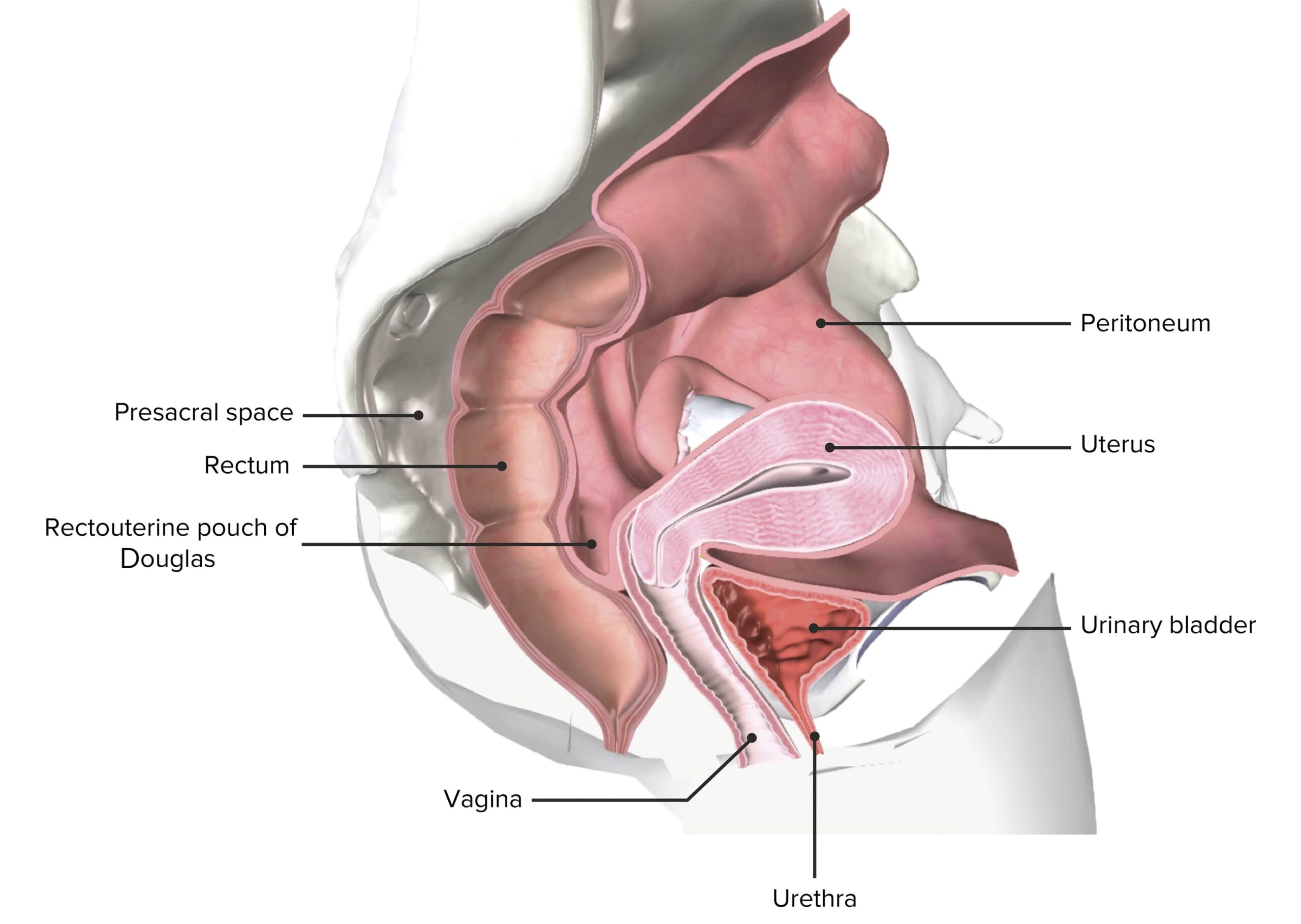
Located between the bladderBladderA musculomembranous sac along the urinary tract. Urine flows from the kidneys into the bladder via the ureters, and is held there until urination.Pyelonephritis and Perinephric Abscess and rectumRectumThe rectum and anal canal are the most terminal parts of the lower GI tract/large intestine that form a functional unit and control defecation. Fecal continence is maintained by several important anatomic structures including rectal folds, anal valves, the sling-like puborectalis muscle, and internal and external anal sphincters. Rectum and Anal Canal: Anatomy in the female pelvisPelvisThe pelvis consists of the bony pelvic girdle, the muscular and ligamentous pelvic floor, and the pelvic cavity, which contains viscera, vessels, and multiple nerves and muscles. The pelvic girdle, composed of 2 “hip” bones and the sacrum, is a ring-like bony structure of the axial skeleton that links the vertebral column with the lower extremities.Pelvis: Anatomy
A sectioned female pelvis depicting the uterus in situ
Vaginal malignancies may be either primary (originating from the vaginaVaginaThe vagina is the female genital canal, extending from the vulva externally to the cervix uteri internally. The structures have sexual, reproductive, and urinary functions and a rich blood supply, mainly arising from the internal iliac artery.Vagina, Vulva, and Pelvic Floor: Anatomy itself) or metastatic to the vaginaVaginaThe vagina is the female genital canal, extending from the vulva externally to the cervix uteri internally. The structures have sexual, reproductive, and urinary functions and a rich blood supply, mainly arising from the internal iliac artery.Vagina, Vulva, and Pelvic Floor: Anatomy from other primary sites.
Primary vaginal cancers:
The most common subtypes of primary vaginal cancer (originating in the vaginaVaginaThe vagina is the female genital canal, extending from the vulva externally to the cervix uteri internally. The structures have sexual, reproductive, and urinary functions and a rich blood supply, mainly arising from the internal iliac artery.Vagina, Vulva, and Pelvic Floor: Anatomy) include:
Squamous cell carcinomaSquamous cell carcinomaCutaneous squamous cell carcinoma (cSCC) is caused by malignant proliferation of atypical keratinocytes. This condition is the 2nd most common skin malignancy and usually affects sun-exposed areas of fair-skinned patients. The cancer presents as a firm, erythematous, keratotic plaque or papule. Squamous Cell Carcinoma (SCC) (SCC): approximately 80%–85%
“Typical” SCC (most common primary vaginal cancer)
Adenocarcinoma (glandular malignancies): approximately 10%
Clear cell adenocarcinoma (2nd most common primary vaginal cancer)
Endodermal sinus tumorTumorInflammation (also called a yolk sacYolk SacThe first of four extra-embryonic membranes to form during embryogenesis. In reptiles and birds, it arises from endoderm and mesoderm to incorporate the egg yolk into the digestive tract for nourishing the embryo. In placental mammals, its nutritional function is vestigial; however, it is the source of intestinal mucosa; blood cells; and germ cells. It is sometimes called the vitelline sac, which should not be confused with the vitelline membrane of the egg.Embryoblast and Trophoblast DevelopmenttumorTumorInflammation; tumorTumorInflammation derived from germ cellsGerm CellsThe reproductive cells in multicellular organisms at various stages during gametogenesis.Gametogenesis)
Sarcoma botryoides (an embryonal rhabdomyosarcoma, which is a malignant tumorTumorInflammation of skeletal muscle)
LeiomyosarcomaLeiomyosarcomaUterine leiomyomas (or uterine fibroids) are benign tumors arising from smooth muscle cells in the uterine myometrium. Leiomyosarcomas, however, are malignant tumors, arising de novo (not from fibroids). Uterine Leiomyoma and Leiomyosarcoma (smooth muscle malignancyMalignancyHemothorax, very rare)
MelanomaMelanomaMelanoma is a malignant tumor arising from melanocytes, the melanin-producing cells of the epidermis. These tumors are most common in fair-skinned individuals with a history of excessive sun exposure and sunburns. Melanoma: approximately 2%
Accounts for 80% of all malignancies in the vaginaVaginaThe vagina is the female genital canal, extending from the vulva externally to the cervix uteri internally. The structures have sexual, reproductive, and urinary functions and a rich blood supply, mainly arising from the internal iliac artery.Vagina, Vulva, and Pelvic Floor: Anatomy
Most common primary sites that metastasize to the vaginaVaginaThe vagina is the female genital canal, extending from the vulva externally to the cervix uteri internally. The structures have sexual, reproductive, and urinary functions and a rich blood supply, mainly arising from the internal iliac artery.Vagina, Vulva, and Pelvic Floor: Anatomy:
CervixCervixThe uterus, cervix, and fallopian tubes are part of the internal female reproductive system. The most inferior portion of the uterus is the cervix, which connects the uterine cavity to the vagina. Externally, the cervix is lined by stratified squamous cells; however, the cervical canal is lined by columnar epithelium.Uterus, Cervix, and Fallopian Tubes: Anatomy
EndometriumEndometriumThe mucous membrane lining of the uterine cavity that is hormonally responsive during the menstrual cycle and pregnancy. The endometrium undergoes cyclic changes that characterize menstruation. After successful fertilization, it serves to sustain the developing embryo.Embryoblast and Trophoblast Development/uterusUterusThe uterus, cervix, and fallopian tubes are part of the internal female reproductive system. The uterus has a thick wall made of smooth muscle (the myometrium) and an inner mucosal layer (the endometrium). The most inferior portion of the uterus is the cervix, which connects the uterine cavity to the vagina.Uterus, Cervix, and Fallopian Tubes: Anatomy
VulvaVulvaThe vulva is the external genitalia of the female and includes the mons pubis, labia majora, labia minora, clitoris, vestibule, vestibular bulb, and greater vestibular glands. Vagina, Vulva, and Pelvic Floor: Anatomy
Ovary
Breast
RectumRectumThe rectum and anal canal are the most terminal parts of the lower GI tract/large intestine that form a functional unit and control defecation. Fecal continence is maintained by several important anatomic structures including rectal folds, anal valves, the sling-like puborectalis muscle, and internal and external anal sphincters. Rectum and Anal Canal: Anatomy
Kidney
Epidemiology
Primary vaginal cancer:
Rare: accounts for only 1%–2% of all gynecologic malignancies
Age-adjusted incidenceIncidenceThe number of new cases of a given disease during a given period in a specified population. It also is used for the rate at which new events occur in a defined population. It is differentiated from prevalence, which refers to all cases in the population at a given time.Measures of Disease Frequency in the United States: approximately 1 per 100,000 population
Sarcoma botryoides: most common vaginal cancer in children
Mean age at diagnosis:
SCC: 60 years
Clear cell adenocarcinoma:
15–20 years (girls who were exposed to diethylstilbestrolDiethylstilbestrolA synthetic nonsteroidal estrogen used in the treatment of menopausal and postmenopausal disorders. It was also used formerly as a growth promoter in animals. According to the fourth annual report on carcinogens, diethylstilbestrol has been listed as a known carcinogen.Noncontraceptive Estrogen and Progestins (DES) in utero)
Late 60s–70s (women who were not exposed to DES in utero)
Sarcoma botryoides: < 5 years (though it is possible in older girls and women)
Infection with high-risk HPVHPVHuman papillomavirus (HPV) is a nonenveloped, circular, double-stranded DNA virus belonging to the Papillomaviridae family. Humans are the only reservoir, and transmission occurs through close skin-to-skin or sexual contact. Human papillomaviruses infect basal epithelial cells and can affect cell-regulatory proteins to result in cell proliferation. Papillomavirus (HPV) types:
Most important risk factor
High-risk types: 16 and 18
Squamous cell atypiaAtypiaFibrocystic Change of the cervixCervixThe uterus, cervix, and fallopian tubes are part of the internal female reproductive system. The most inferior portion of the uterus is the cervix, which connects the uterine cavity to the vagina. Externally, the cervix is lined by stratified squamous cells; however, the cervical canal is lined by columnar epithelium.Uterus, Cervix, and Fallopian Tubes: Anatomy or vaginaVaginaThe vagina is the female genital canal, extending from the vulva externally to the cervix uteri internally. The structures have sexual, reproductive, and urinary functions and a rich blood supply, mainly arising from the internal iliac artery.Vagina, Vulva, and Pelvic Floor: Anatomy (see Vaginal squamous intraepithelial lesions (SILs) and vaginal intraepithelial neoplasia (VaIN) below)
SmokingSmokingWillful or deliberate act of inhaling and exhaling smoke from burning substances or agents held by hand.Interstitial Lung Diseases
Multiple sexual partners
Increasing age
History of cervical or vulvar carcinoma
History of pelvic radiationRadiationEmission or propagation of acoustic waves (sound), electromagnetic energy waves (such as light; radio waves; gamma rays; or x-rays), or a stream of subatomic particles (such as electrons; neutrons; protons; or alpha particles).Osteosarcoma
Immunosuppression
Clear cell adenocarcinoma: exposure in utero to DES
The synthetic estrogenEstrogenCompounds that interact with estrogen receptors in target tissues to bring about the effects similar to those of estradiol. Estrogens stimulate the female reproductive organs, and the development of secondary female sex characteristics. Estrogenic chemicals include natural, synthetic, steroidal, or non-steroidal compounds.Ovaries: Anatomy DES was given to pregnant women to prevent miscarriageMiscarriageSpontaneous abortion, also known as miscarriage, is the loss of a pregnancy before 20 weeks’ gestation. However, the layperson use of the term “abortion” is often intended to refer to induced termination of a pregnancy, whereas “miscarriage” is preferred for spontaneous loss.Spontaneous Abortion in the 1950 and 1960s.
The daughters of women who took DES during pregnancyPregnancyThe status during which female mammals carry their developing young (embryos or fetuses) in utero before birth, beginning from fertilization to birth.Pregnancy: Diagnosis, Physiology, and Care are at risk for clear cell adenocarcinoma.
Because of this association, DES was discontinued in 1971.
Vaginal squamous intraepithelial lesions (SILs) and vaginal intraepithelial neoplasia (VaIN)
Traditionally referred to as VaIN; however, revised terminology was recommended in 2012
Classified according to the depth of epithelial involvement
SIL/VaIN considered a premalignant lesion:
Possible to resolve spontaneously
Risk of malignant transformationTransformationChange brought about to an organism’s genetic composition by unidirectional transfer (transfection; transduction, genetic; conjugation, genetic, etc.) and incorporation of foreign DNA into prokaryotic or eukaryotic cells by recombination of part or all of that DNA into the cell’s genome.Bacteriology from SIL/VaIN to invasive vaginal carcinoma: approximately 10%
Low-grade squamous intraepithelial lesion (LSIL):
Involves < ⅓ of the vaginal epitheliumEpitheliumThe epithelium is a complex of specialized cellular organizations arranged into sheets and lining cavities and covering the surfaces of the body. The cells exhibit polarity, having an apical and a basal pole. Structures important for the epithelial integrity and function involve the basement membrane, the semipermeable sheet on which the cells rest, and interdigitations, as well as cellular junctions. Surface Epithelium: Histology (depth)
Involves > ⅓ of the vaginal epitheliumEpitheliumThe epithelium is a complex of specialized cellular organizations arranged into sheets and lining cavities and covering the surfaces of the body. The cells exhibit polarity, having an apical and a basal pole. Structures important for the epithelial integrity and function involve the basement membrane, the semipermeable sheet on which the cells rest, and interdigitations, as well as cellular junctions. Surface Epithelium: Histology (depth)
Traditional nomenclature: VaIN-II and VaIN-III
Higher risk of malignant progression
Carcinoma in situ: involves the full thickness of the epitheliumEpitheliumThe epithelium is a complex of specialized cellular organizations arranged into sheets and lining cavities and covering the surfaces of the body. The cells exhibit polarity, having an apical and a basal pole. Structures important for the epithelial integrity and function involve the basement membrane, the semipermeable sheet on which the cells rest, and interdigitations, as well as cellular junctions. Surface Epithelium: Histology
Pathogenesis
The pathogenesis of SCC is usually related to HPVHPVHuman papillomavirus (HPV) is a nonenveloped, circular, double-stranded DNA virus belonging to the Papillomaviridae family. Humans are the only reservoir, and transmission occurs through close skin-to-skin or sexual contact. Human papillomaviruses infect basal epithelial cells and can affect cell-regulatory proteins to result in cell proliferation. Papillomavirus (HPV)infectionsInfectionsInvasion of the host organism by microorganisms or their toxins or by parasites that can cause pathological conditions or diseases.Chronic Granulomatous Disease. The pathogenesis of other types is less well characterized.
HPVHPVHuman papillomavirus (HPV) is a nonenveloped, circular, double-stranded DNA virus belonging to the Papillomaviridae family. Humans are the only reservoir, and transmission occurs through close skin-to-skin or sexual contact. Human papillomaviruses infect basal epithelial cells and can affect cell-regulatory proteins to result in cell proliferation. Papillomavirus (HPV) infection → LSIL/VaIN-I → HSIL/VaIN-II/III→ carcinoma in situ → invasion through the basement membraneBasement membraneA darkly stained mat-like extracellular matrix (ecm) that separates cell layers, such as epithelium from endothelium or a layer of connective tissue. The ecm layer that supports an overlying epithelium or endothelium is called basal lamina. Basement membrane (bm) can be formed by the fusion of either two adjacent basal laminae or a basal lamina with an adjacent reticular lamina of connective tissue. Bm, composed mainly of type IV collagen; glycoprotein laminin; and proteoglycan, provides barriers as well as channels between interacting cell layers.Thin Basement Membrane Nephropathy (TBMN) = invasive cancer
Most commonly due to HPV-16
HPVHPVHuman papillomavirus (HPV) is a nonenveloped, circular, double-stranded DNA virus belonging to the Papillomaviridae family. Humans are the only reservoir, and transmission occurs through close skin-to-skin or sexual contact. Human papillomaviruses infect basal epithelial cells and can affect cell-regulatory proteins to result in cell proliferation. Papillomavirus (HPV) has 2 major oncoproteins:
After cells lose tumorTumorInflammation suppressor proteinsProteinsLinear polypeptides that are synthesized on ribosomes and may be further modified, crosslinked, cleaved, or assembled into complex proteins with several subunits. The specific sequence of amino acids determines the shape the polypeptide will take, during protein folding, and the function of the protein.Energy Homeostasis → unregulated proliferation → HSIL
EmbolizationEmbolizationA method of hemostasis utilizing various agents such as gelfoam, silastic, metal, glass, or plastic pellets, autologous clot, fat, and muscle as emboli. It has been used in the treatment of spinal cord and intracranial arteriovenous malformations, renal arteriovenous fistulas, gastrointestinal bleeding, epistaxis, hypersplenism, certain highly vascular tumors, traumatic rupture of blood vessels, and control of operative hemorrhage.Gastrointestinal Bleeding into lymph nodesLymph NodesThey are oval or bean shaped bodies (1 – 30 mm in diameter) located along the lymphatic system.Lymphatic Drainage System: Anatomy:
Upper vaginaVaginaThe vagina is the female genital canal, extending from the vulva externally to the cervix uteri internally. The structures have sexual, reproductive, and urinary functions and a rich blood supply, mainly arising from the internal iliac artery.Vagina, Vulva, and Pelvic Floor: Anatomy: communicates with lymphatic drainage from cervixCervixThe uterus, cervix, and fallopian tubes are part of the internal female reproductive system. The most inferior portion of the uterus is the cervix, which connects the uterine cavity to the vagina. Externally, the cervix is lined by stratified squamous cells; however, the cervical canal is lined by columnar epithelium.Uterus, Cervix, and Fallopian Tubes: Anatomy → pelvic nodes → para-aortic nodes
Lower vaginaVaginaThe vagina is the female genital canal, extending from the vulva externally to the cervix uteri internally. The structures have sexual, reproductive, and urinary functions and a rich blood supply, mainly arising from the internal iliac artery.Vagina, Vulva, and Pelvic Floor: Anatomy: drains into the inguinal and femoral nodes → pelvic nodes → para-aortic nodes
Dissemination via hematologic and lymphatic vesselsLymphatic VesselsTubular vessels that are involved in the transport of lymph and lymphocytes.Lymphatic Drainage System: Anatomy (usually late manifestation):
LungsLungsLungs are the main organs of the respiratory system. Lungs are paired viscera located in the thoracic cavity and are composed of spongy tissue. The primary function of the lungs is to oxygenate blood and eliminate CO2. Lungs: Anatomy
LiverLiverThe liver is the largest gland in the human body. The liver is found in the superior right quadrant of the abdomen and weighs approximately 1.5 kilograms. Its main functions are detoxification, metabolism, nutrient storage (e.g., iron and vitamins), synthesis of coagulation factors, formation of bile, filtration, and storage of blood. Liver: Anatomy
BoneBoneBone is a compact type of hardened connective tissue composed of bone cells, membranes, an extracellular mineralized matrix, and central bone marrow. The 2 primary types of bone are compact and spongy. Bones: Structure and Types
Clinical Presentation
Symptoms
Vaginal bleeding (most common symptom), which is typically:
Postcoital
Postmenopausal
Abnormal vaginal discharge, which may be:
Watery
Malodorous
Pelvic painPainAn unpleasant sensation induced by noxious stimuli which are detected by nerve endings of nociceptive neurons.Pain: Types and Pathways
Lesions involving the anterior vaginal wall may present with:
Lesions involving the posterior wall may present with:
ConstipationConstipationConstipation is common and may be due to a variety of causes. Constipation is generally defined as bowel movement frequency < 3 times per week. Patients who are constipated often strain to pass hard stools. The condition is classified as primary (also known as idiopathic or functional constipation) or secondary, and as acute or chronic. Constipation
About 20% of women are asymptomatic at diagnosis (detected on screeningScreeningPreoperative Care pelvic exams).
Findings on exam
Vaginal massMassThree-dimensional lesion that occupies a space within the breastImaging of the Breast:
Irregular shape
Solid components
Friable (bleeds easily)
Fungating massMassThree-dimensional lesion that occupies a space within the breastImaging of the Breast
May constrict anatomy
Most commonly located on the posterior wall in the upper ⅓ of the vaginaVaginaThe vagina is the female genital canal, extending from the vulva externally to the cervix uteri internally. The structures have sexual, reproductive, and urinary functions and a rich blood supply, mainly arising from the internal iliac artery.Vagina, Vulva, and Pelvic Floor: Anatomy, but may occur anywhere
Sarcoma botryoides: massMassThree-dimensional lesion that occupies a space within the breastImaging of the Breast protruding from the vaginaVaginaThe vagina is the female genital canal, extending from the vulva externally to the cervix uteri internally. The structures have sexual, reproductive, and urinary functions and a rich blood supply, mainly arising from the internal iliac artery.Vagina, Vulva, and Pelvic Floor: Anatomy appearing as a grape-like cluster of tissue
Plaques
Ulcers
ExtensionExtensionExamination of the Upper Limbs of lesion to cervixCervixThe uterus, cervix, and fallopian tubes are part of the internal female reproductive system. The most inferior portion of the uterus is the cervix, which connects the uterine cavity to the vagina. Externally, the cervix is lined by stratified squamous cells; however, the cervical canal is lined by columnar epithelium.Uterus, Cervix, and Fallopian Tubes: Anatomy
Histologic examination of a biopsyBiopsyRemoval and pathologic examination of specimens from the living body.Ewing Sarcoma is required for a formal diagnosis of vaginal cancer. Imaging findings help with stagingStagingMethods which attempt to express in replicable terms the extent of the neoplasm in the patient.Grading, Staging, and Metastasis and surgical planning. Lab assessment (aside from cytology/histology) is generally not helpful.
Examinations and cytology
Pelvic exam:
Carefully assess all walls of the vaginaVaginaThe vagina is the female genital canal, extending from the vulva externally to the cervix uteri internally. The structures have sexual, reproductive, and urinary functions and a rich blood supply, mainly arising from the internal iliac artery.Vagina, Vulva, and Pelvic Floor: Anatomy (requires rotationRotationMotion of an object in which either one or more points on a line are fixed. It is also the motion of a particle about a fixed point.X-rays of the speculum blades).
Palpate for any raised or hardened areas.
Identify any abnormal masses or lesions that should be biopsied.
Measure the size of any lesions.
Palpate lymph nodesLymph NodesThey are oval or bean shaped bodies (1 – 30 mm in diameter) located along the lymphatic system.Lymphatic Drainage System: Anatomy for lymphadenopathyLymphadenopathyLymphadenopathy is lymph node enlargement (> 1 cm) and is benign and self-limited in most patients. Etiologies include malignancy, infection, and autoimmune disorders, as well as iatrogenic causes such as the use of certain medications. Generalized lymphadenopathy often indicates underlying systemic disease. Lymphadenopathy.
Cytology:
Vaginal cytology (i.e., vaginal Pap smearPap smearCytological preparation of cells collected from a mucosal surface and stained with Papanicolaou stain.Cervical Cancer Screening): should be obtained on any abnormal areas (or on the vaginal cuff if the woman has had a hysterectomy)
Cervical Pap smearPap smearCytological preparation of cells collected from a mucosal surface and stained with Papanicolaou stain.Cervical Cancer Screening: should be up to date per screeningScreeningPreoperative Care guidelines owing to high rate of concurrent cervical pathology
ColposcopyColposcopyThe examination, therapy or surgery of the cervix and vagina by means of a specially designed endoscope introduced vaginally.Cervical Cancer Screening:
Performed on the cervixCervixThe uterus, cervix, and fallopian tubes are part of the internal female reproductive system. The most inferior portion of the uterus is the cervix, which connects the uterine cavity to the vagina. Externally, the cervix is lined by stratified squamous cells; however, the cervical canal is lined by columnar epithelium.Uterus, Cervix, and Fallopian Tubes: Anatomy, vaginaVaginaThe vagina is the female genital canal, extending from the vulva externally to the cervix uteri internally. The structures have sexual, reproductive, and urinary functions and a rich blood supply, mainly arising from the internal iliac artery.Vagina, Vulva, and Pelvic Floor: Anatomy, and potentially the vulvaVulvaThe vulva is the external genitalia of the female and includes the mons pubis, labia majora, labia minora, clitoris, vestibule, vestibular bulb, and greater vestibular glands. Vagina, Vulva, and Pelvic Floor: Anatomy (depending on presentation)
Soak the cervixCervixThe uterus, cervix, and fallopian tubes are part of the internal female reproductive system. The most inferior portion of the uterus is the cervix, which connects the uterine cavity to the vagina. Externally, the cervix is lined by stratified squamous cells; however, the cervical canal is lined by columnar epithelium.Uterus, Cervix, and Fallopian Tubes: Anatomy and vaginaVaginaThe vagina is the female genital canal, extending from the vulva externally to the cervix uteri internally. The structures have sexual, reproductive, and urinary functions and a rich blood supply, mainly arising from the internal iliac artery.Vagina, Vulva, and Pelvic Floor: Anatomy in acetic acid and examine under magnification with a colposcopeColposcopeInstruments inserted into the vagina for examination of the tissues of the vagina and cervix by means of a magnifying lens.Diagnostic Procedures in Gynecology.
Cystoscopy: if there is concern for bladderBladderA musculomembranous sac along the urinary tract. Urine flows from the kidneys into the bladder via the ureters, and is held there until urination.Pyelonephritis and Perinephric Abscess involvement
Proctoscopy: if there is concern for rectal involvement
BiopsyBiopsyRemoval and pathologic examination of specimens from the living body.Ewing Sarcoma
Required for diagnosis (gold standard)
May be obtained with an in-office punch biopsyPunch BiopsyActinic Keratosis, though may require an exam under anesthesiaAnesthesiaA state characterized by loss of feeling or sensation. This depression of nerve function is usually the result of pharmacologic action and is induced to allow performance of surgery or other painful procedures.Anesthesiology: History and Basic Concepts in cases such as:
Imaging of the abdominopelvic and/or thoracic cavities is indicated to complement the physical exam and assist in stagingStagingMethods which attempt to express in replicable terms the extent of the neoplasm in the patient.Grading, Staging, and Metastasis and surgical planning.
LiverLiverThe liver is the largest gland in the human body. The liver is found in the superior right quadrant of the abdomen and weighs approximately 1.5 kilograms. Its main functions are detoxification, metabolism, nutrient storage (e.g., iron and vitamins), synthesis of coagulation factors, formation of bile, filtration, and storage of blood. Liver: Anatomy or lung metastasisMetastasisThe transfer of a neoplasm from one organ or part of the body to another remote from the primary site.Grading, Staging, and Metastasis
Imaging methods:
Pelvic MRI
PETPETAn imaging technique that combines a positron-emission tomography (PET) scanner and a ct X ray scanner. This establishes a precise anatomic localization in the same session.Nuclear Imaging/CT scan
Chest radiography
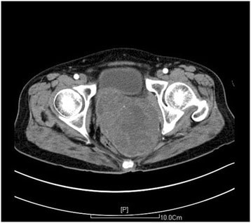
CT scan of the pelvis showing a mass in the posterior vaginal wall (approximately 11.2 x 9.0 cm): The mass was diagnosed as a vaginal leiomyosarcoma on histology.
Image: “A large primary retroperitoneal vaginal leiomyosarcoma: a case report.” by Xu Z, Zeng R, Liu J. License: CC BY 4.0
TNM stagingStagingMethods which attempt to express in replicable terms the extent of the neoplasm in the patient.Grading, Staging, and Metastasis takes into account:
Clinical stagingStagingMethods which attempt to express in replicable terms the extent of the neoplasm in the patient.Grading, Staging, and Metastasis uses findings from:
Physical exam
Cystoscopy and proctoscopy
Chest and skeletal radiography
Surgical stagingStagingMethods which attempt to express in replicable terms the extent of the neoplasm in the patient.Grading, Staging, and Metastasis includes information obtained from:
Individuals are staged based on their “highest” findings. For example, tumorTumorInflammation in an individual with a tumorTumorInflammation confined to the vaginaVaginaThe vagina is the female genital canal, extending from the vulva externally to the cervix uteri internally. The structures have sexual, reproductive, and urinary functions and a rich blood supply, mainly arising from the internal iliac artery.Vagina, Vulva, and Pelvic Floor: Anatomy but with positive lymph nodesLymph NodesThey are oval or bean shaped bodies (1 – 30 mm in diameter) located along the lymphatic system.Lymphatic Drainage System: Anatomy is classified as stage III. Similarly, direct tumorTumorInflammation invasion into the bladderBladderA musculomembranous sac along the urinary tract. Urine flows from the kidneys into the bladder via the ureters, and is held there until urination.Pyelonephritis and Perinephric Abscess mucosa is stage IV even if there is no lymphLymphThe interstitial fluid that is in the lymphatic system.Secondary Lymphatic Organs node involvement or distant metastasisMetastasisThe transfer of a neoplasm from one organ or part of the body to another remote from the primary site.Grading, Staging, and Metastasis.
Table: Vaginal cancer stagingStagingMethods which attempt to express in replicable terms the extent of the neoplasm in the patient.Grading, Staging, and Metastasis
TumorTumorInflammation is confined to the vaginaVaginaThe vagina is the female genital canal, extending from the vulva externally to the cervix uteri internally. The structures have sexual, reproductive, and urinary functions and a rich blood supply, mainly arising from the internal iliac artery.Vagina, Vulva, and Pelvic Floor: Anatomy.
None
II
TumorTumorInflammation invades into paravaginal tissue, but does not extend all the way to the pelvic sidewall.
TumorTumorInflammation causes hydronephrosisHydronephrosisHydronephrosis is dilation of the renal collecting system as a result of the obstruction of urine outflow. Hydronephrosis can be unilateral or bilateral. Nephrolithiasis is the most common cause of hydronephrosis in young adults, while prostatic hyperplasia and neoplasm are seen in older patients. Hydronephrosis or a nonfunctioning kidney owing to compressionCompressionBlunt Chest Trauma.
Direct tumorTumorInflammation invasion into the mucosa of the bladderBladderA musculomembranous sac along the urinary tract. Urine flows from the kidneys into the bladder via the ureters, and is held there until urination.Pyelonephritis and Perinephric Abscess or rectumRectumThe rectum and anal canal are the most terminal parts of the lower GI tract/large intestine that form a functional unit and control defecation. Fecal continence is maintained by several important anatomic structures including rectal folds, anal valves, the sling-like puborectalis muscle, and internal and external anal sphincters. Rectum and Anal Canal: Anatomy
Direct tumorTumorInflammation invasion outside the true pelvisPelvisThe pelvis consists of the bony pelvic girdle, the muscular and ligamentous pelvic floor, and the pelvic cavity, which contains viscera, vessels, and multiple nerves and muscles. The pelvic girdle, composed of 2 “hip” bones and the sacrum, is a ring-like bony structure of the axial skeleton that links the vertebral column with the lower extremities.Pelvis: Anatomy
MetastasisMetastasisThe transfer of a neoplasm from one organ or part of the body to another remote from the primary site.Grading, Staging, and Metastasis to distant structures
Management and Prognosis
Management
Vaginal cancer is rare, so evidence on optimal treatment approaches is lacking. Treatment recommendations are generally adopted from similar cervical and anal cancers (which are more common).
Treatment is individualized based on tumorTumorInflammation location, size, and clinical stage.
Stage I disease is typically treated with:
Surgical excision +/– radiationRadiationEmission or propagation of acoustic waves (sound), electromagnetic energy waves (such as light; radio waves; gamma rays; or x-rays), or a stream of subatomic particles (such as electrons; neutrons; protons; or alpha particles).Osteosarcoma therapy
RadiationRadiationEmission or propagation of acoustic waves (sound), electromagnetic energy waves (such as light; radio waves; gamma rays; or x-rays), or a stream of subatomic particles (such as electrons; neutrons; protons; or alpha particles).Osteosarcoma therapy alone
Stages II–IV are typically treated with radiationRadiationEmission or propagation of acoustic waves (sound), electromagnetic energy waves (such as light; radio waves; gamma rays; or x-rays), or a stream of subatomic particles (such as electrons; neutrons; protons; or alpha particles).Osteosarcoma + chemotherapyChemotherapyOsteosarcoma:
FluorouracilFluorouracilA pyrimidine analog that is an antineoplastic antimetabolite. It interferes with DNA synthesis by blocking the thymidylate synthetase conversion of deoxyuridylic acid to thymidylic acid.Bowen Disease and Erythroplasia of Queyrat
CisplatinCisplatinAn inorganic and water-soluble platinum complex. After undergoing hydrolysis, it reacts with DNA to produce both intra and interstrand crosslinks. These cross links appear to impair replication and transcription of DNA. The cytotoxicity of cisplatin correlates with cellular arrest in the g2 phase of the cell cycle.Alkylating Agents and Platinum
RadiationRadiationEmission or propagation of acoustic waves (sound), electromagnetic energy waves (such as light; radio waves; gamma rays; or x-rays), or a stream of subatomic particles (such as electrons; neutrons; protons; or alpha particles).Osteosarcoma therapy typically involves a combination of:
Brachytherapy
External-beam radiationRadiationEmission or propagation of acoustic waves (sound), electromagnetic energy waves (such as light; radio waves; gamma rays; or x-rays), or a stream of subatomic particles (such as electrons; neutrons; protons; or alpha particles).Osteosarcoma therapy
ColposcopyColposcopyThe examination, therapy or surgery of the cervix and vagina by means of a specially designed endoscope introduced vaginally.Cervical Cancer Screening and biopsyBiopsyRemoval and pathologic examination of specimens from the living body.Ewing Sarcoma if abnormalities are detected.
Surgical management in vaginal cancer
Surgery is associated with worse outcomes in SCC stages II–IV → generally avoided
Owing to local invasion, typically involves removal of adjacent structures, including bladderBladderA musculomembranous sac along the urinary tract. Urine flows from the kidneys into the bladder via the ureters, and is held there until urination.Pyelonephritis and Perinephric Abscess and bowel
Complication rates can be as high as 50% in total pelvic exenteration procedures.
MelanomaMelanomaMelanoma is a malignant tumor arising from melanocytes, the melanin-producing cells of the epidermis. These tumors are most common in fair-skinned individuals with a history of excessive sun exposure and sunburns. Melanoma
Palliative indications in advanced disease
Fertility preservation: surgical transposition of the ovariesOvariesOvaries are the paired gonads of the female reproductive system that contain haploid gametes known as oocytes. The ovaries are located intraperitoneally in the pelvis, just posterior to the broad ligament, and are connected to the pelvic sidewall and to the uterus by ligaments. These organs function to secrete hormones (estrogen and progesterone) and to produce the female germ cells (oocytes).Ovaries: Anatomy out of the pelvisPelvisThe pelvis consists of the bony pelvic girdle, the muscular and ligamentous pelvic floor, and the pelvic cavity, which contains viscera, vessels, and multiple nerves and muscles. The pelvic girdle, composed of 2 “hip” bones and the sacrum, is a ring-like bony structure of the axial skeleton that links the vertebral column with the lower extremities.Pelvis: Anatomy in young women prior to initiating radiationRadiationEmission or propagation of acoustic waves (sound), electromagnetic energy waves (such as light; radio waves; gamma rays; or x-rays), or a stream of subatomic particles (such as electrons; neutrons; protons; or alpha particles).Osteosarcoma therapy
PrognosisPrognosisA prediction of the probable outcome of a disease based on a individual’s condition and the usual course of the disease as seen in similar situations.Non-Hodgkin Lymphomas
The presenting symptom in vaginal cancer is typically postcoital or postemenopausal bleeding. The differential diagnosis for these presenting symptoms includes:
Cervical cancerCervical cancerCervical cancer, or invasive cervical carcinoma (ICC), is the 3rd most common cancer in women in the world, with > 50% of the cases being fatal. In the United States, ICC is the 13th most common cancer and the cause of < 3% of all cancer deaths due to the slow progression of precursor lesions and, more importantly, effective cancer screening. Cervical Cancer: invasive cancer of the cervixCervixThe uterus, cervix, and fallopian tubes are part of the internal female reproductive system. The most inferior portion of the uterus is the cervix, which connects the uterine cavity to the vagina. Externally, the cervix is lined by stratified squamous cells; however, the cervical canal is lined by columnar epithelium.Uterus, Cervix, and Fallopian Tubes: Anatomy (and the most common gynecologic cancer worldwide). There are 2 major histologic types of cervical cancerCervical cancerCervical cancer, or invasive cervical carcinoma (ICC), is the 3rd most common cancer in women in the world, with > 50% of the cases being fatal. In the United States, ICC is the 13th most common cancer and the cause of < 3% of all cancer deaths due to the slow progression of precursor lesions and, more importantly, effective cancer screening. Cervical Cancer: SCC and adenocarcinoma, the vast majority of which are caused by high-risk HPVHPVHuman papillomavirus (HPV) is a nonenveloped, circular, double-stranded DNA virus belonging to the Papillomaviridae family. Humans are the only reservoir, and transmission occurs through close skin-to-skin or sexual contact. Human papillomaviruses infect basal epithelial cells and can affect cell-regulatory proteins to result in cell proliferation. Papillomavirus (HPV)infectionsInfectionsInvasion of the host organism by microorganisms or their toxins or by parasites that can cause pathological conditions or diseases.Chronic Granulomatous Disease. Early cervical neoplasia is asymptomatic, though more advanced disease may present with abnormal bleeding (especially bleeding on contact). Diagnosis is made by Pap testing with cytology, HPV testingHPV testingCervical Cancer Screening, and biopsyBiopsyRemoval and pathologic examination of specimens from the living body.Ewing Sarcoma.
Endometrial cancerEndometrial CancerEndometrial carcinoma (EC) is the most common gynecologic malignancy in the developed world, and it has several histologic types. Endometrioid carcinoma (known as type 1 EC) typically develops from atypical endometrial hyperplasia, is hormonally responsive, and carries a favorable prognosis.Endometrial Hyperplasia and Endometrial Cancer: cancer of the inner lining of the uterusUterusThe uterus, cervix, and fallopian tubes are part of the internal female reproductive system. The uterus has a thick wall made of smooth muscle (the myometrium) and an inner mucosal layer (the endometrium). The most inferior portion of the uterus is the cervix, which connects the uterine cavity to the vagina.Uterus, Cervix, and Fallopian Tubes: Anatomy (and the most common gynecologic cancer in the United States). Anything that increases estrogenEstrogenCompounds that interact with estrogen receptors in target tissues to bring about the effects similar to those of estradiol. Estrogens stimulate the female reproductive organs, and the development of secondary female sex characteristics. Estrogenic chemicals include natural, synthetic, steroidal, or non-steroidal compounds.Ovaries: Anatomy exposure will increase the risk for endometrial cancerEndometrial CancerEndometrial carcinoma (EC) is the most common gynecologic malignancy in the developed world, and it has several histologic types. Endometrioid carcinoma (known as type 1 EC) typically develops from atypical endometrial hyperplasia, is hormonally responsive, and carries a favorable prognosis.Endometrial Hyperplasia and Endometrial Cancer; these risks include obesityObesityObesity is a condition associated with excess body weight, specifically with the deposition of excessive adipose tissue. Obesity is considered a global epidemic. Major influences come from the western diet and sedentary lifestyles, but the exact mechanisms likely include a mixture of genetic and environmental factors. Obesity, chronic anovulationAnovulationSuspension or cessation of ovulation in animals or humans with follicle-containing ovaries (ovarian follicle). Depending on the etiology, ovulation may be induced with appropriate therapy.Polycystic Ovarian Syndrome in reproductive-aged women, hormone replacement therapyHormone Replacement TherapyHormone replacement therapy (HRT) is used to treat symptoms associated with female menopause and in combination to suppress ovulation. Risks and side effects include uterine bleeding, predisposition to cancer, breast tenderness, hyperpigmentation, migraine headaches, hypertension, bloating, and mood changes.Noncontraceptive Estrogen and Progestins, and tamoxifenTamoxifenOne of the selective estrogen receptor modulators with tissue-specific activities. Tamoxifen acts as an anti-estrogen (inhibiting agent) in the mammary tissue, but as an estrogen (stimulating agent) in cholesterol metabolism, bone density, and cell proliferation in the endometrium.Antiestrogens use. Endometrial cancerEndometrial CancerEndometrial carcinoma (EC) is the most common gynecologic malignancy in the developed world, and it has several histologic types. Endometrioid carcinoma (known as type 1 EC) typically develops from atypical endometrial hyperplasia, is hormonally responsive, and carries a favorable prognosis.Endometrial Hyperplasia and Endometrial Cancer is diagnosed with an endometrial biopsyEndometrial BiopsyDiagnostic Procedures in Gynecology; ultrasonography may show a thickened endometrial lining in postmenopausal women. Management is primarily surgical.
Endometrial atrophyAtrophyDecrease in the size of a cell, tissue, organ, or multiple organs, associated with a variety of pathological conditions such as abnormal cellular changes, ischemia, malnutrition, or hormonal changes.Cellular Adaptation: benignBenignFibroadenoma condition in which the endometrial lining becomes thin and atrophic because of prolonged states of low estrogenEstrogenCompounds that interact with estrogen receptors in target tissues to bring about the effects similar to those of estradiol. Estrogens stimulate the female reproductive organs, and the development of secondary female sex characteristics. Estrogenic chemicals include natural, synthetic, steroidal, or non-steroidal compounds.Ovaries: Anatomy. With little to no fluid in the cavity, friction may lead to micro-erosions and a subsequent inflammatory reaction that typically presents with postmenopausal light bleeding or spotting. Endometrial atrophyAtrophyDecrease in the size of a cell, tissue, organ, or multiple organs, associated with a variety of pathological conditions such as abnormal cellular changes, ischemia, malnutrition, or hormonal changes.Cellular Adaptation is diagnosed on ultrasonography (which shows a thin endometrial lining) in the setting of a negative endometrial biopsyEndometrial BiopsyDiagnostic Procedures in Gynecology. No treatment is required.
Endometrial or cervical polyps: pedunculated or sessile projections of the endometriumEndometriumThe mucous membrane lining of the uterine cavity that is hormonally responsive during the menstrual cycle and pregnancy. The endometrium undergoes cyclic changes that characterize menstruation. After successful fertilization, it serves to sustain the developing embryo.Embryoblast and Trophoblast Development that result from overgrowth of endometrial glands and stroma around a central vascular stalk. Although these polyps are usually benignBenignFibroadenoma, they can be malignant, particularly in postmenopausal women. Endometrial or cervical polyps present with abnormal uterine or postmenopausal bleeding, though many are asymptomatic. Endometrial polypsEndometrial polypsEndometrial polyps are pedunculated or sessile projections of the endometrium that result from overgrowth of endometrial glands and stroma around a central vascular stalk. Endometrial polyps are a few millimeters to a few centimeters in size, can occur anywhere within the uterine cavity, and, while usually benign, can be malignant, particularly in postmenopausal women. Endometrial Polyps are best diagnosed with saline-infusion sonographySaline-Infusion SonographyCongenital Malformations of the Female Reproductive System (SISSISInfertility) and are usually treated with hysteroscopic resection.
Leiomyomas (uterine fibroidsUterine FibroidsGynecological Imaging): common, benignBenignFibroadenoma tumors arising from smooth muscle cells in the uterine myometrium. Leiomyomas typically present with abnormal bleeding, pelvic painPainAn unpleasant sensation induced by noxious stimuli which are detected by nerve endings of nociceptive neurons.Pain: Types and Pathways, and/or bulk symptoms. FibroidsFibroidsA benign tumor derived from smooth muscle tissue, also known as a fibroid tumor. They rarely occur outside of the uterus and the gastrointestinal tract but can occur in the skin and subcutaneous tissue, probably arising from the smooth muscle of small blood vessels in these tissues.Infertility are identified as a hypoechoicHypoechoicA structure that produces a low-amplitude echo (darker grays)Ultrasound (Sonography), well-circumscribed, round massMassThree-dimensional lesion that occupies a space within the breastImaging of the Breast on pelvic ultrasonography. Leiomyomas of the vaginal wall are also possible, though extremely rare.
AdenomyosisAdenomyosisAdenomyosis is a benign uterine condition characterized by the presence of ectopic endometrial glands and stroma within the myometrium. Adenomyosis is a common condition, affecting 20%-35% of women, and typically presents with heavy menstrual bleeding and dysmenorrhea. Adenomyosis: very common benignBenignFibroadenoma uterine condition characterized by the presence of ectopic endometrial glands and stroma within the myometrium. AdenomyosisAdenomyosisAdenomyosis is a benign uterine condition characterized by the presence of ectopic endometrial glands and stroma within the myometrium. Adenomyosis is a common condition, affecting 20%-35% of women, and typically presents with heavy menstrual bleeding and dysmenorrhea. Adenomyosis typically presents with heavy menstrual bleedingHeavy menstrual bleedingExcessive menstrual blood loss (objectively defined as > 80 mL blood loss/cycle). Can be based on heavy flow, as determined by the patientAbnormal Uterine Bleeding and dysmenorrhea. Diagnosis is either clinical or assisted with pelvic imaging, usually transvaginal ultrasonography or, occasionally, MRI. Management is based on the woman’s preference regarding future childbearing and may include hysterectomy, other surgical options, or medical hormonal suppressionSuppressionDefense Mechanisms with progestinsProgestinsCompounds that interact with progesterone receptors in target tissues to bring about the effects similar to those of progesterone. Primary actions of progestins, including natural and synthetic steroids, are on the uterus and the mammary gland in preparation for and in maintenance of pregnancy.Hormonal Contraceptives.
VulvovaginitisVulvovaginitisThe term vulvovaginitis is used to describe an acute inflammation of the vulva and vagina. Vulvovaginitis can be caused by several infectious and non-infectious etiologies, and results from disruption of the normal vaginal environment. Common signs and symptoms include pain, pruritus, erythema, edema, vaginal discharge and dyspareunia. Vulvovaginitis:acute inflammationAcute InflammationInflammation of the vulvaVulvaThe vulva is the external genitalia of the female and includes the mons pubis, labia majora, labia minora, clitoris, vestibule, vestibular bulb, and greater vestibular glands. Vagina, Vulva, and Pelvic Floor: Anatomy and vaginaVaginaThe vagina is the female genital canal, extending from the vulva externally to the cervix uteri internally. The structures have sexual, reproductive, and urinary functions and a rich blood supply, mainly arising from the internal iliac artery.Vagina, Vulva, and Pelvic Floor: Anatomy, most commonly due to Candida albicansCandida albicansA unicellular budding fungus which is the principal pathogenic species causing candidiasis (moniliasis).Candida/Candidiasis, bacterial vaginosisBacterial vaginosisPolymicrobial, nonspecific vaginitis associated with positive cultures of gardnerella vaginalis and other anaerobic organisms and a decrease in lactobacilli. It remains unclear whether the initial pathogenic event is caused by the growth of anaerobes or a primary decrease in lactobacilli.Vulvovaginitis, and TrichomonasTrichomonasA genus of parasitic flagellate eukaryotes distinguished by the presence of four anterior flagella, an undulating membrane, and a trailing flagellum.Nitroimidazoles vaginalisinfectionsInfectionsInvasion of the host organism by microorganisms or their toxins or by parasites that can cause pathological conditions or diseases.Chronic Granulomatous Disease. Noninfectious causes include atrophic vaginitisAtrophic vaginitisInflammation of the vagina due to thinning of the vaginal wall and decreased lubrication associated with reduced estrogen levels at menopause.Vulvovaginitis and contact dermatitisContact dermatitisA type of acute or chronic skin reaction in which sensitivity is manifested by reactivity to materials or substances coming in contact with the skin. It may involve allergic or non-allergic mechanisms.Male Genitourinary Examination. Common signs and symptoms include abnormal discharge, painPainAn unpleasant sensation induced by noxious stimuli which are detected by nerve endings of nociceptive neurons.Pain: Types and Pathways/dyspareuniaDyspareuniaRecurrent genital pain occurring during, before, or after sexual intercourse in either the male or the female.Primary Ovarian Insufficiency, pruritusPruritusAn intense itching sensation that produces the urge to rub or scratch the skin to obtain relief.Atopic Dermatitis (Eczema), erythemaErythemaRedness of the skin produced by congestion of the capillaries. This condition may result from a variety of disease processes.Chalazion, and edemaEdemaEdema is a condition in which excess serous fluid accumulates in the body cavity or interstitial space of connective tissues. Edema is a symptom observed in several medical conditions. It can be categorized into 2 types, namely, peripheral (in the extremities) and internal (in an organ or body cavity). Edema of the affected region. Management depends on the etiology.
CervicitisCervicitisInflammation of the uterine cervix.Gonorrhea: inflammationInflammationInflammation is a complex set of responses to infection and injury involving leukocytes as the principal cellular mediators in the body’s defense against pathogenic organisms. Inflammation is also seen as a response to tissue injury in the process of wound healing. The 5 cardinal signs of inflammation are pain, heat, redness, swelling, and loss of function. Inflammation of the cervixCervixThe uterus, cervix, and fallopian tubes are part of the internal female reproductive system. The most inferior portion of the uterus is the cervix, which connects the uterine cavity to the vagina. Externally, the cervix is lined by stratified squamous cells; however, the cervical canal is lined by columnar epithelium.Uterus, Cervix, and Fallopian Tubes: Anatomy, most commonly due to infectionsInfectionsInvasion of the host organism by microorganisms or their toxins or by parasites that can cause pathological conditions or diseases.Chronic Granulomatous Disease with Chlamydia trachomatisChlamydia trachomatisType species of Chlamydia causing a variety of ocular and urogenital diseases.Chlamydia and/or Neisseria gonorrhoeaeNeisseria gonorrhoeaeA species of gram-negative, aerobic bacteria primarily found in purulent venereal discharges. It is the causative agent of gonorrhea.Neisseria. Individuals are often asymptomatic, but they may present with a purulent abnormal discharge, pelvic painPainAn unpleasant sensation induced by noxious stimuli which are detected by nerve endings of nociceptive neurons.Pain: Types and Pathways, and irregular bleeding (especially contact bleeding). Diagnosis is with a nucleic acid amplificationNucleic acid amplificationLaboratory techniques that involve the in-vitro synthesis of many copies of DNA or RNA from one original template.Septic Arthritis test (NAAT) and management is with antibiotics.
Vaginal massMassThree-dimensional lesion that occupies a space within the breastImaging of the Breast
Vaginal inclusion or epidermal cystsCystsAny fluid-filled closed cavity or sac that is lined by an epithelium. Cysts can be of normal, abnormal, non-neoplastic, or neoplastic tissues.Fibrocystic Change:benignBenignFibroadenoma, small (approximately 1 cm) white or yellow cystsCystsAny fluid-filled closed cavity or sac that is lined by an epithelium. Cysts can be of normal, abnormal, non-neoplastic, or neoplastic tissues.Fibrocystic Change that can be located in the vaginaVaginaThe vagina is the female genital canal, extending from the vulva externally to the cervix uteri internally. The structures have sexual, reproductive, and urinary functions and a rich blood supply, mainly arising from the internal iliac artery.Vagina, Vulva, and Pelvic Floor: Anatomy or on the vulvaVulvaThe vulva is the external genitalia of the female and includes the mons pubis, labia majora, labia minora, clitoris, vestibule, vestibular bulb, and greater vestibular glands. Vagina, Vulva, and Pelvic Floor: Anatomy. Inclusion cystsCystsAny fluid-filled closed cavity or sac that is lined by an epithelium. Cysts can be of normal, abnormal, non-neoplastic, or neoplastic tissues.Fibrocystic Change occur when epithelial tissue becomes trapped under the surface after trauma. Epidermal cystsCystsAny fluid-filled closed cavity or sac that is lined by an epithelium. Cysts can be of normal, abnormal, non-neoplastic, or neoplastic tissues.Fibrocystic Change occur when sebaceous glandSebaceous GlandSmall, sacculated organs found within the dermis. Each gland has a single duct that emerges from a cluster of oval alveoli. Each alveolus consists of a transparent basement membrane enclosing epithelial cells. The ducts from most sebaceous glands open into a hair follicle, but some open on the general surface of the skin. Sebaceous glands secrete sebum.Hordeolum (Stye) ducts become obstructed, causing secretions to accumulate under the skinSkinThe skin, also referred to as the integumentary system, is the largest organ of the body. The skin is primarily composed of the epidermis (outer layer) and dermis (deep layer). The epidermis is primarily composed of keratinocytes that undergo rapid turnover, while the dermis contains dense layers of connective tissue.Skin: Structure and Functions. These cystsCystsAny fluid-filled closed cavity or sac that is lined by an epithelium. Cysts can be of normal, abnormal, non-neoplastic, or neoplastic tissues.Fibrocystic Change are usually asymptomatic, but they may cause dyspareuniaDyspareuniaRecurrent genital pain occurring during, before, or after sexual intercourse in either the male or the female.Primary Ovarian Insufficiency if they enlarge or become infected.
Gartner duct cystsCystsAny fluid-filled closed cavity or sac that is lined by an epithelium. Cysts can be of normal, abnormal, non-neoplastic, or neoplastic tissues.Fibrocystic Change: Gartner ducts are the embryologic remnants of the Wolffian (mesonephric) ducts, which typically regress in females in utero. If Gartner ducts persist and fill with fluid, they can become cystsCystsAny fluid-filled closed cavity or sac that is lined by an epithelium. Cysts can be of normal, abnormal, non-neoplastic, or neoplastic tissues.Fibrocystic Change (usually < 2 cm) on the anterolateral wall of the upper vaginaVaginaThe vagina is the female genital canal, extending from the vulva externally to the cervix uteri internally. The structures have sexual, reproductive, and urinary functions and a rich blood supply, mainly arising from the internal iliac artery.Vagina, Vulva, and Pelvic Floor: Anatomy. These cystsCystsAny fluid-filled closed cavity or sac that is lined by an epithelium. Cysts can be of normal, abnormal, non-neoplastic, or neoplastic tissues.Fibrocystic Change are typically asymptomatic and discovered as incidental findings on gynecologic exams or imaging studies. If symptoms are present, they most commonly include dyspareuniaDyspareuniaRecurrent genital pain occurring during, before, or after sexual intercourse in either the male or the female.Primary Ovarian Insufficiency and voiding disturbances.
Urethral diverticulumDiverticulumA pouch or sac opening from the colon.Diverticular Disease: focal outpouchings of the urethraUrethraA tube that transports urine from the urinary bladder to the outside of the body in both the sexes. It also has a reproductive function in the male by providing a passage for sperm.Urinary Tract: Anatomy that present as a vaginal massMassThree-dimensional lesion that occupies a space within the breastImaging of the Breast on the anterior wall of the lower vaginaVaginaThe vagina is the female genital canal, extending from the vulva externally to the cervix uteri internally. The structures have sexual, reproductive, and urinary functions and a rich blood supply, mainly arising from the internal iliac artery.Vagina, Vulva, and Pelvic Floor: Anatomy. Individuals will typically present with dysuriaDysuriaPainful urination. It is often associated with infections of the lower urinary tract.Urinary Tract Infections (UTIs), postvoid dribbling, dyspareuniaDyspareuniaRecurrent genital pain occurring during, before, or after sexual intercourse in either the male or the female.Primary Ovarian Insufficiency, recurrent urinary tractUrinary tractThe urinary tract is located in the abdomen and pelvis and consists of the kidneys, ureters, urinary bladder, and urethra. The structures permit the excretion of urine from the body. Urine flows from the kidneys through the ureters to the urinary bladder and out through the urethra.Urinary Tract: AnatomyinfectionsInfectionsInvasion of the host organism by microorganisms or their toxins or by parasites that can cause pathological conditions or diseases.Chronic Granulomatous Disease, and/or hematuriaHematuriaPresence of blood in the urine.Renal Cell Carcinoma. PalpationPalpationApplication of fingers with light pressure to the surface of the body to determine consistency of parts beneath in physical diagnosis; includes palpation for determining the outlines of organs.Dermatologic Examination of the massMassThree-dimensional lesion that occupies a space within the breastImaging of the Breast may cause leakage of urine. Diagnosed clinically and with cystoscopy.
Vaginal endometriosisEndometriosisEndometriosis is a common disease in which patients have endometrial tissue implanted outside of the uterus. Endometrial implants can occur anywhere in the pelvis, including the ovaries, the broad and uterosacral ligaments, the pelvic peritoneum, and the urinary and gastrointestinal tracts.Endometriosis:EndometriosisEndometriosisEndometriosis is a common disease in which patients have endometrial tissue implanted outside of the uterus. Endometrial implants can occur anywhere in the pelvis, including the ovaries, the broad and uterosacral ligaments, the pelvic peritoneum, and the urinary and gastrointestinal tracts.Endometriosis is the ectopic implantationImplantationEndometrial implantation of embryo, mammalian at the blastocyst stage.Fertilization and First Week of endometrial tissueEndometrial tissueThe mucous membrane lining of the uterine cavity that is hormonally responsive during the menstrual cycle and pregnancy. The endometrium undergoes cyclic changes that characterize menstruation. After successful fertilization, it serves to sustain the developing embryo.Endometriosis outside the uterine cavity. Although rare, it is possible for endometrial tissueEndometrial tissueThe mucous membrane lining of the uterine cavity that is hormonally responsive during the menstrual cycle and pregnancy. The endometrium undergoes cyclic changes that characterize menstruation. After successful fertilization, it serves to sustain the developing embryo.Endometriosis to implant in the vaginaVaginaThe vagina is the female genital canal, extending from the vulva externally to the cervix uteri internally. The structures have sexual, reproductive, and urinary functions and a rich blood supply, mainly arising from the internal iliac artery.Vagina, Vulva, and Pelvic Floor: Anatomy. The implant may present as a small blue, black, brown, or white lesion or as a larger cyst filled with dark fluid (known as a “chocolate cyst”). Other symptoms may include dyspareuniaDyspareuniaRecurrent genital pain occurring during, before, or after sexual intercourse in either the male or the female.Primary Ovarian Insufficiency, dysmenorrhea, abnormal bleeding, and urinary/defecatory symptoms.