Aortic dissection Aortic dissection Aortic dissection occurs due to shearing stress from pulsatile pressure causing a tear in the tunica intima of the aortic wall. This tear allows blood to flow into the media, creating a "false lumen." Aortic dissection is most commonly caused by uncontrolled hypertension. Aortic Dissection is an emergency that occurs due to shearing stress from pulsatile pressure causing a tear in the tunica intima Tunica intima The innermost layer of an artery or vein, made up of one layer of endothelial cells and supported by an internal elastic lamina. Arteries: Histology of the aortic wall. This tear, often associated with uncontrolled hypertension Uncontrolled hypertension Although hypertension is defined as a blood pressure of > 130/80 mm Hg, individuals can present with comorbidities of severe asymptomatic or "uncontrolled" hypertension (≥ 180 mm Hg systolic and/or ≥ 120 mm Hg diastolic) that carries with it a significant risk of morbidity and mortality. Uncontrolled Hypertension, allows blood to flow Flow Blood flows through the heart, arteries, capillaries, and veins in a closed, continuous circuit. Flow is the movement of volume per unit of time. Flow is affected by the pressure gradient and the resistance fluid encounters between 2 points. Vascular resistance is the opposition to flow, which is caused primarily by blood friction against vessel walls. Vascular Resistance, Flow, and Mean Arterial Pressure into the medial layer, thus creating a false lumen False lumen Aortic Dissection.[1] Patients Patients Individuals participating in the health care system for the purpose of receiving therapeutic, diagnostic, or preventive procedures. Clinician–Patient Relationship often present with acute, severe chest or back pain Pain An unpleasant sensation induced by noxious stimuli which are detected by nerve endings of nociceptive neurons. Pain: Types and Pathways, described as “tearing.” Complications arise due to partial occlusion of vital branches of the aorta Aorta The main trunk of the systemic arteries. Mediastinum and Great Vessels: Anatomy with reduced blood flow Blood flow Blood flow refers to the movement of a certain volume of blood through the vasculature over a given unit of time (e.g., mL per minute). Vascular Resistance, Flow, and Mean Arterial Pressure to the brain Brain The part of central nervous system that is contained within the skull (cranium). Arising from the neural tube, the embryonic brain is comprised of three major parts including prosencephalon (the forebrain); mesencephalon (the midbrain); and rhombencephalon (the hindbrain). The developed brain consists of cerebrum; cerebellum; and other structures in the brain stem. Nervous System: Anatomy, Structure, and Classification, visceral organs, and extremities. Computed tomography is the diagnostic imaging modality of choice. All type A or proximal dissections ( ascending aorta Ascending aorta Mediastinum and Great Vessels: Anatomy) are surgical emergencies due to the risk of imminent rupture. Type B or distal dissections ( descending aorta Descending aorta Mediastinum and Great Vessels: Anatomy) can be managed medically with beta-blockers Beta-blockers Drugs that bind to but do not activate beta-adrenergic receptors thereby blocking the actions of beta-adrenergic agonists. Adrenergic beta-antagonists are used for treatment of hypertension, cardiac arrhythmias, angina pectoris, glaucoma, migraine headaches, and anxiety. Class 2 Antiarrhythmic Drugs (Beta Blockers) and calcium Calcium A basic element found in nearly all tissues. It is a member of the alkaline earth family of metals with the atomic symbol ca, atomic number 20, and atomic weight 40. Calcium is the most abundant mineral in the body and combines with phosphorus to form calcium phosphate in the bones and teeth. It is essential for the normal functioning of nerves and muscles and plays a role in blood coagulation (as factor IV) and in many enzymatic processes. Electrolytes channel blockers. If there is evidence of decreased perfusion to visceral organs or extremities, aneurysm Aneurysm An aneurysm is a bulging, weakened area of a blood vessel that causes an abnormal widening of its diameter > 1.5 times the size of the native vessel. Aneurysms occur more often in arteries than in veins and are at risk of dissection and rupture, which can be life-threatening. Thoracic Aortic Aneurysms dilation > 5 cm, retrograde extension Extension Examination of the Upper Limbs into the ascending aorta Ascending aorta Mediastinum and Great Vessels: Anatomy, or intractable pain Pain An unpleasant sensation induced by noxious stimuli which are detected by nerve endings of nociceptive neurons. Pain: Types and Pathways, then evaluation for endovascular or open repair is required.[2, 3] For further review of this topic, including links to lectures by specialists in the field, follow this link: https://www.lecturio.com/concepts/aortic-dissection/
Last updated: Dec 15, 2025
Authors: Ahmed Elsherif 1 ; Michelle Wyatt 2
Peer Reviewers: Stanley Oiseth 3 ; Joseph Alpert 4
Affiliations: 1 Suez Canal University; 2 Medical Editor at Lecturio; 3 Chief Medical Editor at Lecturio; 4 Tucson University, Arizona
This article is not intended to substitute for professional medical advice and should not be relied on as health or personal advice. Always seek the guidance of your doctor or other qualified health professional with any questions you may have regarding your health or a medical condition.
An aortic dissection Aortic dissection Aortic dissection occurs due to shearing stress from pulsatile pressure causing a tear in the tunica intima of the aortic wall. This tear allows blood to flow into the media, creating a “false lumen.” Aortic dissection is most commonly caused by uncontrolled hypertension. Aortic Dissection ( AD AD The term advance directive (AD) refers to treatment preferences and/or the designation of a surrogate decision-maker in the event that a person becomes unable to make medical decisions on their own behalf. Advance directives represent the ethical principle of autonomy and may take the form of a living will, health care proxy, durable power of attorney for health care, and/or a physician’s order for life-sustaining treatment. Advance Directives) occurs due to longitudinal cleavage of the medial layer of the vessel wall, creating a false lumen False lumen Aortic Dissection in the aorta Aorta The main trunk of the systemic arteries. Mediastinum and Great Vessels: Anatomy. It is a surgical emergency Surgical Emergency Acute Abdomen as the dissection causes reduced blood flow Blood flow Blood flow refers to the movement of a certain volume of blood through the vasculature over a given unit of time (e.g., mL per minute). Vascular Resistance, Flow, and Mean Arterial Pressure to vital organs. In severe cases, the aorta Aorta The main trunk of the systemic arteries. Mediastinum and Great Vessels: Anatomy can rupture and be fatal.
Aortic dissection Aortic dissection Aortic dissection occurs due to shearing stress from pulsatile pressure causing a tear in the tunica intima of the aortic wall. This tear allows blood to flow into the media, creating a “false lumen.” Aortic dissection is most commonly caused by uncontrolled hypertension. Aortic Dissection is classified among acute aortic syndromes (AASs), a group of severe conditions affecting the thoracic and abdominal aorta Abdominal Aorta The aorta from the diaphragm to the bifurcation into the right and left common iliac arteries. Posterior Abdominal Wall: Anatomy that often present urgently and require immediate surgical evaluation. These syndromes, which include aortic dissection Aortic dissection Aortic dissection occurs due to shearing stress from pulsatile pressure causing a tear in the tunica intima of the aortic wall. This tear allows blood to flow into the media, creating a “false lumen.” Aortic dissection is most commonly caused by uncontrolled hypertension. Aortic Dissection, intramural hematoma Intramural hematoma Dissection of the Carotid and Vertebral Arteries, penetrating atherosclerotic ulcer, and traumatic AD AD The term advance directive (AD) refers to treatment preferences and/or the designation of a surrogate decision-maker in the event that a person becomes unable to make medical decisions on their own behalf. Advance directives represent the ethical principle of autonomy and may take the form of a living will, health care proxy, durable power of attorney for health care, and/or a physician’s order for life-sustaining treatment. Advance Directives, are potentially life-threatening and typically manifest with aortic discomfort. AASs are characterized by a shared spectrum of signs and symptoms, with aortic pain Pain An unpleasant sensation induced by noxious stimuli which are detected by nerve endings of nociceptive neurons. Pain: Types and Pathways being the most notable.[26]
Aortic dissection
Aortic dissection
Aortic dissection occurs due to shearing stress from pulsatile pressure causing a tear in the tunica intima of the aortic wall. This tear allows blood to flow into the media, creating a “false lumen.” Aortic dissection is most commonly caused by uncontrolled hypertension.
Aortic Dissection (
AD
AD
The term advance directive (AD) refers to treatment preferences and/or the designation of a surrogate decision-maker in the event that a person becomes unable to make medical decisions on their own behalf. Advance directives represent the ethical principle of autonomy and may take the form of a living will, health care proxy, durable power of attorney for health care, and/or a physician’s order for life-sustaining treatment.
Advance Directives) is an infrequent occurrence, with new cases reported at 2–3.5 per 100,000 people every year. It is more common in men (65%) than women and is often associated with
hypertension
Hypertension
Hypertension, or high blood pressure, is a common disease that manifests as elevated systemic arterial pressures. Hypertension is most often asymptomatic and is found incidentally as part of a routine physical examination or during triage for an unrelated medical encounter.
Hypertension.
The age of presentation of
AD
AD
The term advance directive (AD) refers to treatment preferences and/or the designation of a surrogate decision-maker in the event that a person becomes unable to make medical decisions on their own behalf. Advance directives represent the ethical principle of autonomy and may take the form of a living will, health care proxy, durable power of attorney for health care, and/or a physician’s order for life-sustaining treatment.
Advance Directives depends on underlying risk factors. Age, male
sex
Sex
The totality of characteristics of reproductive structure, functions, phenotype, and genotype, differentiating the male from the female organism.
Gender Dysphoria, and
hypertension
Hypertension
Hypertension, or high blood pressure, is a common disease that manifests as elevated systemic arterial pressures. Hypertension is most often asymptomatic and is found incidentally as part of a routine physical examination or during triage for an unrelated medical encounter.
Hypertension confer the most significant risks in adults over 40, but genetic
connective tissue
Connective tissue
Connective tissues originate from embryonic mesenchyme and are present throughout the body except inside the brain and spinal cord. The main function of connective tissues is to provide structural support to organs. Connective tissues consist of cells and an extracellular matrix.
Connective Tissue: Histology diseases increase the risk of
AD
AD
The term advance directive (AD) refers to treatment preferences and/or the designation of a surrogate decision-maker in the event that a person becomes unable to make medical decisions on their own behalf. Advance directives represent the ethical principle of autonomy and may take the form of a living will, health care proxy, durable power of attorney for health care, and/or a physician’s order for life-sustaining treatment.
Advance Directives in younger
patients
Patients
Individuals participating in the health care system for the purpose of receiving therapeutic, diagnostic, or preventive procedures.
Clinician–Patient Relationship. It is more common in the African American population than whites, similar to
hypertension
Hypertension
Hypertension, or high blood pressure, is a common disease that manifests as elevated systemic arterial pressures. Hypertension is most often asymptomatic and is found incidentally as part of a routine physical examination or during triage for an unrelated medical encounter.
Hypertension, and Asians have the lowest
incidence
Incidence
The number of new cases of a given disease during a given period in a specified population. It also is used for the rate at which new events occur in a defined population. It is differentiated from prevalence, which refers to all cases in the population at a given time.
Measures of Disease Frequency. Dissections in younger individuals ages 30 – 40 are usually associated with genetic or
connective tissue
Connective tissue
Connective tissues originate from embryonic mesenchyme and are present throughout the body except inside the brain and spinal cord. The main function of connective tissues is to provide structural support to organs. Connective tissues consist of cells and an extracellular matrix.
Connective Tissue: Histology diseases such as
Marfan syndrome
Marfan syndrome
Marfan syndrome is a genetic condition with autosomal dominant inheritance. Marfan syndrome affects the elasticity of connective tissues throughout the body, most notably in the cardiovascular, ocular, and musculoskeletal systems.
Marfan Syndrome.[4,5]
There are several systems of classification for aortic dissection Aortic dissection Aortic dissection occurs due to shearing stress from pulsatile pressure causing a tear in the tunica intima of the aortic wall. This tear allows blood to flow into the media, creating a “false lumen.” Aortic dissection is most commonly caused by uncontrolled hypertension. Aortic Dissection based on anatomy or duration of onset of symptoms.
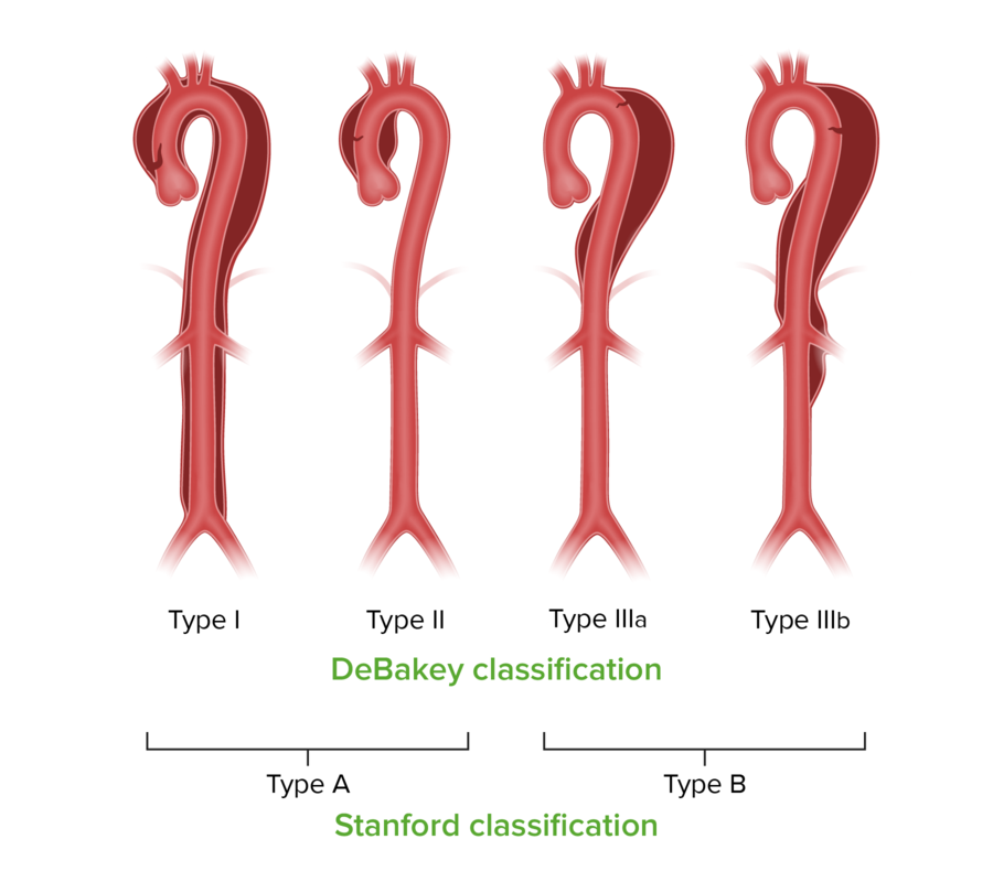
Two classifications are used for aortic dissections; the Stanford classification is used more often. Both the DeBakey classification and the Stanford classification are used to separate aortic dissections into those that need surgical repair and those that usually require only medical management. DeBakey Type I involves the ascending aorta, arch, and descending thoracic aorta and may progress to involve the abdominal aorta); DeBakey Type II is limited to the ascending aorta; DeBakey Type IIIa involves the descending thoracic aorta distal to the left subclavian artery and proximal to the celiac artery; DeBakey Type IIIb involves the thoracic and abdominal aorta distal to the left subclavian artery. In the Stanford classification, Type A involves the ascending aorta and may progress to involve the arch and thoracoabdominal aorta. Type B involves the descending thoracic or thoracoabdominal aorta distal to the left subclavian artery without the involvement of the ascending aorta. The treatment of type A dissections is surgical unless the patient would not survive the surgery. Type B dissections can usually be managed medically, but surgery or endovascular intervention may be used if there are complications or progressive symptoms. Other classifications of aortic dissections are also used to improve specificity and reporting standards [10].
Image by Lecturio.Mnemonic:
Stanford A = affects ascending aorta
Stanford B = begins beyond brachiocephalic vessels
The Stanford classification Stanford classification Aortic Dissection is the most commonly used in aortic dissection Aortic dissection Aortic dissection occurs due to shearing stress from pulsatile pressure causing a tear in the tunica intima of the aortic wall. This tear allows blood to flow into the media, creating a “false lumen.” Aortic dissection is most commonly caused by uncontrolled hypertension. Aortic Dissection.
| Type A 70%–75% | Ascending aorta Ascending aorta Mediastinum and Great Vessels: Anatomy +/- aortic arch Aortic arch Mediastinum and Great Vessels: Anatomy, possibly descending aorta Descending aorta Mediastinum and Great Vessels: Anatomy. Can involve the aortic valve Aortic valve The valve between the left ventricle and the ascending aorta which prevents backflow into the left ventricle. Heart: Anatomy. | Requires primary surgical treatment |
| Type B 25%–30% | Descending aorta Descending aorta Mediastinum and Great Vessels: Anatomy or distal to the left subclavian artery without involvement of the ascending aorta Ascending aorta Mediastinum and Great Vessels: Anatomy. May be acute, subacute (onset 14 to 90 days), or chronic (onset > 90 days). | It is generally treated conservatively by controlling blood pressure and heart rate Heart rate The number of times the heart ventricles contract per unit of time, usually per minute. Cardiac Physiology. Surgery is indicated in complicated cases only.[2] |
Note: The 301 Classification modifies the Stanford Type B Aortic Dissection Aortic dissection Aortic dissection occurs due to shearing stress from pulsatile pressure causing a tear in the tunica intima of the aortic wall. This tear allows blood to flow into the media, creating a “false lumen.” Aortic dissection is most commonly caused by uncontrolled hypertension. Aortic Dissection classification to improve prognostication for thoracic endovascular aortic repair (TEVAR). It introduces three subtypes (B1, B2, B3) based on anatomical and clinical features, which helps in better risk stratification and management, with types B2 and B3 associated with higher risks for adverse events post-TEVAR. [28]
In contrast, the DeBakey system is based on anatomy:
|
Type 1
Type 1
Spinal Muscular Atrophy 60% | Origin — ascending aorta Ascending aorta Mediastinum and Great Vessels: Anatomy extends to the aortic arch Aortic arch Mediastinum and Great Vessels: Anatomy and often beyond. Most lethal and often seen in patients Patients Individuals participating in the health care system for the purpose of receiving therapeutic, diagnostic, or preventive procedures. Clinician–Patient Relationship < age 65. |
| Type 2 30%–35% | Origin — ascending aorta Ascending aorta Mediastinum and Great Vessels: Anatomy and is confined here. |
|
Type 3
Type 3
Spinal Muscular Atrophy 10%–15% | Origin — descending aorta Descending aorta Mediastinum and Great Vessels: Anatomy — rarely goes proximally but commonly goes distally. Elderly with hypertension Hypertension Hypertension, or high blood pressure, is a common disease that manifests as elevated systemic arterial pressures. Hypertension is most often asymptomatic and is found incidentally as part of a routine physical examination or during triage for an unrelated medical encounter. Hypertension and atherosclerosis Atherosclerosis Atherosclerosis is a common form of arterial disease in which lipid deposition forms a plaque in the blood vessel walls. Atherosclerosis is an incurable disease, for which there are clearly defined risk factors that often can be reduced through a change in lifestyle and behavior of the patient. Atherosclerosis. |
In patients Patients Individuals participating in the health care system for the purpose of receiving therapeutic, diagnostic, or preventive procedures. Clinician–Patient Relationship with AD AD The term advance directive (AD) refers to treatment preferences and/or the designation of a surrogate decision-maker in the event that a person becomes unable to make medical decisions on their own behalf. Advance directives represent the ethical principle of autonomy and may take the form of a living will, health care proxy, durable power of attorney for health care, and/or a physician’s order for life-sustaining treatment. Advance Directives, blood enters the intima from the media layers. The high pressure exerted by blood tears the media apart in a laminated plane. The plane is usually between the inner 2/3rds and the outer 1/3rd. The dissection can extend proximally or distally for variable Variable Variables represent information about something that can change. The design of the measurement scales, or of the methods for obtaining information, will determine the data gathered and the characteristics of that data. As a result, a variable can be qualitative or quantitative, and may be further classified into subgroups. Types of Variables distances and establishes a connection between the media and intima through a false lumen False lumen Aortic Dissection. [13]
Most dissections originate in the ascending aorta Ascending aorta Mediastinum and Great Vessels: Anatomy, usually within 10 cm of the aortic valve Aortic valve The valve between the left ventricle and the ascending aorta which prevents backflow into the left ventricle. Heart: Anatomy. These tears are commonly 1–5 cm long and are transverse or oblique in orientation Orientation Awareness of oneself in relation to time, place and person. Psychiatric Assessment, with rough edges.
Sometimes, the dissection can spread through the intima, media, and adventitia causing external rupture. This results in huge internal bleeding or cardiac tamponade Tamponade Pericardial effusion, usually of rapid onset, exceeding ventricular filling pressures and causing collapse of the heart with a markedly reduced cardiac output. Pericarditis if the dissection extends through the adventitia but into the pericardial sac, forming a hemopericardium. Both scenarios are life-threatening and can rapidly lead to death.
When the blood enters the intima and tears through the media, it creates a false lumen False lumen Aortic Dissection. The true lumen True lumen Aortic Dissection is the natural physiological lumen of the vessel. In between both of these lumens is a layer of intima which is known as the intimal flap. As stated above, the false lumen False lumen Aortic Dissection may recanalize into the true lumen True lumen Aortic Dissection.
There are different types of aortic dissection Aortic dissection Aortic dissection occurs due to shearing stress from pulsatile pressure causing a tear in the tunica intima of the aortic wall. This tear allows blood to flow into the media, creating a “false lumen.” Aortic dissection is most commonly caused by uncontrolled hypertension. Aortic Dissection. The majority originate in the ascending aorta Ascending aorta Mediastinum and Great Vessels: Anatomy, about 10% in the aortic arch Aortic arch Mediastinum and Great Vessels: Anatomy, and 15% in the descending thoracic aorta Aorta The main trunk of the systemic arteries. Mediastinum and Great Vessels: Anatomy (distal to the ligamentum arteriosum Ligamentum arteriosum Prenatal and Postnatal Physiology of the Neonate).[12]
The reason an intimal tear occurs is unknown. It can occur due to intimal ischemia Ischemia A hypoperfusion of the blood through an organ or tissue caused by a pathologic constriction or obstruction of its blood vessels, or an absence of blood circulation. Ischemic Cell Damage from increased shear forces due to hypertension Hypertension Hypertension, or high blood pressure, is a common disease that manifests as elevated systemic arterial pressures. Hypertension is most often asymptomatic and is found incidentally as part of a routine physical examination or during triage for an unrelated medical encounter. Hypertension or genetic connective tissue Connective tissue Connective tissues originate from embryonic mesenchyme and are present throughout the body except inside the brain and spinal cord. The main function of connective tissues is to provide structural support to organs. Connective tissues consist of cells and an extracellular matrix. Connective Tissue: Histology diseases such as Marfan syndrome Marfan syndrome Marfan syndrome is a genetic condition with autosomal dominant inheritance. Marfan syndrome affects the elasticity of connective tissues throughout the body, most notably in the cardiovascular, ocular, and musculoskeletal systems. Marfan Syndrome. In patients Patients Individuals participating in the health care system for the purpose of receiving therapeutic, diagnostic, or preventive procedures. Clinician–Patient Relationship with Marfan syndrome Marfan syndrome Marfan syndrome is a genetic condition with autosomal dominant inheritance. Marfan syndrome affects the elasticity of connective tissues throughout the body, most notably in the cardiovascular, ocular, and musculoskeletal systems. Marfan Syndrome, the collagen Collagen A polypeptide substance comprising about one third of the total protein in mammalian organisms. It is the main constituent of skin; connective tissue; and the organic substance of bones (bone and bones) and teeth (tooth). Connective Tissue: Histology and elastin within the media are degenerative, unstructured, and dysfunctional—causing cystic Cystic Fibrocystic Change medial necrosis Necrosis The death of cells in an organ or tissue due to disease, injury or failure of the blood supply. Ischemic Cell Damage.
In approximately 10% of cases, there is no evidence of an intimal tear. These dissections may be caused by bleeding within the medial layer of the vessel resulting in secondary aortic dissection Aortic dissection Aortic dissection occurs due to shearing stress from pulsatile pressure causing a tear in the tunica intima of the aortic wall. This tear allows blood to flow into the media, creating a “false lumen.” Aortic dissection is most commonly caused by uncontrolled hypertension. Aortic Dissection.
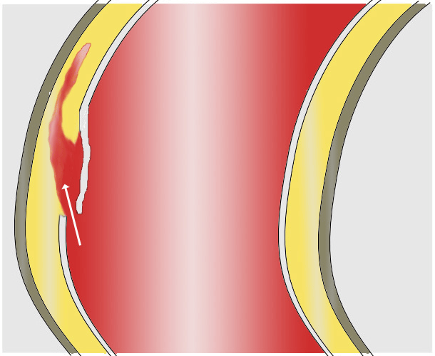
This drawing illustrates how an intimal tear leads to bleeding into the media, creating a false lumen.
Image: “aortic dissection – Aortendissektion Scheme” by J. Heuser. License: CC BY-SA 3.0The most commonly identified lesion within the aortic wall is cystic Cystic Fibrocystic Change medial degeneration, manifesting as decreased smooth muscle, necrosis Necrosis The death of cells in an organ or tissue due to disease, injury or failure of the blood supply. Ischemic Cell Damage, elastic Elastic Connective Tissue: Histology tissue fragmentation Fragmentation Chronic Apophyseal Injury, and proteoglycan-rich extracellular matrix Extracellular matrix A meshwork-like substance found within the extracellular space and in association with the basement membrane of the cell surface. It promotes cellular proliferation and provides a supporting structure to which cells or cell lysates in culture dishes adhere. Hypertrophic and Keloid Scars deposition. Cystic Cystic Fibrocystic Change medial degeneration is usually related to genetic diseases like Marfan syndrome Marfan syndrome Marfan syndrome is a genetic condition with autosomal dominant inheritance. Marfan syndrome affects the elasticity of connective tissues throughout the body, most notably in the cardiovascular, ocular, and musculoskeletal systems. Marfan Syndrome. There may be further evidence of atherosclerosis Atherosclerosis Atherosclerosis is a common form of arterial disease in which lipid deposition forms a plaque in the blood vessel walls. Atherosclerosis is an incurable disease, for which there are clearly defined risk factors that often can be reduced through a change in lifestyle and behavior of the patient. Atherosclerosis and abnormal connective tissue Connective tissue Connective tissues originate from embryonic mesenchyme and are present throughout the body except inside the brain and spinal cord. The main function of connective tissues is to provide structural support to organs. Connective tissues consist of cells and an extracellular matrix. Connective Tissue: Histology structure in genetic conditions. Inflammation Inflammation Inflammation is a complex set of responses to infection and injury involving leukocytes as the principal cellular mediators in the body’s defense against pathogenic organisms. Inflammation is also seen as a response to tissue injury in the process of wound healing. The 5 cardinal signs of inflammation are pain, heat, redness, swelling, and loss of function. Inflammation is absent. Dissection can also be spontaneous and occur when no identifiable histologic lesions are present. [11]
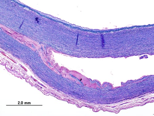
This is a histopathological photo of a dissecting thoracic aortic aneurysm in a patient without evidence of Marfan trait. The damaged aorta was surgically removed and replaced by an artificial vessel.
Image: “Aortic dissection (1) Victoria blue” by KGH. License: CC BY-SA 3.0The diagnosis of thoracic aortic dissection Aortic dissection Aortic dissection occurs due to shearing stress from pulsatile pressure causing a tear in the tunica intima of the aortic wall. This tear allows blood to flow into the media, creating a “false lumen.” Aortic dissection is most commonly caused by uncontrolled hypertension. Aortic Dissection should be considered in all patients Patients Individuals participating in the health care system for the purpose of receiving therapeutic, diagnostic, or preventive procedures. Clinician–Patient Relationship with chest pain Pain An unpleasant sensation induced by noxious stimuli which are detected by nerve endings of nociceptive neurons. Pain: Types and Pathways. This pain Pain An unpleasant sensation induced by noxious stimuli which are detected by nerve endings of nociceptive neurons. Pain: Types and Pathways usually has the following characteristics: [12,14]
Neurological symptoms are the presenting complaint in 20% of cases:
Additionally, other types of symptoms may occur with AD AD The term advance directive (AD) refers to treatment preferences and/or the designation of a surrogate decision-maker in the event that a person becomes unable to make medical decisions on their own behalf. Advance directives represent the ethical principle of autonomy and may take the form of a living will, health care proxy, durable power of attorney for health care, and/or a physician’s order for life-sustaining treatment. Advance Directives:
This list comprises the most common signs of aortic dissection Aortic dissection Aortic dissection occurs due to shearing stress from pulsatile pressure causing a tear in the tunica intima of the aortic wall. This tear allows blood to flow into the media, creating a “false lumen.” Aortic dissection is most commonly caused by uncontrolled hypertension. Aortic Dissection: [12,14]
Diagnosis of aortic dissection Aortic dissection Aortic dissection occurs due to shearing stress from pulsatile pressure causing a tear in the tunica intima of the aortic wall. This tear allows blood to flow into the media, creating a “false lumen.” Aortic dissection is most commonly caused by uncontrolled hypertension. Aortic Dissection needs to be rapid and accurate. As previously explained, the diagnosis should be suspected from the history and physical examination.
When there is a high clinical suspicion of aortic dissection Aortic dissection Aortic dissection occurs due to shearing stress from pulsatile pressure causing a tear in the tunica intima of the aortic wall. This tear allows blood to flow into the media, creating a “false lumen.” Aortic dissection is most commonly caused by uncontrolled hypertension. Aortic Dissection, imaging studies must be done emergently to confirm or exclude the diagnosis.[14] Each has its advantages and disadvantages, and selection Selection Lymphocyte activation by a specific antigen thus triggering clonal expansion of lymphocytes already capable of mounting an immune response to the antigen. B cells: Types and Functions depends on test availability and the patient’s presentation.

This chest X-ray shows mediastinal widening (line labeled as 1) and a prominent aortic knob (line labeled as 2) in a patient with a Stanford type A aortic dissection. Small pleural effusions are also noted.
Image: “Aortic dissection Xray” by J. Heuser. License: CC BY-SA 3.0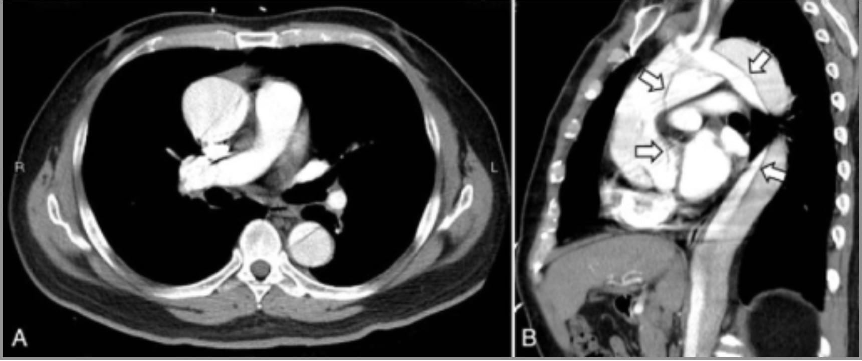
The initial presenting signs and symptoms of acute AD are diverse, and early diagnosis can be challenging. This CT is from a 61-year-old man who presented by ambulance to the emergency department with an atypical history of transient loss of consciousness and a suspected seizure. Loss of consciousness was again seen in the emergency department with an ECG monitor recording transient cardiac asystole followed by spontaneous recovery of sinus rhythm. His chest X-ray revealed a widened mediastinum, and a CT (above) demonstrated a Stanford type A aortic dissection from the aortic root. (A) Axial view; (B) sagittal view. [18] Arrows indicate the intimal flap.
Image: “CT of aortic dissection” by Medicine. License: CC BY 4.0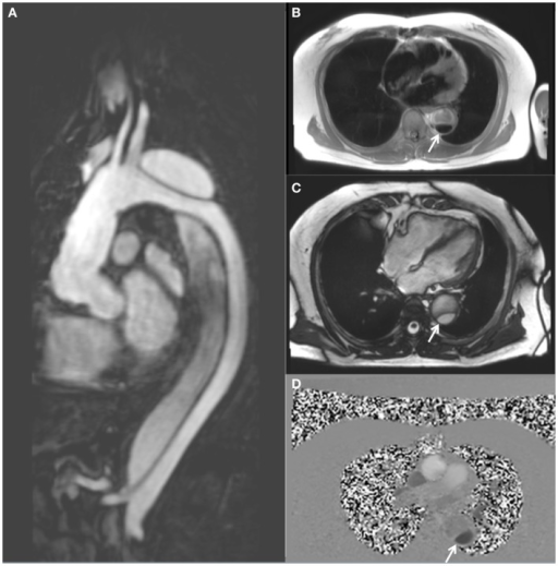
Magnetic resonance imaging demonstrating a descending thoracic aortic dissection:
A: sagittal gadolinium-contrast-enhanced MRA view
B: axial black blood view of the proximal descending thoracic aorta
C: axial true FISP (steady state-free precession) cine view
D: axial phase-contrast view, showing flow patterns in the true and false lumens of the descending aorta (the true lumen is indicated by the white arrow)
Myocarditis Myocarditis Myocarditis is an inflammatory disease of the myocardium, which may occur alone or in association with a systemic process. There are numerous etiologies of myocarditis, but all lead to inflammation and myocyte injury, most often leading to signs and symptoms of heart failure. Myocarditis, myocardial infarction Myocardial infarction MI is ischemia and death of an area of myocardial tissue due to insufficient blood flow and oxygenation, usually from thrombus formation on a ruptured atherosclerotic plaque in the epicardial arteries. Clinical presentation is most commonly with chest pain, but women and patients with diabetes may have atypical symptoms. Myocardial Infarction, aortic aneurysmal rupture Aneurysmal rupture The tearing or bursting of the weakened wall of the aneurysmal sac, usually heralded by sudden worsening pain. The great danger of a ruptured aneurysm is the large amount of blood spilling into the surrounding tissues and cavities, causing hemorrhagic shock. Thoracic Aortic Aneurysms, and mechanical chest pain Pain An unpleasant sensation induced by noxious stimuli which are detected by nerve endings of nociceptive neurons. Pain: Types and Pathways (e.g., costochondritis) are other conditions in the differential diagnosis of aortic dissection Aortic dissection Aortic dissection occurs due to shearing stress from pulsatile pressure causing a tear in the tunica intima of the aortic wall. This tear allows blood to flow into the media, creating a “false lumen.” Aortic dissection is most commonly caused by uncontrolled hypertension. Aortic Dissection.
Type A (DeBakey 1+2) acute ascending aortic dissection Ascending aortic dissection Aortic Dissection is treated emergently with open surgery, less often by endovascular stent-grafting if there are major comorbidities Comorbidities The presence of co-existing or additional diseases with reference to an initial diagnosis or with reference to the index condition that is the subject of study. Comorbidity may affect the ability of affected individuals to function and also their survival; it may be used as a prognostic indicator for length of hospital stay, cost factors, and outcome or survival. St. Louis Encephalitis Virus, and a hybrid approach has been used (surgical repair of the ascending aorta Ascending aorta Mediastinum and Great Vessels: Anatomy, and endovascular stent-graft for the descending aorta Descending aorta Mediastinum and Great Vessels: Anatomy). As soon as acute type A aortic dissection Aortic dissection Aortic dissection occurs due to shearing stress from pulsatile pressure causing a tear in the tunica intima of the aortic wall. This tear allows blood to flow into the media, creating a “false lumen.” Aortic dissection is most commonly caused by uncontrolled hypertension. Aortic Dissection is diagnosed, immediate cardiac surgical consultation is required. If experienced cardiac surgical services are not available, the patient should be promptly transferred for definitive care. [20,22,25]
Type B (DeBakey3) descending aortic dissection Descending aortic dissection Aortic Dissection is initially treated by beta-blockers Beta-blockers Drugs that bind to but do not activate beta-adrenergic receptors thereby blocking the actions of beta-adrenergic agonists. Adrenergic beta-antagonists are used for treatment of hypertension, cardiac arrhythmias, angina pectoris, glaucoma, migraine headaches, and anxiety. Class 2 Antiarrhythmic Drugs (Beta Blockers), vasodilators Vasodilators Drugs used to cause dilation of the blood vessels. Thromboangiitis Obliterans (Buerger Disease), or calcium Calcium A basic element found in nearly all tissues. It is a member of the alkaline earth family of metals with the atomic symbol ca, atomic number 20, and atomic weight 40. Calcium is the most abundant mineral in the body and combines with phosphorus to form calcium phosphate in the bones and teeth. It is essential for the normal functioning of nerves and muscles and plays a role in blood coagulation (as factor IV) and in many enzymatic processes. Electrolytes channel blockers. Surgical or thoracic endovascular repair are indicated if there are complications (occlusion of a major branch, severe hypertension Severe hypertension A confirmed blood pressure ≥ 180 mm Hg systolic and/or ≥ 120 mm Hg diastolic. Uncontrolled Hypertension, chest pain Pain An unpleasant sensation induced by noxious stimuli which are detected by nerve endings of nociceptive neurons. Pain: Types and Pathways, propagation Propagation Propagation refers to how the electrical signal spreads to every myocyte in the heart. Cardiac Physiology of the dissection aneurysmal expansion, expanding hematoma Hematoma A collection of blood outside the blood vessels. Hematoma can be localized in an organ, space, or tissue. Intussusception, or rupture). Complicated type B dissections can be treated with thoracic endovascular aortic repair, now considered the gold standard intervention in this situation. [21]
IV beta blockers are the first-line early treatment of AD AD The term advance directive (AD) refers to treatment preferences and/or the designation of a surrogate decision-maker in the event that a person becomes unable to make medical decisions on their own behalf. Advance directives represent the ethical principle of autonomy and may take the form of a living will, health care proxy, durable power of attorney for health care, and/or a physician’s order for life-sustaining treatment. Advance Directives to decrease the heart rate Heart rate The number of times the heart ventricles contract per unit of time, usually per minute. Cardiac Physiology to a goal of 60 bpm. IV labetalol Labetalol A salicylamide derivative that is a non-cardioselective blocker of beta-adrenergic receptors and alpha-1 adrenergic receptors. Subarachnoid Hemorrhage, esmolol Esmolol Antiadrenergic Drugs, and propranolol Propranolol A widely used non-cardioselective beta-adrenergic antagonist. Propranolol has been used for myocardial infarction; arrhythmia; angina pectoris; hypertension; hyperthyroidism; migraine; pheochromocytoma; and anxiety but adverse effects instigate replacement by newer drugs. Antiadrenergic Drugs are used in this setting. The next step is to control hypertension Hypertension Hypertension, or high blood pressure, is a common disease that manifests as elevated systemic arterial pressures. Hypertension is most often asymptomatic and is found incidentally as part of a routine physical examination or during triage for an unrelated medical encounter. Hypertension if not achieved with beta blockers alone; add-on therapy with nitroprusside Nitroprusside A powerful vasodilator used in emergencies to lower blood pressure or to improve cardiac function. It is also an indicator for free sulfhydryl groups in proteins. Nitrates (for systolic BP > 120 mm Hg) or calcium Calcium A basic element found in nearly all tissues. It is a member of the alkaline earth family of metals with the atomic symbol ca, atomic number 20, and atomic weight 40. Calcium is the most abundant mineral in the body and combines with phosphorus to form calcium phosphate in the bones and teeth. It is essential for the normal functioning of nerves and muscles and plays a role in blood coagulation (as factor IV) and in many enzymatic processes. Electrolytes channel blockers can be used if beta blockers are not tolerated.
Note: Initial treatment should be beta-blockers Beta-blockers Drugs that bind to but do not activate beta-adrenergic receptors thereby blocking the actions of beta-adrenergic agonists. Adrenergic beta-antagonists are used for treatment of hypertension, cardiac arrhythmias, angina pectoris, glaucoma, migraine headaches, and anxiety. Class 2 Antiarrhythmic Drugs (Beta Blockers) before vasodilators Vasodilators Drugs used to cause dilation of the blood vessels. Thromboangiitis Obliterans (Buerger Disease) to avoid reflex tachycardia Tachycardia Abnormally rapid heartbeat, usually with a heart rate above 100 beats per minute for adults. Tachycardia accompanied by disturbance in the cardiac depolarization (cardiac arrhythmia) is called tachyarrhythmia. Sepsis in Children.
Note: Beta-blockers Beta-blockers Drugs that bind to but do not activate beta-adrenergic receptors thereby blocking the actions of beta-adrenergic agonists. Adrenergic beta-antagonists are used for treatment of hypertension, cardiac arrhythmias, angina pectoris, glaucoma, migraine headaches, and anxiety. Class 2 Antiarrhythmic Drugs (Beta Blockers) decrease the heart rate Heart rate The number of times the heart ventricles contract per unit of time, usually per minute. Cardiac Physiology, reducing shearing forces Shearing forces Vascular Resistance, Flow, and Mean Arterial Pressure with the aorta Aorta The main trunk of the systemic arteries. Mediastinum and Great Vessels: Anatomy.
Aortic dissection Aortic dissection Aortic dissection occurs due to shearing stress from pulsatile pressure causing a tear in the tunica intima of the aortic wall. This tear allows blood to flow into the media, creating a “false lumen.” Aortic dissection is most commonly caused by uncontrolled hypertension. Aortic Dissection involving the ascending aorta Ascending aorta Mediastinum and Great Vessels: Anatomy is a surgical emergency Surgical Emergency Acute Abdomen. The surgery involves excision of the intimal tear, obliteration of the proximal entry point into the false lumen False lumen Aortic Dissection, reconstitution of the aorta Aorta The main trunk of the systemic arteries. Mediastinum and Great Vessels: Anatomy with a synthetic graft Graft A piece of living tissue that is surgically transplanted Organ Transplantation, and repair or replacement of the aortic valve Aortic valve The valve between the left ventricle and the ascending aorta which prevents backflow into the left ventricle. Heart: Anatomy.
The 2022 ACC/AHA guidelines for the diagnosis and management of aortic disease provide updated recommendations to improve patient outcomes. Key changes include lowering the threshold Threshold Minimum voltage necessary to generate an action potential (an all-or-none response) Skeletal Muscle Contraction for surgical intervention in sporadic Sporadic Selective IgA Deficiency aortic root and ascending aortic aneurysms from 5.5 cm to 5.0 cm for selected patients Patients Individuals participating in the health care system for the purpose of receiving therapeutic, diagnostic, or preventive procedures. Clinician–Patient Relationship and even lower for those with heritable conditions. The guidelines also recommend indexing the aortic diameter to patient body surface area or height for significantly smaller or taller individuals and emphasize the role of thoracic endovascular aortic repair (TEVAR) in managing uncomplicated type B aortic dissection Aortic dissection Aortic dissection occurs due to shearing stress from pulsatile pressure causing a tear in the tunica intima of the aortic wall. This tear allows blood to flow into the media, creating a “false lumen.” Aortic dissection is most commonly caused by uncontrolled hypertension. Aortic Dissection. Furthermore, they stress the importance of genetic screening Genetic Screening Physical Examination of the Newborn and the potential benefits of valve-sparing aortic root replacement in appropriate cases.[27]
Aortic dissection Aortic dissection Aortic dissection occurs due to shearing stress from pulsatile pressure causing a tear in the tunica intima of the aortic wall. This tear allows blood to flow into the media, creating a “false lumen.” Aortic dissection is most commonly caused by uncontrolled hypertension. Aortic Dissection may cause these complications:[22]
Acute aortic dissection Aortic dissection Aortic dissection occurs due to shearing stress from pulsatile pressure causing a tear in the tunica intima of the aortic wall. This tear allows blood to flow into the media, creating a “false lumen.” Aortic dissection is most commonly caused by uncontrolled hypertension. Aortic Dissection has a high mortality Mortality All deaths reported in a given population. Measures of Health Status rate, about 1% per hour for type A dissection if untreated, which highlights the importance of rapid diagnosis and referral for surgical repair or medical treatment if indicated. In-hospital mortality Mortality All deaths reported in a given population. Measures of Health Status, including operative deaths, was 22% in a review of 487 patients Patients Individuals participating in the health care system for the purpose of receiving therapeutic, diagnostic, or preventive procedures. Clinician–Patient Relationship. [20] Type A aortic dissections have a much worse prognosis Prognosis A prediction of the probable outcome of a disease based on a individual’s condition and the usual course of the disease as seen in similar situations. Non-Hodgkin Lymphomas than descending thoracic aortic dissections.
In a meta-analysis comparing thoracic endovascular repair versus medical management for acute uncomplicated type B aortic dissection Aortic dissection Aortic dissection occurs due to shearing stress from pulsatile pressure causing a tear in the tunica intima of the aortic wall. This tear allows blood to flow into the media, creating a “false lumen.” Aortic dissection is most commonly caused by uncontrolled hypertension. Aortic Dissection, no difference was seen in short-term (1 month) and mid-term (2.5 years) mortality Mortality All deaths reported in a given population. Measures of Health Status.[23] Patients Patients Individuals participating in the health care system for the purpose of receiving therapeutic, diagnostic, or preventive procedures. Clinician–Patient Relationship with complicated type B dissections have a higher mortality Mortality All deaths reported in a given population. Measures of Health Status rate than uncomplicated, and thoracic endovascular aortic repair is considered the gold standard intervention in this situation.[24]
Medically treated patients Patients Individuals participating in the health care system for the purpose of receiving therapeutic, diagnostic, or preventive procedures. Clinician–Patient Relationship must be closely followed with serial imaging ( MR MR Calculated as the ratio of the total number of people who die due to all causes over a specific time period to the total number of people in the selected population. Measures of Health Status or CT) every 6 months for the first 2 years, then annually.
Risk factors that affect the prognosis Prognosis A prediction of the probable outcome of a disease based on a individual’s condition and the usual course of the disease as seen in similar situations. Non-Hodgkin Lymphomas postoperatively:
High-risk individuals with a family history Family History Adult Health Maintenance of collagen-vascular disease or AD AD The term advance directive (AD) refers to treatment preferences and/or the designation of a surrogate decision-maker in the event that a person becomes unable to make medical decisions on their own behalf. Advance directives represent the ethical principle of autonomy and may take the form of a living will, health care proxy, durable power of attorney for health care, and/or a physician’s order for life-sustaining treatment. Advance Directives should be screened with imaging, especially if they have hypertension Hypertension Hypertension, or high blood pressure, is a common disease that manifests as elevated systemic arterial pressures. Hypertension is most often asymptomatic and is found incidentally as part of a routine physical examination or during triage for an unrelated medical encounter. Hypertension.
Answers: 1-1, 2-4, 3-2