An aneurysm Aneurysm An aneurysm is a bulging, weakened area of a blood vessel that causes an abnormal widening of its diameter > 1.5 times the size of the native vessel. Aneurysms occur more often in arteries than in veins and are at risk of dissection and rupture, which can be life-threatening. Thoracic Aortic Aneurysms is a bulging, weakened area of a blood vessel that causes an abnormal widening of its diameter > 1.5 times the size of the native vessel. Aneurysms occur more often in arteries Arteries Arteries are tubular collections of cells that transport oxygenated blood and nutrients from the heart to the tissues of the body. The blood passes through the arteries in order of decreasing luminal diameter, starting in the largest artery (the aorta) and ending in the small arterioles. Arteries are classified into 3 types: large elastic arteries, medium muscular arteries, and small arteries and arterioles. Arteries: Histology than in veins Veins Veins are tubular collections of cells, which transport deoxygenated blood and waste from the capillary beds back to the heart. Veins are classified into 3 types: small veins/venules, medium veins, and large veins. Each type contains 3 primary layers: tunica intima, tunica media, and tunica adventitia. Veins: Histology and are at risk of dissection and rupture, which can be life-threatening. Peripheral aneurysms affect arteries Arteries Arteries are tubular collections of cells that transport oxygenated blood and nutrients from the heart to the tissues of the body. The blood passes through the arteries in order of decreasing luminal diameter, starting in the largest artery (the aorta) and ending in the small arterioles. Arteries are classified into 3 types: large elastic arteries, medium muscular arteries, and small arteries and arterioles. Arteries: Histology of the extremities or the viscera (organs). Aneurysms in the upper and lower limbs may present with symptoms of ischemia Ischemia A hypoperfusion of the blood through an organ or tissue caused by a pathologic constriction or obstruction of its blood vessels, or an absence of blood circulation. Ischemic Cell Damage due to the development of an intramural thrombus. Visceral aneurysms affect the celiac, splenic, or superior mesenteric arteries Arteries Arteries are tubular collections of cells that transport oxygenated blood and nutrients from the heart to the tissues of the body. The blood passes through the arteries in order of decreasing luminal diameter, starting in the largest artery (the aorta) and ending in the small arterioles. Arteries are classified into 3 types: large elastic arteries, medium muscular arteries, and small arteries and arterioles. Arteries: Histology and may present with vague abdominal symptoms or signs of an acute abdomen Acute Abdomen Acute abdomen, which is in many cases a surgical emergency, is the sudden onset of abdominal pain that may be caused by inflammation, infection, perforation, ischemia, or obstruction. The location of the pain, its characteristics, and associated symptoms (e.g., jaundice) are important tools that help narrow the differential diagnosis. Acute Abdomen. Management is usually surgical to prevent rupture; however, observation in asymptomatic nonpregnant individuals may be appropriate for smaller aneurysms.
Last updated: Dec 15, 2025
An aneurysm Aneurysm An aneurysm is a bulging, weakened area of a blood vessel that causes an abnormal widening of its diameter > 1.5 times the size of the native vessel. Aneurysms occur more often in arteries than in veins and are at risk of dissection and rupture, which can be life-threatening. Thoracic Aortic Aneurysms is a bulging, weakened area of a blood vessel, usually an artery, that causes an abnormal widening of its diameter > 1.5 times the size of the native vessel.
Upper-limb aneurysms are rare and can involve the branches of the aortic arch Aortic arch Mediastinum and Great Vessels: Anatomy extending to the hand Hand The hand constitutes the distal part of the upper limb and provides the fine, precise movements needed in activities of daily living. It consists of 5 metacarpal bones and 14 phalanges, as well as numerous muscles innervated by the median and ulnar nerves. Hand: Anatomy. Symptoms are caused by thrombus formation, distal emboli, or local expansion. Most upper-extremity aneurysms should be surgically repaired to reduce the risk of stroke.
Location:
Etiology:
Clinical presentation:
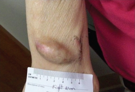
Right-upper-arm (brachiocephalic) arteriovenous (AV) fistula aneurysm
Image: “Right upper arm AV fistula aneurysm of the patient” by College of Medicine at Urbana-Champaign, University of Illinois. License: CC BY 3.0Diagnosis:
Management:
Epidemiology:
Risk factors (for atherosclerosis Atherosclerosis Atherosclerosis is a common form of arterial disease in which lipid deposition forms a plaque in the blood vessel walls. Atherosclerosis is an incurable disease, for which there are clearly defined risk factors that often can be reduced through a change in lifestyle and behavior of the patient. Atherosclerosis):
Clinical presentation:
Diagnosis:
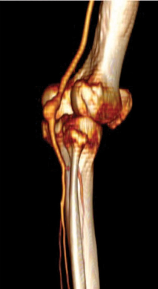
Preoperative CT angiography with a 3-dimensional reconstruction of popliteal artery aneurysm
Image: “ Preoperative CTA angiography three-dimensional reconstruction showing popliteal artey aneurysm location” by Department of Vascular Surgery, San Filippo Neri Hospital, Rome. License: CC BY 4.0Management:
Epidemiology:
Clinical presentation:
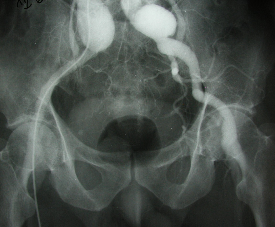
Left iliofemoral aneurysm
Image: “True aneurysm” by Jojo. License: Public DomainManagement:
Visceral artery aneurysms (VAAs) can affect the celiac artery, the superior and inferior mesenteric arteries Arteries Arteries are tubular collections of cells that transport oxygenated blood and nutrients from the heart to the tissues of the body. The blood passes through the arteries in order of decreasing luminal diameter, starting in the largest artery (the aorta) and ending in the small arterioles. Arteries are classified into 3 types: large elastic arteries, medium muscular arteries, and small arteries and arterioles. Arteries: Histology, or their branches.
Epidemiology:
Risk factors:
Clinical presentation:
Diagnosis:
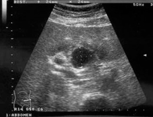
Ruptured hepatic artery aneurysm presenting as abdominal pain:
Ultrasound scan shows aneurysmal sac to the left of the common bile duct and portal vein.
Management:
Prognosis Prognosis A prediction of the probable outcome of a disease based on a individual’s condition and the usual course of the disease as seen in similar situations. Non-Hodgkin Lymphomas: