Overview
- Hypersensitivity reaction
- A “hyper” or exaggerated response to what should be considered harmless environmental antigens
- Types I, II, and III are immediate reactions occurring within 24 hours.
- Type IV reaction develops over several days.
- Type III hypersensitivity reaction
- Immune complex (IC)-mediated hypersensitivity: triggered by antigen-antibody (Ag-Ab) complexes deposited in tissues
- Ag: intrinsic (part of the host) or extrinsic (exogenous source such as bacteria, virus)
- As with type II hypersensitivity, cell injury is similar: Complement system leads to a reaction that produces cellular damage.
- Unlike type II hypersensitivity, in type III reactions:
- Antigens are not bound to cell surfaces.
- Ag-Ab complexes form in circulation.
- Target of the immune response is not the tissue or cell.
- Target is the IC deposited in the tissue.
Pathophysiology
Physiology
- Immune complex formation normally results in antigen neutralization.
- The complement system reduces pathologic IC accumulation.
- Antibodies (Ab) have 2 regions:
- Fab region: attaches to antigens
- Fc region: interacts with complement and Fc-bearing receptor (FcR) cells
- C1q: activates complement system and binds Ab Fc region, mediating IC clearance by FcR-bearing cells.
- C3b: makes ICs soluble and tags them for phagocytosis (opsonization).
- Antibodies (Ab) have 2 regions:
Pathogenesis
Immune complex formation: IC formed by the binding of Ag and Ab
Immune complex deposition
Immune complex deposition depends on:
- Physical properties of the IC:
- Affinity of Ab to complement, size, and charge of the IC
- Increased rate of IC formation → overwhelms clearing mechanism → IC freely circulate out to organs
- Antigen-to-antibody ratio:
- Low antibodies or excess antibodies → decreased effector activation
- Tissue-specific hemodynamics
- ICs first localize within blood vessels → vasculitis
- Common areas affected are “permeability”-susceptible tissues:
- Glomeruli (nephritis)
- Joints/synovium (arthritis)
Immune complex inflammatory reaction
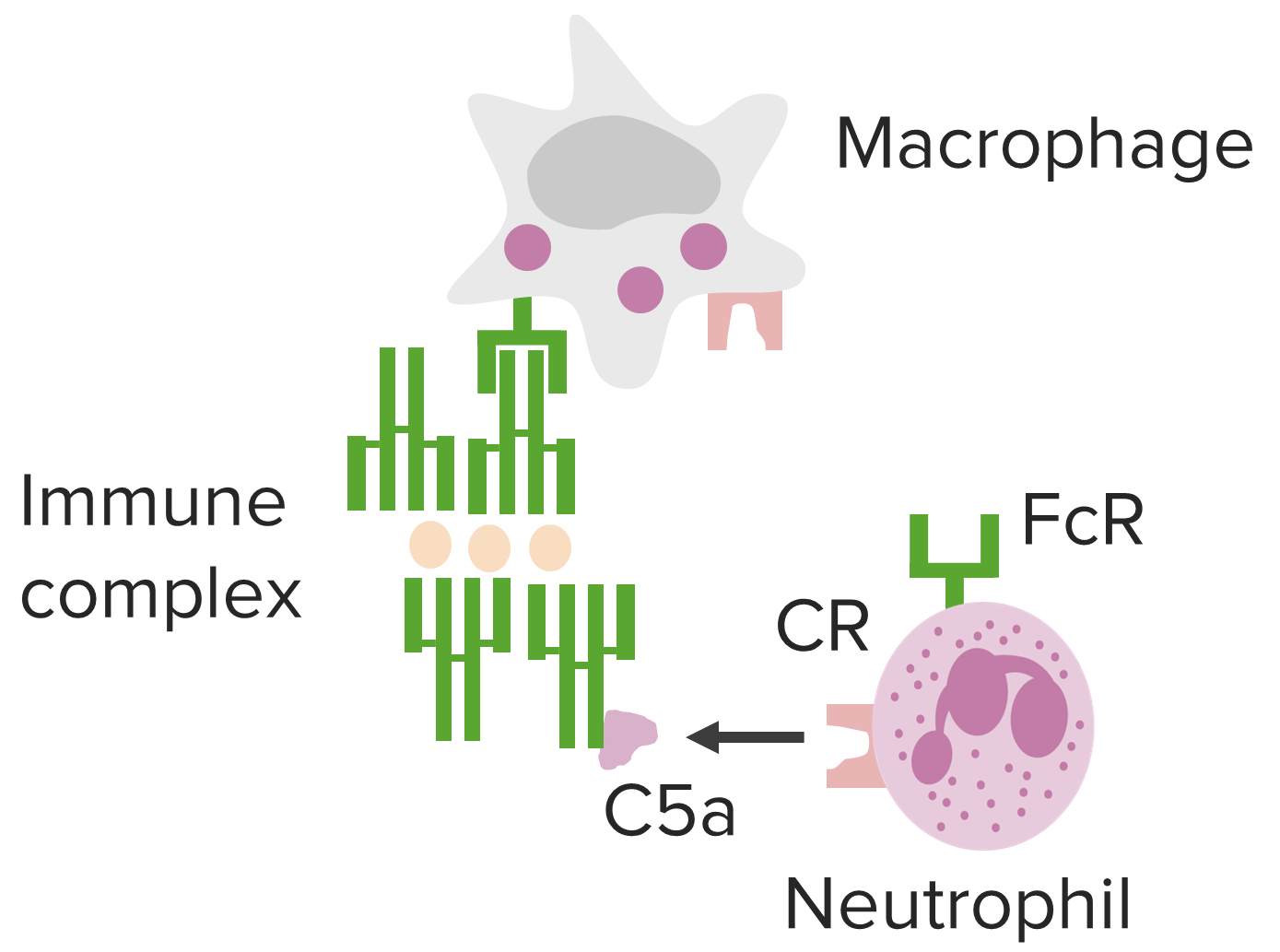
- IC deposits activate the complement cascade.
- C3a initiates mast cell degranulation:
- Histamine increases vascular permeability in the involved tissue.
- ICs enter tissue → normal tissue with IC becomes a target for inflammatory response
- C5a (chemoattractant) recruits neutrophils → release lysozymes and inflammatory mediators → cell death and tissue injury
- C3b opsonizes the tissue → phagocytosis and membrane attack complex (MAC)-mediated cell lysis
- Macrophages, natural killer cells → release lytic mediators and injure tissue cells
- Platelet aggregation can occur → micro thrombus formation
Related videos
Clinical Presentation
Manifestations are affected by the route of entry, site(s) of IC deposition, and persistence of antigen(s).
Arthus phenomenon /Arthus reaction
- A locally injected antigen (e.g., immunization like Tdap) causes a localized reaction.
- Due to antigen excess and IC deposition on vascular walls
- Necrosis of affected tissues: pain, redness, induration, and edema at the site of injection
- Self-limited
Systemic lupus erythematosus (SLE)
- Antibodies directed against parts of the nucleus: antinuclear antibodies (ANA), a universal finding in SLE
- Antibodies to other parts of the nucleus:
- Histone (anti-histone Ab)
- DNA (anti-dsDNA Ab)
- Ribonucleoprotein (anti-RNP Ab)
- Extractable nuclear antigen/Smith antigen (Anti-Smith Ab)
- Other: Antibodies against phospholipids in cells (antiphospholipid Ab)
- Present in 30%–40% of patients with SLE
- Increased risk of thrombosis
- Ag-Ab complexes deposit in multiple areas:
- Skin/mucocutaneous (malar rash, photosensitivity, oral ulcers)
- Kidneys (glomerulonephritis)
- Joints (arthritis)
- Blood vessels (vasculitis, Raynaud’s phenomenon, blood clots)
- Pleura (effusion)
- Pericardium (pericarditis)
- Blood cells (anemia, leukopenia, thrombocytopenia)
- Central nervous system (strokes, seizures)
Post-streptococcal glomerulonephritis (PSGN)
- Reaction to nephritogenic antigens of group A beta-hemolytic streptococcus (GAS)
- Can be from streptococcal throat infection or skin infection
- Presentation: hematuria, proteinuria, hypertension, edema, and elevated creatinine
Serum sickness
- Reaction to foreign antisera (e.g., antivenom)
- Higher doses of administered agent more likely to result in a reaction
- 1–2 weeks after exposure: fever, rash, arthritis; proteinuria occurs with renal involvement
Hypersensitivity pneumonitis
- Extrinsic allergic alveolitis
- Antigen: a microbe, protein, or chemical
- IC deposition in alveoli, interstitium, bronchioli, and lung parenchyma
- Farmer’s lung: Ag from thermophilic molds on crops
- Bird fancier’s disease: Ag from intestinal mucin in droppings or feathers
- Note: can also be T-lymphocyte–mediated (type IV reaction)
Polyarteritis nodosa (PAN)
- Etiology mostly idiopathic but can be a reaction to hepatitis B virus surface Ag
- Medium-sized arterial inflammation → ↓ blood flow, ↑ thrombosis; spares the veins
- Kidneys: most commonly involved organ
- Presentation:
- Tender erythematous nodules, hypertension, renal insufficiency, and neuropathy
- Some have abdominal symptoms (mesenteric arteritis)
Type III hypersensitivity immune complex mediated tissue injury in blood vessel wall, vasculitis, which is associated with various conditions.
Image by Lecturio..
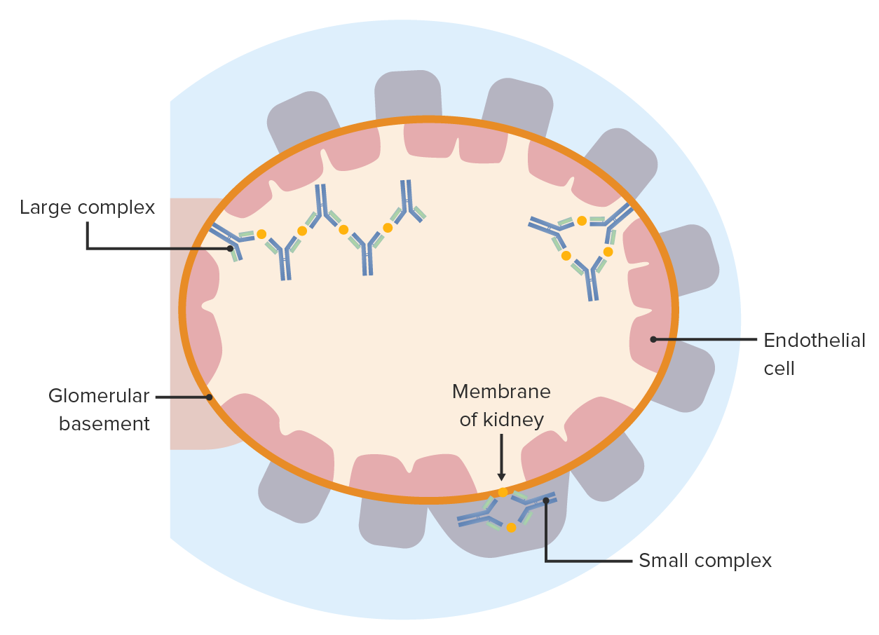
Immune complex disease: Antigen-antibody complexes deposit on either side of the glomerular basement membrane. These elicit an inflammatory reaction and cause glomerular injury.
Image by Lecturio.
Diagnosis and Management
Diagnosis
- Clinical history and findings (Arthus reaction, serum sickness often diagnosed clinically)
- Laboratory tests:
- CBC
- Anemia, thrombocytopenia, leukopenia: SLE
- Eosinophilia: serum sickness, hypersensitivity pneumonitis
- Metabolic panel
- Creatinine is abnormal in reactions involving the kidneys
- Serologies and culture
- Hepatitis panel for polyarteritis nodosa
- Culture: streptococcal infection (only 25% of patients will have positive results because PSGN occurs weeks after the infection)
- Complement levels
- Generally found in low levels in associated conditions
- Antibodies
- ANA, anti-dsDNA Ab, antiSmith Ab, anti-RNP Ab, antiphospholipid Ab: SLE
- PSGN: antistreptolysin O (ASO), streptozyme test
- Urinalysis
- Proteinuria: SLE, PAN, serum sickness
- PSGN: hematuria, proteinuria
- Inflammatory markers
- Generally elevated C-reactive protein (CRP), erythrocyte sedimentation rate (ESR) during active inflammation
- Imaging
- High-resolution computed tomography (CT) (lung): hypersensitivity pneumonitis
- Arteriography or CT/magnetic resonance angiography (MRA): for polyarteritis nodosa
- X-ray: joint involvement
- Surgical procedures
- Renal biopsy: for renal involvement
- Skin biopsy: for cutaneous involvement
- Bronchoscopy: for lung involvement
- CBC
A 27-year-old female patient with a history of exposure to mold. High-resolution CT scans of the chest (lung window) at the level of the lower lobes show extensive ground-glass opacities (asterisks), with overlapping foci of lobular air trapping (arrows).
Image: “Hypersensitivity Pneumonitis” by Torres PP, Moreira MA, Silva DG, da Gama RR, Sugita DM, Moreira MA. License: CC BY 4.0Management
- Remove or avoid offending agent
- Antihistamines, nonsteroidal anti-inflammatory drugs (NSAIDs) for symptom relief (rash, itching, joint pains)
- In reactions due to infection (PSGN), antibiotic therapy if infection is still present
- Control of complications such as hypertension (PSGN, PAN), edema (PSGN, SLE), airway symptoms (hypersensitivity pneumonitis)
- Glucocorticoids used in severe cases to suppress inflammation
- Therapeutic options for specific conditions:
- Hypersensitivity pneumonitis: immunosuppressive therapy (mycophenolate, azathioprine)
- SLE:
- Antimalarials (hydroxychloroquine, chloroquine)
- Immunosuppressive therapy: mycophenolate, azathioprine, cyclophosphamide, rituximab
- Long-term anticoagulation if with thrombosis: warfarin, low-molecular-weight heparin
- PAN:
- Methotrexate, a disease-modifying anti-rheumatic drug (DMARD)
- Immunosuppressive therapy (cyclophosphamide, azathioprine)
- Dialysis for end-stage renal disease from SLE
- Organ transplantation:
- Lung transplantation for advanced lung disease in hypersensitivity pneumonitis
- Renal transplantation for end-stage renal disease in SLE
References
- King, T., Flaherty, K. & Hollingsworth, H. (Eds.). (2019). Hypersensitivity pneumonitis (extrinsic allergic alveolitis): Clinical manifestations and diagnosis. UpToDate. Retrieved Aug 17, 2020, from https://www.uptodate.com/contents/hypersensitivity-pneumonitis-extrinsic-allergic-alveolitis-treatment-prognosis-and-prevention
- King, T., Flaherty, K. & Hollingsworth, H. (Eds.). (2020). Hypersensitivity pneumonitis (extrinsic allergic alveolitis): Treatment, prognosis, and prevention. UpToDate. Retrieved Aug 17, 2020, from https://www.uptodate.com/contents/hypersensitivity-pneumonitis-extrinsic-allergic-alveolitis-treatment-prognosis-and-prevention
- Lu, L., Suscovich, T., Fortune, S., Galit, A. (2017). Beyond Binding: antibody effector functions in infectious diseases. Nature Reviews Immunology, 18, 46–61. https://doi.org/10.1038/nri.2017.106
- Mak, T., Saunders, M., Jett, B. (2007). Primer to the Immune response. Elsevier, 2nd ed.
- Mayadas, T., Tsokos, G., Tsuboi, N. (2009). Mechanisms of Immune Complex-mediated Neutrophil Recruitment and Tissue Injury : official publication of the Circulation, 120(20), 2012–2014. https://www.ahajournals.org/doi/10.1161/CIRCULATIONAHA.108.771170
- Merkel, P., Hunder, G. & Ramirez Curtis, M. (Eds.) (2019). Clinical manifestations and diagnosis of polyarteritis arteritis nodosa in adults. UpToDate. Retrieved 17 Aug 2020, from https://www.uptodate.com/contents/clinical-manifestations-and-diagnosis-of-polyarteritis-nodosa-in-adults
- Wallace, D., Gladman, D., Pisetsky, D.,Shur, P & Ramirez Curtis, M. (Eds.) (2019). Overview of the management and prognosis of systemic lupus erythematosus in adults. UpToDate. Retrieved 17 Aug 2020, from https://www.uptodate.com/contents/clinical-manifestations-and-diagnosis-of-systemic-lupus-erythematosus-in-adults
- Wallace, D., Pisetsky, D., Shur, P & Ramirez Curtis, M. (Eds.) (2020,). Overview of the management and prognosis of systemic lupus erythematosus in adults. UpToDate. Retrieved 17 Aug 2020, from https://www.uptodate.com/contents/overview-of-the-management-and-prognosis-of-systemic-lupus-erythematosus-in-adults