Pericardial effusion is the accumulation of excess fluid in the pericardial space around the heart. The pericardiumPericardiumA conical fibroserous sac surrounding the heart and the roots of the great vessels (aorta; venae cavae; pulmonary artery). Pericardium consists of two sacs: the outer fibrous pericardium and the inner serous pericardium. The latter consists of an outer parietal layer facing the fibrous pericardium, and an inner visceral layer (epicardium) resting next to the heart, and a pericardial cavity between these two layers.Heart: Anatomy does not easily expand; thus, rapid fluid accumulation leads to increased pressure around the heart. The increase in pressure restricts cardiac filling, resulting in decreased cardiac outputCardiac outputThe volume of blood passing through the heart per unit of time. It is usually expressed as liters (volume) per minute so as not to be confused with stroke volume (volume per beat).Cardiac Mechanics and cardiac tamponadeTamponadePericardial effusion, usually of rapid onset, exceeding ventricular filling pressures and causing collapse of the heart with a markedly reduced cardiac output.Pericarditis. Signs and symptoms usually occur in the setting of cardiac tamponadeTamponadePericardial effusion, usually of rapid onset, exceeding ventricular filling pressures and causing collapse of the heart with a markedly reduced cardiac output.Pericarditis and include dyspneaDyspneaDyspnea is the subjective sensation of breathing discomfort. Dyspnea is a normal manifestation of heavy physical or psychological exertion, but also may be caused by underlying conditions (both pulmonary and extrapulmonary). Dyspnea, hypotensionHypotensionHypotension is defined as low blood pressure, specifically < 90/60 mm Hg, and is most commonly a physiologic response. Hypotension may be mild, serious, or life threatening, depending on the cause. Hypotension, muffled heart soundsHeart soundsHeart sounds are brief, transient sounds produced by valve opening and closure and by movement of blood in the heart. They are divided into systolic and diastolic sounds. In most cases, only the first (S1) and second (S2) heart sounds are heard. These are high-frequency sounds and arise from aortic and pulmonary valve closure (S1), as well as mitral and tricuspid valve closure (S2).Heart Sounds, jugular venous distensionJugular Venous DistensionCardiovascular Examination, and pulsus paradoxus. The diagnosis of pericardial effusion is confirmed with echocardiographyEchocardiographyUltrasonic recording of the size, motion, and composition of the heart and surrounding tissues. The standard approach is transthoracic.Tricuspid Valve Atresia (TVA). Small effusions in stable patientsStable PatientsBlunt Chest Trauma are treated medically. Larger effusions and cardiac tamponadeTamponadePericardial effusion, usually of rapid onset, exceeding ventricular filling pressures and causing collapse of the heart with a markedly reduced cardiac output.Pericarditis may require pericardiocentesisPericardiocentesisPuncture and aspiration of fluid from the pericardium.Cardiac Surgery or pericardiotomy.
Pericardial effusion is the accumulation of fluid in the pericardial space.
Cardiac tamponadeTamponadePericardial effusion, usually of rapid onset, exceeding ventricular filling pressures and causing collapse of the heart with a markedly reduced cardiac output.Pericarditis is the accumulation of pericardial fluidPericardial fluidWatery fluid produced in the serous and visceral pericardium surrounding the surface of the heart.Heart: Anatomy sufficient to impair cardiac filling and cause hemodynamic compromise. The rate of fluid accumulation, and not necessarily the amount, is most important.
Epidemiology
Pericardial effusion:
The incidenceIncidenceThe number of new cases of a given disease during a given period in a specified population. It also is used for the rate at which new events occur in a defined population. It is differentiated from prevalence, which refers to all cases in the population at a given time.Measures of Disease Frequency is unknown.
Has been observed in approximately 3% of autopsy subjects in studies
Age:
Can occur in all age groups
Mean: 50–60 years
Cardiac tamponadeTamponadePericardial effusion, usually of rapid onset, exceeding ventricular filling pressures and causing collapse of the heart with a markedly reduced cardiac output.Pericarditis:
IncidenceIncidenceThe number of new cases of a given disease during a given period in a specified population. It also is used for the rate at which new events occur in a defined population. It is differentiated from prevalence, which refers to all cases in the population at a given time.Measures of Disease Frequency: 2 cases per 10,000 people in the United States
Many conditions are associated with pericardial effusion, including:
Infection:
Viral (most common):
CoxsackievirusCoxsackievirusCoxsackievirus is a member of a family of viruses called Picornaviridae and the genus Enterovirus. Coxsackieviruses are single-stranded, positive-sense RNA viruses, and are divided into coxsackie group A and B viruses. Both groups of viruses cause upper respiratory infections, rashes, aseptic meningitis, or encephalitis. Coxsackievirus group B
InfluenzaInfluenzaInfluenza viruses are members of the Orthomyxoviridae family and the causative organisms of influenza, a highly contagious febrile respiratory disease. There are 3 primary influenza viruses (A, B, and C) and various subtypes, which are classified based on their virulent surface antigens, hemagglutinin (HA) and neuraminidase (NA). Influenza typically presents with a fever, myalgia, headache, and symptoms of an upper respiratory infection. Influenza Viruses/Influenza
EchovirusEchovirusEchoviruses are single-stranded positive-sense RNA viruses belonging to the genus Enterovirus. Transmission is most commonly through the fecal-oral route. The majority of patients are asymptomatic. Patients who are symptomatic can exhibit a wide range of illnesses ranging from nonspecific URIs and exanthems to severe and life-threatening illnesses.Echovirus
EBVEBVEpstein-barr virus (EBV) is a linear, double-stranded DNA virus belonging to the herpesviridae family. This highly prevalent virus is mostly transmitted through contact with oropharyngeal secretions from an infected individual. The virus can infect epithelial cells and B lymphocytes, where it can undergo lytic replication or latency.Epstein-Barr Virus
CMV
Parvovirus B19Parvovirus B19Primate erythroparvovirus 1 (generally referred to as parvovirus B19, B19 virus, or sometimes erythrovirus B19) ranks among the smallest DNA viruses. Parvovirus B19 is of the family Parvoviridae and genus Erythrovirus. In immunocompetent humans, parvovirus B19 classically results in erythema infectiosum (5th disease) or “slapped cheek syndrome.”Parvovirus B19
Varicella
Bacterial:
Staphylococcus aureusStaphylococcus aureusPotentially pathogenic bacteria found in nasal membranes, skin, hair follicles, and perineum of warm-blooded animals. They may cause a wide range of infections and intoxications.Brain Abscess
StreptococcusStreptococcusStreptococcus is one of the two medically important genera of gram-positive cocci, the other being Staphylococcus. Streptococci are identified as different species on blood agar on the basis of their hemolytic pattern and sensitivity to optochin and bacitracin. There are many pathogenic species of streptococci, including S. pyogenes, S. agalactiae, S. pneumoniae, and the viridans streptococci.Streptococcus
NeisseriaNeisseriaNeisseria is a genus of bacteria commonly present on mucosal surfaces. Several species exist, but only 2 are pathogenic to humans: N. gonorrhoeae and N. meningitidis. Neisseria species are non-motile, gram-negative diplococci most commonly isolated on modified Thayer-Martin (MTM) agar. Neisseria
LegionellaLegionellaLegionella is a facultative intracellular, gram-negative bacilli. Legionella does not grow on common culture media because it requires certain supplementation (cysteine and iron). Legionella pneumophila (L. pneumophila) accounts for the majority of human infections.Legionella/Legionellosis
Treponema pallidumTreponema pallidumThe causative agent of venereal and non-venereal syphilis as well as yaws.Treponema
Mycobacterium tuberculosisMycobacterium tuberculosisTuberculosis (TB) is an infectious disease caused by Mycobacterium tuberculosis complex bacteria. The bacteria usually attack the lungs but can also damage other parts of the body. Approximately 30% of people around the world are infected with this pathogen, with the majority harboring a latent infection. Tuberculosis spreads through the air when a person with active pulmonary infection coughs or sneezes.Tuberculosis
Fungal:
CandidaCandidaCandida is a genus of dimorphic, opportunistic fungi. Candida albicans is part of the normal human flora and is the most common cause of candidiasis. The clinical presentation varies and can include localized mucocutaneous infections (e.g., oropharyngeal, esophageal, intertriginous, and vulvovaginal candidiasis) and invasive disease (e.g., candidemia, intraabdominal abscess, pericarditis, and meningitis). Candida/Candidiasis
HistoplasmosisHistoplasmosisHistoplasmosis is an infection caused by Histoplasma capsulatum, a dimorphic fungus. Transmission is through inhalation, and exposure to soils containing bird or bat droppings increases the risk of infection. Most infections are asymptomatic; however, immunocompromised individuals generally develop acute pulmonary infection, chronic infection, or even disseminated disease.Histoplasma/Histoplasmosis
CoccidioidomycosisCoccidioidomycosisCoccidioidomycosis, commonly known as San Joaquin Valley fever, is a fungal disease caused by Coccidioides immitis or Coccidioides posadasii. When Coccidioides spores are inhaled, they transform into spherules that result in infection. Coccidioidomycosis is also a common cause of community-acquired pneumonia and can cause severe disease in the immunocompromised.Coccidioides/Coccidioidomycosis
Cardiac surgeryCardiac surgeryCardiac surgery is the surgical management of cardiac abnormalities and of the great vessels of the thorax. In general terms, surgical intervention of the heart is performed to directly restore adequate pump function, correct inherent structural issues, and reestablish proper blood supply via the coronary circulation.Cardiac Surgery (postpericardiotomy syndrome)
RadiationRadiationEmission or propagation of acoustic waves (sound), electromagnetic energy waves (such as light; radio waves; gamma rays; or x-rays), or a stream of subatomic particles (such as electrons; neutrons; protons; or alpha particles).Osteosarcoma
Autoimmune and connective tissueConnective tissueConnective tissues originate from embryonic mesenchyme and are present throughout the body except inside the brain and spinal cord. The main function of connective tissues is to provide structural support to organs. Connective tissues consist of cells and an extracellular matrix.Connective Tissue: Histology disease:
Systemic lupus erythematosusSystemic lupus erythematosusSystemic lupus erythematosus (SLE) is a chronic autoimmune, inflammatory condition that causes immune-complex deposition in organs, resulting in systemic manifestations. Women, particularly those of African American descent, are more commonly affected.Systemic Lupus Erythematosus
Ankylosing spondylitisAnkylosing spondylitisAnkylosing spondylitis (also known as Bechterew’s disease or Marie-Strümpell disease) is a seronegative spondyloarthropathy characterized by chronic and indolent inflammation of the axial skeleton. Severe disease can lead to fusion and rigidity of the spine. Ankylosing Spondylitis
SclerodermaSclerodermaScleroderma (systemic sclerosis) is an autoimmune condition characterized by diffuse collagen deposition and fibrosis. The clinical presentation varies from limited skin involvement to diffuse involvement of internal organs. Scleroderma
SarcoidosisSarcoidosisSarcoidosis is a multisystem inflammatory disease that causes noncaseating granulomas. The exact etiology is unknown. Sarcoidosis usually affects the lungs and thoracic lymph nodes, but it can also affect almost every system in the body, including the skin, heart, and eyes, most commonly. Sarcoidosis
VasculitisVasculitisInflammation of any one of the blood vessels, including the arteries; veins; and rest of the vasculature system in the body.Systemic Lupus Erythematosus
Heart failureHeart FailureA heterogeneous condition in which the heart is unable to pump out sufficient blood to meet the metabolic need of the body. Heart failure can be caused by structural defects, functional abnormalities (ventricular dysfunction), or a sudden overload beyond its capacity. Chronic heart failure is more common than acute heart failure which results from sudden insult to cardiac function, such as myocardial infarction.Total Anomalous Pulmonary Venous Return (TAPVR)
Aortic dissectionAortic dissectionAortic dissection occurs due to shearing stress from pulsatile pressure causing a tear in the tunica intima of the aortic wall. This tear allows blood to flow into the media, creating a “false lumen.” Aortic dissection is most commonly caused by uncontrolled hypertension.Aortic Dissection (type A)
UremiaUremiaA clinical syndrome associated with the retention of renal waste products or uremic toxins in the blood. It is usually the result of renal insufficiency. Most uremic toxins are end products of protein or nitrogen catabolism, such as urea or creatinine. Severe uremia can lead to multiple organ dysfunctions with a constellation of symptoms.Acute Kidney Injury (chronic renal failureRenal failureConditions in which the kidneys perform below the normal level in the ability to remove wastes, concentrate urine, and maintain electrolyte balance; blood pressure; and calcium metabolism. Renal insufficiency can be classified by the degree of kidney damage (as measured by the level of proteinuria) and reduction in glomerular filtration rate.Crush Syndrome)
MyxedemaMyxedemaA condition characterized by a dry, waxy type of swelling (edema) with abnormal deposits of mucopolysaccharides in the skin and other tissues. It is caused by a deficiency of thyroid hormones. The skin becomes puffy around the eyes and on the cheeks. The face is dull and expressionless with thickened nose and lips.Edema
AmyloidosisAmyloidosisAmyloidosis is a disease caused by abnormal extracellular tissue deposition of fibrils composed of various misfolded low-molecular-weight protein subunits. These proteins are frequently byproducts of other pathological processes (e.g., multiple myeloma). Amyloidosis
IsoniazidIsoniazidAntibacterial agent used primarily as a tuberculostatic. It remains the treatment of choice for tuberculosis.Antimycobacterial Drugs
Minoxidil
PhenytoinPhenytoinAn anticonvulsant that is used to treat a wide variety of seizures. The mechanism of therapeutic action is not clear, although several cellular actions have been described including effects on ion channels, active transport, and general membrane stabilization. Phenytoin has been proposed for several other therapeutic uses, but its use has been limited by its many adverse effects and interactions with other drugs.First-Generation Anticonvulsant Drugs
AnticoagulantsAnticoagulantsAnticoagulants are drugs that retard or interrupt the coagulation cascade. The primary classes of available anticoagulants include heparins, vitamin K-dependent antagonists (e.g., warfarin), direct thrombin inhibitors, and factor Xa inhibitors. Anticoagulants
The pericardial space normally contains a small volume of serous fluid.
Under normal circumstances, the pericardial fluidPericardial fluidWatery fluid produced in the serous and visceral pericardium surrounding the surface of the heart.Heart: Anatomy:
Cushions the heart
Provides a low-friction environment
Allows the heart to move easily
Pericardial effusion and cardiac tamponadeTamponadePericardial effusion, usually of rapid onset, exceeding ventricular filling pressures and causing collapse of the heart with a markedly reduced cardiac output.Pericarditis
The pericardiumPericardiumA conical fibroserous sac surrounding the heart and the roots of the great vessels (aorta; venae cavae; pulmonary artery). Pericardium consists of two sacs: the outer fibrous pericardium and the inner serous pericardium. The latter consists of an outer parietal layer facing the fibrous pericardium, and an inner visceral layer (epicardium) resting next to the heart, and a pericardial cavity between these two layers.Heart: Anatomy has limited elasticityElasticityResistance and recovery from distortion of shape.Skeletal Muscle Contraction.
Accumulation of pericardial fluidPericardial fluidWatery fluid produced in the serous and visceral pericardium surrounding the surface of the heart.Heart: Anatomy → ↑ pressure in the pericardial sac
↓ Stroke volumeStroke volumeThe amount of blood pumped out of the heart per beat, not to be confused with cardiac output (volume/time). It is calculated as the difference between the end-diastolic volume and the end-systolic volume.Cardiac Cycle
↓ Cardiac outputCardiac outputThe volume of blood passing through the heart per unit of time. It is usually expressed as liters (volume) per minute so as not to be confused with stroke volume (volume per beat).Cardiac Mechanics → hypotensionHypotensionHypotension is defined as low blood pressure, specifically < 90/60 mm Hg, and is most commonly a physiologic response. Hypotension may be mild, serious, or life threatening, depending on the cause. Hypotension and obstructive (cardiogenic) shockShockShock is a life-threatening condition associated with impaired circulation that results in tissue hypoxia. The different types of shock are based on the underlying cause: distributive (↑ cardiac output (CO), ↓ systemic vascular resistance (SVR)), cardiogenic (↓ CO, ↑ SVR), hypovolemic (↓ CO, ↑ SVR), obstructive (↓ CO), and mixed. Types of Shock
↑ HR to maintain cardiac outputCardiac outputThe volume of blood passing through the heart per unit of time. It is usually expressed as liters (volume) per minute so as not to be confused with stroke volume (volume per beat).Cardiac Mechanics as a compensatory mechanism
The rate of fluid accumulation is important:
If fluid were to fill the pericardial space rapidly (e.g., chest trauma), as little as 150 mL could lead to tamponadeTamponadePericardial effusion, usually of rapid onset, exceeding ventricular filling pressures and causing collapse of the heart with a markedly reduced cardiac output.Pericarditis.
If fluid accumulates slowly, the pericardial sac can stretch to accommodate approximately 2 L of fluid.
Clinical Presentation
Symptoms
Without cardiac tamponadeTamponadePericardial effusion, usually of rapid onset, exceeding ventricular filling pressures and causing collapse of the heart with a markedly reduced cardiac output.Pericarditis:
Usually no symptoms specific to effusion
Symptoms may be related to the underlying condition (e.g., infection, uremiaUremiaA clinical syndrome associated with the retention of renal waste products or uremic toxins in the blood. It is usually the result of renal insufficiency. Most uremic toxins are end products of protein or nitrogen catabolism, such as urea or creatinine. Severe uremia can lead to multiple organ dysfunctions with a constellation of symptoms.Acute Kidney Injury, autoimmune disease).
Cardiac tamponadeTamponadePericardial effusion, usually of rapid onset, exceeding ventricular filling pressures and causing collapse of the heart with a markedly reduced cardiac output.Pericarditis:
DyspneaDyspneaDyspnea is the subjective sensation of breathing discomfort. Dyspnea is a normal manifestation of heavy physical or psychological exertion, but also may be caused by underlying conditions (both pulmonary and extrapulmonary). Dyspnea
Chest painPainAn unpleasant sensation induced by noxious stimuli which are detected by nerve endings of nociceptive neurons.Pain: Types and Pathways (pericarditisPericarditisPericarditis is an inflammation of the pericardium, often with fluid accumulation. It can be caused by infection (often viral), myocardial infarction, drugs, malignancies, metabolic disorders, autoimmune disorders, or trauma. Acute, subacute, and chronic forms exist. Pericarditis):
SyncopeSyncopeSyncope is a short-term loss of consciousness and loss of postural stability followed by spontaneous return of consciousness to the previous neurologic baseline without the need for resuscitation. The condition is caused by transient interruption of cerebral blood flow that may be benign or related to a underlying life-threatening condition. Syncope
AnxietyAnxietyFeelings or emotions of dread, apprehension, and impending disaster but not disabling as with anxiety disorders.Generalized Anxiety Disorder or confusion
FatigueFatigueThe state of weariness following a period of exertion, mental or physical, characterized by a decreased capacity for work and reduced efficiency to respond to stimuli.Fibromyalgia
Hiccups
Physical exam
The following may be seen with large pericardial effusions and cardiac tamponadeTamponadePericardial effusion, usually of rapid onset, exceeding ventricular filling pressures and causing collapse of the heart with a markedly reduced cardiac output.Pericarditis:
Vital signs:
HypotensionHypotensionHypotension is defined as low blood pressure, specifically < 90/60 mm Hg, and is most commonly a physiologic response. Hypotension may be mild, serious, or life threatening, depending on the cause. Hypotension
TachycardiaTachycardiaAbnormally rapid heartbeat, usually with a heart rate above 100 beats per minute for adults. Tachycardia accompanied by disturbance in the cardiac depolarization (cardiac arrhythmia) is called tachyarrhythmia.Sepsis in Children
Cardiovascular:
Pericardial friction rubPericardial friction rubA rasping, scratching, or grating sound with up to 3 components per cardiac cycle and best heard during expiration with the patient leaning forward.Pericarditis (pericarditisPericarditisPericarditis is an inflammation of the pericardium, often with fluid accumulation. It can be caused by infection (often viral), myocardial infarction, drugs, malignancies, metabolic disorders, autoimmune disorders, or trauma. Acute, subacute, and chronic forms exist. Pericarditis)
Muffled heart soundsHeart soundsHeart sounds are brief, transient sounds produced by valve opening and closure and by movement of blood in the heart. They are divided into systolic and diastolic sounds. In most cases, only the first (S1) and second (S2) heart sounds are heard. These are high-frequency sounds and arise from aortic and pulmonary valve closure (S1), as well as mitral and tricuspid valve closure (S2).Heart Sounds
Dullness to percussionPercussionAct of striking a part with short, sharp blows as an aid in diagnosing the condition beneath the sound obtained.Pulmonary Examination beneath the angle of the left scapula
Tubular breath sounds
Egophony
Diminished breath sounds (if pleural effusionPleural EffusionPleural effusion refers to the accumulation of fluid between the layers of the parietal and visceral pleura. Common causes of this condition include infection, malignancy, autoimmune disorders, or volume overload. Clinical manifestations include chest pain, cough, and dyspnea. Pleural Effusion is present)
Peripheral:
EdemaEdemaEdema is a condition in which excess serous fluid accumulates in the body cavity or interstitial space of connective tissues. Edema is a symptom observed in several medical conditions. It can be categorized into 2 types, namely, peripheral (in the extremities) and internal (in an organ or body cavity). Edema
CyanosisCyanosisA bluish or purplish discoloration of the skin and mucous membranes due to an increase in the amount of deoxygenated hemoglobin in the blood or a structural defect in the hemoglobin molecule.Pulmonary Examination
Beck triad
The triad describes the classic findings in cardiac tamponadeTamponadePericardial effusion, usually of rapid onset, exceeding ventricular filling pressures and causing collapse of the heart with a markedly reduced cardiac output.Pericarditis:
HypotensionHypotensionHypotension is defined as low blood pressure, specifically < 90/60 mm Hg, and is most commonly a physiologic response. Hypotension may be mild, serious, or life threatening, depending on the cause. Hypotension
Muffled heart soundsHeart soundsHeart sounds are brief, transient sounds produced by valve opening and closure and by movement of blood in the heart. They are divided into systolic and diastolic sounds. In most cases, only the first (S1) and second (S2) heart sounds are heard. These are high-frequency sounds and arise from aortic and pulmonary valve closure (S1), as well as mitral and tricuspid valve closure (S2).Heart Sounds on auscultation
Diagnosis
Imaging
ECGECGAn electrocardiogram (ECG) is a graphic representation of the electrical activity of the heart plotted against time. Adhesive electrodes are affixed to the skin surface allowing measurement of cardiac impulses from many angles. The ECG provides 3-dimensional information about the conduction system of the heart, the myocardium, and other cardiac structures. Electrocardiogram (ECG):
Sinus tachycardiaTachycardiaAbnormally rapid heartbeat, usually with a heart rate above 100 beats per minute for adults. Tachycardia accompanied by disturbance in the cardiac depolarization (cardiac arrhythmia) is called tachyarrhythmia.Sepsis in Children
Low voltage of QRS complexes
Diffuse ST elevation with PR depression (pericarditisPericarditisPericarditis is an inflammation of the pericardium, often with fluid accumulation. It can be caused by infection (often viral), myocardial infarction, drugs, malignancies, metabolic disorders, autoimmune disorders, or trauma. Acute, subacute, and chronic forms exist. Pericarditis)
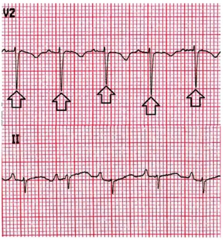
Electrical alternans:
Consecutive QRS complexes that alternate in height
A motion artifact due to the pendular swinging of the heart within the pericardial space
Seen in large pericardial effusion or cardiac tamponadeTamponadePericardial effusion, usually of rapid onset, exceeding ventricular filling pressures and causing collapse of the heart with a markedly reduced cardiac output.Pericarditis
Electrical alternans on an ECG in a patient with a large pericardial effusion: The arrows point to the alternating amplitude of the QRS complex.
Image: “Electrical alternans” by Eric Williams Medical Sciences Complex, The University of the West Indies, Champs Fleurs, Trinidad and Tobago. License: CC BY 4.0
Chest X-rayX-rayPenetrating electromagnetic radiation emitted when the inner orbital electrons of an atom are excited and release radiant energy. X-ray wavelengths range from 1 pm to 10 nm. Hard x-rays are the higher energy, shorter wavelength x-rays. Soft x-rays or grenz rays are less energetic and longer in wavelength. The short wavelength end of the x-ray spectrum overlaps the gamma rays wavelength range. The distinction between gamma rays and x-rays is based on their radiation source.Pulmonary Function Tests:
Might appear normal in conditions with low fluid accumulation
Enlargement of the cardiac silhouette:
Occurs when > 250 mL of fluid has accumulated
Takes on a “water bottle” shape
Lung fields are typically clear.
Cardiomegaly due to pericardial effusion before and after drainage: (a) Chest X-ray demonstrating cardiomegaly due to the accumulation of pericardial effusion (b) There is resolution of this cardiomegaly after drainage of the fluid.
Image: “CXR” by Division of Cardiology, Saint Luke’s University Health Network, Bethlehem, PA 18015, USA. License: CC BY 3.0
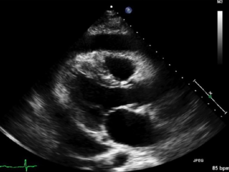
High sensitivity and specificitySensitivity and SpecificityBinary classification measures to assess test results. Sensitivity or recall rate is the proportion of true positives. Specificity is the probability of correctly determining the absence of a condition.Epidemiological Values of Diagnostic Tests
Provides hemodynamic information
Pericardial effusion appears as an echolucent space in the pericardial sac.
Cardiac tamponadeTamponadePericardial effusion, usually of rapid onset, exceeding ventricular filling pressures and causing collapse of the heart with a markedly reduced cardiac output.Pericarditis findings:
Right atrial free-wall collapse during systoleSystolePeriod of contraction of the heart, especially of the heart ventricles.Cardiac Cycle
Right ventricle collapse during diastoleDiastolePost-systolic relaxation of the heart, especially the heart ventricles.Cardiac Cycle
Transthoracic echocardiography showing pericardial effusion (echolucent region around the heart)
Image: “Transthoracic echocardiography” by Department of Internal Medicine, The University of New Mexico, Albuquerque, NM 87106, USA. License: CC BY 4.0
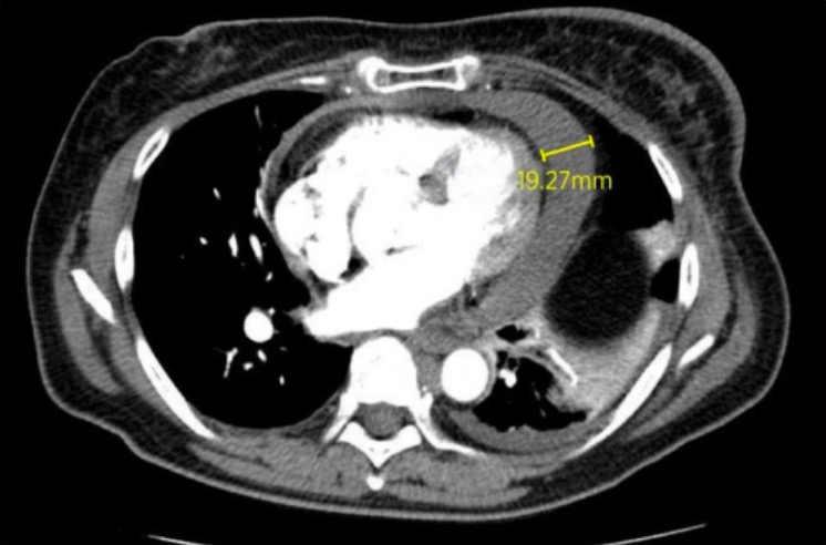
CT and MRI:
Not the diagnostic modalities of choice
May be used if echo imaging is not diagnostic
Can evaluate for pericardial pathology
May be more sensitive for identifying loculated effusions
A CT scan demonstrating pericardial effusion, measuring 19.27 mm
Image: “CT pulmonary embolus” by Stanford Hospital and Clinics, Stanford, California. License: CC BY 2.0
Pericardial fluidPericardial fluidWatery fluid produced in the serous and visceral pericardium surrounding the surface of the heart.Heart: Anatomy analysis and pericardial biopsyBiopsyRemoval and pathologic examination of specimens from the living body.Ewing Sarcoma
Pericardial fluidPericardial fluidWatery fluid produced in the serous and visceral pericardium surrounding the surface of the heart.Heart: Anatomy analysis and pericardial biopsyBiopsyRemoval and pathologic examination of specimens from the living body.Ewing Sarcoma may be performed to determine the cause of the pericardial effusion. The following tests may be conducted on the pericardial fluidPericardial fluidWatery fluid produced in the serous and visceral pericardium surrounding the surface of the heart.Heart: Anatomy:
Acid-fast bacillusBacillusBacillus are aerobic, spore-forming, gram-positive bacilli. Two pathogenic species are Bacillus anthracis (B. anthracis) and B. cereus. Bacillus stain and culture
Viral PCRPCRPolymerase chain reaction (PCR) is a technique that amplifies DNA fragments exponentially for analysis. The process is highly specific, allowing for the targeting of specific genomic sequences, even with minuscule sample amounts. The PCR cycles multiple times through 3 phases: denaturation of the template DNA, annealing of a specific primer to the individual DNA strands, and synthesis/elongation of new DNA molecules.Polymerase Chain Reaction (PCR) panel
Laboratory evaluation
The following tests may be performed to ascertain the etiology of a pericardial effusion:
Thyroid-stimulating hormoneThyroid-stimulating hormoneA glycoprotein hormone secreted by the adenohypophysis. Thyrotropin stimulates thyroid gland by increasing the iodide transport, synthesis and release of thyroid hormones (thyroxine and triiodothyronine).Thyroid Hormones (TSH)
Rheumatoid factorRheumatoid factorAntibodies found in adult rheumatoid arthritis patients that are directed against gamma-chain immunoglobulins.Autoimmune Hepatitis levels
Depends on the patient’s stability and the underlying cause of effusion
Identify and treat the underlying conditions.
Medical therapy for inflammatory effusions or associated pericarditisPericarditisPericarditis is an inflammation of the pericardium, often with fluid accumulation. It can be caused by infection (often viral), myocardial infarction, drugs, malignancies, metabolic disorders, autoimmune disorders, or trauma. Acute, subacute, and chronic forms exist. Pericarditis:
ColchicineColchicineA major alkaloid from colchicum autumnale l. And found also in other colchicum species. Its primary therapeutic use is in the treatment of gout.Gout Drugs
Small effusions in a stable patient are usually self resolving → no need for any intervention
Pericardial drainage can be considered in:
Large symptomatic effusions
Uncertain etiology
Management of cardiac tamponadeTamponadePericardial effusion, usually of rapid onset, exceeding ventricular filling pressures and causing collapse of the heart with a markedly reduced cardiac output.Pericarditis
General considerations:
Administer oxygen.
Measures to ↑ cardiac outputCardiac outputThe volume of blood passing through the heart per unit of time. It is usually expressed as liters (volume) per minute so as not to be confused with stroke volume (volume per beat).Cardiac Mechanics:
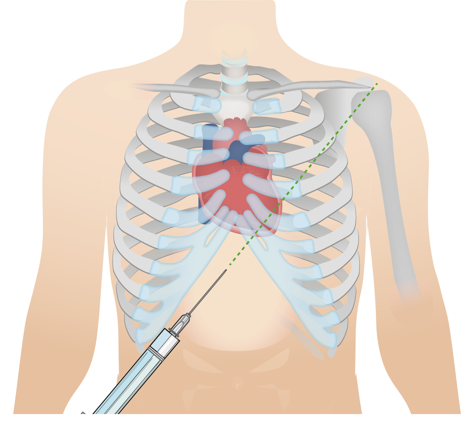
Fluid is removed to relieve pressure on the heart.
A catheter can be placed for periodic drainage.
Subxiphoid approach for pericardiocentesis: This approach allows the drainage of pericardial fluid.
Image by Lecturio.
Surgical management:
Allows for pericardial biopsyBiopsyRemoval and pathologic examination of specimens from the living body.Ewing Sarcoma
Preferred in traumatic pericardial effusions
Options:
Pericardiotomy
Pericardial windowPericardial windowSurgical construction of an opening or window in the pericardium. It is often called subxiphoid pericardial window technique.Cardiac Surgery
Differential Diagnosis
PericarditisPericarditisPericarditis is an inflammation of the pericardium, often with fluid accumulation. It can be caused by infection (often viral), myocardial infarction, drugs, malignancies, metabolic disorders, autoimmune disorders, or trauma. Acute, subacute, and chronic forms exist. Pericarditis: an inflammationInflammationInflammation is a complex set of responses to infection and injury involving leukocytes as the principal cellular mediators in the body’s defense against pathogenic organisms. Inflammation is also seen as a response to tissue injury in the process of wound healing. The 5 cardinal signs of inflammation are pain, heat, redness, swelling, and loss of function. Inflammation of the pericardiumPericardiumA conical fibroserous sac surrounding the heart and the roots of the great vessels (aorta; venae cavae; pulmonary artery). Pericardium consists of two sacs: the outer fibrous pericardium and the inner serous pericardium. The latter consists of an outer parietal layer facing the fibrous pericardium, and an inner visceral layer (epicardium) resting next to the heart, and a pericardial cavity between these two layers.Heart: Anatomy resulting from infection, autoimmune disease, radiationRadiationEmission or propagation of acoustic waves (sound), electromagnetic energy waves (such as light; radio waves; gamma rays; or x-rays), or a stream of subatomic particles (such as electrons; neutrons; protons; or alpha particles).Osteosarcoma, surgery, myocardial infarctionMyocardial infarctionMI is ischemia and death of an area of myocardial tissue due to insufficient blood flow and oxygenation, usually from thrombus formation on a ruptured atherosclerotic plaque in the epicardial arteries. Clinical presentation is most commonly with chest pain, but women and patients with diabetes may have atypical symptoms.Myocardial Infarction, or cardiac surgeryCardiac surgeryCardiac surgery is the surgical management of cardiac abnormalities and of the great vessels of the thorax. In general terms, surgical intervention of the heart is performed to directly restore adequate pump function, correct inherent structural issues, and reestablish proper blood supply via the coronary circulation.Cardiac Surgery. PatientsPatientsIndividuals participating in the health care system for the purpose of receiving therapeutic, diagnostic, or preventive procedures.Clinician–Patient Relationship may have feverFeverFever is defined as a measured body temperature of at least 38°C (100.4°F). Fever is caused by circulating endogenous and/or exogenous pyrogens that increase levels of prostaglandin E2 in the hypothalamus. Fever is commonly associated with chills, rigors, sweating, and flushing of the skin. Fever, pleuritic chest painPainAn unpleasant sensation induced by noxious stimuli which are detected by nerve endings of nociceptive neurons.Pain: Types and Pathways, and a pericardial rub on cardiac auscultation. The diagnosis is confirmed based on diffuse ST elevation on ECGECGAn electrocardiogram (ECG) is a graphic representation of the electrical activity of the heart plotted against time. Adhesive electrodes are affixed to the skin surface allowing measurement of cardiac impulses from many angles. The ECG provides 3-dimensional information about the conduction system of the heart, the myocardium, and other cardiac structures. Electrocardiogram (ECG), and findings of pericardial thickening and effusion on echocardiographyEchocardiographyUltrasonic recording of the size, motion, and composition of the heart and surrounding tissues. The standard approach is transthoracic.Tricuspid Valve Atresia (TVA). Management may include NSAIDsNSAIDSPrimary vs Secondary Headaches, colchicineColchicineA major alkaloid from colchicum autumnale l. And found also in other colchicum species. Its primary therapeutic use is in the treatment of gout.Gout Drugs, and steroidsSteroidsA group of polycyclic compounds closely related biochemically to terpenes. They include cholesterol, numerous hormones, precursors of certain vitamins, bile acids, alcohols (sterols), and certain natural drugs and poisons. Steroids have a common nucleus, a fused, reduced 17-carbon atom ring system, cyclopentanoperhydrophenanthrene. Most steroids also have two methyl groups and an aliphatic side-chain attached to the nucleus.Benign Liver Tumors.
MyocarditisMyocarditisMyocarditis is an inflammatory disease of the myocardium, which may occur alone or in association with a systemic process. There are numerous etiologies of myocarditis, but all lead to inflammation and myocyte injury, most often leading to signs and symptoms of heart failure. Myocarditis: an inflammatory disease of the myocardiumMyocardiumThe muscle tissue of the heart. It is composed of striated, involuntary muscle cells connected to form the contractile pump to generate blood flow.Heart: Anatomy. MyocarditisMyocarditisMyocarditis is an inflammatory disease of the myocardium, which may occur alone or in association with a systemic process. There are numerous etiologies of myocarditis, but all lead to inflammation and myocyte injury, most often leading to signs and symptoms of heart failure. Myocarditis most often leads to signs and symptoms of heart failureHeart FailureA heterogeneous condition in which the heart is unable to pump out sufficient blood to meet the metabolic need of the body. Heart failure can be caused by structural defects, functional abnormalities (ventricular dysfunction), or a sudden overload beyond its capacity. Chronic heart failure is more common than acute heart failure which results from sudden insult to cardiac function, such as myocardial infarction.Total Anomalous Pulmonary Venous Return (TAPVR). The course of myocarditisMyocarditisMyocarditis is an inflammatory disease of the myocardium, which may occur alone or in association with a systemic process. There are numerous etiologies of myocarditis, but all lead to inflammation and myocyte injury, most often leading to signs and symptoms of heart failure. Myocarditis may vary based on the etiology and the timeline of symptom progression. The diagnosis is supported by clinical findings, laboratory evaluation, and cardiac imaging. A definitive diagnosis using endomyocardial biopsyEndomyocardial biopsyMyocarditis is rarely required. Management is supportive and aimed at addressing complications.
Pulmonary embolismPulmonary EmbolismPulmonary embolism (PE) is a potentially fatal condition that occurs as a result of intraluminal obstruction of the main pulmonary artery or its branches. The causative factors include thrombi, air, amniotic fluid, and fat. In PE, gas exchange is impaired due to the decreased return of deoxygenated blood to the lungs. Pulmonary Embolism: an obstruction of the pulmonary arteriesArteriesArteries are tubular collections of cells that transport oxygenated blood and nutrients from the heart to the tissues of the body. The blood passes through the arteries in order of decreasing luminal diameter, starting in the largest artery (the aorta) and ending in the small arterioles. Arteries are classified into 3 types: large elastic arteries, medium muscular arteries, and small arteries and arterioles. Arteries: Histology, most often due to thrombus migration from the deep venous system. Signs and symptoms include pleuritic chest painPainAn unpleasant sensation induced by noxious stimuli which are detected by nerve endings of nociceptive neurons.Pain: Types and Pathways, dyspneaDyspneaDyspnea is the subjective sensation of breathing discomfort. Dyspnea is a normal manifestation of heavy physical or psychological exertion, but also may be caused by underlying conditions (both pulmonary and extrapulmonary). Dyspnea, tachypneaTachypneaIncreased respiratory rate.Pulmonary Examination, and tachycardiaTachycardiaAbnormally rapid heartbeat, usually with a heart rate above 100 beats per minute for adults. Tachycardia accompanied by disturbance in the cardiac depolarization (cardiac arrhythmia) is called tachyarrhythmia.Sepsis in Children. Severe cases can result in hemodynamic instability or cardiopulmonary arrestCardiopulmonary arrestCardiac arrest is the sudden, complete cessation of cardiac output with hemodynamic collapse. Patients present as pulseless, unresponsive, and apneic. Rhythms associated with cardiac arrest are ventricular fibrillation/tachycardia, asystole, or pulseless electrical activity.Cardiac Arrest. A chest CT with angiographyAngiographyRadiography of blood vessels after injection of a contrast medium.Cardiac Surgery is the primary method of diagnosis. Management includes oxygenation, anticoagulationAnticoagulationPulmonary Hypertension Drugs, and thrombolytic therapy for unstable patientsUnstable PatientsBlunt Chest Trauma.
PneumothoraxPneumothoraxA pneumothorax is a life-threatening condition in which air collects in the pleural space, causing partial or full collapse of the lung. A pneumothorax can be traumatic or spontaneous. Patients present with a sudden onset of sharp chest pain, dyspnea, and diminished breath sounds on exam.Pneumothorax: a life-threatening condition in which air collects in the pleural spacePleural spaceThe thin serous membrane enveloping the lungs (lung) and lining the thoracic cavity. Pleura consist of two layers, the inner visceral pleura lying next to the pulmonary parenchyma and the outer parietal pleura. Between the two layers is the pleural cavity which contains a thin film of liquid.Pleuritis, causing a partial or complete collapse of the lung. A pneumothoraxPneumothoraxA pneumothorax is a life-threatening condition in which air collects in the pleural space, causing partial or full collapse of the lung. A pneumothorax can be traumatic or spontaneous. Patients present with a sudden onset of sharp chest pain, dyspnea, and diminished breath sounds on exam.Pneumothorax can be traumatic or spontaneous. PatientsPatientsIndividuals participating in the health care system for the purpose of receiving therapeutic, diagnostic, or preventive procedures.Clinician–Patient Relationship present with a sudden onset of sharp chest painSharp Chest PainChest Pain, dyspneaDyspneaDyspnea is the subjective sensation of breathing discomfort. Dyspnea is a normal manifestation of heavy physical or psychological exertion, but also may be caused by underlying conditions (both pulmonary and extrapulmonary). Dyspnea, and diminished breath sounds on exam. A large or tension pneumothoraxTension PneumothoraxPneumothorax can result in cardiopulmonary collapse. The diagnosis is made based on imaging findings. Management includes needle decompressionNeedle DecompressionPneumothorax and placement of a chest tube (thoracostomyThoracostomySurgical procedure involving the creation of an opening (stoma) into the chest cavity for drainage; used in the treatment of pleural effusion; pneumothorax; hemothorax; and empyema.Hemothorax).
Sagristà-Sauleda, J., Angel, J., Sambola, A., Permanyer-Miralda, G. (2008). Hemodynamic effects of volume expansion in patients with cardiac tamponade. Circulation. 117:1545–1549. https://doi.org/10.1161/CIRCULATIONAHA.107.737841