Epididymitis and orchitis are characterized by acute inflammation of the epididymis and the testicle, respectively, due to viral or bacterial infections. Patients typically present with gradually worsening testicular pain and scrotal swelling along with systemic symptoms such as fever, depending on severity. Patients with concomitant sexually transmitted diseases (STDs) may present with lower urinary tract symptoms. Diagnosis is based on clinical findings and urinalysis with culture. Scrotal ultrasound may show increased blood flow to the affected epididymis or testicle. Treatment is with empiric gram-negative coverage antibiotics and culture-directed therapy. Supportive care includes scrotal support and non-steroidal anti-inflammatory drugs.
EpididymitisEpididymitisEpididymitis and orchitis are characterized by acute inflammation of the epididymis and the testicle, respectively, due to viral or bacterial infections. Patients typically present with gradually worsening testicular pain and scrotal swelling along with systemic symptoms such as fever, depending on severity. Epididymitis and Orchitis: acute inflammationAcute InflammationInflammation of the epididymisEpididymisThe convoluted cordlike structure attached to the posterior of the testis. Epididymis consists of the head (caput), the body (corpus), and the tail (cauda). A network of ducts leaving the testis joins into a common epididymal tubule proper which provides the transport, storage, and maturation of spermatozoa.Testicles: Anatomy due to viral or bacterial infectionsInfectionsInvasion of the host organism by microorganisms or their toxins or by parasites that can cause pathological conditions or diseases.Chronic Granulomatous Disease
OrchitisOrchitisInflammation of a testis. It has many features of epididymitis, such as swollen scrotum; pain; pyuria; and fever. It is usually related to infections in the urinary tract, which likely spread to the epididymis and then the testis through either the vas deferens or the lymphatics of the spermatic cord.Epididymitis and Orchitis: acute inflammationAcute InflammationInflammation of the testis due to viral or bacterial infectionsInfectionsInvasion of the host organism by microorganisms or their toxins or by parasites that can cause pathological conditions or diseases.Chronic Granulomatous Disease
Disease spectrum: EpididymitisEpididymitisEpididymitis and orchitis are characterized by acute inflammation of the epididymis and the testicle, respectively, due to viral or bacterial infections. Patients typically present with gradually worsening testicular pain and scrotal swelling along with systemic symptoms such as fever, depending on severity. Epididymitis and Orchitis can often progress to epididymo-orchitisEpididymo-OrchitisEpididymitis and Orchitis if untreated.
Epidemiology[1,14]
EpididymitisEpididymitisEpididymitis and orchitis are characterized by acute inflammation of the epididymis and the testicle, respectively, due to viral or bacterial infections. Patients typically present with gradually worsening testicular pain and scrotal swelling along with systemic symptoms such as fever, depending on severity. Epididymitis and Orchitis and orchitisOrchitisInflammation of a testis. It has many features of epididymitis, such as swollen scrotum; pain; pyuria; and fever. It is usually related to infections in the urinary tract, which likely spread to the epididymis and then the testis through either the vas deferens or the lymphatics of the spermatic cord.Epididymitis and Orchitis are the most common causes of scrotal painPainAn unpleasant sensation induced by noxious stimuli which are detected by nerve endings of nociceptive neurons.Pain: Types and Pathways in adults (> 600,000 cases per year in the United States).
< 35 years old: Chlamydia trachomatisChlamydia trachomatisType species of Chlamydia causing a variety of ocular and urogenital diseases.Chlamydia (C. trachomatis), NeisseriaNeisseriaNeisseria is a genus of bacteria commonly present on mucosal surfaces. Several species exist, but only 2 are pathogenic to humans: N. gonorrhoeae and N. meningitidis. Neisseria species are non-motile, gram-negative diplococci most commonly isolated on modified Thayer-Martin (MTM) agar. Neisseriagonorrhoeae (N. gonorrhoeae)
> 35 years old: Escherichia coliEscherichia coliThe gram-negative bacterium Escherichia coli is a key component of the human gut microbiota. Most strains of E. coli are avirulent, but occasionally they escape the GI tract, infecting the urinary tract and other sites. Less common strains of E. coli are able to cause disease within the GI tract, most commonly presenting as abdominal pain and diarrhea. Escherichia coli (E. coli) and PseudomonasPseudomonasPseudomonas is a non-lactose-fermenting, gram-negative bacillus that produces pyocyanin, which gives it a characteristic blue-green color. Pseudomonas is found ubiquitously in the environment, as well as in moist reservoirs, such as hospital sinks and respiratory equipment. Pseudomonas related to urinary tractUrinary tractThe urinary tract is located in the abdomen and pelvis and consists of the kidneys, ureters, urinary bladder, and urethra. The structures permit the excretion of urine from the body. Urine flows from the kidneys through the ureters to the urinary bladder and out through the urethra.Urinary Tract: Anatomy infection (UTIUTIUrinary tract infections (UTIs) represent a wide spectrum of diseases, from self-limiting simple cystitis to severe pyelonephritis that can result in sepsis and death. Urinary tract infections are most commonly caused by Escherichia coli, but may also be caused by other bacteria and fungi. Urinary Tract Infections (UTIs)) or prostatitisProstatitisProstatitis is inflammation or an irritative condition of the prostate that presents as different syndromes: acute bacterial, chronic bacterial, chronic prostatitis/chronic pelvic pain, and asymptomatic. Bacterial prostatitis is easier to identify clinically and the management (antibiotics) is better established. Prostatitis
In men with recent instrumentation or urinary tractUrinary tractThe urinary tract is located in the abdomen and pelvis and consists of the kidneys, ureters, urinary bladder, and urethra. The structures permit the excretion of urine from the body. Urine flows from the kidneys through the ureters to the urinary bladder and out through the urethra.Urinary Tract: Anatomy abnormalities, the condition is typically caused by enteric gram-negative bacteriagram-negative bacteriaBacteria which lose crystal violet stain but are stained pink when treated by gram’s method.Bacteriology as well.
Acute epididymitisAcute EpididymitisEpididymitis and Orchitis in children < 14 years of age is due to urinary reflux into the ejaculatory ductsEjaculatory DuctsPaired ducts in the human male through which semen is ejaculated into the urethra..[11]
Symptom duration is usually ≥ 3 months (but can be seen as early as ≥ 6 weeks)[3,6,14]
Genitourinary traumaGenitourinary traumaTraumatic injuries to the genitourinary (GU) tract include injuries to the kidneys, ureter, bladder, urethra, or genitals. Typically, injuries to the GU tract alone are not life threatening, but can be associated with other potentially more significant injuries. Genitourinary Trauma history
Systemic disease
Mycobacterium tuberculosisMycobacterium tuberculosisTuberculosis (TB) is an infectious disease caused by Mycobacterium tuberculosis complex bacteria. The bacteria usually attack the lungs but can also damage other parts of the body. Approximately 30% of people around the world are infected with this pathogen, with the majority harboring a latent infection. Tuberculosis spreads through the air when a person with active pulmonary infection coughs or sneezes.Tuberculosis infection → causes a granulomatous reaction
OrchitisOrchitisInflammation of a testis. It has many features of epididymitis, such as swollen scrotum; pain; pyuria; and fever. It is usually related to infections in the urinary tract, which likely spread to the epididymis and then the testis through either the vas deferens or the lymphatics of the spermatic cord.Epididymitis and Orchitis:[10,15]
Often occurs as an extensionExtensionExamination of the Upper Limbs of epididymitisEpididymitisEpididymitis and orchitis are characterized by acute inflammation of the epididymis and the testicle, respectively, due to viral or bacterial infections. Patients typically present with gradually worsening testicular pain and scrotal swelling along with systemic symptoms such as fever, depending on severity. Epididymitis and Orchitis (epididymo-orchitisEpididymo-OrchitisEpididymitis and Orchitis)
Isolated orchitisOrchitisInflammation of a testis. It has many features of epididymitis, such as swollen scrotum; pain; pyuria; and fever. It is usually related to infections in the urinary tract, which likely spread to the epididymis and then the testis through either the vas deferens or the lymphatics of the spermatic cord.Epididymitis and Orchitis:
Rare
Can rarely be caused by mumpsMumpsMumps is caused by a single-stranded, linear, negative-sense RNA virus of the family Paramyxoviridae. Mumps is typically a disease of childhood, which manifests initially with fever, muscle pain, headache, poor appetite, and a general feeling of malaise, and is classically followed by parotitis. Mumps Virus/Mumps or other virusesVirusesMinute infectious agents whose genomes are composed of DNA or RNA, but not both. They are characterized by a lack of independent metabolism and the inability to replicate outside living host cells.Virology (in nonimmune individuals)
Causes:
Infectious causes:[3,11,13–15]
Bacterial:
C. trachomatis
N. gonorrhoeaeN. gonorrhoeaeA species of gram-negative, aerobic bacteria primarily found in purulent venereal discharges. It is the causative agent of gonorrhea.Neisseria
E. coli
Pseudomonas aeruginosaPseudomonas aeruginosaA species of gram-negative, aerobic, rod-shaped bacteria commonly isolated from clinical specimens (wound, burn, and urinary tract infections). It is also found widely distributed in soil and water. P. Aeruginosa is a major agent of nosocomial infection.Pseudomonas
Klebsiella pneumoniaeKlebsiella PneumoniaeGram-negative, non-motile, capsulated, gas-producing rods found widely in nature and associated with urinary and respiratory infections in humans.Aminoglycosides
Haemophilus influenzaeHaemophilus InfluenzaeA species of Haemophilus found on the mucous membranes of humans and a variety of animals. The species is further divided into biotypes I through viii.Haemophilus
Proteus mirabilisProteus mirabilisA species of gram-negative, facultatively anaerobic, rod-shaped bacteria that is frequently isolated from clinical specimens. Its most common site of infection is the urinary tract.Proteus
Ureaplasma urealyticum
Mycoplasma genitaliumMycoplasma genitaliumA species of gram-negative bacteria originally isolated from urethral specimens of patients with non-gonococcal urethritis. In primates it exists in parasitic association with ciliated epithelial cells in the genital and respiratory tracts.Mycoplasma
M. tuberculosisTuberculosisTuberculosis (TB) is an infectious disease caused by Mycobacterium tuberculosis complex bacteria. The bacteria usually attack the lungs but can also damage other parts of the body. Approximately 30% of people around the world are infected with this pathogen, with the majority harboring a latent infection. Tuberculosis spreads through the air when a person with active pulmonary infection coughs or sneezes. Tuberculosis
BrucellaBrucellaBrucellosis (also known as undulant fever, Mediterranean fever, or Malta fever) is a zoonotic infection that spreads predominantly through ingestion of unpasteurized dairy products or direct contact with infected animal products. Clinical manifestations include fever, arthralgias, malaise, lymphadenopathy, and hepatosplenomegaly. Brucella/Brucellosis spp.
Viral (most common cause in children):
AdenovirusAdenovirusAdenovirus (member of the family Adenoviridae) is a nonenveloped, double-stranded DNA virus. Adenovirus is transmitted in a variety of ways, and it can have various presentations based on the site of entry. Presentation can include febrile pharyngitis, conjunctivitis, acute respiratory disease, atypical pneumonia, and gastroenteritis. Adenovirus
EnterovirusEnterovirusA genus of the family picornaviridae whose members preferentially inhabit the intestinal tract of a variety of hosts. The genus contains many species. Newly described members of human enteroviruses are assigned continuous numbers with the species designated ‘human enterovirus’.Coxsackievirus
Coxsackie virusVirusViruses are infectious, obligate intracellular parasites composed of a nucleic acid core surrounded by a protein capsid. Viruses can be either naked (non-enveloped) or enveloped. The classification of viruses is complex and based on many factors, including type and structure of the nucleoid and capsid, the presence of an envelope, the replication cycle, and the host range. Virology
Epstein-Barr virusEpstein-Barr VirusEpstein-Barr virus (EBV) is a linear, double-stranded DNA virus belonging to the Herpesviridae family. This highly prevalent virus is mostly transmitted through contact with oropharyngeal secretions from an infected individual. The virus can infect epithelial cells and B lymphocytes, where it can undergo lytic replication or latency. Epstein-Barr Virus
CytomegalovirusCytomegalovirusCMV is a ubiquitous double-stranded DNA virus belonging to the Herpesviridae family. CMV infections can be transmitted in bodily fluids, such as blood, saliva, urine, semen, and breast milk. The initial infection is usually asymptomatic in the immunocompetent host, or it can present with symptoms of mononucleosis. Cytomegalovirus
Varicella zoster virusVirusViruses are infectious, obligate intracellular parasites composed of a nucleic acid core surrounded by a protein capsid. Viruses can be either naked (non-enveloped) or enveloped. The classification of viruses is complex and based on many factors, including type and structure of the nucleoid and capsid, the presence of an envelope, the replication cycle, and the host range. Virology
RubellaRubellaAn acute infectious disease caused by the rubella virus. The virus enters the respiratory tract via airborne droplet and spreads to the lymphatic system.Rubella VirusvirusVirusViruses are infectious, obligate intracellular parasites composed of a nucleic acid core surrounded by a protein capsid. Viruses can be either naked (non-enveloped) or enveloped. The classification of viruses is complex and based on many factors, including type and structure of the nucleoid and capsid, the presence of an envelope, the replication cycle, and the host range. Virology
MumpsMumpsMumps is caused by a single-stranded, linear, negative-sense RNA virus of the family Paramyxoviridae. Mumps is typically a disease of childhood, which manifests initially with fever, muscle pain, headache, poor appetite, and a general feeling of malaise, and is classically followed by parotitis. Mumps Virus/MumpsvirusVirusViruses are infectious, obligate intracellular parasites composed of a nucleic acid core surrounded by a protein capsid. Viruses can be either naked (non-enveloped) or enveloped. The classification of viruses is complex and based on many factors, including type and structure of the nucleoid and capsid, the presence of an envelope, the replication cycle, and the host range. Virology
Fungal:
BlastomycosisBlastomycosisBlastomycosis is an infection caused by inhalation of the spores of the fungus, Blastomyces. Blastomyces species thrive in moist soil and decaying material and are common in the Ohio and Mississippi River valleys and the Great Lakes regions of the United States and Canada. Although most patients are asymptomatic, some can develop pneumonia.Blastomyces/Blastomycosis
HistoplasmosisHistoplasmosisHistoplasmosis is an infection caused by Histoplasma capsulatum, a dimorphic fungus. Transmission is through inhalation, and exposure to soils containing bird or bat droppings increases the risk of infection. Most infections are asymptomatic; however, immunocompromised individuals generally develop acute pulmonary infection, chronic infection, or even disseminated disease.Histoplasma/Histoplasmosis
CoccidioidomycosisCoccidioidomycosisCoccidioidomycosis, commonly known as San Joaquin Valley fever, is a fungal disease caused by Coccidioides immitis or Coccidioides posadasii. When Coccidioides spores are inhaled, they transform into spherules that result in infection. Coccidioidomycosis is also a common cause of community-acquired pneumonia and can cause severe disease in the immunocompromised.Coccidioides/Coccidioidomycosis
CandidiasisCandidiasisCandida is a genus of dimorphic, opportunistic fungi. Candida albicans is part of the normal human flora and is the most common cause of candidiasis. The clinical presentation varies and can include localized mucocutaneous infections (e.g., oropharyngeal, esophageal, intertriginous, and vulvovaginal candidiasis) and invasive disease (e.g., candidemia, intraabdominal abscess, pericarditis, and meningitis). Candida/Candidiasis
Urinary tractUrinary tractThe urinary tract is located in the abdomen and pelvis and consists of the kidneys, ureters, urinary bladder, and urethra. The structures permit the excretion of urine from the body. Urine flows from the kidneys through the ureters to the urinary bladder and out through the urethra.Urinary Tract: Anatomy surgery or instrumentation
Autoimmune and inflammatory conditions:
Immunoglobulin A (IgAIgARepresents 15-20% of the human serum immunoglobulins, mostly as the 4-chain polymer in humans or dimer in other mammals. Secretory iga is the main immunoglobulin in secretions.Immunoglobulins: Types and Functions) vasculitisVasculitisInflammation of any one of the blood vessels, including the arteries; veins; and rest of the vasculature system in the body.Systemic Lupus Erythematosus
Behçet disease
SarcoidosisSarcoidosisSarcoidosis is a multisystem inflammatory disease that causes noncaseating granulomas. The exact etiology is unknown. Sarcoidosis usually affects the lungs and thoracic lymph nodes, but it can also affect almost every system in the body, including the skin, heart, and eyes, most commonly. Sarcoidosis
AmiodaroneAmiodaroneAn antianginal and class III antiarrhythmic drug. It increases the duration of ventricular and atrial muscle action by inhibiting potassium channels and voltage-gated sodium channels. There is a resulting decrease in heart rate and in vascular resistance.Pulmonary Fibrosis:
Secondary to high drug concentrations at the head of the epididymisEpididymisThe convoluted cordlike structure attached to the posterior of the testis. Epididymis consists of the head (caput), the body (corpus), and the tail (cauda). A network of ducts leaving the testis joins into a common epididymal tubule proper which provides the transport, storage, and maturation of spermatozoa.Testicles: Anatomy
Occurs in about 3%–11% of patientsPatientsIndividuals participating in the health care system for the purpose of receiving therapeutic, diagnostic, or preventive procedures.Clinician–Patient Relationship
Gradual onset of testicular painPainAn unpleasant sensation induced by noxious stimuli which are detected by nerve endings of nociceptive neurons.Pain: Types and Pathways, swellingSwellingInflammation, and tenderness:
Usually unilateral
Important to differentiate from testicular torsionTesticular torsionTesticular torsion is the sudden rotation of the testicle, specifically the spermatic cord, around its axis in the inguinal canal or below. The acute rotation results in compromised blood flow to and from the testicle, which puts the testicle at risk for necrosis. Testicular Torsion (where painPainAn unpleasant sensation induced by noxious stimuli which are detected by nerve endings of nociceptive neurons.Pain: Types and Pathways is usually sudden, severe, and without signs of infection)
PainPainAn unpleasant sensation induced by noxious stimuli which are detected by nerve endings of nociceptive neurons.Pain: Types and Pathways may radiate to the lower abdomen.
InflammationInflammationInflammation is a complex set of responses to infection and injury involving leukocytes as the principal cellular mediators in the body’s defense against pathogenic organisms. Inflammation is also seen as a response to tissue injury in the process of wound healing. The 5 cardinal signs of inflammation are pain, heat, redness, swelling, and loss of function. Inflammation and swellingSwellingInflammation often start at the tail of the epididymisEpididymisThe convoluted cordlike structure attached to the posterior of the testis. Epididymis consists of the head (caput), the body (corpus), and the tail (cauda). A network of ducts leaving the testis joins into a common epididymal tubule proper which provides the transport, storage, and maturation of spermatozoa.Testicles: Anatomy.
When the inflammationInflammationInflammation is a complex set of responses to infection and injury involving leukocytes as the principal cellular mediators in the body’s defense against pathogenic organisms. Inflammation is also seen as a response to tissue injury in the process of wound healing. The 5 cardinal signs of inflammation are pain, heat, redness, swelling, and loss of function. Inflammation spreads to the rest of the epididymisEpididymisThe convoluted cordlike structure attached to the posterior of the testis. Epididymis consists of the head (caput), the body (corpus), and the tail (cauda). A network of ducts leaving the testis joins into a common epididymal tubule proper which provides the transport, storage, and maturation of spermatozoa.Testicles: Anatomy and testicle → epididymo-orchitisEpididymo-OrchitisEpididymitis and Orchitis
Isolated orchitisOrchitisInflammation of a testis. It has many features of epididymitis, such as swollen scrotum; pain; pyuria; and fever. It is usually related to infections in the urinary tract, which likely spread to the epididymis and then the testis through either the vas deferens or the lymphatics of the spermatic cord.Epididymitis and Orchitis is rare in adults (consider mumpsMumpsMumps is caused by a single-stranded, linear, negative-sense RNA virus of the family Paramyxoviridae. Mumps is typically a disease of childhood, which manifests initially with fever, muscle pain, headache, poor appetite, and a general feeling of malaise, and is classically followed by parotitis. Mumps Virus/Mumps as an etiology in this case).
Lower urinary tractUrinary tractThe urinary tract is located in the abdomen and pelvis and consists of the kidneys, ureters, urinary bladder, and urethra. The structures permit the excretion of urine from the body. Urine flows from the kidneys through the ureters to the urinary bladder and out through the urethra.Urinary Tract: Anatomy symptoms:
Noninfectious epididymitisEpididymitisEpididymitis and orchitis are characterized by acute inflammation of the epididymis and the testicle, respectively, due to viral or bacterial infections. Patients typically present with gradually worsening testicular pain and scrotal swelling along with systemic symptoms such as fever, depending on severity. Epididymitis and Orchitis can have other clinical findings (e.g., rashRashRocky Mountain Spotted Fever in vasculitisVasculitisInflammation of any one of the blood vessels, including the arteries; veins; and rest of the vasculature system in the body.Systemic Lupus Erythematosus).
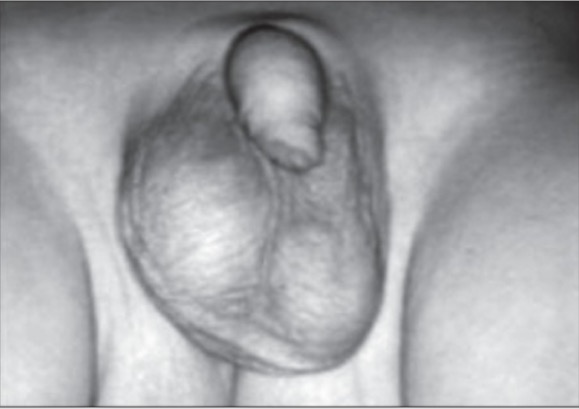
Epididymo-orchitis: presenting with swollen and tender right testicle, which appears larger compared to the normal left testicle
Image: “Painless mass” by Division of Pediatric Surgery, Oita Prefectural Hospital, 476 Bunyou Oita, Japan. License: CC BY 2.0
Chronic testicular painPainAn unpleasant sensation induced by noxious stimuli which are detected by nerve endings of nociceptive neurons.Pain: Types and Pathways
Tuberculous epididymitisEpididymitisEpididymitis and orchitis are characterized by acute inflammation of the epididymis and the testicle, respectively, due to viral or bacterial infections. Patients typically present with gradually worsening testicular pain and scrotal swelling along with systemic symptoms such as fever, depending on severity. Epididymitis and Orchitis can present as a nonresponsive acute epididymitisAcute EpididymitisEpididymitis and Orchitis:
Persistent painPainAn unpleasant sensation induced by noxious stimuli which are detected by nerve endings of nociceptive neurons.Pain: Types and Pathways 2–4 weeks after the initial presentation
Can have a hard, painless massMassThree-dimensional lesion that occupies a space within the breastImaging of the Breast
Scrotal painPainAn unpleasant sensation induced by noxious stimuli which are detected by nerve endings of nociceptive neurons.Pain: Types and Pathways:
Location, severity, duration
Exacerbating factors
Medical (e.g., immunosuppressed state, systemic diseases) and surgical (e.g., urologic instrumentation/surgery) history
Sexual history:
Unprotected intercourse (↑ risk of sexually transmitted infectionsInfectionsInvasion of the host organism by microorganisms or their toxins or by parasites that can cause pathological conditions or diseases.Chronic Granulomatous Disease)
Anal intercourseAnal intercourseHemorrhoids (↑ risk of infectionsInfectionsInvasion of the host organism by microorganisms or their toxins or by parasites that can cause pathological conditions or diseases.Chronic Granulomatous Disease secondary to enteric organisms)
Medications (e.g., amiodaroneAmiodaroneAn antianginal and class III antiarrhythmic drug. It increases the duration of ventricular and atrial muscle action by inhibiting potassium channels and voltage-gated sodium channels. There is a resulting decrease in heart rate and in vascular resistance.Pulmonary Fibrosis)
Other risk factors:
Exposure to tuberculosisTuberculosisTuberculosis (TB) is an infectious disease caused by Mycobacterium tuberculosis complex bacteria. The bacteria usually attack the lungs but can also damage other parts of the body. Approximately 30% of people around the world are infected with this pathogen, with the majority harboring a latent infection. Tuberculosis spreads through the air when a person with active pulmonary infection coughs or sneezes. Tuberculosis or travel to an endemic area
Risk of mumpsMumpsMumps is caused by a single-stranded, linear, negative-sense RNA virus of the family Paramyxoviridae. Mumps is typically a disease of childhood, which manifests initially with fever, muscle pain, headache, poor appetite, and a general feeling of malaise, and is classically followed by parotitis. Mumps Virus/Mumps infection
Clinical findings[3,11,13,14]
Examination: if condition is unilateral, it is best to examine the unaffected side 1st:
Varying levels of painPainAn unpleasant sensation induced by noxious stimuli which are detected by nerve endings of nociceptive neurons.Pain: Types and Pathways intensity
Can have a tender or normal epididymisEpididymisThe convoluted cordlike structure attached to the posterior of the testis. Epididymis consists of the head (caput), the body (corpus), and the tail (cauda). A network of ducts leaving the testis joins into a common epididymal tubule proper which provides the transport, storage, and maturation of spermatozoa.Testicles: Anatomy on palpationPalpationApplication of fingers with light pressure to the surface of the body to determine consistency of parts beneath in physical diagnosis; includes palpation for determining the outlines of organs.Dermatologic Examination
Positive Prehn’s sign: decreased painPainAn unpleasant sensation induced by noxious stimuli which are detected by nerve endings of nociceptive neurons.Pain: Types and Pathways with elevation of testicle
Cremasteric reflexCremasteric ReflexMale Genitourinary Examination: Present (testis pulls up upon stimulation/stroking of the ipsilateral thighThighThe thigh is the region of the lower limb found between the hip and the knee joint. There is a single bone in the thigh called the femur, which is surrounded by large muscles grouped into 3 fascial compartments. Thigh: Anatomy; absence of this reflex suggests possibility of torsion)[1,11]
Laboratory findings[3,7–12,14,15]
Laboratory studies aim to identify a causative infection and should be guided by clinical suspicion. Testing may include:
UrinalysisUrinalysisExamination of urine by chemical, physical, or microscopic means. Routine urinalysis usually includes performing chemical screening tests, determining specific gravity, observing any unusual color or odor, screening for bacteriuria, and examining the sediment microscopically.Urinary Tract Infections (UTIs) in Children: usually with pyuriaPyuriaThe presence of white blood cells (leukocytes) in the urine. It is often associated with bacterial infections of the urinary tract. Pyuria without bacteriuria can be caused by tuberculosis, stones, or cancer.Urinary Tract Infections (UTIs) and/or bacteriuriaBacteriuriaThe presence of bacteria in the urine which is normally bacteria-free. These bacteria are from the urinary tract and are not contaminants of the surrounding tissues. Bacteriuria can be symptomatic or asymptomatic. Significant bacteriuria is an indicator of urinary tract infection.Urinary Tract Infections (UTIs) in Children
Sexually transmitted diseaseSexually Transmitted DiseaseSexually transmitted diseases (STDs) are infections that spread either by vaginal intercourse, anal sex, or oral sex. Symptoms and signs may include vaginal discharge, penile discharge, dysuria, skin lesions (e.g., warts, ulcers) on or around the genitals, and pelvic pain. Some infections can lead to infertility and chronic debilitating disease.Sexually Transmitted Infections (STIs) (STD) screeningScreeningPreoperative Care:
Urine nucleic acid amplificationNucleic acid amplificationLaboratory techniques that involve the in-vitro synthesis of many copies of DNA or RNA from one original template.Septic Arthritis test (NAAT) for N. gonorrhoeaeN. gonorrhoeaeA species of gram-negative, aerobic bacteria primarily found in purulent venereal discharges. It is the causative agent of gonorrhea.Neisseriaand C. trachomatis
Consider treating partner when applicable
If with positive results for gonorrheaGonorrheaGonorrhea is a sexually transmitted infection (STI) caused by the gram-negative bacteria Neisseria gonorrhoeae (N. gonorrhoeae). Gonorrhea may be asymptomatic but commonly manifests as cervicitis or urethritis with less common presentations such as proctitis, conjunctivitis, or pharyngitis. Gonorrhea or chlamydiaChlamydiaChlamydiae are obligate intracellular gram-negative bacteria. They lack a peptidoglycan layer and are best visualized using Giemsa stain. The family of Chlamydiaceae comprises 3 pathogens that can infect humans: Chlamydia trachomatis, Chlamydia psittaci, and Chlamydia pneumoniae.Chlamydia, test the patient for HIVHIVAnti-HIV Drugs and syphilisSyphilisSyphilis is a bacterial infection caused by the spirochete Treponema pallidum pallidum (T. p. pallidum), which is usually spread through sexual contact. Syphilis has 4 clinical stages: primary, secondary, latent, and tertiary. Syphilis.
If Mycoplasma genitaliumMycoplasma genitaliumA species of gram-negative bacteria originally isolated from urethral specimens of patients with non-gonococcal urethritis. In primates it exists in parasitic association with ciliated epithelial cells in the genital and respiratory tracts.Mycoplasma(a slow-growing organism) is suspected → NAAT:
Testing has limited availability.
Use urine, urethral swab
If tuberculosisTuberculosisTuberculosis (TB) is an infectious disease caused by Mycobacterium tuberculosis complex bacteria. The bacteria usually attack the lungs but can also damage other parts of the body. Approximately 30% of people around the world are infected with this pathogen, with the majority harboring a latent infection. Tuberculosis spreads through the air when a person with active pulmonary infection coughs or sneezes. Tuberculosis is suspected:
NAAT for Mycobacterium tuberculosisMycobacterium tuberculosisTuberculosis (TB) is an infectious disease caused by Mycobacterium tuberculosis complex bacteria. The bacteria usually attack the lungs but can also damage other parts of the body. Approximately 30% of people around the world are infected with this pathogen, with the majority harboring a latent infection. Tuberculosis spreads through the air when a person with active pulmonary infection coughs or sneezes.TuberculosisDNADNAA deoxyribonucleotide polymer that is the primary genetic material of all cells. Eukaryotic and prokaryotic organisms normally contain DNA in a double-stranded state, yet several important biological processes transiently involve single-stranded regions. DNA, which consists of a polysugar-phosphate backbone possessing projections of purines (adenine and guanine) and pyrimidines (thymine and cytosine), forms a double helix that is held together by hydrogen bonds between these purines and pyrimidines (adenine to thymine and guanine to cytosine).DNA Types and Structure/RNARNAA polynucleotide consisting essentially of chains with a repeating backbone of phosphate and ribose units to which nitrogenous bases are attached. RNA is unique among biological macromolecules in that it can encode genetic information, serve as an abundant structural component of cells, and also possesses catalytic activity.RNA Types and Structure
If mumpsMumpsMumps is caused by a single-stranded, linear, negative-sense RNA virus of the family Paramyxoviridae. Mumps is typically a disease of childhood, which manifests initially with fever, muscle pain, headache, poor appetite, and a general feeling of malaise, and is classically followed by parotitis. Mumps Virus/Mumps is suspected:
SerologySerologyThe study of serum, especially of antigen-antibody reactions in vitro.Yellow Fever Virus for IgMIgMA class of immunoglobulin bearing mu chains (immunoglobulin mu-chains). Igm can fix complement. The name comes from its high molecular weight and originally being called a macroglobulin.Immunoglobulins: Types and FunctionsantibodiesAntibodiesImmunoglobulins (Igs), also known as antibodies, are glycoprotein molecules produced by plasma cells that act in immune responses by recognizing and binding particular antigens. The various Ig classes are IgG (the most abundant), IgM, IgE, IgD, and IgA, which differ in their biologic features, structure, target specificity, and distribution.Immunoglobulins: Types and Functions
Acute and convalescent IgGIgGThe major immunoglobulin isotype class in normal human serum. There are several isotype subclasses of igg, for example, igg1, igg2a, and igg2b.Hypersensitivity PneumonitisserologySerologyThe study of serum, especially of antigen-antibody reactions in vitro.Yellow Fever Virus
Note: Tissue aspirate/biopsyBiopsyRemoval and pathologic examination of specimens from the living body.Ewing Sarcoma may also be considered to obtain specimens for culture.
Table: Diagnostic testsDiagnostic testsDiagnostic tests are important aspects in making a diagnosis. Some of the most important epidemiological values of diagnostic tests include sensitivity and specificity, false positives and false negatives, positive and negative predictive values, likelihood ratios, and pre-test and post-test probabilities. Epidemiological Values of Diagnostic Tests for some infectious etiologiesInfectious EtiologiesHigh-Risk Headaches of epididymitisEpididymitisEpididymitis and orchitis are characterized by acute inflammation of the epididymis and the testicle, respectively, due to viral or bacterial infections. Patients typically present with gradually worsening testicular pain and scrotal swelling along with systemic symptoms such as fever, depending on severity. Epididymitis and Orchitis and orchitisOrchitisInflammation of a testis. It has many features of epididymitis, such as swollen scrotum; pain; pyuria; and fever. It is usually related to infections in the urinary tract, which likely spread to the epididymis and then the testis through either the vas deferens or the lymphatics of the spermatic cord.Epididymitis and Orchitis[7,15]
Category
Organisms
Test
Bacterial
Chlamydia trachomatisChlamydia trachomatisType species of Chlamydia causing a variety of ocular and urogenital diseases.Chlamydia
NAAT Culture
Neisseria gonorrhoeaeNeisseria gonorrhoeaeA species of gram-negative, aerobic bacteria primarily found in purulent venereal discharges. It is the causative agent of gonorrhea.Neisseria
Enteric bacteriaBacteriaBacteria are prokaryotic single-celled microorganisms that are metabolically active and divide by binary fission. Some of these organisms play a significant role in the pathogenesis of diseases. Bacteriology
Culture
Staphylococcus aureusStaphylococcus aureusPotentially pathogenic bacteria found in nasal membranes, skin, hair follicles, and perineum of warm-blooded animals. They may cause a wide range of infections and intoxications.Brain Abscess
Mycobacterium tuberculosisMycobacterium tuberculosisTuberculosis (TB) is an infectious disease caused by Mycobacterium tuberculosis complex bacteria. The bacteria usually attack the lungs but can also damage other parts of the body. Approximately 30% of people around the world are infected with this pathogen, with the majority harboring a latent infection. Tuberculosis spreads through the air when a person with active pulmonary infection coughs or sneezes.Tuberculosis
Mycoplasma genitaliumMycoplasma genitaliumA species of gram-negative bacteria originally isolated from urethral specimens of patients with non-gonococcal urethritis. In primates it exists in parasitic association with ciliated epithelial cells in the genital and respiratory tracts.Mycoplasma
NAAT
BrucellaBrucellaBrucellosis (also known as undulant fever, Mediterranean fever, or Malta fever) is a zoonotic infection that spreads predominantly through ingestion of unpasteurized dairy products or direct contact with infected animal products. Clinical manifestations include fever, arthralgias, malaise, lymphadenopathy, and hepatosplenomegaly. Brucella/Brucellosis
SerologySerologyThe study of serum, especially of antigen-antibody reactions in vitro.Yellow Fever Virus
Viral
Coxsackie
SerologySerologyThe study of serum, especially of antigen-antibody reactions in vitro.Yellow Fever Virus
MumpsMumpsMumps is caused by a single-stranded, linear, negative-sense RNA virus of the family Paramyxoviridae. Mumps is typically a disease of childhood, which manifests initially with fever, muscle pain, headache, poor appetite, and a general feeling of malaise, and is classically followed by parotitis. Mumps Virus/Mumps
EBVEBVEpstein-barr virus (EBV) is a linear, double-stranded DNA virus belonging to the herpesviridae family. This highly prevalent virus is mostly transmitted through contact with oropharyngeal secretions from an infected individual. The virus can infect epithelial cells and B lymphocytes, where it can undergo lytic replication or latency.Epstein-Barr Virus
Culture (if available)
RubellaRubellaAn acute infectious disease caused by the rubella virus. The virus enters the respiratory tract via airborne droplet and spreads to the lymphatic system.Rubella Virus
Scrotal color DopplerDopplerUltrasonography applying the doppler effect, with frequency-shifted ultrasound reflections produced by moving targets (usually red blood cells) in the bloodstream along the ultrasound axis in direct proportion to the velocity of movement of the targets, to determine both direction and velocity of blood flow.Ultrasound (Sonography) ultrasonography[6,7,11,13,14]
Obtain urgently, preferably within an hour of presentation
Typical findings:
Increased blood flowBlood flowBlood flow refers to the movement of a certain volume of blood through the vasculature over a given unit of time (e.g., mL per minute).Vascular Resistance, Flow, and Mean Arterial Pressure and inflammationInflammationInflammation is a complex set of responses to infection and injury involving leukocytes as the principal cellular mediators in the body’s defense against pathogenic organisms. Inflammation is also seen as a response to tissue injury in the process of wound healing. The 5 cardinal signs of inflammation are pain, heat, redness, swelling, and loss of function. Inflammation of epididymisEpididymisThe convoluted cordlike structure attached to the posterior of the testis. Epididymis consists of the head (caput), the body (corpus), and the tail (cauda). A network of ducts leaving the testis joins into a common epididymal tubule proper which provides the transport, storage, and maturation of spermatozoa.Testicles: Anatomy or testis
Also detects complications:
AbscessAbscessAccumulation of purulent material in tissues, organs, or circumscribed spaces, usually associated with signs of infection.Chronic Granulomatous Disease formation
Testicular torsionTesticular torsionTesticular torsion is the sudden rotation of the testicle, specifically the spermatic cord, around its axis in the inguinal canal or below. The acute rotation results in compromised blood flow to and from the testicle, which puts the testicle at risk for necrosis. Testicular Torsion (with decreased testicular blood flowBlood flowBlood flow refers to the movement of a certain volume of blood through the vasculature over a given unit of time (e.g., mL per minute).Vascular Resistance, Flow, and Mean Arterial Pressure)
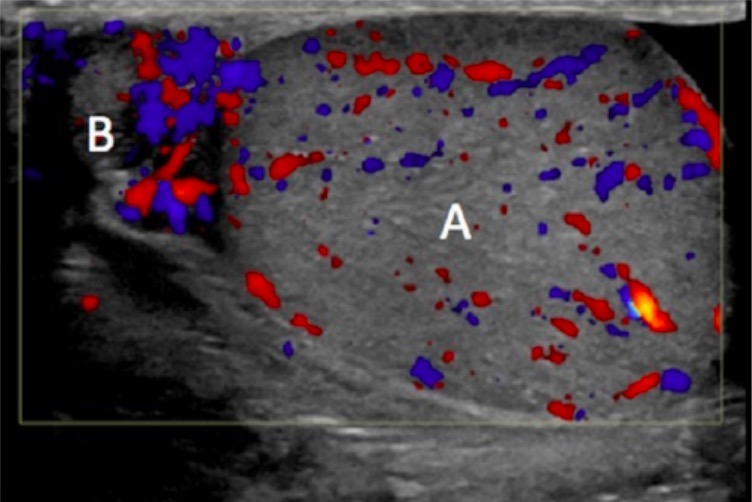
Testicular ultrasound depicting the inflamed epididymis (B) with increased blood flow; testicle (A) with normal blood flow
Image: “Right scrotal ultrasound with color Doppler” by University of Pittsburgh, Graduate School of Medicine, 401 Scaife Hall, 3550 Terrace Street, Pittsburgh, PA 15261, USA. License: CC BY 3.0
Management
Management may vary depending on practice location. The following information is based on US, European, and UK guidelines and literature. See your local guidelines for further recommendations.
Prevent complications (e.g., infertilityInfertilityInfertility is the inability to conceive in the context of regular intercourse. The most common causes of infertility in women are related to ovulatory dysfunction or tubal obstruction, whereas, in men, abnormal sperm is a common cause. Infertility, chronic painChronic painAching sensation that persists for more than a few months. It may or may not be associated with trauma or disease, and may persist after the initial injury has healed. Its localization, character, and timing are more vague than with acute pain.Pain Management)
Indications for hospitalizationHospitalizationThe confinement of a patient in a hospital.Delirium:[11,14]
Severe infection
BacteremiaBacteremiaThe presence of viable bacteria circulating in the blood. Fever, chills, tachycardia, and tachypnea are common acute manifestations of bacteremia. The majority of cases are seen in already hospitalized patients, most of whom have underlying diseases or procedures which render their bloodstreams susceptible to invasion.Glycopeptides
SepsisSepsisSystemic inflammatory response syndrome with a proven or suspected infectious etiology. When sepsis is associated with organ dysfunction distant from the site of infection, it is called severe sepsis. When sepsis is accompanied by hypotension despite adequate fluid infusion, it is called septic shock.Sepsis and Septic Shock
Intractable painPainAn unpleasant sensation induced by noxious stimuli which are detected by nerve endings of nociceptive neurons.Pain: Types and Pathways
Unable to tolerate oral medications
Need for surgery (e.g., abscessAbscessAccumulation of purulent material in tissues, organs, or circumscribed spaces, usually associated with signs of infection.Chronic Granulomatous Disease)
For patientsPatientsIndividuals participating in the health care system for the purpose of receiving therapeutic, diagnostic, or preventive procedures.Clinician–Patient Relationship at risk for STDs, consider STD coverage even before laboratory results are available:
Regimen: ceftriaxoneCeftriaxoneA broad-spectrum cephalosporin antibiotic and cefotaxime derivative with a very long half-life and high penetrability to meninges, eyes and inner ears.Cephalosporins + doxycycline
FluoroquinolonesFluoroquinolonesFluoroquinolones are a group of broad-spectrum, bactericidal antibiotics inhibiting bacterial DNA replication. Fluoroquinolones cover gram-negative, anaerobic, and atypical organisms, as well as some gram-positive and multidrug-resistant (MDR) organisms. Fluoroquinolones not an option for gonococcal coverage (due to increased resistanceResistancePhysiologically, the opposition to flow of air caused by the forces of friction. As a part of pulmonary function testing, it is the ratio of driving pressure to the rate of air flow.Ventilation: Mechanics of Breathing)
AzithromycinAzithromycinA semi-synthetic macrolide antibiotic structurally related to erythromycin. It has been used in the treatment of Mycobacterium avium intracellulare infections, toxoplasmosis, and cryptosporidiosis.Macrolides and Ketolides 1 g (1 dose) is a potential alternative to doxycycline if it is contraindicated.[10]
For patientsPatientsIndividuals participating in the health care system for the purpose of receiving therapeutic, diagnostic, or preventive procedures.Clinician–Patient Relationship most likely infected with enteric pathogens (e.g., urologic instrumentation), and STD is ruled out, consider coverage with:
LevofloxacinLevofloxacinThe l-isomer of ofloxacin.Fluoroquinolones (or ofloxacinOfloxacinA synthetic fluoroquinolone antibacterial agent that inhibits the supercoiling activity of bacterial DNA gyrase, halting DNA replication.Fluoroquinolones outside the US)
TrimethoprimTrimethoprimThe sulfonamides are a class of antimicrobial drugs inhibiting folic acid synthesize in pathogens. The prototypical drug in the class is sulfamethoxazole. Although not technically sulfonamides, trimethoprim, dapsone, and pyrimethamine are also important antimicrobial agents inhibiting folic acid synthesis. The agents are often combined with sulfonamides, resulting in a synergistic effect. Sulfonamides and Trimethoprim–sulfamethoxazoleSulfamethoxazoleA bacteriostatic antibacterial agent that interferes with folic acid synthesis in susceptible bacteria. Its broad spectrum of activity has been limited by the development of resistance.Sulfonamides and Trimethoprim if unable to tolerate fluoroquinolonesFluoroquinolonesFluoroquinolones are a group of broad-spectrum, bactericidal antibiotics inhibiting bacterial DNA replication. Fluoroquinolones cover gram-negative, anaerobic, and atypical organisms, as well as some gram-positive and multidrug-resistant (MDR) organisms. Fluoroquinolones
Once culture data are available, antibiotics can be adjusted (as necessary).
If testing for Mycoplasma genitaliumMycoplasma genitaliumA species of gram-negative bacteria originally isolated from urethral specimens of patients with non-gonococcal urethritis. In primates it exists in parasitic association with ciliated epithelial cells in the genital and respiratory tracts.Mycoplasmais available, and the organism is identified:
Treat with moxifloxacinMoxifloxacinA fluoroquinolone that acts as an inhibitor of DNA topoisomerase II and is used as a broad-spectrum antibacterial agent.Fluoroquinolones 400 mg once daily for 7 days.
Alternative: azithromycinAzithromycinA semi-synthetic macrolide antibiotic structurally related to erythromycin. It has been used in the treatment of Mycobacterium avium intracellulare infections, toxoplasmosis, and cryptosporidiosis.Macrolides and Ketolides 1 g once, then 500 mg daily for the next 3 days (resistanceResistancePhysiologically, the opposition to flow of air caused by the forces of friction. As a part of pulmonary function testing, it is the ratio of driving pressure to the rate of air flow.Ventilation: Mechanics of Breathing is high, so this is given only if susceptibility is proven)
ChlamydiaChlamydiaChlamydiae are obligate intracellular gram-negative bacteria. They lack a peptidoglycan layer and are best visualized using Giemsa stain. The family of Chlamydiaceae comprises 3 pathogens that can infect humans: Chlamydia trachomatis, Chlamydia psittaci, and Chlamydia pneumoniae.Chlamydia and/or gonorrheaGonorrheaGonorrhea is a sexually transmitted infection (STI) caused by the gram-negative bacteria Neisseria gonorrhoeae (N. gonorrhoeae). Gonorrhea may be asymptomatic but commonly manifests as cervicitis or urethritis with less common presentations such as proctitis, conjunctivitis, or pharyngitis. Gonorrhea infection
CeftriaxoneCeftriaxoneA broad-spectrum cephalosporin antibiotic and cefotaxime derivative with a very long half-life and high penetrability to meninges, eyes and inner ears.Cephalosporins 500 mg IM* once, PLUS Doxycycline 100 mg orally twice a day for 10 days
ChlamydiaChlamydiaChlamydiae are obligate intracellular gram-negative bacteria. They lack a peptidoglycan layer and are best visualized using Giemsa stain. The family of Chlamydiaceae comprises 3 pathogens that can infect humans: Chlamydia trachomatis, Chlamydia psittaci, and Chlamydia pneumoniae.Chlamydia, gonorrheaGonorrheaGonorrhea is a sexually transmitted infection (STI) caused by the gram-negative bacteria Neisseria gonorrhoeae (N. gonorrhoeae). Gonorrhea may be asymptomatic but commonly manifests as cervicitis or urethritis with less common presentations such as proctitis, conjunctivitis, or pharyngitis. Gonorrhea, and/or infection from enteric organism/s (e.g., involved in anal sexSexThe totality of characteristics of reproductive structure, functions, phenotype, and genotype, differentiating the male from the female organism.Gender Dysphoria)
CeftriaxoneCeftriaxoneA broad-spectrum cephalosporin antibiotic and cefotaxime derivative with a very long half-life and high penetrability to meninges, eyes and inner ears.Cephalosporins 500 mg IM* once, PLUS LevofloxacinLevofloxacinThe l-isomer of ofloxacin.Fluoroquinolones 500 mg orally once a day for 10 days
*Higher dose if weight is ≥ 150 kg: ceftriaxone 1 g IM
Other recommendations for infectionsInfectionsInvasion of the host organism by microorganisms or their toxins or by parasites that can cause pathological conditions or diseases.Chronic Granulomatous Disease:[3,7–9,12,14]
PatientsPatientsIndividuals participating in the health care system for the purpose of receiving therapeutic, diagnostic, or preventive procedures.Clinician–Patient Relationship with confirmed or suspected STDs should:
Refer sexSexThe totality of characteristics of reproductive structure, functions, phenotype, and genotype, differentiating the male from the female organism.Gender Dysphoria partners within preceding 60 days for evaluation and treatment
Abstain from sexual intercourse until they and their partners are:
Adequately treated
No longer have symptoms
Report STD positivity to local health department
If symptoms persist for ≥ 48–72 hours after initiating treatment, reevaluation is recommended.
Follow up in 1–2 weeks to ensure treatment complianceComplianceDistensibility measure of a chamber such as the lungs (lung compliance) or bladder. Compliance is expressed as a change in volume per unit change in pressure.Veins: Histology.
For noninfectious causes:[4,12]
Management is typically directed at symptoms.
Treat the underlying disease if systemic illness is diagnosed.
Decrease or discontinue amiodaroneAmiodaroneAn antianginal and class III antiarrhythmic drug. It increases the duration of ventricular and atrial muscle action by inhibiting potassium channels and voltage-gated sodium channels. There is a resulting decrease in heart rate and in vascular resistance.Pulmonary Fibrosis (as indicated).
Referral to urology:[3,11]
Children < 14 years of age: Evaluate potential anatomic abnormalities.
Men > 50 years of age: Evaluate urinary tractUrinary tractThe urinary tract is located in the abdomen and pelvis and consists of the kidneys, ureters, urinary bladder, and urethra. The structures permit the excretion of urine from the body. Urine flows from the kidneys through the ureters to the urinary bladder and out through the urethra.Urinary Tract: Anatomy obstruction (e.g., prostateProstateThe prostate is a gland in the male reproductive system. The gland surrounds the bladder neck and a portion of the urethra. The prostate is an exocrine gland that produces a weakly acidic secretion, which accounts for roughly 20% of the seminal fluid. enlargement).
If surgery may be required
If the diagnosis is unclear (e.g., cannot rule out testicular torsionTesticular torsionTesticular torsion is the sudden rotation of the testicle, specifically the spermatic cord, around its axis in the inguinal canal or below. The acute rotation results in compromised blood flow to and from the testicle, which puts the testicle at risk for necrosis. Testicular Torsion)
Surgery is indicated for:[13,14]
Large abscesses
Small abscessAbscessAccumulation of purulent material in tissues, organs, or circumscribed spaces, usually associated with signs of infection.Chronic Granulomatous Disease that fails antibiotic therapy
PainPainAn unpleasant sensation induced by noxious stimuli which are detected by nerve endings of nociceptive neurons.Pain: Types and Pathways management:
Additional painPainAn unpleasant sensation induced by noxious stimuli which are detected by nerve endings of nociceptive neurons.Pain: Types and Pathways treatments to consider:[11]
Neuroleptics (gabapentinGabapentinA cyclohexane-gamma-aminobutyric acid derivative that is used for the treatment of partial seizures; neuralgia; and restless legs syndrome.Second-Generation Anticonvulsant Drugs)
Tricyclic antidepressantsTricyclic antidepressantsTricyclic antidepressants (TCAs) are a class of medications used in the management of mood disorders, primarily depression. These agents, named after their 3-ring chemical structure, act via reuptake inhibition of neurotransmitters (particularly norepinephrine and serotonin) in the brain.Tricyclic Antidepressants
Treat any underlying infection.
Refer to urology if etiology is unclear.
Surgery (epididymectomy) is rarely used but can be beneficial in those for whom conservative measures fail.
Differential Diagnosis
Urinary tractUrinary tractThe urinary tract is located in the abdomen and pelvis and consists of the kidneys, ureters, urinary bladder, and urethra. The structures permit the excretion of urine from the body. Urine flows from the kidneys through the ureters to the urinary bladder and out through the urethra.Urinary Tract: Anatomy infection (UTIUTIUrinary tract infections (UTIs) represent a wide spectrum of diseases, from self-limiting simple cystitis to severe pyelonephritis that can result in sepsis and death. Urinary tract infections are most commonly caused by Escherichia coli, but may also be caused by other bacteria and fungi. Urinary Tract Infections (UTIs)): very common bacterial infection of the lower genitourinary system, which is effectively treated with antibiotics. The patient presents with dysuriaDysuriaPainful urination. It is often associated with infections of the lower urinary tract.Urinary Tract Infections (UTIs), hematuriaHematuriaPresence of blood in the urine.Renal Cell Carcinoma, suprapubic tenderness, or sometimes flank painFlank painPain emanating from below the ribs and above the ilium.Renal Cell Carcinoma. Diagnosis is clinical with urinalysisUrinalysisExamination of urine by chemical, physical, or microscopic means. Routine urinalysis usually includes performing chemical screening tests, determining specific gravity, observing any unusual color or odor, screening for bacteriuria, and examining the sediment microscopically.Urinary Tract Infections (UTIs) in Children and urine cultureUrine cultureUrinary Tract Infections (UTIs).
Testicular torsionTesticular torsionTesticular torsion is the sudden rotation of the testicle, specifically the spermatic cord, around its axis in the inguinal canal or below. The acute rotation results in compromised blood flow to and from the testicle, which puts the testicle at risk for necrosis. Testicular Torsion: acute-onset severe testicular painPainAn unpleasant sensation induced by noxious stimuli which are detected by nerve endings of nociceptive neurons.Pain: Types and Pathways in an adolescent boy, typically without an inciting event, is highly suspicious for torsion. Additional clinical findings include a hard testicle, swollen scrotumScrotumA cutaneous pouch of skin containing the testicles and spermatic cords.Testicles: Anatomy, and negative cremasteric reflexCremasteric ReflexMale Genitourinary Examination. DopplerDopplerUltrasonography applying the doppler effect, with frequency-shifted ultrasound reflections produced by moving targets (usually red blood cells) in the bloodstream along the ultrasound axis in direct proportion to the velocity of movement of the targets, to determine both direction and velocity of blood flow.Ultrasound (Sonography) ultrasound (showing no blood flowBlood flowBlood flow refers to the movement of a certain volume of blood through the vasculature over a given unit of time (e.g., mL per minute).Vascular Resistance, Flow, and Mean Arterial Pressure to the affected testicle) should not delay definitive management, which is acute surgical exploration and orchidopexy.
ProstatitisProstatitisProstatitis is inflammation or an irritative condition of the prostate that presents as different syndromes: acute bacterial, chronic bacterial, chronic prostatitis/chronic pelvic pain, and asymptomatic. Bacterial prostatitis is easier to identify clinically and the management (antibiotics) is better established. Prostatitis: bacterial infection of the prostateProstateThe prostate is a gland in the male reproductive system. The gland surrounds the bladder neck and a portion of the urethra. The prostate is an exocrine gland that produces a weakly acidic secretion, which accounts for roughly 20% of the seminal fluid. gland leading to lower urinary tractUrinary tractThe urinary tract is located in the abdomen and pelvis and consists of the kidneys, ureters, urinary bladder, and urethra. The structures permit the excretion of urine from the body. Urine flows from the kidneys through the ureters to the urinary bladder and out through the urethra.Urinary Tract: Anatomy symptoms. PatientsPatientsIndividuals participating in the health care system for the purpose of receiving therapeutic, diagnostic, or preventive procedures.Clinician–Patient Relationship often present with dysuriaDysuriaPainful urination. It is often associated with infections of the lower urinary tract.Urinary Tract Infections (UTIs), pelvic painPainAn unpleasant sensation induced by noxious stimuli which are detected by nerve endings of nociceptive neurons.Pain: Types and Pathways, urinary frequency, urgency, irritative voiding, and fevers. Diagnosis is clinical with a urinalysisUrinalysisExamination of urine by chemical, physical, or microscopic means. Routine urinalysis usually includes performing chemical screening tests, determining specific gravity, observing any unusual color or odor, screening for bacteriuria, and examining the sediment microscopically.Urinary Tract Infections (UTIs) in Children, culture, and, if needed, a transrectal ultrasound. Treatment consists of long-term gram-negative coverage antibiotics.
Billing and Coding
Diagnosis Codes:
These codes are for epididymitisEpididymitisEpididymitis and orchitis are characterized by acute inflammation of the epididymis and the testicle, respectively, due to viral or bacterial infections. Patients typically present with gradually worsening testicular pain and scrotal swelling along with systemic symptoms such as fever, depending on severity. Epididymitis and Orchitis (inflammationInflammationInflammation is a complex set of responses to infection and injury involving leukocytes as the principal cellular mediators in the body’s defense against pathogenic organisms. Inflammation is also seen as a response to tissue injury in the process of wound healing. The 5 cardinal signs of inflammation are pain, heat, redness, swelling, and loss of function. Inflammation of the epididymisEpididymisThe convoluted cordlike structure attached to the posterior of the testis. Epididymis consists of the head (caput), the body (corpus), and the tail (cauda). A network of ducts leaving the testis joins into a common epididymal tubule proper which provides the transport, storage, and maturation of spermatozoa.Testicles: Anatomy) and orchitisOrchitisInflammation of a testis. It has many features of epididymitis, such as swollen scrotum; pain; pyuria; and fever. It is usually related to infections in the urinary tract, which likely spread to the epididymis and then the testis through either the vas deferens or the lymphatics of the spermatic cord.Epididymitis and Orchitis (inflammationInflammationInflammation is a complex set of responses to infection and injury involving leukocytes as the principal cellular mediators in the body’s defense against pathogenic organisms. Inflammation is also seen as a response to tissue injury in the process of wound healing. The 5 cardinal signs of inflammation are pain, heat, redness, swelling, and loss of function. Inflammation of the testicle), which often occur together (epididymo-orchitisEpididymo-OrchitisEpididymitis and Orchitis).
Coding System
Code
Description
ICD-10-CM
N45.1
EpididymitisEpididymitisEpididymitis and orchitis are characterized by acute inflammation of the epididymis and the testicle, respectively, due to viral or bacterial infections. Patients typically present with gradually worsening testicular pain and scrotal swelling along with systemic symptoms such as fever, depending on severity. Epididymitis and Orchitis
ICD-10-CM
N45.2
OrchitisOrchitisInflammation of a testis. It has many features of epididymitis, such as swollen scrotum; pain; pyuria; and fever. It is usually related to infections in the urinary tract, which likely spread to the epididymis and then the testis through either the vas deferens or the lymphatics of the spermatic cord.Epididymitis and Orchitis
This CPT code is for a scrotal ultrasound, an important imaging test to confirm inflammationInflammationInflammation is a complex set of responses to infection and injury involving leukocytes as the principal cellular mediators in the body’s defense against pathogenic organisms. Inflammation is also seen as a response to tissue injury in the process of wound healing. The 5 cardinal signs of inflammation are pain, heat, redness, swelling, and loss of function. Inflammation, assess blood flowBlood flowBlood flow refers to the movement of a certain volume of blood through the vasculature over a given unit of time (e.g., mL per minute).Vascular Resistance, Flow, and Mean Arterial Pressure, and rule out other causes of testicular painPainAn unpleasant sensation induced by noxious stimuli which are detected by nerve endings of nociceptive neurons.Pain: Types and Pathways like torsion.
Coding System
Code
Description
CPT
76870
Ultrasound, scrotumScrotumA cutaneous pouch of skin containing the testicles and spermatic cords.Testicles: Anatomy and contents
Medications:
These codes are for the antibiotic combination of ceftriaxoneCeftriaxoneA broad-spectrum cephalosporin antibiotic and cefotaxime derivative with a very long half-life and high penetrability to meninges, eyes and inner ears.Cephalosporins and doxycycline, the recommended treatment for epididymitisEpididymitisEpididymitis and orchitis are characterized by acute inflammation of the epididymis and the testicle, respectively, due to viral or bacterial infections. Patients typically present with gradually worsening testicular pain and scrotal swelling along with systemic symptoms such as fever, depending on severity. Epididymitis and Orchitis in men under 35 to cover for sexually transmitted infectionsInfectionsInvasion of the host organism by microorganisms or their toxins or by parasites that can cause pathological conditions or diseases.Chronic Granulomatous Disease like gonorrheaGonorrheaGonorrhea is a sexually transmitted infection (STI) caused by the gram-negative bacteria Neisseria gonorrhoeae (N. gonorrhoeae). Gonorrhea may be asymptomatic but commonly manifests as cervicitis or urethritis with less common presentations such as proctitis, conjunctivitis, or pharyngitis. Gonorrhea and chlamydiaChlamydiaChlamydiae are obligate intracellular gram-negative bacteria. They lack a peptidoglycan layer and are best visualized using Giemsa stain. The family of Chlamydiaceae comprises 3 pathogens that can infect humans: Chlamydia trachomatis, Chlamydia psittaci, and Chlamydia pneumoniae.Chlamydia.
Coding System
Code
Description
RxNorm
2240
CeftriaxoneCeftriaxoneA broad-spectrum cephalosporin antibiotic and cefotaxime derivative with a very long half-life and high penetrability to meninges, eyes and inner ears.Cephalosporins (ingredient)
Nickel, J. C. (2003). Chronic epididymitis: a practical approach to understanding and managing a difficult urologic enigma. Reviews in Urology, 5(4), 209–215. https://www.ncbi.nlm.nih.gov/pmc/articles/PMC1553215/
Chirwa, M., et al. (2021). United Kingdom British association for sexual health and HIV national guideline for the management of epididymo-orchitis, 2020. International Journal of STD & AIDS, 32(10), 884-895. https://www.bashhguidelines.org/media/1291/eo-2020.pdf