Ovarian cysts are defined as collections of fluid or semiliquid material, often walled off by a membrane, located in the ovary. These cysts are broadly categorized as either neoplastic or nonneoplastic. Neoplastic ovarian cysts are subcategorized as either benign or malignant and classified according to their cell of origin. Nonneoplastic cysts are benign and include functional cysts (such as follicular and corpus luteal cysts, which result from normal physiologic processes), hemorrhagic cysts, and endometriomas. In women of reproductive age, neoplastic ovarian cysts are typically benign; however, the risk of malignancy increases in the postmenopausal period. While most ovarian cysts do not cause symptoms, some women report vague symptoms such as lower abdominal pain or abdominal fullness. Complications of cysts include torsion and rupture. Treatment is dependent on the etiology of the ovarian cyst and may range from surgical intervention to supportive care only.
Functional cystsCystsAny fluid-filled closed cavity or sac that is lined by an epithelium. Cysts can be of normal, abnormal, non-neoplastic, or neoplastic tissues.Fibrocystic Change (follicular cystsCystsAny fluid-filled closed cavity or sac that is lined by an epithelium. Cysts can be of normal, abnormal, non-neoplastic, or neoplastic tissues.Fibrocystic Change, corpus luteal cystsCystsAny fluid-filled closed cavity or sac that is lined by an epithelium. Cysts can be of normal, abnormal, non-neoplastic, or neoplastic tissues.Fibrocystic Change, theca lutein cystsCystsAny fluid-filled closed cavity or sac that is lined by an epithelium. Cysts can be of normal, abnormal, non-neoplastic, or neoplastic tissues.Fibrocystic Change)[1]
Simple fluid collections without septations or internal debris
May be a single cyst or multiple cystsCystsAny fluid-filled closed cavity or sac that is lined by an epithelium. Cysts can be of normal, abnormal, non-neoplastic, or neoplastic tissues.Fibrocystic Change
VariableVariableVariables represent information about something that can change. The design of the measurement scales, or of the methods for obtaining information, will determine the data gathered and the characteristics of that data. As a result, a variable can be qualitative or quantitative, and may be further classified into subgroups.Types of Variables size (normal follicles < 3 cm; pathologic follicular cystsCystsAny fluid-filled closed cavity or sac that is lined by an epithelium. Cysts can be of normal, abnormal, non-neoplastic, or neoplastic tissues.Fibrocystic Change are typically < 10 cm)
Lined with granulosa and theca cellsTheca cellsThe flattened stroma cells forming a sheath or theca outside the basal lamina lining the mature ovarian follicle. Thecal interstitial or stromal cells are steroidogenic, and produce primarily androgens which serve as precursors of estrogens in the granulosa cells.Puberty
Corpus luteal cystCorpus luteal cystFollowing ovulation, follicles become corpus luteal cysts. Secrete progesteroneOvarian Cysts:
Following ovulationOvulationThe discharge of an ovum from a rupturing follicle in the ovary.Menstrual Cycle, follicles become corpus luteal cystsCystsAny fluid-filled closed cavity or sac that is lined by an epithelium. Cysts can be of normal, abnormal, non-neoplastic, or neoplastic tissues.Fibrocystic Change
Normal, physiologic structures in the second half of the menstrual cycleMenstrual cycleThe menstrual cycle is the cyclic pattern of hormonal and tissular activity that prepares a suitable uterine environment for the fertilization and implantation of an ovum. The menstrual cycle involves both an endometrial and ovarian cycle that are dependent on one another for proper functioning. There are 2 phases of the ovarian cycle and 3 phases of the endometrial cycle.Menstrual Cycle and 1st trimester of pregnancyPregnancyThe status during which female mammals carry their developing young (embryos or fetuses) in utero before birth, beginning from fertilization to birth.Pregnancy: Diagnosis, Physiology, and Care
Abnormal when they persist (outside of pregnancyPregnancyThe status during which female mammals carry their developing young (embryos or fetuses) in utero before birth, beginning from fertilization to birth.Pregnancy: Diagnosis, Physiology, and Care) or enlarge beyond about 3 cm
Secrete progesteroneProgesteroneThe major progestational steroid that is secreted primarily by the corpus luteum and the placenta. Progesterone acts on the uterus, the mammary glands and the brain. It is required in embryo implantation; pregnancy maintenance, and the development of mammary tissue for milk production. Progesterone, converted from pregnenolone, also serves as an intermediate in the biosynthesis of gonadal steroid hormones and adrenal corticosteroids.Gonadal Hormones required to maintain the endometriumEndometriumThe mucous membrane lining of the uterine cavity that is hormonally responsive during the menstrual cycle and pregnancy. The endometrium undergoes cyclic changes that characterize menstruation. After successful fertilization, it serves to sustain the developing embryo.Embryoblast and Trophoblast Development in pregnancyPregnancyThe status during which female mammals carry their developing young (embryos or fetuses) in utero before birth, beginning from fertilization to birth.Pregnancy: Diagnosis, Physiology, and Care
Characteristics:
Unilateral
Typically, 2–3 cm size (but can be up to 8 cm)
Uniloculated cystsCystsAny fluid-filled closed cavity or sac that is lined by an epithelium. Cysts can be of normal, abnormal, non-neoplastic, or neoplastic tissues.Fibrocystic Change, which can contain some internal debris.
Theca lutein cystsCystsAny fluid-filled closed cavity or sac that is lined by an epithelium. Cysts can be of normal, abnormal, non-neoplastic, or neoplastic tissues.Fibrocystic Change (rare):[2]
Luteinized follicular cystsCystsAny fluid-filled closed cavity or sac that is lined by an epithelium. Cysts can be of normal, abnormal, non-neoplastic, or neoplastic tissues.Fibrocystic Change resulting from overstimulation with human chorionic gonadotropin (hCG)
Associated with:
PregnancyPregnancyThe status during which female mammals carry their developing young (embryos or fetuses) in utero before birth, beginning from fertilization to birth.Pregnancy: Diagnosis, Physiology, and Care, especially multiple gestations (e.g., twins)
Ovarian hyperstimulation during fertility treatments
Gestational trophoblastic diseaseGestational trophoblastic diseaseGestational trophoblastic diseases are a spectrum of placental disorders resulting from abnormal placental trophoblastic growth. These disorders range from benign molar pregnancies (complete and partial moles) to neoplastic conditions such as invasive moles and choriocarcinoma. Gestational Trophoblastic Disease
Clinical presentation of functional cystsCystsAny fluid-filled closed cavity or sac that is lined by an epithelium. Cysts can be of normal, abnormal, non-neoplastic, or neoplastic tissues.Fibrocystic Change:
Usually asymptomatic if < 6 cm
Symptoms may include:
PainPainAn unpleasant sensation induced by noxious stimuli which are detected by nerve endings of nociceptive neurons.Pain: Types and Pathways
Peritoneal irritation
Delayed mensesMensesThe periodic shedding of the endometrium and associated menstrual bleeding in the menstrual cycle of humans and primates. Menstruation is due to the decline in circulating progesterone, and occurs at the late luteal phase when luteolysis of the corpus luteum takes place.Menstrual Cycle
Abnormal uterine bleedingAbnormal Uterine BleedingAbnormal uterine bleeding is the medical term for abnormalities in the frequency, volume, duration, and regularity of the menstrual cycle. Abnormal uterine bleeding is classified using the acronym PALM-COEIN, with PALM representing the structural causes and COEIN indicating the non-structural causes. Abnormal Uterine Bleeding
Theca lutein cystsCystsAny fluid-filled closed cavity or sac that is lined by an epithelium. Cysts can be of normal, abnormal, non-neoplastic, or neoplastic tissues.Fibrocystic Change may also present with:[2]
Maternal virilization
Hyperemesis gravidarum
ThyroidThyroidThe thyroid gland is one of the largest endocrine glands in the human body. The thyroid gland is a highly vascular, brownish-red gland located in the visceral compartment of the anterior region of the neck.Thyroid Gland: Anatomy dysfunction
PreeclampsiaPreeclampsiaA complication of pregnancy, characterized by a complex of symptoms including maternal hypertension and proteinuria with or without pathological edema. Symptoms may range between mild and severe. Pre-eclampsia usually occurs after the 20th week of gestation, but may develop before this time in the presence of trophoblastic disease.Hypertensive Pregnancy Disorders
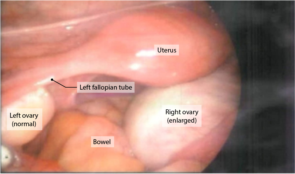
Laparoscopic image showing an enlarged right ovary possibly representing an ovarian cyst
Image: “Laparoscopic finding shows an enlarged right ovary” by Department of Obstetrics and Gynecology, Research Institute of Clinical Medicine, Chonbuk National University Medical School, Jeonju, South Korea. License: CC BY 2.0
Hemorrhagic cystsCystsAny fluid-filled closed cavity or sac that is lined by an epithelium. Cysts can be of normal, abnormal, non-neoplastic, or neoplastic tissues.Fibrocystic Change and endometriomas[1,6]
Hemorrhagic cystsCystsAny fluid-filled closed cavity or sac that is lined by an epithelium. Cysts can be of normal, abnormal, non-neoplastic, or neoplastic tissues.Fibrocystic Change
Follicular or corpus luteal cystsCystsAny fluid-filled closed cavity or sac that is lined by an epithelium. Cysts can be of normal, abnormal, non-neoplastic, or neoplastic tissues.Fibrocystic Change that rupture and bleed into themselves
Present with a relatively sudden onset of acute painAcute painIntensely discomforting, distressful, or agonizing sensation associated with trauma or disease, with well-defined location, character, and timing.Pain Management (often, but not always, unilateral)
Typically self-limited, but on occasion they may present with potentially significant intraperitonealIntraperitonealPeritoneum: Anatomy hemorrhage, seen clinically as:
Severe painPainAn unpleasant sensation induced by noxious stimuli which are detected by nerve endings of nociceptive neurons.Pain: Types and Pathways
Signs of an acute abdomenAcute AbdomenAcute abdomen, which is in many cases a surgical emergency, is the sudden onset of abdominal pain that may be caused by inflammation, infection, perforation, ischemia, or obstruction. The location of the pain, its characteristics, and associated symptoms (e.g., jaundice) are important tools that help narrow the differential diagnosis.Acute Abdomen
Arise from ectopic growth of endometrial tissueEndometrial tissueThe mucous membrane lining of the uterine cavity that is hormonally responsive during the menstrual cycle and pregnancy. The endometrium undergoes cyclic changes that characterize menstruation. After successful fertilization, it serves to sustain the developing embryo.Endometriosis on the ovary
A form of endometriosisEndometriosisEndometriosis is a common disease in which patients have endometrial tissue implanted outside of the uterus. Endometrial implants can occur anywhere in the pelvis, including the ovaries, the broad and uterosacral ligaments, the pelvic peritoneum, and the urinary and gastrointestinal tracts.Endometriosis
Endometriomas are much more likely to be symptomatic, regardless of size, typically presenting with:
Dysmenorrhea
DyspareuniaDyspareuniaRecurrent genital pain occurring during, before, or after sexual intercourse in either the male or the female.Primary Ovarian Insufficiency
InfertilityInfertilityInfertility is the inability to conceive in the context of regular intercourse. The most common causes of infertility in women are related to ovulatory dysfunction or tubal obstruction, whereas, in men, abnormal sperm is a common cause. Infertility
Neoplastic masses are classified according to their cell of origin, with 3 main types:
Epithelial cell tumors
Germ cell tumorsGerm cell tumorsA true neoplasm composed of a number of different types of tissue, none of which is native to the area in which it occurs. It is composed of tissues that are derived from three germinal layers, the endoderm, mesoderm, and ectoderm. They are classified histologically as mature (benign) or immature (malignant).Ovarian Cancer
Epithelial tumors can also be classified as “borderline” or being of “low malignant potential” (managed differently from disease that is clearly benignBenignFibroadenoma or clearly malignant)
Histology, rather than radiographic appearance, determines if a tumorTumorInflammation is benignBenignFibroadenoma, borderline, or malignant.
Table: Classification of neoplastic cystsCystsAny fluid-filled closed cavity or sac that is lined by an epithelium. Cysts can be of normal, abnormal, non-neoplastic, or neoplastic tissues.Fibrocystic Change
70% of malignant epithelial tumors are high-grade serous.
Typically affect postmenopausal women
Germ cell tumorsGerm cell tumorsA true neoplasm composed of a number of different types of tissue, none of which is native to the area in which it occurs. It is composed of tissues that are derived from three germinal layers, the endoderm, mesoderm, and ectoderm. They are classified histologically as mature (benign) or immature (malignant).Ovarian Cancer
Mature cysticCysticFibrocystic ChangeteratomaTeratomaA true neoplasm composed of a number of different types of tissue, none of which is native to the area in which it occurs. It is composed of tissues that are derived from three germinal layers, the endoderm, mesoderm, and ectoderm. They are classified histologically as mature (benign) or immature (malignant).Imaging of the Mediastinum (also called a dermoid cystDermoid cystA tumor consisting of displaced ectodermal structures along the lines of embryonic fusion, the wall being formed of epithelium-lined connective tissue, including skin appendages, and containing keratin, sebum, and hair.Gynecological Imaging)
Malignant degeneration of a mature teratomaTeratomaA true neoplasm composed of a number of different types of tissue, none of which is native to the area in which it occurs. It is composed of tissues that are derived from three germinal layers, the endoderm, mesoderm, and ectoderm. They are classified histologically as mature (benign) or immature (malignant).Imaging of the Mediastinum
Immature teratomasTeratomasA true neoplasm composed of a number of different types of tissue, none of which is native to the area in which it occurs. It is composed of tissues that are derived from three germinal layers, the endoderm, mesoderm, and ectoderm. They are classified histologically as mature (benign) or immature (malignant).Ovarian Cancer
Dysgerminomas
Carcinomas:
Endodermal sinus tumors
Embryonal
Nongestational choriocarcinomaChoriocarcinomaA malignant metastatic form of trophoblastic tumors. Unlike the hydatidiform mole, choriocarcinoma contains no chorionic villi but rather sheets of undifferentiated cytotrophoblasts and syncytiotrophoblasts (trophoblasts). It is characterized by the large amounts of chorionic gonadotropin produced. Tissue origins can be determined by DNA analyses: placental (fetal) origin or non-placental origin.Gestational Trophoblastic Disease
Arise from germ cellsGerm CellsThe reproductive cells in multicellular organisms at various stages during gametogenesis.Gametogenesis (i.e., prematurePrematureChildbirth before 37 weeks of pregnancy (259 days from the first day of the mother’s last menstrual period, or 245 days after fertilization).Necrotizing EnterocolitisoocytesOocytesFemale germ cells derived from oogonia and termed oocytes when they enter meiosis. The primary oocytes begin meiosis but are arrested at the diplotene state until ovulation at puberty to give rise to haploid secondary oocytes or ova (ovum).Ovaries: Anatomy)
5% of primary malignant ovarian tumors
Typically affect younger women (ages 10–30 years)
Sex-cord stromal tumors
Fibroma
Thecoma
Fibrothecoma
Granulosa cellGranulosa cellSupporting cells for the developing female gamete in the ovary. They are derived from the coelomic epithelial cells of the gonadal ridge. Granulosa cells form a single layer around the oocyte in the primordial ovarian follicle and advance to form a multilayered cumulus oophorus surrounding the ovum in the graafian follicle. The major functions of granulosa cells include the production of steroids and LH receptors.Puberty tumors
Theca cell tumors
Fibrosarcoma
Arise from cells in the ovarian stroma
5% of primary malignant ovarian tumors
Typically affect middle-aged/perimenopausal women
May secrete hormonesHormonesHormones are messenger molecules that are synthesized in one part of the body and move through the bloodstream to exert specific regulatory effects on another part of the body. Hormones play critical roles in coordinating cellular activities throughout the body in response to the constant changes in both the internal and external environments. Hormones: Overview and Types
Metastatic tumors
—
Most commonly from:
EndometriumEndometriumThe mucous membrane lining of the uterine cavity that is hormonally responsive during the menstrual cycle and pregnancy. The endometrium undergoes cyclic changes that characterize menstruation. After successful fertilization, it serves to sustain the developing embryo.Embryoblast and Trophoblast Development
Breast
ColonColonThe large intestines constitute the last portion of the digestive system. The large intestine consists of the cecum, appendix, colon (with ascending, transverse, descending, and sigmoid segments), rectum, and anal canal. The primary function of the colon is to remove water and compact the stool prior to expulsion from the body via the rectum and anal canal. Colon, Cecum, and Appendix: Anatomy
CervixCervixThe uterus, cervix, and fallopian tubes are part of the internal female reproductive system. The most inferior portion of the uterus is the cervix, which connects the uterine cavity to the vagina. Externally, the cervix is lined by stratified squamous cells; however, the cervical canal is lined by columnar epithelium.Uterus, Cervix, and Fallopian Tubes: Anatomy
—
Clinical presentation[1,6,12]
Neoplastic ovarian masses are frequently asymptomatic. If they are symptomatic, they typically present with vague, nonspecific symptoms which can include:
Lower abdominal and/or pelvic painPainAn unpleasant sensation induced by noxious stimuli which are detected by nerve endings of nociceptive neurons.Pain: Types and Pathways:
Typically vague, noncyclic painPainAn unpleasant sensation induced by noxious stimuli which are detected by nerve endings of nociceptive neurons.Pain: Types and Pathways of insidious onset
Acute-onset painPainAn unpleasant sensation induced by noxious stimuli which are detected by nerve endings of nociceptive neurons.Pain: Types and Pathways associated with ovarian torsionOvarian torsionOvarian torsion is a clinical emergency in which the ovaries (with or without the fallopian tubes) twist along their axis, leading to partial or complete obstruction of their blood supply. Ovarian torsion is also called adnexal or tubo-ovarian torsion, especially if a fallopian tube is also involved. Ovarian Torsion, or bleeding
Abnormal uterine bleedingAbnormal Uterine BleedingAbnormal uterine bleeding is the medical term for abnormalities in the frequency, volume, duration, and regularity of the menstrual cycle. Abnormal uterine bleeding is classified using the acronym PALM-COEIN, with PALM representing the structural causes and COEIN indicating the non-structural causes. Abnormal Uterine Bleeding
Urinary frequency
ConstipationConstipationConstipation is common and may be due to a variety of causes. Constipation is generally defined as bowel movement frequency < 3 times per week. Patients who are constipated often strain to pass hard stools. The condition is classified as primary (also known as idiopathic or functional constipation) or secondary, and as acute or chronic. Constipation
LegLegThe lower leg, or just “leg” in anatomical terms, is the part of the lower limb between the knee and the ankle joint. The bony structure is composed of the tibia and fibula bones, and the muscles of the leg are grouped into the anterior, lateral, and posterior compartments by extensions of fascia.Leg: AnatomyswellingSwellingInflammation
Sex-cord stromal tumors in particular may secrete hormonesHormonesHormones are messenger molecules that are synthesized in one part of the body and move through the bloodstream to exert specific regulatory effects on another part of the body. Hormones play critical roles in coordinating cellular activities throughout the body in response to the constant changes in both the internal and external environments. Hormones: Overview and Types:
Granulosa cellGranulosa cellSupporting cells for the developing female gamete in the ovary. They are derived from the coelomic epithelial cells of the gonadal ridge. Granulosa cells form a single layer around the oocyte in the primordial ovarian follicle and advance to form a multilayered cumulus oophorus surrounding the ovum in the graafian follicle. The major functions of granulosa cells include the production of steroids and LH receptors.Puberty tumors: estrogenEstrogenCompounds that interact with estrogen receptors in target tissues to bring about the effects similar to those of estradiol. Estrogens stimulate the female reproductive organs, and the development of secondary female sex characteristics. Estrogenic chemicals include natural, synthetic, steroidal, or non-steroidal compounds.Ovaries: Anatomy → may present with:
SuppressionSuppressionDefense Mechanisms of mensesMensesThe periodic shedding of the endometrium and associated menstrual bleeding in the menstrual cycle of humans and primates. Menstruation is due to the decline in circulating progesterone, and occurs at the late luteal phase when luteolysis of the corpus luteum takes place.Menstrual Cycle/abnormal bleeding
Breast enlargement
Theca cell tumors: testosteroneTestosteroneA potent androgenic steroid and major product secreted by the leydig cells of the testis. Its production is stimulated by luteinizing hormone from the pituitary gland. In turn, testosterone exerts feedback control of the pituitary LH and FSH secretion. Depending on the tissues, testosterone can be further converted to dihydrotestosterone or estradiol.Androgens and Antiandrogens → may present with:
HirsutismHirsutismA condition observed in women and children when there is excess coarse body hair of an adult male distribution pattern, such as facial and chest areas. It is the result of elevated androgens from the ovaries, the adrenal glands, or exogenous sources. The concept does not include hypertrichosis, which is an androgen-independent excessive hair growth.Polycystic Ovarian Syndrome
Hair loss
SuppressionSuppressionDefense Mechanisms of mensesMensesThe periodic shedding of the endometrium and associated menstrual bleeding in the menstrual cycle of humans and primates. Menstruation is due to the decline in circulating progesterone, and occurs at the late luteal phase when luteolysis of the corpus luteum takes place.Menstrual Cycle
Diagnosis
The key to diagnosing ovarian cystsCystsAny fluid-filled closed cavity or sac that is lined by an epithelium. Cysts can be of normal, abnormal, non-neoplastic, or neoplastic tissues.Fibrocystic Change is to accurately identify benignBenignFibroadenomacystsCystsAny fluid-filled closed cavity or sac that is lined by an epithelium. Cysts can be of normal, abnormal, non-neoplastic, or neoplastic tissues.Fibrocystic Change that will resolve spontaneously (e.g., follicular, corpus luteal, and hemorrhagic cystsCystsAny fluid-filled closed cavity or sac that is lined by an epithelium. Cysts can be of normal, abnormal, non-neoplastic, or neoplastic tissues.Fibrocystic Change), differentiating them from cystsCystsAny fluid-filled closed cavity or sac that is lined by an epithelium. Cysts can be of normal, abnormal, non-neoplastic, or neoplastic tissues.Fibrocystic Change that are benignBenignFibroadenoma but will not resolve spontaneously (e.g., endometriomas, benignBenignFibroadenoma neoplastic cystsCystsAny fluid-filled closed cavity or sac that is lined by an epithelium. Cysts can be of normal, abnormal, non-neoplastic, or neoplastic tissues.Fibrocystic Change) and cystsCystsAny fluid-filled closed cavity or sac that is lined by an epithelium. Cysts can be of normal, abnormal, non-neoplastic, or neoplastic tissues.Fibrocystic Change that are malignant.
History and exam[1,12]
Consistent clinical symptoms
Palpable adnexal massMassThree-dimensional lesion that occupies a space within the breastImaging of the Breast on pelvic examination
BenignBenignFibroadenomacystsCystsAny fluid-filled closed cavity or sac that is lined by an epithelium. Cysts can be of normal, abnormal, non-neoplastic, or neoplastic tissues.Fibrocystic Change:
Unilocular
Thin-walled
Absence of solid components and vascularity within the lesion
Follicular cystsCystsAny fluid-filled closed cavity or sac that is lined by an epithelium. Cysts can be of normal, abnormal, non-neoplastic, or neoplastic tissues.Fibrocystic Change: simple, anechoicAnechoicA structure that produces no echo at all (looks completely black)Ultrasound (Sonography) structures
Corpus luteal cystsCystsAny fluid-filled closed cavity or sac that is lined by an epithelium. Cysts can be of normal, abnormal, non-neoplastic, or neoplastic tissues.Fibrocystic Change: may have a complex appearance with internal echoes and slightly thicker walls
Hemorrhagic cystsCystsAny fluid-filled closed cavity or sac that is lined by an epithelium. Cysts can be of normal, abnormal, non-neoplastic, or neoplastic tissues.Fibrocystic Change and endometriomas:
Differentiated from one another with follow-up ultrasonography in 8‒12 weeks; a hemorrhagic cystHemorrhagic CystGynecological Imaging will resolve (or be resolving), whereas an endometriomaEndometriomaEndometriosis will persist without significant changes.
Malignant tumors:
Worrisome size: > 10 cm
Multilocular (thicker and/or vascularized walls are more worrisome)
Increased vascularity within and surrounding the lesion
Presence of ascitesAscitesAscites is the pathologic accumulation of fluid within the peritoneal cavity that occurs due to an osmotic and/or hydrostatic pressure imbalance secondary to portal hypertension (cirrhosis, heart failure) or non-portal hypertension (hypoalbuminemia, malignancy, infection).Ascites
Abdominal involvement
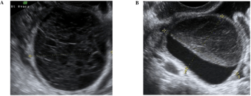
A resolving hemorrhagic corpus luteum cyst. A) demonstrates the typical appearance of an early hemorrhagic corpus luteal cyst, with the lace-like or cob-web appearance of the fibrin strings of the recently formed clot. B) Demonstrates the appearance of the cyst after clot retraction.
Image: “f2-ijo-46-02-0445” by Ahmad Sayasneh et al. License: CC BY 3.0
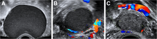
Sonographic appearance of a typical endometrioma. [A] shows a unilocular cyst with the typical “ground glass appearance. [B] and [C] demonstrate minimal vascularity within/around the mass.
Image: “Fig1” by Katie Pateman et al. License: CC BY 4.0
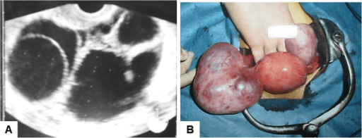
(A) Transvaginal ultrasound of an enlarged ovary with multiple simple cysts caused by a follicle stimulating hormone (FSH)-secreting pituitary adenoma.
(B) Photograph of the enlarged ovary at surgery.
Image: “F1” by Tomohiro Kawaguchi et al. License: CC BY 2.0
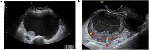
Ultrasounds of malignant ovarian epithelial tumors.
A) Stage 1 clear cell carcinoma of the ovary. Note the solid components in the bottom left of the image.
B) Demonstrates a unilocular invasive epithelial cancer with increased vascularity within the cyst on color doppler, a finding concerning for malignancy.
Image: “f22-ijo-46-02-0445” by Ahmad Sayasenh et al. License: CC BY 3.0
MRI: 2nd-line imaging used for surgical planning and/or when diagnosis is unclear after ultrasonography[1,12]
MRI usually reveals a thin and featureless wall that enhances the uptake of gadoliniumGadoliniumAn element of the rare earth family of metals. It has the atomic symbol gd, atomic number 64, and atomic weight 157. 25. Its oxide is used in the control rods of some nuclear reactors.Magnetic Resonance Imaging (MRI).
T2 shortening is not seen in corpus luteal cystsCystsAny fluid-filled closed cavity or sac that is lined by an epithelium. Cysts can be of normal, abnormal, non-neoplastic, or neoplastic tissues.Fibrocystic Change, in contrast to endometrial chocolate cystsChocolate CystsEndometriosis.
Malignant lesions (similar features are seen on ultrasonography):
Heterogeneous
Faster contrast enhancement
Calcification
Multilocular cyst
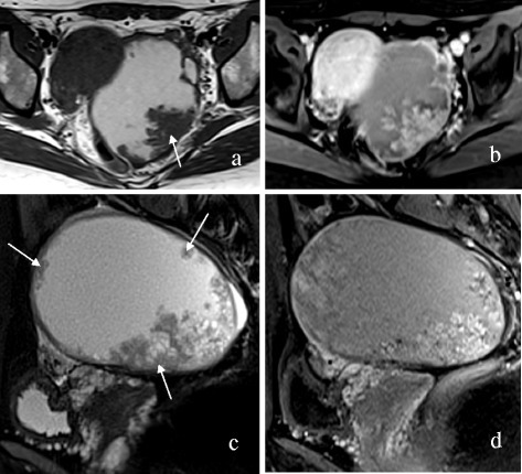
MRI demonstrating a left-sided ovarian epithelial carcinoma. T2-weighted images (a: axial; c: sagittal) demonstrate a mainly cystic mass with multiple mural nodules (arrows). T1-weighted images with fat suppression (b: axial; d: sagittal) show that the nodules moderately enhance
Image: “Fig2: A 32-year-old woman with left-sided OEC. ” by Hai Ming Li et al. License: CC BY 4.0CC BY 1.0
Laboratory and pathology
Definitive diagnosis of an ovarian cyst/massMassThree-dimensional lesion that occupies a space within the breastImaging of the Breast is made on histologic examination (see Ovarian cancerOvarian cancerOvarian cancer is a malignant tumor arising from the ovarian tissue and is classified according to the type of tissue from which it originates. The 3 major types of ovarian cancer are epithelial ovarian carcinomas (EOCs), ovarian germ cell tumors (OGCTs), and sex cord-stromal tumors (SCSTs). Ovarian Cancer concept for additional details). Several serum tests are available to help assess the risk of malignancyMalignancyHemothorax prior to surgery.
PregnancyPregnancyThe status during which female mammals carry their developing young (embryos or fetuses) in utero before birth, beginning from fertilization to birth.Pregnancy: Diagnosis, Physiology, and Care test in individuals of reproductive age:[1,12]
Rules out ectopic pregnancyEctopic pregnancyEctopic pregnancy refers to the implantation of a fertilized egg (embryo) outside the uterine cavity. The main cause is disruption of the normal anatomy of the fallopian tube. Ectopic Pregnancy
A normal intrauterine pregnancyPregnancyThe status during which female mammals carry their developing young (embryos or fetuses) in utero before birth, beginning from fertilization to birth.Pregnancy: Diagnosis, Physiology, and Care affects management.
Cancer antigen 125Cancer antigen 125A carbohydrate antigen that occurs in tumors of the ovary as well as in breast, kidney, and gastrointestinal tract tumors and normal tissue. While it is tumor-associated, it is not tumor-specific and may have a protective function against particles and infectious agents at mucosal surfaces.Serum Tumor Markers (CA-125CA-125A carbohydrate antigen that occurs in tumors of the ovary as well as in breast, kidney, and gastrointestinal tract tumors and normal tissue. While it is tumor-associated, it is not tumor-specific and may have a protective function against particles and infectious agents at mucosal surfaces.Serum Tumor Markers) blood test:[1,4,9,10,12]
Indications:
Postmenopausal women with an adnexal massMassThree-dimensional lesion that occupies a space within the breastImaging of the Breast
Premenopausal women with ultrasound findings suspicious for malignancyMalignancyHemothorax
Complex cystsCystsAny fluid-filled closed cavity or sac that is lined by an epithelium. Cysts can be of normal, abnormal, non-neoplastic, or neoplastic tissues.Fibrocystic Change and high CA-125CA-125A carbohydrate antigen that occurs in tumors of the ovary as well as in breast, kidney, and gastrointestinal tract tumors and normal tissue. While it is tumor-associated, it is not tumor-specific and may have a protective function against particles and infectious agents at mucosal surfaces.Serum Tumor Markers associated with increased risk of malignancyMalignancyHemothorax[1]
CA-125CA-125A carbohydrate antigen that occurs in tumors of the ovary as well as in breast, kidney, and gastrointestinal tract tumors and normal tissue. While it is tumor-associated, it is not tumor-specific and may have a protective function against particles and infectious agents at mucosal surfaces.Serum Tumor Markers is ↑ in 80% of individuals with epithelial ovarian cancerOvarian cancerOvarian cancer is a malignant tumor arising from the ovarian tissue and is classified according to the type of tissue from which it originates. The 3 major types of ovarian cancer are epithelial ovarian carcinomas (EOCs), ovarian germ cell tumors (OGCTs), and sex cord-stromal tumors (SCSTs). Ovarian Cancer.
CA-125CA-125A carbohydrate antigen that occurs in tumors of the ovary as well as in breast, kidney, and gastrointestinal tract tumors and normal tissue. While it is tumor-associated, it is not tumor-specific and may have a protective function against particles and infectious agents at mucosal surfaces.Serum Tumor Markers is not used as a screeningScreeningPreoperative Care test, however, because only 50% of individuals with stage I epithelial ovarian cancerOvarian cancerOvarian cancer is a malignant tumor arising from the ovarian tissue and is classified according to the type of tissue from which it originates. The 3 major types of ovarian cancer are epithelial ovarian carcinomas (EOCs), ovarian germ cell tumors (OGCTs), and sex cord-stromal tumors (SCSTs). Ovarian Cancer have ↑ CA-125CA-125A carbohydrate antigen that occurs in tumors of the ovary as well as in breast, kidney, and gastrointestinal tract tumors and normal tissue. While it is tumor-associated, it is not tumor-specific and may have a protective function against particles and infectious agents at mucosal surfaces.Serum Tumor Markers.
Note: CA-125CA-125A carbohydrate antigen that occurs in tumors of the ovary as well as in breast, kidney, and gastrointestinal tract tumors and normal tissue. While it is tumor-associated, it is not tumor-specific and may have a protective function against particles and infectious agents at mucosal surfaces.Serum Tumor Markers may be elevated in many conditions, including:
EndometriosisEndometriosisEndometriosis is a common disease in which patients have endometrial tissue implanted outside of the uterus. Endometrial implants can occur anywhere in the pelvis, including the ovaries, the broad and uterosacral ligaments, the pelvic peritoneum, and the urinary and gastrointestinal tracts.Endometriosis
Leiomyomas
PregnancyPregnancyThe status during which female mammals carry their developing young (embryos or fetuses) in utero before birth, beginning from fertilization to birth.Pregnancy: Diagnosis, Physiology, and Care
Pelvic inflammatory diseasePelvic inflammatory diseasePelvic inflammatory disease (PID) is defined as a polymicrobial infection of the upper female reproductive system. The disease can affect the uterus, fallopian tubes, ovaries, and adjacent structures. Pelvic inflammatory disease is closely linked with sexually transmitted diseases, most commonly caused by Chlamydia trachomatis, Neisseria gonorrhoeae, and Gardnerella vaginalis. Pelvic Inflammatory Disease (PIDPIDPelvic inflammatory disease (PID) is defined as a polymicrobial infection of the upper female reproductive system. The disease can affect the uterus, fallopian tubes, ovaries, and adjacent structures. Pelvic inflammatory disease is closely linked with sexually transmitted diseases, most commonly caused by Chlamydia trachomatis, Neisseria gonorrhoeae, and gardnerella vaginalis.Pelvic Inflammatory Disease)
Nongynecologic cancers
The FDA has approved tumorTumorInflammation marker serum panels to assess the risk of ovarian malignancyMalignancyHemothorax in adult women with pelvic masses.[1]
Multivariate index assay (MIA), which uses:[3]
CA-125CA-125A carbohydrate antigen that occurs in tumors of the ovary as well as in breast, kidney, and gastrointestinal tract tumors and normal tissue. While it is tumor-associated, it is not tumor-specific and may have a protective function against particles and infectious agents at mucosal surfaces.Serum Tumor Markers II
TransferrinTransferrinAn iron-binding beta1-globulin that is synthesized in the liver and secreted into the blood. It plays a central role in the transport of iron throughout the circulation.Heme Metabolism
Risk of Ovarian MalignancyMalignancyHemothorax Algorithm (ROMA), which uses:[10]
CA-125CA-125A carbohydrate antigen that occurs in tumors of the ovary as well as in breast, kidney, and gastrointestinal tract tumors and normal tissue. While it is tumor-associated, it is not tumor-specific and may have a protective function against particles and infectious agents at mucosal surfaces.Serum Tumor Markers
Human epididymisEpididymisThe convoluted cordlike structure attached to the posterior of the testis. Epididymis consists of the head (caput), the body (corpus), and the tail (cauda). A network of ducts leaving the testis joins into a common epididymal tubule proper which provides the transport, storage, and maturation of spermatozoa.Testicles: Anatomy protein 4 (HE4)
Menopausal status
Both use specific algorithms to determine an overall score; specific cutoffs have been identified that indicate that an individual is at higher risk for malignancyMalignancyHemothorax.
In women < 40 years of age with a complex ovarian massMassThree-dimensional lesion that occupies a space within the breastImaging of the Breast, additional biomarkers are recommended to screen for germ cell tumorsGerm cell tumorsA true neoplasm composed of a number of different types of tissue, none of which is native to the area in which it occurs. It is composed of tissues that are derived from three germinal layers, the endoderm, mesoderm, and ectoderm. They are classified histologically as mature (benign) or immature (malignant).Ovarian Cancer, including:[12]
Table: Patterns of serum biomarkers in different ovarian germ cell tumorsGerm cell tumorsA true neoplasm composed of a number of different types of tissue, none of which is native to the area in which it occurs. It is composed of tissues that are derived from three germinal layers, the endoderm, mesoderm, and ectoderm. They are classified histologically as mature (benign) or immature (malignant).Ovarian Cancer[1]
CA-125CA-125A carbohydrate antigen that occurs in tumors of the ovary as well as in breast, kidney, and gastrointestinal tract tumors and normal tissue. While it is tumor-associated, it is not tumor-specific and may have a protective function against particles and infectious agents at mucosal surfaces.Serum Tumor Markers
Immature teratomaTeratomaA true neoplasm composed of a number of different types of tissue, none of which is native to the area in which it occurs. It is composed of tissues that are derived from three germinal layers, the endoderm, mesoderm, and ectoderm. They are classified histologically as mature (benign) or immature (malignant).Imaging of the Mediastinum
Nongestational choriocarcinomaChoriocarcinomaA malignant metastatic form of trophoblastic tumors. Unlike the hydatidiform mole, choriocarcinoma contains no chorionic villi but rather sheets of undifferentiated cytotrophoblasts and syncytiotrophoblasts (trophoblasts). It is characterized by the large amounts of chorionic gonadotropin produced. Tissue origins can be determined by DNA analyses: placental (fetal) origin or non-placental origin.Gestational Trophoblastic Disease
+
‒
‒
‒
AFP: α-fetoprotein β-hCG: β human chorionic gonadotropin CA-125: cancer antigen 125 LDH: lactate dehydrogenase
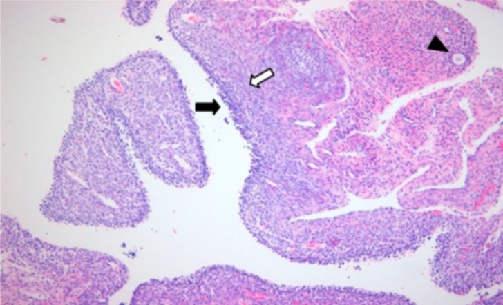
Follicular cyst
Image: “Histopathology of follicular cyst wall” by Department of Obstetrics and Gynecology, Research Institute of Clinical Medicine, Chonbuk National University Medical School, Jeonju, South Korea. License: CC BY 2.0
RMI calculator: clinical tool used to determine risk of cancer for ovarian tumors and to guide management
Considers 3 features:
RMI = (menopausal status score) x (ultrasound score) x serum CA-125CA-125A carbohydrate antigen that occurs in tumors of the ovary as well as in breast, kidney, and gastrointestinal tract tumors and normal tissue. While it is tumor-associated, it is not tumor-specific and may have a protective function against particles and infectious agents at mucosal surfaces.Serum Tumor Markers (U/mL)
Ultrasound score: points given for specific findings of concern:
Worrisome features:
Multiloculated (instead of uniloculated) cystsCystsAny fluid-filled closed cavity or sac that is lined by an epithelium. Cysts can be of normal, abnormal, non-neoplastic, or neoplastic tissues.Fibrocystic Change
Solid areas
Bilateral lesions
AscitesAscitesAscites is the pathologic accumulation of fluid within the peritoneal cavity that occurs due to an osmotic and/or hydrostatic pressure imbalance secondary to portal hypertension (cirrhosis, heart failure) or non-portal hypertension (hypoalbuminemia, malignancy, infection).Ascites
Intra-abdominal metastases
If 0‒1 worrisome features are present: 1 point
If ≥ 2 worrisome features are present: 3 points
Presence of CA-125CA-125A carbohydrate antigen that occurs in tumors of the ovary as well as in breast, kidney, and gastrointestinal tract tumors and normal tissue. While it is tumor-associated, it is not tumor-specific and may have a protective function against particles and infectious agents at mucosal surfaces.Serum Tumor Markers in serum
CystsCystsAny fluid-filled closed cavity or sac that is lined by an epithelium. Cysts can be of normal, abnormal, non-neoplastic, or neoplastic tissues.Fibrocystic Change/masses with an elevated RMI (> 200) should be referred to a gynecologic oncologist.[1,12]
Indications for surgical removal:
Size > 10 cm
Suspected ovarian torsionOvarian torsionOvarian torsion is a clinical emergency in which the ovaries (with or without the fallopian tubes) twist along their axis, leading to partial or complete obstruction of their blood supply. Ovarian torsion is also called adnexal or tubo-ovarian torsion, especially if a fallopian tube is also involved. Ovarian Torsion
Persistent adnexal massMassThree-dimensional lesion that occupies a space within the breastImaging of the Breast
Symptomatic (e.g., painPainAn unpleasant sensation induced by noxious stimuli which are detected by nerve endings of nociceptive neurons.Pain: Types and Pathways)
Concern for active internal hemorrhage (e.g., hemodynamic instability) → emergency
In general, if surgery is indicated:
CystsCystsAny fluid-filled closed cavity or sac that is lined by an epithelium. Cysts can be of normal, abnormal, non-neoplastic, or neoplastic tissues.Fibrocystic Change should be removed via cystectomy or oophorectomy.[12]
Laparoscopic approach is recommended.
An attempt should be made intraoperatively to avoid inadvertent cyst rupture in order not to spill undiagnosed malignant cells into the peritoneal cavityPeritoneal CavityThe space enclosed by the peritoneum. It is divided into two portions, the greater sac and the lesser sac or omental bursa, which lies behind the stomach. The two sacs are connected by the foramen of winslow, or epiploic foramen.Peritoneum: Anatomy.
Cyst can be removed intact within a tissue retrieval bag, ideally through the umbilical port (less painPainAn unpleasant sensation induced by noxious stimuli which are detected by nerve endings of nociceptive neurons.Pain: Types and Pathways than with lateral ports).
CystsCystsAny fluid-filled closed cavity or sac that is lined by an epithelium. Cysts can be of normal, abnormal, non-neoplastic, or neoplastic tissues.Fibrocystic Change should not be aspirated because of a very high risk of recurrence.[12]
Specific management[1,5,12]
Follicular, corpus luteal, and hemorrhagic cystsCystsAny fluid-filled closed cavity or sac that is lined by an epithelium. Cysts can be of normal, abnormal, non-neoplastic, or neoplastic tissues.Fibrocystic Change:
Typically no treatment is required unless complications occur.
Usually resolve spontaneously within 1–2 menstrual cycles
Resolution occurs after cyst fluid resorption or spontaneous rupture.
CystsCystsAny fluid-filled closed cavity or sac that is lined by an epithelium. Cysts can be of normal, abnormal, non-neoplastic, or neoplastic tissues.Fibrocystic Change that do not resolve require further investigation.
Use of oral contraceptives has not proved to be helpful in resolving the current cyst; however, may be helpful in preventing recurrent cystsCystsAny fluid-filled closed cavity or sac that is lined by an epithelium. Cysts can be of normal, abnormal, non-neoplastic, or neoplastic tissues.Fibrocystic Change.
Transvaginal ultrasonography may be required to monitor cyst changes; typically repeated after 6–12 weeks.
> 7 cm: MRI imaging for better evaluation owing to difficulties examining the entire cyst with ultrasonography OR consider surgery
Endometriomas:
Do not resolve spontaneously
Require surgical excision
Hormonal contraceptivesHormonal contraceptivesHormonal contraceptives (HCs) contain synthetic analogs of the reproductive hormones estrogen and progesterone, which may be used either in combination or in progestin-only formulations for contraception. Hormonal Contraceptives are helpful to suppress endometriosisEndometriosisEndometriosis is a common disease in which patients have endometrial tissue implanted outside of the uterus. Endometrial implants can occur anywhere in the pelvis, including the ovaries, the broad and uterosacral ligaments, the pelvic peritoneum, and the urinary and gastrointestinal tracts.Endometriosis.
Neoplastic cystsCystsAny fluid-filled closed cavity or sac that is lined by an epithelium. Cysts can be of normal, abnormal, non-neoplastic, or neoplastic tissues.Fibrocystic Change/masses:
Complications from ovarian cystsCystsAny fluid-filled closed cavity or sac that is lined by an epithelium. Cysts can be of normal, abnormal, non-neoplastic, or neoplastic tissues.Fibrocystic Change include:[1,6]
Typically presents with acute onset of moderate to severe painPainAn unpleasant sensation induced by noxious stimuli which are detected by nerve endings of nociceptive neurons.Pain: Types and Pathways, often unilateral
Typically resolves spontaneously over several weeks → treat with analgesics
If actively hemorrhaging (less common) → requires emergent surgery
Ovarian torsionOvarian torsionOvarian torsion is a clinical emergency in which the ovaries (with or without the fallopian tubes) twist along their axis, leading to partial or complete obstruction of their blood supply. Ovarian torsion is also called adnexal or tubo-ovarian torsion, especially if a fallopian tube is also involved. Ovarian Torsion:
Twisting of the ovary and fallopian tubeFallopian TubeA pair of highly specialized canals extending from the uterus to its corresponding ovary. They provide the means for ovum transport from the ovaries and they are the site of the ovum’s final maturation and fertilization. The fallopian tube consists of an interstitium, an isthmus, an ampulla, an infundibulum, and fimbriae. Its wall consists of three layers: serous, muscular, and an internal mucosal layer lined with both ciliated and secretory cells.Uterus, Cervix, and Fallopian Tubes: Anatomy, obstructing blood supply
Occurs more often in larger, heterogeneous (i.e., lopsided) cystsCystsAny fluid-filled closed cavity or sac that is lined by an epithelium. Cysts can be of normal, abnormal, non-neoplastic, or neoplastic tissues.Fibrocystic Change (e.g., dermoids)
Infection (e.g., iatrogenicIatrogenicAny adverse condition in a patient occurring as the result of treatment by a physician, surgeon, or other health professional, especially infections acquired by a patient during the course of treatment.Anterior Cord Syndrome introduction of bacteriaBacteriaBacteria are prokaryotic single-celled microorganisms that are metabolically active and divide by binary fission. Some of these organisms play a significant role in the pathogenesis of diseases. Bacteriology following aspiration biopsyBiopsyRemoval and pathologic examination of specimens from the living body.Ewing Sarcoma)
Bowel or ureteral obstructionUreteral obstructionBlockage in any part of the ureter causing obstruction of urine flow from the kidney to the urinary bladder. The obstruction may be congenital, acquired, unilateral, bilateral, complete, partial, acute, or chronic. Depending on the degree and duration of the obstruction, clinical features vary greatly such as hydronephrosis and obstructive nephropathy.Vesicoureteral Reflux (due either to massMassThree-dimensional lesion that occupies a space within the breastImaging of the Breast effect of a fixed ovarian cyst or local invasion of malignant lesions)
Malignant lesions also predispose to deep vein thrombosisThrombosisFormation and development of a thrombus or blood clot in the blood vessel.Epidemic Typhus (DVTDVTDeep vein thrombosis (DVT) usually occurs in the deep veins of the lower extremities. The affected veins include the femoral, popliteal, iliofemoral, and pelvic veins. Proximal DVT is more likely to cause a pulmonary embolism (PE) and is generally considered more serious. Deep Vein Thrombosis)/pulmonary embolismPulmonary EmbolismPulmonary embolism (PE) is a potentially fatal condition that occurs as a result of intraluminal obstruction of the main pulmonary artery or its branches. The causative factors include thrombi, air, amniotic fluid, and fat. In PE, gas exchange is impaired due to the decreased return of deoxygenated blood to the lungs. Pulmonary Embolism (PE).
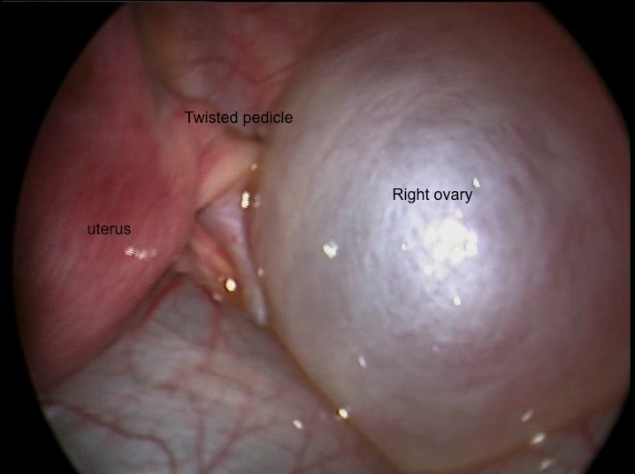
Right ovarian torsion demonstrated by twisted pedicle and swollen ovary
Image: “Twisted right ovarian cyst” by Department of Pediatric Surgery, Division of Pediatric Urology, Cukurova University, Faculty of Medicine, Adana, Turkey. License: CC BY 3.0
In addition to ovarian cystsCystsAny fluid-filled closed cavity or sac that is lined by an epithelium. Cysts can be of normal, abnormal, non-neoplastic, or neoplastic tissues.Fibrocystic Change, the differential diagnosis of a pelvic massMassThree-dimensional lesion that occupies a space within the breastImaging of the Breast includes:
Ectopic pregnancyEctopic pregnancyEctopic pregnancy refers to the implantation of a fertilized egg (embryo) outside the uterine cavity. The main cause is disruption of the normal anatomy of the fallopian tube. Ectopic Pregnancy: a pregnancyPregnancyThe status during which female mammals carry their developing young (embryos or fetuses) in utero before birth, beginning from fertilization to birth.Pregnancy: Diagnosis, Physiology, and Care that implants outside the uterusUterusThe uterus, cervix, and fallopian tubes are part of the internal female reproductive system. The uterus has a thick wall made of smooth muscle (the myometrium) and an inner mucosal layer (the endometrium). The most inferior portion of the uterus is the cervix, which connects the uterine cavity to the vagina.Uterus, Cervix, and Fallopian Tubes: Anatomy, most commonly in the fallopian tubesFallopian tubesThe uterus, cervix, and fallopian tubes are part of the internal female reproductive system. The fallopian tubes receive an ovum after ovulation and help move it and/or a fertilized embryo toward the uterus via ciliated cells lining the tubes and peristaltic movements of its smooth muscle. Uterus, Cervix, and Fallopian Tubes: Anatomy. Ectopic pregnancies can grow large enough to rupture, which can lead to life-threatening hemorrhage. For this reason, all women of reproductive age who present with a pelvic massMassThree-dimensional lesion that occupies a space within the breastImaging of the Breast and/or unilateral pelvic painPainAn unpleasant sensation induced by noxious stimuli which are detected by nerve endings of nociceptive neurons.Pain: Types and Pathways should have a urine pregnancyPregnancyThe status during which female mammals carry their developing young (embryos or fetuses) in utero before birth, beginning from fertilization to birth.Pregnancy: Diagnosis, Physiology, and Care test to rule out ectopic pregnancyEctopic pregnancyEctopic pregnancy refers to the implantation of a fertilized egg (embryo) outside the uterine cavity. The main cause is disruption of the normal anatomy of the fallopian tube. Ectopic Pregnancy. Management is typically either medical (with methotrexateMethotrexateAn antineoplastic antimetabolite with immunosuppressant properties. It is an inhibitor of tetrahydrofolate dehydrogenase and prevents the formation of tetrahydrofolate, necessary for synthesis of thymidylate, an essential component of DNA.Antimetabolite Chemotherapy) or surgical, depending on the situation.
Uterine leiomyomas (fibroidsFibroidsA benign tumor derived from smooth muscle tissue, also known as a fibroid tumor. They rarely occur outside of the uterus and the gastrointestinal tract but can occur in the skin and subcutaneous tissue, probably arising from the smooth muscle of small blood vessels in these tissues.Infertility): benignBenignFibroadenoma tumors arising from the smooth muscle layer of the uterusUterusThe uterus, cervix, and fallopian tubes are part of the internal female reproductive system. The uterus has a thick wall made of smooth muscle (the myometrium) and an inner mucosal layer (the endometrium). The most inferior portion of the uterus is the cervix, which connects the uterine cavity to the vagina.Uterus, Cervix, and Fallopian Tubes: Anatomy. FibroidsFibroidsA benign tumor derived from smooth muscle tissue, also known as a fibroid tumor. They rarely occur outside of the uterus and the gastrointestinal tract but can occur in the skin and subcutaneous tissue, probably arising from the smooth muscle of small blood vessels in these tissues.Infertility can also be pedunculated (i.e., on a stalk) and may be confused for a solid adnexal massMassThree-dimensional lesion that occupies a space within the breastImaging of the Breast. FibroidsFibroidsA benign tumor derived from smooth muscle tissue, also known as a fibroid tumor. They rarely occur outside of the uterus and the gastrointestinal tract but can occur in the skin and subcutaneous tissue, probably arising from the smooth muscle of small blood vessels in these tissues.Infertility typically present with heavy menstrual bleedingHeavy menstrual bleedingExcessive menstrual blood loss (objectively defined as > 80 mL blood loss/cycle). Can be based on heavy flow, as determined by the patientAbnormal Uterine Bleeding and/or pelvic painPainAn unpleasant sensation induced by noxious stimuli which are detected by nerve endings of nociceptive neurons.Pain: Types and Pathways/bulk symptoms and are usually diagnosed on ultrasound; management is typically surgical.
Polycystic ovarian syndromePolycystic ovarian syndromePolycystic ovarian syndrome (PCOS) is the most common endocrine disorder of reproductive-age women, affecting nearly 5%-10% of women in the age group. It is characterized by hyperandrogenism, chronic anovulation leading to oligomenorrhea (or amenorrhea), and metabolic dysfunction.Polycystic Ovarian Syndrome: heterogeneous multisystem endocrinopathyEndocrinopathyIPEX Syndrome that is characterized by hyperandrogenismHyperandrogenismA condition caused by the excessive secretion of androgens from the adrenal cortex; the ovaries; or the testes. The clinical significance in males is negligible. In women, the common manifestations are hirsutism and virilism as seen in patients with polycystic ovary syndrome and adrenocortical hyperfunction.Potassium-sparing Diuretics, ovarian dysfunction, and multiple cystsCystsAny fluid-filled closed cavity or sac that is lined by an epithelium. Cysts can be of normal, abnormal, non-neoplastic, or neoplastic tissues.Fibrocystic Change in the ovariesOvariesOvaries are the paired gonads of the female reproductive system that contain haploid gametes known as oocytes. The ovaries are located intraperitoneally in the pelvis, just posterior to the broad ligament, and are connected to the pelvic sidewall and to the uterus by ligaments. These organs function to secrete hormones (estrogen and progesterone) and to produce the female germ cells (oocytes).Ovaries: Anatomy, which can cause the ovariesOvariesOvaries are the paired gonads of the female reproductive system that contain haploid gametes known as oocytes. The ovaries are located intraperitoneally in the pelvis, just posterior to the broad ligament, and are connected to the pelvic sidewall and to the uterus by ligaments. These organs function to secrete hormones (estrogen and progesterone) and to produce the female germ cells (oocytes).Ovaries: Anatomy to become enlarged. The condition is also associated with metabolic syndromeMetabolic syndromeMetabolic syndrome is a cluster of conditions that significantly increases the risk for several secondary diseases, notably cardiovascular disease, type 2 diabetes, and nonalcoholic fatty liver. In general, it is agreed that hypertension, insulin resistance/hyperglycemia, and hyperlipidemia, along with central obesity, are components of the metabolic syndrome. Metabolic Syndrome, hyperinsulinemiaHyperinsulinemiaDiabetes Mellitus, and insulin resistanceInsulin resistanceDiminished effectiveness of insulin in lowering blood sugar levels: requiring the use of 200 units or more of insulin per day to prevent hyperglycemia or ketosis.Diabetes Mellitus.Diagnosis is one of exclusion, so other causes of abnormal uterine bleedingAbnormal Uterine BleedingAbnormal uterine bleeding is the medical term for abnormalities in the frequency, volume, duration, and regularity of the menstrual cycle. Abnormal uterine bleeding is classified using the acronym PALM-COEIN, with PALM representing the structural causes and COEIN indicating the non-structural causes. Abnormal Uterine Bleeding and hirsutismHirsutismA condition observed in women and children when there is excess coarse body hair of an adult male distribution pattern, such as facial and chest areas. It is the result of elevated androgens from the ovaries, the adrenal glands, or exogenous sources. The concept does not include hypertrichosis, which is an androgen-independent excessive hair growth.Polycystic Ovarian Syndrome should be ruled out. Management includes attempting to restore normal ovulationOvulationThe discharge of an ovum from a rupturing follicle in the ovary.Menstrual Cycle through weight lossWeight lossDecrease in existing body weight.Bariatric Surgery, oral contraceptiveOral contraceptiveCompounds, usually hormonal, taken orally in order to block ovulation and prevent the occurrence of pregnancy. The hormones are generally estrogen or progesterone or both.Benign Liver Tumors pills, and assistance with fertility.
Tubo-ovarian abscessAbscessAccumulation of purulent material in tissues, organs, or circumscribed spaces, usually associated with signs of infection.Chronic Granulomatous Disease (TOA): an infected collection in the adnexa, typically arising in the setting of pelvic inflammatory diseasePelvic inflammatory diseasePelvic inflammatory disease (PID) is defined as a polymicrobial infection of the upper female reproductive system. The disease can affect the uterus, fallopian tubes, ovaries, and adjacent structures. Pelvic inflammatory disease is closely linked with sexually transmitted diseases, most commonly caused by Chlamydia trachomatis, Neisseria gonorrhoeae, and Gardnerella vaginalis. Pelvic Inflammatory Disease (PIDPIDPelvic inflammatory disease (PID) is defined as a polymicrobial infection of the upper female reproductive system. The disease can affect the uterus, fallopian tubes, ovaries, and adjacent structures. Pelvic inflammatory disease is closely linked with sexually transmitted diseases, most commonly caused by Chlamydia trachomatis, Neisseria gonorrhoeae, and gardnerella vaginalis.Pelvic Inflammatory Disease). Diagnosis can usually be made using ultrasonography, so all patientsPatientsIndividuals participating in the health care system for the purpose of receiving therapeutic, diagnostic, or preventive procedures.Clinician–Patient Relationship with PIDPIDPelvic inflammatory disease (PID) is defined as a polymicrobial infection of the upper female reproductive system. The disease can affect the uterus, fallopian tubes, ovaries, and adjacent structures. Pelvic inflammatory disease is closely linked with sexually transmitted diseases, most commonly caused by Chlamydia trachomatis, Neisseria gonorrhoeae, and gardnerella vaginalis.Pelvic Inflammatory Disease and with worrisome findings (such as those who are acutely ill, have a tender adnexal massMassThree-dimensional lesion that occupies a space within the breastImaging of the Breast, or have a poor response to initial therapy), should undergo imaging to look for a TOA. Management is with antibiotics and, potentially, surgical drainage.
Müllerian anomalies: congenital anomalies of the internalfemale genital organs can result in the presence of a pelvic massMassThree-dimensional lesion that occupies a space within the breastImaging of the Breast. Presentation and management depend on the anomaly, which can typically be identified with an exam and pelvic imaging.
GI masses: since the GI system also occupies space in the pelvisPelvisThe pelvis consists of the bony pelvic girdle, the muscular and ligamentous pelvic floor, and the pelvic cavity, which contains viscera, vessels, and multiple nerves and muscles. The pelvic girdle, composed of 2 “hip” bones and the sacrum, is a ring-like bony structure of the axial skeleton that links the vertebral column with the lower extremities.Pelvis: Anatomy, GI masses should be considered in the differential diagnosis of any pelvic massMassThree-dimensional lesion that occupies a space within the breastImaging of the Breast. This includes GI tumors (e.g., colorectal cancers), as well as diverticular and appendiceal abscesses. Symptoms depend on the etiology and may include nauseaNauseaAn unpleasant sensation in the stomach usually accompanied by the urge to vomit. Common causes are early pregnancy, sea and motion sickness, emotional stress, intense pain, food poisoning, and various enteroviruses.Antiemetics, vomitingVomitingThe forcible expulsion of the contents of the stomach through the mouth.Hypokalemia, and/or abnormal stooling. A CT scan is typically the best test to identify GI pathology.
Ureteral or bladderBladderA musculomembranous sac along the urinary tract. Urine flows from the kidneys into the bladder via the ureters, and is held there until urination.Pyelonephritis and Perinephric AbscessdiverticulumDiverticulumA pouch or sac opening from the colon.Diverticular Disease: like the GI tract, masses in the urinary tractUrinary tractThe urinary tract is located in the abdomen and pelvis and consists of the kidneys, ureters, urinary bladder, and urethra. The structures permit the excretion of urine from the body. Urine flows from the kidneys through the ureters to the urinary bladder and out through the urethra.Urinary Tract: Anatomy should also be part of the differential diagnosis of a pelvic massMassThree-dimensional lesion that occupies a space within the breastImaging of the Breast. Imaging can typically clarify the diagnosis.
Pelvic kidney: during embryologic development, the kidneysKidneysThe kidneys are a pair of bean-shaped organs located retroperitoneally against the posterior wall of the abdomen on either side of the spine. As part of the urinary tract, the kidneys are responsible for blood filtration and excretion of water-soluble waste in the urine.Kidneys: Anatomy initially develop in the pelvic region. Abnormal development may result in failure of the kidneysKidneysThe kidneys are a pair of bean-shaped organs located retroperitoneally against the posterior wall of the abdomen on either side of the spine. As part of the urinary tract, the kidneys are responsible for blood filtration and excretion of water-soluble waste in the urine.Kidneys: Anatomy to ascend into their typical position, resulting in a normal functioning kidney located within the pelvisPelvisThe pelvis consists of the bony pelvic girdle, the muscular and ligamentous pelvic floor, and the pelvic cavity, which contains viscera, vessels, and multiple nerves and muscles. The pelvic girdle, composed of 2 “hip” bones and the sacrum, is a ring-like bony structure of the axial skeleton that links the vertebral column with the lower extremities.Pelvis: Anatomy.
Billing and Coding
Diagnosis Codes:
These codes are used to classify different types of ovarian cystsCystsAny fluid-filled closed cavity or sac that is lined by an epithelium. Cysts can be of normal, abnormal, non-neoplastic, or neoplastic tissues.Fibrocystic Change, which are fluid-filled sacs within or on the surface of an ovary. Specific codes exist for follicular, corpus luteumCorpus LuteumThe yellow body derived from the ruptured ovarian follicle after ovulation. The process of corpus luteum formation, luteinization, is regulated by luteinizing hormone.Ovaries: Anatomy, and other types of cystsCystsAny fluid-filled closed cavity or sac that is lined by an epithelium. Cysts can be of normal, abnormal, non-neoplastic, or neoplastic tissues.Fibrocystic Change.
Domain
Code
Description
ICD-10-CM
N83.01
Follicular cystFollicular cystUnruptured graafian follicle that continues to grow in the ovariesOvarian Cysts of right ovary
These codes are used to order a pelvic ultrasound, the primary imaging tool to characterize the size and features of an ovarian cyst, and a CA-125CA-125A carbohydrate antigen that occurs in tumors of the ovary as well as in breast, kidney, and gastrointestinal tract tumors and normal tissue. While it is tumor-associated, it is not tumor-specific and may have a protective function against particles and infectious agents at mucosal surfaces.Serum Tumor Markers blood test, which may be ordered in postmenopausal women to assess for malignancyMalignancyHemothorax risk.
Domain
Code
Description
CPT
76856
Ultrasound, pelvic (nonobstetric), real time with image documentationDocumentationSystematic organization, storage, retrieval, and dissemination of specialized information, especially of a scientific or technical nature. It often involves authenticating or validating information.Advance Directives; complete
CPT
86304
Immunoassay for tumorTumorInflammationantigenAntigenSubstances that are recognized by the immune system and induce an immune reaction.Vaccination, quantitative; CACACondylomata acuminata are a clinical manifestation of genital HPV infection. Condylomata acuminata are described as raised, pearly, flesh-colored, papular, cauliflower-like lesions seen in the anogenital region that may cause itching, pain, or bleeding.Condylomata Acuminata (Genital Warts) 125
Procedures/Interventions:
This code is used for a surgical ovarian cystectomy, a procedure to remove a cyst while preserving the ovary. This is typically performed via laparoscopyLaparoscopyLaparoscopy is surgical exploration and interventions performed through small incisions with a camera and long instruments. Laparotomy and Laparoscopy for cystsCystsAny fluid-filled closed cavity or sac that is lined by an epithelium. Cysts can be of normal, abnormal, non-neoplastic, or neoplastic tissues.Fibrocystic Change that are large, persistent, painful, or suspicious.
Domain
Code
Description
CPT
58925
Ovarian cystectomy, unilateral or bilateral
Complications & Supportive Procedures:
These codes are used to document the acute complications that can arise from an ovarian cyst, including ovarian torsionOvarian torsionOvarian torsion is a clinical emergency in which the ovaries (with or without the fallopian tubes) twist along their axis, leading to partial or complete obstruction of their blood supply. Ovarian torsion is also called adnexal or tubo-ovarian torsion, especially if a fallopian tube is also involved. Ovarian Torsion (twisting) or rupture of the cyst, which can cause severe painPainAn unpleasant sensation induced by noxious stimuli which are detected by nerve endings of nociceptive neurons.Pain: Types and Pathways and internal bleeding.
Domain
Code
Description
ICD-10-CM
N83.519
Torsion of unspecified ovary and ovarian pedicle
ICD-10-CM
N83.8
Other noninflammatory disorders of ovary, fallopian tubeFallopian TubeA pair of highly specialized canals extending from the uterus to its corresponding ovary. They provide the means for ovum transport from the ovaries and they are the site of the ovum’s final maturation and fertilization. The fallopian tube consists of an interstitium, an isthmus, an ampulla, an infundibulum, and fimbriae. Its wall consists of three layers: serous, muscular, and an internal mucosal layer lined with both ciliated and secretory cells.Uterus, Cervix, and Fallopian Tubes: Anatomy and broad ligamentBroad LigamentA broad fold of peritoneum that extends from the side of the uterus to the wall of the pelvis.Uterus, Cervix, and Fallopian Tubes: Anatomy
References
American College of Obstetricians and Gynecologists’ Committee on Practice Bulletins—Gynecology. (2016). Practice bulletin no. 174: evaluation and management of adnexal masses. Obstetrics and Gynecology, 128(5), e210-e226. doi: 10.1097/AOG.0000000000001768. https://pubmed.ncbi.nlm.nih.gov/27776072/
Beckmann C. R. B., Ling, F. W., et al. (Eds.). Obstetric and Gynecology (6th ed., pp. 283-294). Lippincott Williams & Wilkins.
Bristow, R. E., Hodeib, M., Smith, A., Chan, D. W., Zhang, Z., Fung, E. T., Tewari, K. S., Munroe, D. G., Ueland, F. R. (2013). Impact of a multivariate index assay on referral patterns for surgical management of an adnexal mass. American Journal of Obstetrics and Gynecology, 209(6), 581.e1-8. doi: 10.1016/j.ajog.2013.08.009. https://pubmed.ncbi.nlm.nih.gov/23942039/
Jacobs, I., Oram, D., Fairbanks, J., Turner, J., Frost, C., Grudzinskas, J. G. (1990). A risk of malignancy index incorporating CA 125, ultrasound and menopausal status for the accurate preoperative diagnosis of ovarian cancer. British Journal of Obstetrics Gynaecology, 97(10), 922–929. doi: 10.1111/j.1471-0528.1990.tb02448.x. https://pubmed.ncbi.nlm.nih.gov/2223684/
Moolthiya, W., Yuenyao, P. (2009). The risk of malignancy index (RMI) in diagnosis of ovarian malignancy. Asian Pacific Journal of Cancer Prevention, 10, 865–868. https://pubmed.ncbi.nlm.nih.gov/20162854/
Moore, R. G., McMeekin, D. S., Brown, A. K., DiSilvestro, P., Miller, M. C., Allard, W.J., Gajewski, W., Kurman, R., Bast, R. C., Jr., Skates, S. J. (2009). A novel multiple marker bioassay utilizing HE4 and CA125 for the prediction of ovarian cancer in patients with a pelvic mass. Gynecologic Oncology, 112(1), 40-6. doi: 10.1016/j.ygyno.2008.08.031. https://pubmed.ncbi.nlm.nih.gov/18851871/
Royal College of Obstetricians and Gynaecologists. (2011). Management of suspected ovarian masses in premenopausal women. Green-top guideline no. 62. Retrieved October 25, 2022, from https://www.rcog.org.uk/media/yhujmdvr/gtg_62-1.pdf
Create your free account or log in to continue reading!