X-rays are high-energy particles of electromagnetic radiationRadiationEmission or propagation of acoustic waves (sound), electromagnetic energy waves (such as light; radio waves; gamma rays; or x-rays), or a stream of subatomic particles (such as electrons; neutrons; protons; or alpha particles).Osteosarcoma used in the medical field for the generation of anatomical images. X-rays are projected through the body of a patient and onto a film, and this technique is called conventional or projectional radiography. As radiationRadiationEmission or propagation of acoustic waves (sound), electromagnetic energy waves (such as light; radio waves; gamma rays; or x-rays), or a stream of subatomic particles (such as electrons; neutrons; protons; or alpha particles).Osteosarcoma by X-rays can cause adverse effects depending on the absorbed dose, it is necessary to take protective measures to reduce harm. Digital radiography uses the digital data format and allows for the digital manipulation of images. Common uses include evaluation of chest, mediastinal, spinal, and boneBoneBone is a compact type of hardened connective tissue composed of bone cells, membranes, an extracellular mineralized matrix, and central bone marrow. The 2 primary types of bone are compact and spongy. Bones: Structure and Types/joint conditions. While radiography is still used to visualize head and abdominal structures, more advanced modalities (CT and MRI) are now preferred. Radiography remains an essential component of initial tests in many diseases, given its wide availability, low cost, and ease of operation.
An X-rayX-rayPenetrating electromagnetic radiation emitted when the inner orbital electrons of an atom are excited and release radiant energy. X-ray wavelengths range from 1 pm to 10 nm. Hard x-rays are the higher energy, shorter wavelength x-rays. Soft x-rays or grenz rays are less energetic and longer in wavelength. The short wavelength end of the x-ray spectrum overlaps the gamma rays wavelength range. The distinction between gamma rays and x-rays is based on their radiation source.Pulmonary Function Tests is a discrete, high-energy particle of electromagnetic radiationRadiationEmission or propagation of acoustic waves (sound), electromagnetic energy waves (such as light; radio waves; gamma rays; or x-rays), or a stream of subatomic particles (such as electrons; neutrons; protons; or alpha particles).Osteosarcoma (photon) that propagates through space at the speed of light.
X-rayX-rayPenetrating electromagnetic radiation emitted when the inner orbital electrons of an atom are excited and release radiant energy. X-ray wavelengths range from 1 pm to 10 nm. Hard x-rays are the higher energy, shorter wavelength x-rays. Soft x-rays or grenz rays are less energetic and longer in wavelength. The short wavelength end of the x-ray spectrum overlaps the gamma rays wavelength range. The distinction between gamma rays and x-rays is based on their radiation source.Pulmonary Function Tests production
X-rays are produced through different processes:
Characteristic X-rayX-rayPenetrating electromagnetic radiation emitted when the inner orbital electrons of an atom are excited and release radiant energy. X-ray wavelengths range from 1 pm to 10 nm. Hard x-rays are the higher energy, shorter wavelength x-rays. Soft x-rays or grenz rays are less energetic and longer in wavelength. The short wavelength end of the x-ray spectrum overlaps the gamma rays wavelength range. The distinction between gamma rays and x-rays is based on their radiation source.Pulmonary Function TestsradiationRadiationEmission or propagation of acoustic waves (sound), electromagnetic energy waves (such as light; radio waves; gamma rays; or x-rays), or a stream of subatomic particles (such as electrons; neutrons; protons; or alpha particles).Osteosarcoma:
Result from the movement or transition of electrons from an outer shell (orbit) to vacancies in the inner shell
Emission of X-rayX-rayPenetrating electromagnetic radiation emitted when the inner orbital electrons of an atom are excited and release radiant energy. X-ray wavelengths range from 1 pm to 10 nm. Hard x-rays are the higher energy, shorter wavelength x-rays. Soft x-rays or grenz rays are less energetic and longer in wavelength. The short wavelength end of the x-ray spectrum overlaps the gamma rays wavelength range. The distinction between gamma rays and x-rays is based on their radiation source.Pulmonary Function Tests photon is material dependent.
Braking radiationRadiationEmission or propagation of acoustic waves (sound), electromagnetic energy waves (such as light; radio waves; gamma rays; or x-rays), or a stream of subatomic particles (such as electrons; neutrons; protons; or alpha particles).Osteosarcoma (Bremsstrahlung):
Electrons move rapidly toward the anode (positively charged electrode) and decelerate when they collide.
During decelerationDecelerationA decrease in the rate of speed.Blunt Chest Trauma, 99% of the energy dissipates as heatHeatInflammation and 1% is released as X-rayX-rayPenetrating electromagnetic radiation emitted when the inner orbital electrons of an atom are excited and release radiant energy. X-ray wavelengths range from 1 pm to 10 nm. Hard x-rays are the higher energy, shorter wavelength x-rays. Soft x-rays or grenz rays are less energetic and longer in wavelength. The short wavelength end of the x-ray spectrum overlaps the gamma rays wavelength range. The distinction between gamma rays and x-rays is based on their radiation source.Pulmonary Function Tests photons.
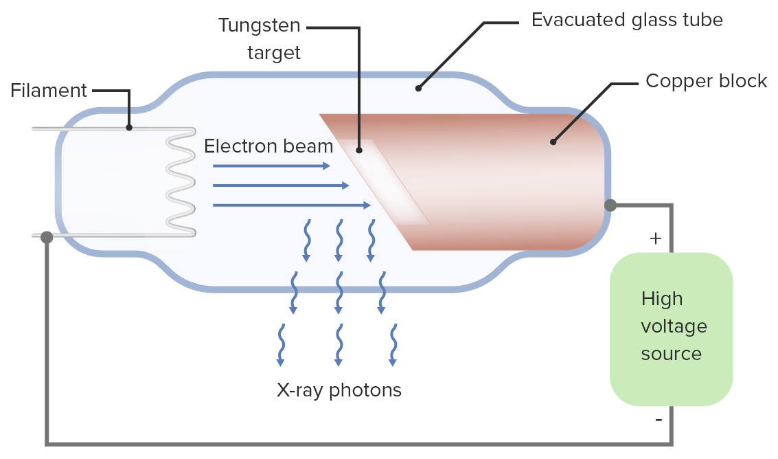
X-rayX-rayPenetrating electromagnetic radiation emitted when the inner orbital electrons of an atom are excited and release radiant energy. X-ray wavelengths range from 1 pm to 10 nm. Hard x-rays are the higher energy, shorter wavelength x-rays. Soft x-rays or grenz rays are less energetic and longer in wavelength. The short wavelength end of the x-ray spectrum overlaps the gamma rays wavelength range. The distinction between gamma rays and x-rays is based on their radiation source.Pulmonary Function Tests imaging utilizes an X-rayX-rayPenetrating electromagnetic radiation emitted when the inner orbital electrons of an atom are excited and release radiant energy. X-ray wavelengths range from 1 pm to 10 nm. Hard x-rays are the higher energy, shorter wavelength x-rays. Soft x-rays or grenz rays are less energetic and longer in wavelength. The short wavelength end of the x-ray spectrum overlaps the gamma rays wavelength range. The distinction between gamma rays and x-rays is based on their radiation source.Pulmonary Function Tests tube consisting of:
A heated filament that emits electrons
A tungsten target/anode where electrons strike, producing X-rays
X-rays penetrate matter and interact with the atomic electrons of the material. During this process, X-rays can be absorbed or scattered.
Not all X-rays can penetrate a patient. Most X-rays are scattered and do not contribute to image creation.
Diagram of an X-ray tube: In the tube, electrons are accelerated toward a tungsten target (anode), which then decelerate after hitting the target, releasing heat and X-ray photons.
Image by Lecturio.
Effects of X-rayX-rayPenetrating electromagnetic radiation emitted when the inner orbital electrons of an atom are excited and release radiant energy. X-ray wavelengths range from 1 pm to 10 nm. Hard x-rays are the higher energy, shorter wavelength x-rays. Soft x-rays or grenz rays are less energetic and longer in wavelength. The short wavelength end of the x-ray spectrum overlaps the gamma rays wavelength range. The distinction between gamma rays and x-rays is based on their radiation source.Pulmonary Function TestsradiationRadiationEmission or propagation of acoustic waves (sound), electromagnetic energy waves (such as light; radio waves; gamma rays; or x-rays), or a stream of subatomic particles (such as electrons; neutrons; protons; or alpha particles).Osteosarcoma
Biological damage by X-rays is attributed to the ionizing radiationRadiationEmission or propagation of acoustic waves (sound), electromagnetic energy waves (such as light; radio waves; gamma rays; or x-rays), or a stream of subatomic particles (such as electrons; neutrons; protons; or alpha particles).Osteosarcoma that is produced as X-rays interact with matter.
The absorbed dose is the energy (from the interaction) deposited in matter.
Absorbed radiationRadiationEmission or propagation of acoustic waves (sound), electromagnetic energy waves (such as light; radio waves; gamma rays; or x-rays), or a stream of subatomic particles (such as electrons; neutrons; protons; or alpha particles).Osteosarcoma: measured in units known as Gray (Gy) or rad (100 rads are equivalent to 1 Gy)
Types of radiationRadiationEmission or propagation of acoustic waves (sound), electromagnetic energy waves (such as light; radio waves; gamma rays; or x-rays), or a stream of subatomic particles (such as electrons; neutrons; protons; or alpha particles).Osteosarcoma effects:
Deterministic effect:
Damage occurs when a thresholdThresholdMinimum voltage necessary to generate an action potential (an all-or-none response)Skeletal Muscle Contraction of radiationRadiationEmission or propagation of acoustic waves (sound), electromagnetic energy waves (such as light; radio waves; gamma rays; or x-rays), or a stream of subatomic particles (such as electrons; neutrons; protons; or alpha particles).Osteosarcoma is crossed, such that the ability of a cell to repair itself is overwhelmed.
Results from very high doses of radiationRadiationEmission or propagation of acoustic waves (sound), electromagnetic energy waves (such as light; radio waves; gamma rays; or x-rays), or a stream of subatomic particles (such as electrons; neutrons; protons; or alpha particles).Osteosarcoma, causing skinSkinThe skin, also referred to as the integumentary system, is the largest organ of the body. The skin is primarily composed of the epidermis (outer layer) and dermis (deep layer). The epidermis is primarily composed of keratinocytes that undergo rapid turnover, while the dermis contains dense layers of connective tissue.Skin: Structure and FunctionserythemaErythemaRedness of the skin produced by congestion of the capillaries. This condition may result from a variety of disease processes.Chalazion, cataracts, and sterility
Stochastic effect: Damage is additive and the probabilityProbabilityProbability is a mathematical tool used to study randomness and provide predictions about the likelihood of something happening. There are several basic rules of probability that can be used to help determine the probability of multiple events happening together, separately, or sequentially.Basics of Probability of the effect increases with increased exposure.
Damage occurs at the genetic level during cell divisionCell DivisionA type of cell nucleus division by means of which the two daughter nuclei normally receive identical complements of the number of chromosomes of the somatic cells of the species.Cell Cycle and can lead to carcinogenesisCarcinogenesisThe origin, production or development of cancer through genotypic and phenotypic changes which upset the normal balance between cell proliferation and cell death. Carcinogenesis generally requires a constellation of steps, which may occur quickly or over a period of many years.Carcinogenesis.
The likelihood of effects increases with radiationRadiationEmission or propagation of acoustic waves (sound), electromagnetic energy waves (such as light; radio waves; gamma rays; or x-rays), or a stream of subatomic particles (such as electrons; neutrons; protons; or alpha particles).OsteosarcomadosageDosageDosage Calculation.
Ultimately, the resulting damage includes:
Formation of free radicalsFree radicalsHighly reactive molecules with an unsatisfied electron valence pair. Free radicals are produced in both normal and pathological processes. They are proven or suspected agents of tissue damage in a wide variety of circumstances including radiation, damage from environment chemicals, and aging. Natural and pharmacological prevention of free radical damage is being actively investigated.Ischemic Cell Damage
Disruption of normal metabolic function and mitosisMitosisA type of cell nucleus division by means of which the two daughter nuclei normally receive identical complements of the number of chromosomes of the somatic cells of the species.Cell Cycle
Cancer induction:
Organs with the most rapidly dividing cells are most susceptible, which also explains why children, overall, are most susceptible.
Most susceptible organs:
Bone marrowBone marrowThe soft tissue filling the cavities of bones. Bone marrow exists in two types, yellow and red. Yellow marrow is found in the large cavities of large bones and consists mostly of fat cells and a few primitive blood cells. Red marrow is a hematopoietic tissue and is the site of production of erythrocytes and granular leukocytes. Bone marrow is made up of a framework of connective tissue containing branching fibers with the frame being filled with marrow cells.Bone Marrow: Composition and Hematopoiesis
ColonColonThe large intestines constitute the last portion of the digestive system. The large intestine consists of the cecum, appendix, colon (with ascending, transverse, descending, and sigmoid segments), rectum, and anal canal. The primary function of the colon is to remove water and compact the stool prior to expulsion from the body via the rectum and anal canal. Colon, Cecum, and Appendix: Anatomy
LungsLungsLungs are the main organs of the respiratory system. Lungs are paired viscera located in the thoracic cavity and are composed of spongy tissue. The primary function of the lungs is to oxygenate blood and eliminate CO2. Lungs: Anatomy
StomachStomachThe stomach is a muscular sac in the upper left portion of the abdomen that plays a critical role in digestion. The stomach develops from the foregut and connects the esophagus with the duodenum. Structurally, the stomach is C-shaped and forms a greater and lesser curvature and is divided grossly into regions: the cardia, fundus, body, and pylorus. Stomach: Anatomy
Moderately susceptible organs:
BladderBladderA musculomembranous sac along the urinary tract. Urine flows from the kidneys into the bladder via the ureters, and is held there until urination.Pyelonephritis and Perinephric Abscess
Breast
LiverLiverThe liver is the largest gland in the human body. The liver is found in the superior right quadrant of the abdomen and weighs approximately 1.5 kilograms. Its main functions are detoxification, metabolism, nutrient storage (e.g., iron and vitamins), synthesis of coagulation factors, formation of bile, filtration, and storage of blood. Liver: Anatomy
EsophagusEsophagusThe esophagus is a muscular tube-shaped organ of around 25 centimeters in length that connects the pharynx to the stomach. The organ extends from approximately the 6th cervical vertebra to the 11th thoracic vertebra and can be divided grossly into 3 parts: the cervical part, the thoracic part, and the abdominal part. Esophagus: Anatomy
ThyroidThyroidThe thyroid gland is one of the largest endocrine glands in the human body. The thyroid gland is a highly vascular, brownish-red gland located in the visceral compartment of the anterior region of the neck.Thyroid Gland: Anatomy
Fetal risk of radiationRadiationEmission or propagation of acoustic waves (sound), electromagnetic energy waves (such as light; radio waves; gamma rays; or x-rays), or a stream of subatomic particles (such as electrons; neutrons; protons; or alpha particles).Osteosarcoma
Table: Fetal risk of radiationRadiationEmission or propagation of acoustic waves (sound), electromagnetic energy waves (such as light; radio waves; gamma rays; or x-rays), or a stream of subatomic particles (such as electrons; neutrons; protons; or alpha particles).Osteosarcoma
Weeks post conception
Effects of major exposure
2
10–50 rads: risk of failure to implant
> 50 rads: high likelihood of implantationImplantationEndometrial implantation of embryo, mammalian at the blastocyst stage.Fertilization and First Week failure
3–5
10–50 rads: growth restriction possible
> 50 rads: congenital abnormalitiesCongenital AbnormalitiesMalformations of organs or body parts during development in utero.Omphalocele, growth restriction, risk of miscarriageMiscarriageSpontaneous abortion, also known as miscarriage, is the loss of a pregnancy before 20 weeks’ gestation. However, the layperson use of the term “abortion” is often intended to refer to induced termination of a pregnancy, whereas “miscarriage” is preferred for spontaneous loss.Spontaneous Abortion
6–13
10–50 rads: growth restriction possible
> 50 rads: growth restriction, risk of miscarriageMiscarriageSpontaneous abortion, also known as miscarriage, is the loss of a pregnancy before 20 weeks’ gestation. However, the layperson use of the term “abortion” is often intended to refer to induced termination of a pregnancy, whereas “miscarriage” is preferred for spontaneous loss.Spontaneous Abortion
14–23
10–50 rads: noncancer health effects unlikely
> 50 rads: growth restriction, risk of miscarriageMiscarriageSpontaneous abortion, also known as miscarriage, is the loss of a pregnancy before 20 weeks’ gestation. However, the layperson use of the term “abortion” is often intended to refer to induced termination of a pregnancy, whereas “miscarriage” is preferred for spontaneous loss.Spontaneous Abortion, possible congenital abnormalitiesCongenital AbnormalitiesMalformations of organs or body parts during development in utero.Omphalocele
24 weeks to term
10–50 rads: noncancer health effects unlikely
> 50 rads: miscarriageMiscarriageSpontaneous abortion, also known as miscarriage, is the loss of a pregnancy before 20 weeks’ gestation. However, the layperson use of the term “abortion” is often intended to refer to induced termination of a pregnancy, whereas “miscarriage” is preferred for spontaneous loss.Spontaneous Abortion, neonatal death (depending on dose)
RadiationRadiationEmission or propagation of acoustic waves (sound), electromagnetic energy waves (such as light; radio waves; gamma rays; or x-rays), or a stream of subatomic particles (such as electrons; neutrons; protons; or alpha particles).Osteosarcoma protection
Minimize the radiationRadiationEmission or propagation of acoustic waves (sound), electromagnetic energy waves (such as light; radio waves; gamma rays; or x-rays), or a stream of subatomic particles (such as electrons; neutrons; protons; or alpha particles).OsteosarcomadosageDosageDosage Calculation whenever possible (ALARA: as low as reasonably achievable).
Measures:
Exposed personnel should be monitored using a film badge.
Lead shielding and increasing the distance from the source
Shielding within rooms
Increasing the kilovoltage of the X-rayX-rayPenetrating electromagnetic radiation emitted when the inner orbital electrons of an atom are excited and release radiant energy. X-ray wavelengths range from 1 pm to 10 nm. Hard x-rays are the higher energy, shorter wavelength x-rays. Soft x-rays or grenz rays are less energetic and longer in wavelength. The short wavelength end of the x-ray spectrum overlaps the gamma rays wavelength range. The distinction between gamma rays and x-rays is based on their radiation source.Pulmonary Function Tests beam and, thus, increasing its penetration
Projectional radiography: generation of a radiographic image by projecting a beam of X-rayX-rayPenetrating electromagnetic radiation emitted when the inner orbital electrons of an atom are excited and release radiant energy. X-ray wavelengths range from 1 pm to 10 nm. Hard x-rays are the higher energy, shorter wavelength x-rays. Soft x-rays or grenz rays are less energetic and longer in wavelength. The short wavelength end of the x-ray spectrum overlaps the gamma rays wavelength range. The distinction between gamma rays and x-rays is based on their radiation source.Pulmonary Function Tests particles through a subject and onto a film:
The X-rayX-rayPenetrating electromagnetic radiation emitted when the inner orbital electrons of an atom are excited and release radiant energy. X-ray wavelengths range from 1 pm to 10 nm. Hard x-rays are the higher energy, shorter wavelength x-rays. Soft x-rays or grenz rays are less energetic and longer in wavelength. The short wavelength end of the x-ray spectrum overlaps the gamma rays wavelength range. The distinction between gamma rays and x-rays is based on their radiation source.Pulmonary Function Tests image is a shadow picture obtained using a single “light” source.
Fluoroscopy: the use of projectional radiography to observe internal structures in real-time (e.g., GI imaging)
CT: generation of a multilayered image by a beam projected by a rotatory X-rayX-rayPenetrating electromagnetic radiation emitted when the inner orbital electrons of an atom are excited and release radiant energy. X-ray wavelengths range from 1 pm to 10 nm. Hard x-rays are the higher energy, shorter wavelength x-rays. Soft x-rays or grenz rays are less energetic and longer in wavelength. The short wavelength end of the x-ray spectrum overlaps the gamma rays wavelength range. The distinction between gamma rays and x-rays is based on their radiation source.Pulmonary Function Tests tube onto radiation detectorsRadiation DetectorsComputed Tomography (CT)
Image generation by X-rays
Order of producing an image with X-rays:
X-rayX-rayPenetrating electromagnetic radiation emitted when the inner orbital electrons of an atom are excited and release radiant energy. X-ray wavelengths range from 1 pm to 10 nm. Hard x-rays are the higher energy, shorter wavelength x-rays. Soft x-rays or grenz rays are less energetic and longer in wavelength. The short wavelength end of the x-ray spectrum overlaps the gamma rays wavelength range. The distinction between gamma rays and x-rays is based on their radiation source.Pulmonary Function Tests tube: X-rays generated after electrons collide with the anode.
Patient: X-rayX-rayPenetrating electromagnetic radiation emitted when the inner orbital electrons of an atom are excited and release radiant energy. X-ray wavelengths range from 1 pm to 10 nm. Hard x-rays are the higher energy, shorter wavelength x-rays. Soft x-rays or grenz rays are less energetic and longer in wavelength. The short wavelength end of the x-ray spectrum overlaps the gamma rays wavelength range. The distinction between gamma rays and x-rays is based on their radiation source.Pulmonary Function Tests beam traverses the patient and is attenuated depending on the tissues in their path.
Antiscatter grid: lead strips that improve image contrast by reducing scattered photons
Image capture is by using an imaging plate in a cassette.
Technologies producing radiographic image:
Conventional radiography:
Screen film is used and the film is developed.
High sensitivity, low cost, and easy handling
Digital radiography (utilizes digital data format, allowing digital manipulation of images):
Computed radiography: Cassette is inserted into a scanner and the image is shown on a monitor.
Direct radiography: no cassette used. X-rays are converted into electrical charges by a photoconductor.
Image of an early X-ray: X-ray of a left hand taken at a public lecture by Wilhelm Röntgen
Image: “An early X-ray” by Wilhelm Röntgen; current version created by Old Moonraker. License: Public Domain
CalciumCalciumA basic element found in nearly all tissues. It is a member of the alkaline earth family of metals with the atomic symbol ca, atomic number 20, and atomic weight 40. Calcium is the most abundant mineral in the body and combines with phosphorus to form calcium phosphate in the bones and teeth. It is essential for the normal functioning of nerves and muscles and plays a role in blood coagulation (as factor IV) and in many enzymatic processes.Electrolytes (boneBoneBone is a compact type of hardened connective tissue composed of bone cells, membranes, an extracellular mineralized matrix, and central bone marrow. The 2 primary types of bone are compact and spongy. Bones: Structure and Types)
Metal
Terminology according to the density of the object
Radiolucent: an object of low density that is permeable to X-rays (looks black)
Radiopaque: an object of high density that blocks X-rays (looks white)
Principles of radiography
Summation of shadows: Images appear more radiopaque due to overlapping densities.
Silhouette sign:
Edges of an object are indistinguishable when densities are adjacent to one another.
Think of pneumoniaPneumoniaPneumonia or pulmonary inflammation is an acute or chronic inflammation of lung tissue. Causes include infection with bacteria, viruses, or fungi. In more rare cases, pneumonia can also be caused through toxic triggers through inhalation of toxic substances, immunological processes, or in the course of radiotherapy.Pneumonia in the right middle lobe, which obscures the right heart borderRight Heart BorderImaging of the Heart and Great Vessels.
Orthogonal imaging: taking 2 projections of the same structure to better document its 3-dimensionality
Elements that reduce the diagnostic yield of an X-rayX-rayPenetrating electromagnetic radiation emitted when the inner orbital electrons of an atom are excited and release radiant energy. X-ray wavelengths range from 1 pm to 10 nm. Hard x-rays are the higher energy, shorter wavelength x-rays. Soft x-rays or grenz rays are less energetic and longer in wavelength. The short wavelength end of the x-ray spectrum overlaps the gamma rays wavelength range. The distinction between gamma rays and x-rays is based on their radiation source.Pulmonary Function Tests
Excessive or insufficient penetration
Rotation of the patient
Image magnification
Movement of the patient
Artifacts, such as dust particles
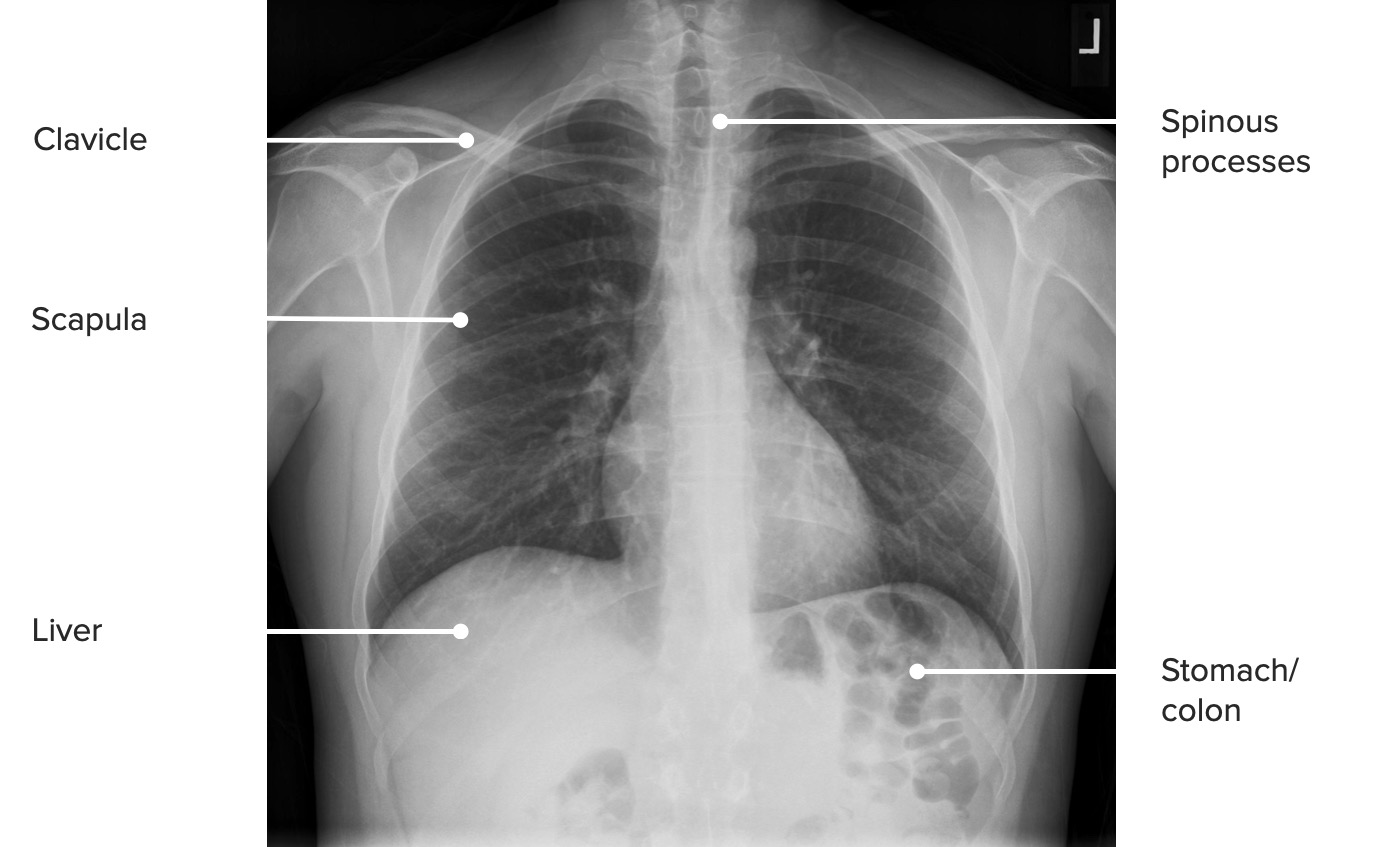
Chest
Projections
X-rayX-rayPenetrating electromagnetic radiation emitted when the inner orbital electrons of an atom are excited and release radiant energy. X-ray wavelengths range from 1 pm to 10 nm. Hard x-rays are the higher energy, shorter wavelength x-rays. Soft x-rays or grenz rays are less energetic and longer in wavelength. The short wavelength end of the x-ray spectrum overlaps the gamma rays wavelength range. The distinction between gamma rays and x-rays is based on their radiation source.Pulmonary Function Tests images of the chest can be produced in the following projections:
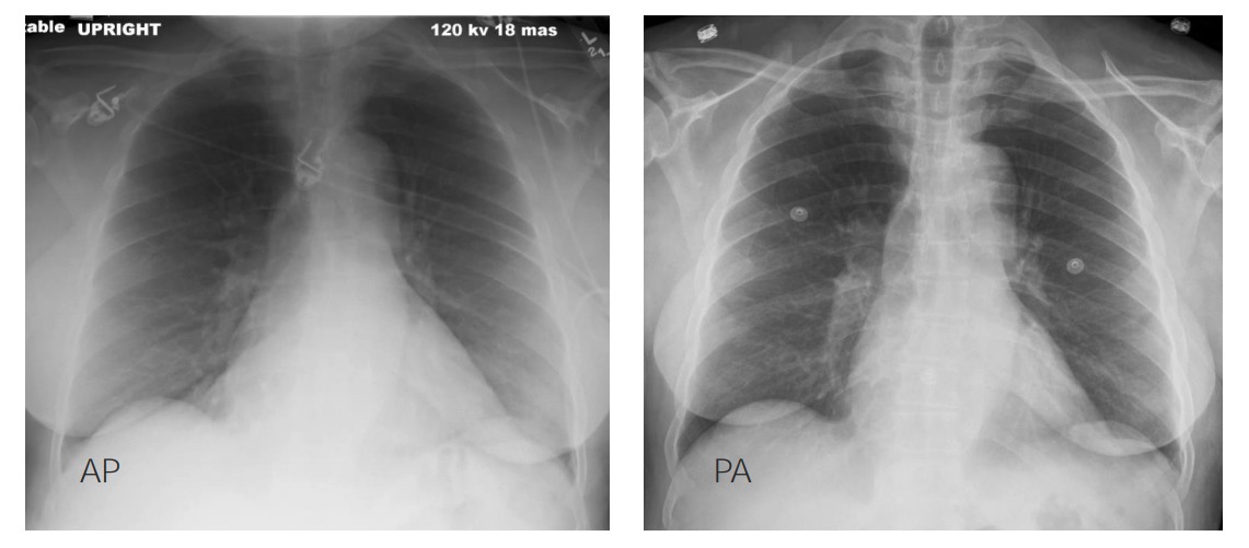
Posteroanterior (PA):
The X-rayX-rayPenetrating electromagnetic radiation emitted when the inner orbital electrons of an atom are excited and release radiant energy. X-ray wavelengths range from 1 pm to 10 nm. Hard x-rays are the higher energy, shorter wavelength x-rays. Soft x-rays or grenz rays are less energetic and longer in wavelength. The short wavelength end of the x-ray spectrum overlaps the gamma rays wavelength range. The distinction between gamma rays and x-rays is based on their radiation source.Pulmonary Function Tests beam initially penetrates the posterior aspect of the body while the cassette is placed in direct contact with the anterior aspect.
Preferred method for evaluating cardiac silhouette size
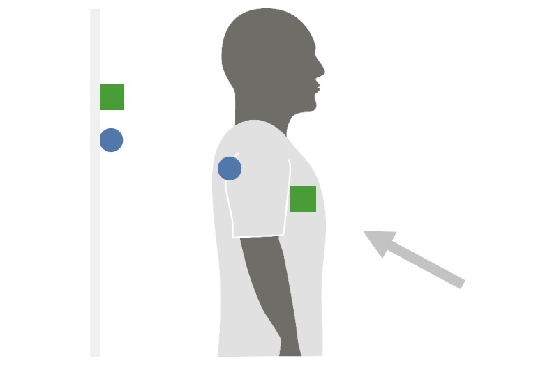
Anteroposterior (AP):
The X-rayX-rayPenetrating electromagnetic radiation emitted when the inner orbital electrons of an atom are excited and release radiant energy. X-ray wavelengths range from 1 pm to 10 nm. Hard x-rays are the higher energy, shorter wavelength x-rays. Soft x-rays or grenz rays are less energetic and longer in wavelength. The short wavelength end of the x-ray spectrum overlaps the gamma rays wavelength range. The distinction between gamma rays and x-rays is based on their radiation source.Pulmonary Function Tests beam initially penetrates the anterior aspect of the body while the cassette is placed in direct contact with the posterior aspect.
Used in portable radiography (very common in hospitalized patientsPatientsIndividuals participating in the health care system for the purpose of receiving therapeutic, diagnostic, or preventive procedures.Clinician–Patient Relationship who are unable to move)
Structures further away from the cassette appear magnified, creating false positives for cardiomegalyCardiomegalyEnlargement of the heart, usually indicated by a cardiothoracic ratio above 0. 50. Heart enlargement may involve the right, the left, or both heart ventricles or heart atria. Cardiomegaly is a nonspecific symptom seen in patients with chronic systolic heart failure (heart failure) or several forms of cardiomyopathies.Ebstein’s Anomaly.
Lateral:
The X-rayX-rayPenetrating electromagnetic radiation emitted when the inner orbital electrons of an atom are excited and release radiant energy. X-ray wavelengths range from 1 pm to 10 nm. Hard x-rays are the higher energy, shorter wavelength x-rays. Soft x-rays or grenz rays are less energetic and longer in wavelength. The short wavelength end of the x-ray spectrum overlaps the gamma rays wavelength range. The distinction between gamma rays and x-rays is based on their radiation source.Pulmonary Function Tests beam is incident on a lateral aspect of the body and the cassette is placed in contact with the other lateral aspect.
Lordotic and semi-upright positioning:
The X-rayX-rayPenetrating electromagnetic radiation emitted when the inner orbital electrons of an atom are excited and release radiant energy. X-ray wavelengths range from 1 pm to 10 nm. Hard x-rays are the higher energy, shorter wavelength x-rays. Soft x-rays or grenz rays are less energetic and longer in wavelength. The short wavelength end of the x-ray spectrum overlaps the gamma rays wavelength range. The distinction between gamma rays and x-rays is based on their radiation source.Pulmonary Function Tests beam penetrates the patient at an angle to display 2 different structures at different levels.
Decubitus:
The patient lays on their right or left side.
A replacement for lateral projection used for patientsPatientsIndividuals participating in the health care system for the purpose of receiving therapeutic, diagnostic, or preventive procedures.Clinician–Patient Relationship who cannot stand up
Chest radiograph projections
Image by Lecturio.
Posteroanterior (PA) X-ray projection: The X-ray beam penetrates the patient from the posterior aspect, and the anterior aspect is placed in direct contact with the receptor.
Image by Lecturio.
Anteroposterior (AP) X-ray projection: The X-ray beam penetrates the patient from the anterior aspect, and the posterior aspect is placed in direct contact with the receptor.
Image by Lecturio.
Apical lordotic X-ray projection: The X-ray beam penetrates the patient at an angle to display 2 different elements at different levels.
Image by Lecturio.
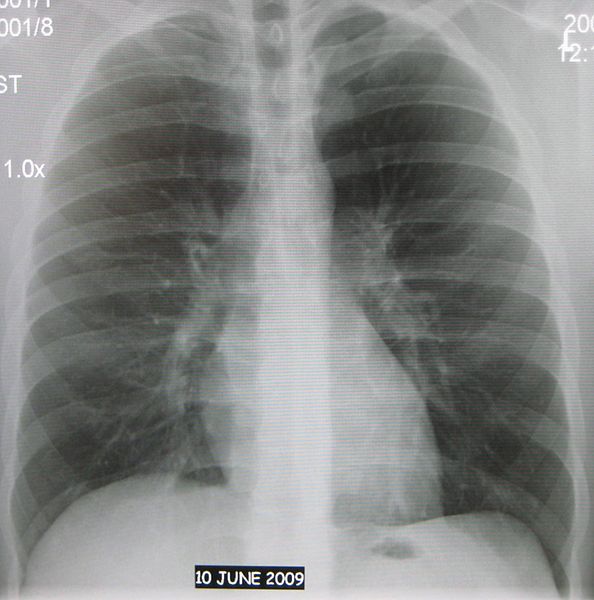
Normal chest X-ray
Image: “Normal AP chest X-ray” by James Heilman, MD. License: CC BY 3.0
Differences between anteroposterior (AP, left) and posteroanterior (PA, right) X-ray projections: In the AP view, the structures that are further away from the cassette, such as the heart, are magnified.
Image by Hetal Verma.
Technical aspects
To obtain an optimal anatomical image:
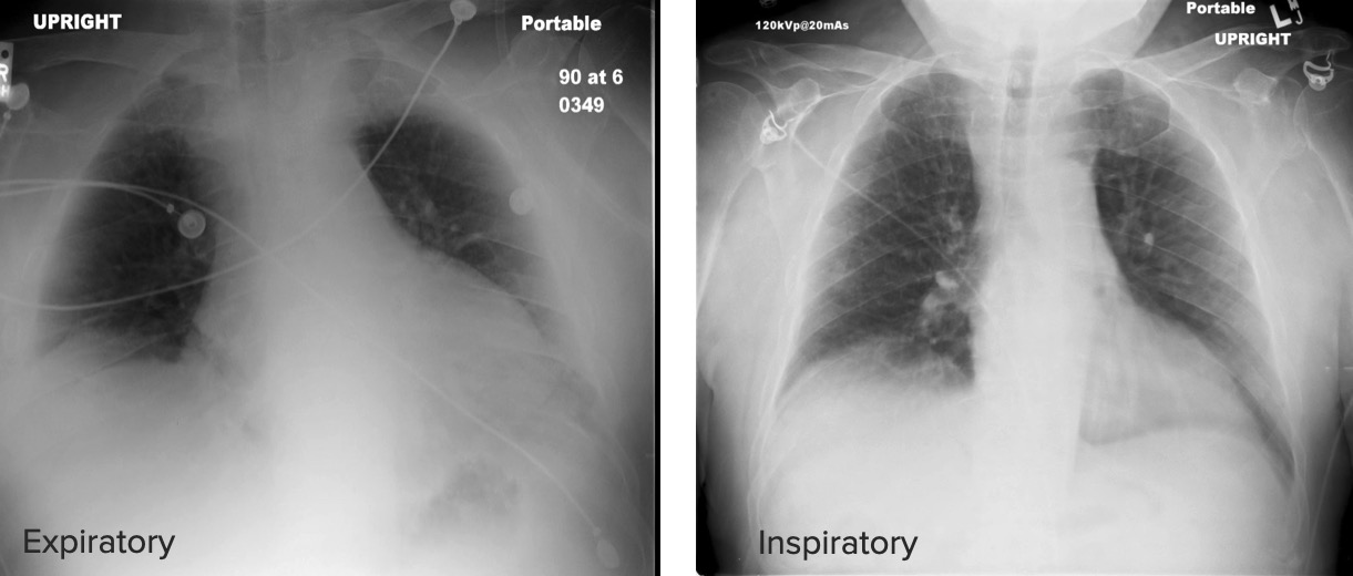
InspirationInspirationVentilation: Mechanics of Breathing: The patient is asked to take a deep breathDeep breathRespiratory Alkalosis while an X-rayX-rayPenetrating electromagnetic radiation emitted when the inner orbital electrons of an atom are excited and release radiant energy. X-ray wavelengths range from 1 pm to 10 nm. Hard x-rays are the higher energy, shorter wavelength x-rays. Soft x-rays or grenz rays are less energetic and longer in wavelength. The short wavelength end of the x-ray spectrum overlaps the gamma rays wavelength range. The distinction between gamma rays and x-rays is based on their radiation source.Pulmonary Function Tests is being obtained.
8–9 posterior ribsRibsA set of twelve curved bones which connect to the vertebral column posteriorly, and terminate anteriorly as costal cartilage. Together, they form a protective cage around the internal thoracic organs.Chest Wall: Anatomy should be made visible for the inspirationInspirationVentilation: Mechanics of Breathing to be optimal.
The following aspects reduce the qualityQualityActivities and programs intended to assure or improve the quality of care in either a defined medical setting or a program. The concept includes the assessment or evaluation of the quality of care; identification of problems or shortcomings in the delivery of care; designing activities to overcome these deficiencies; and follow-up monitoring to ensure effectiveness of corrective steps.Quality Measurement and Improvement of the anatomical image:
Penetration: Excessive or deficient penetration of X-rays through the anatomical structures affects results.
Overpenetrated regions can simulate collections of air (pneumothoraxPneumothoraxA pneumothorax is a life-threatening condition in which air collects in the pleural space, causing partial or full collapse of the lung. A pneumothorax can be traumatic or spontaneous. Patients present with a sudden onset of sharp chest pain, dyspnea, and diminished breath sounds on exam.Pneumothorax).
Underpenetrated regions can simulate consolidations (pneumoniaPneumoniaPneumonia or pulmonary inflammation is an acute or chronic inflammation of lung tissue. Causes include infection with bacteria, viruses, or fungi. In more rare cases, pneumonia can also be caused through toxic triggers through inhalation of toxic substances, immunological processes, or in the course of radiotherapy.Pneumonia).
Rotation: When the patient is not appropriately placed in front of the cassette, the structures are unevenly represented in the anatomical image.
The mediastinumMediastinumThe mediastinum is the thoracic area between the 2 pleural cavities. The mediastinum contains vital structures of the circulatory, respiratory, digestive, and nervous systems including the heart and esophagus, and major thoracic vessels.Mediastinum and Great Vessels: Anatomy and hilumHilumLungs: Anatomy mimic masses.
An image can be rotated to the right or to the left.
Inspect by checking the distance between the medial aspects of the clavicleClavicleA bone on the ventral side of the shoulder girdle, which in humans is commonly called the collar bone.Clavicle Fracture and the spinous processes of the thoracic vertebraeThoracic vertebraeA group of twelve vertebrae connected to the ribs that support the upper trunk region.Vertebral Column: Anatomy.
Differences between an expiratory and an inspiratory chest X-ray: Notice that in the inspiratory X-ray, the posterior ribs and the pulmonary parenchyma are more easily seen, whereas in the expiratory X-ray, the parenchyma looks hazy and lacks definition.
Image by Hetal Verma.
Sequence
The qualityQualityActivities and programs intended to assure or improve the quality of care in either a defined medical setting or a program. The concept includes the assessment or evaluation of the quality of care; identification of problems or shortcomings in the delivery of care; designing activities to overcome these deficiencies; and follow-up monitoring to ensure effectiveness of corrective steps.Quality Measurement and ImprovementinspectionInspectionDermatologic Examination of the image must be included and should be done preferably before the following reading sequence:
Bony structures (ribsRibsA set of twelve curved bones which connect to the vertebral column posteriorly, and terminate anteriorly as costal cartilage. Together, they form a protective cage around the internal thoracic organs.Chest Wall: Anatomy and clavicles)
Upper abdomen
Tubes and lines
The following elements should be checked for adequate placement:
A chest X-ray showing correct endotracheal tube placement and no acute lung pathology
Image: “A chest X-ray showing correct endotracheal tube placement” by Department of Anesthesia, Rutgers New Jersey Medical School, Newark, NJ. License: CC BY 2.0
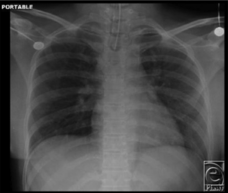
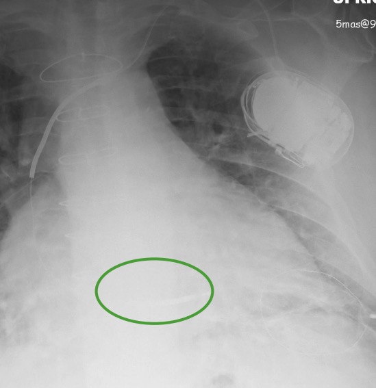
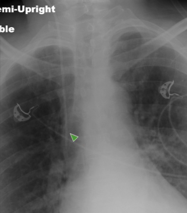
A chest X-ray of a patient with an implanted defibrillator with distal tip (thicker than a pacemaker) in the right ventricular apex (green marker)
Image by Hetal Verma.
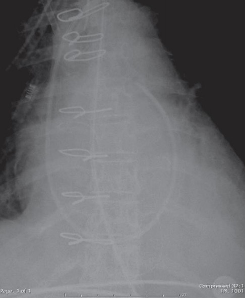
Posteroanterior (PA) and lateral chest X-rays of a patient with an implanted pacemaker: See the 2 leads extending into the heart. Also, see the 2 valvular replacements on the lateral view.
Image by Hetal Verma.
X-ray of a patient with a chest tube
Image by Hetal Verma.
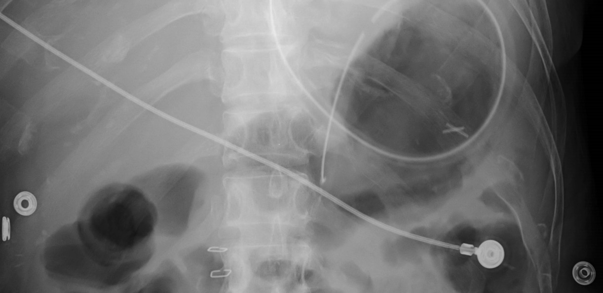
X-ray of a patient with a Swan-Ganz catheter with the tip projecting over the distal pulmonary trunk/proximal right pulmonary artery
Image by Hetal Verma.
X-ray of a patient with a peripherally inserted central catheter
Image by Hetal Verma.
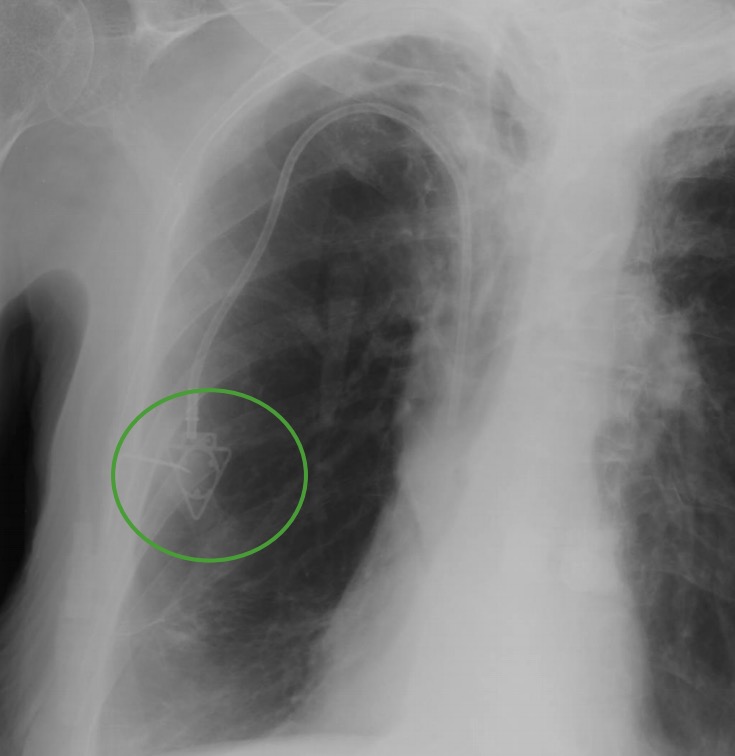
X-ray of a patient with a Port-A-Cath: See its characteristic triangular end on the chest wall (green circle).
Image by Hetal Verma.
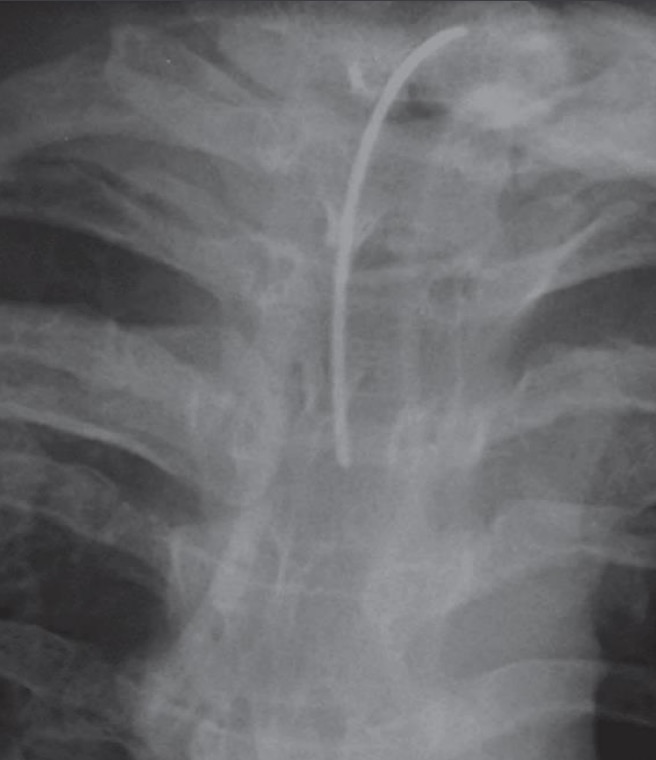
X-ray of a patient with a central line: The line enters and runs through the internal jugular vein until it reaches the superior vena cava (green arrowhead).
Image by Hetal Verma.
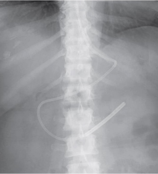
X-ray of a patient with a Dobhoff tube: Notice how the tube bends through the stomach to reach the duodenum and into the proximal jejunum near the ligament of Treitz.
Image by Hetal Verma.
X-ray of a patient with a nasogastric tube: Notice the tube coil within the stomach.
Image by Hetal Verma.
Lung anatomy
The following structures must be identified in a cephalocaudal manner and checked for abnormalities (e.g., cavitations, consolidations):
PA projection:
Right hemithorax:
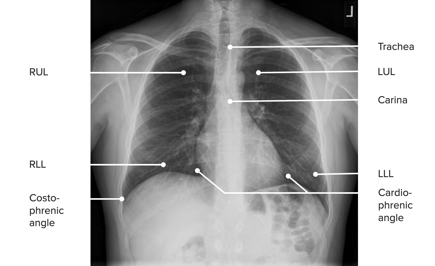
Right upper lobe
Right lower lobe
Right costophrenic angle
Right cardiophrenic angle
Left hemothoraxHemothoraxA hemothorax is a collection of blood in the pleural cavity. Hemothorax most commonly occurs due to damage to the intercostal arteries or from a lung laceration following chest trauma. Hemothorax can also occur as a complication of disease, or hemothorax may be spontaneous or iatrogenic. Hemothorax:
Left upper lobe
Left lower lobe
Left costophrenic angle
Left cardiophrenic angle
TracheaTracheaThe trachea is a tubular structure that forms part of the lower respiratory tract. The trachea is continuous superiorly with the larynx and inferiorly becomes the bronchial tree within the lungs. The trachea consists of a support frame of semicircular, or C-shaped, rings made out of hyaline cartilage and reinforced by collagenous connective tissue. Trachea: Anatomy and carina
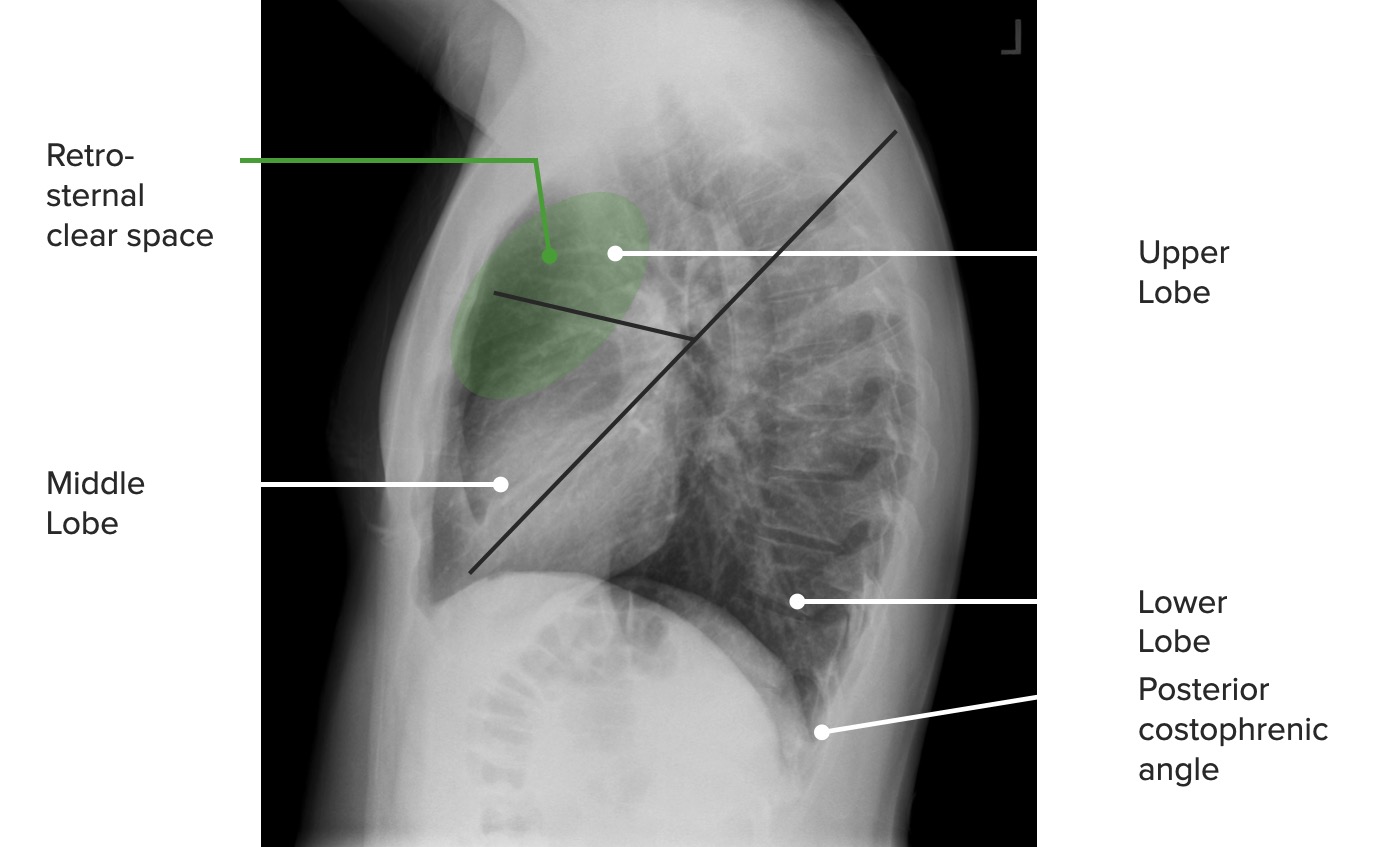
Lateral projection:
The pulmonary lobes are identified by tracing a diagonal line and dividing the lung into:
An upper portion (2 upper lobes on the right, 1 upper lobe on the left)
A lower portion (1 lower lobe each on the right and left)
A posteroanterior (PA) projection of the chest identifying pulmonary structures and landmarks RUL: right upper lobe RLL: right lower lobe LUL: left upper lobe LLL: left lower lobe
Image by Hetal Verma.
A lateral projection of the chest identifying pulmonary structures and landmarks
Image by Hetal Verma.
Heart and mediastinal anatomy
MediastinumMediastinumThe mediastinum is the thoracic area between the 2 pleural cavities. The mediastinum contains vital structures of the circulatory, respiratory, digestive, and nervous systems including the heart and esophagus, and major thoracic vessels.Mediastinum and Great Vessels: Anatomy:
The area between the lungsLungsLungs are the main organs of the respiratory system. Lungs are paired viscera located in the thoracic cavity and are composed of spongy tissue. The primary function of the lungs is to oxygenate blood and eliminate CO2. Lungs: Anatomy and pleural cavities that stands in the middle of the thoracic cavity
Divided into the anterior, middle, and posterior mediastinumPosterior mediastinumMediastinum and Great Vessels: Anatomy, this space houses every structure located medially to the lungsLungsLungs are the main organs of the respiratory system. Lungs are paired viscera located in the thoracic cavity and are composed of spongy tissue. The primary function of the lungs is to oxygenate blood and eliminate CO2. Lungs: Anatomy.
This space contains:
Great vessels, such as the superior vena cavaSuperior vena cavaThe venous trunk which returns blood from the head, neck, upper extremities and chest.Mediastinum and Great Vessels: Anatomy (SVC), inferior vena cavaInferior vena cavaThe venous trunk which receives blood from the lower extremities and from the pelvic and abdominal organs.Mediastinum and Great Vessels: Anatomy, pulmonary arteriesArteriesArteries are tubular collections of cells that transport oxygenated blood and nutrients from the heart to the tissues of the body. The blood passes through the arteries in order of decreasing luminal diameter, starting in the largest artery (the aorta) and ending in the small arterioles. Arteries are classified into 3 types: large elastic arteries, medium muscular arteries, and small arteries and arterioles. Arteries: Histology, pulmonary veinsPulmonary veinsThe veins that return the oxygenated blood from the lungs to the left atrium of the heart.Lungs: Anatomy, and the aortaAortaThe main trunk of the systemic arteries.Mediastinum and Great Vessels: Anatomy
The thymusThymusA single, unpaired primary lymphoid organ situated in the mediastinum, extending superiorly into the neck to the lower edge of the thyroid gland and inferiorly to the fourth costal cartilage. It is necessary for normal development of immunologic function early in life. By puberty, it begins to involute and much of the tissue is replaced by fat.Lymphatic Drainage System: Anatomy can be seen in the anterior mediastinumAnterior mediastinumMediastinum and Great Vessels: Anatomy in children and young adults.
The following structures must be identified in a cephalocaudal manner and checked for abnormalities in size or shape:
PA projection:
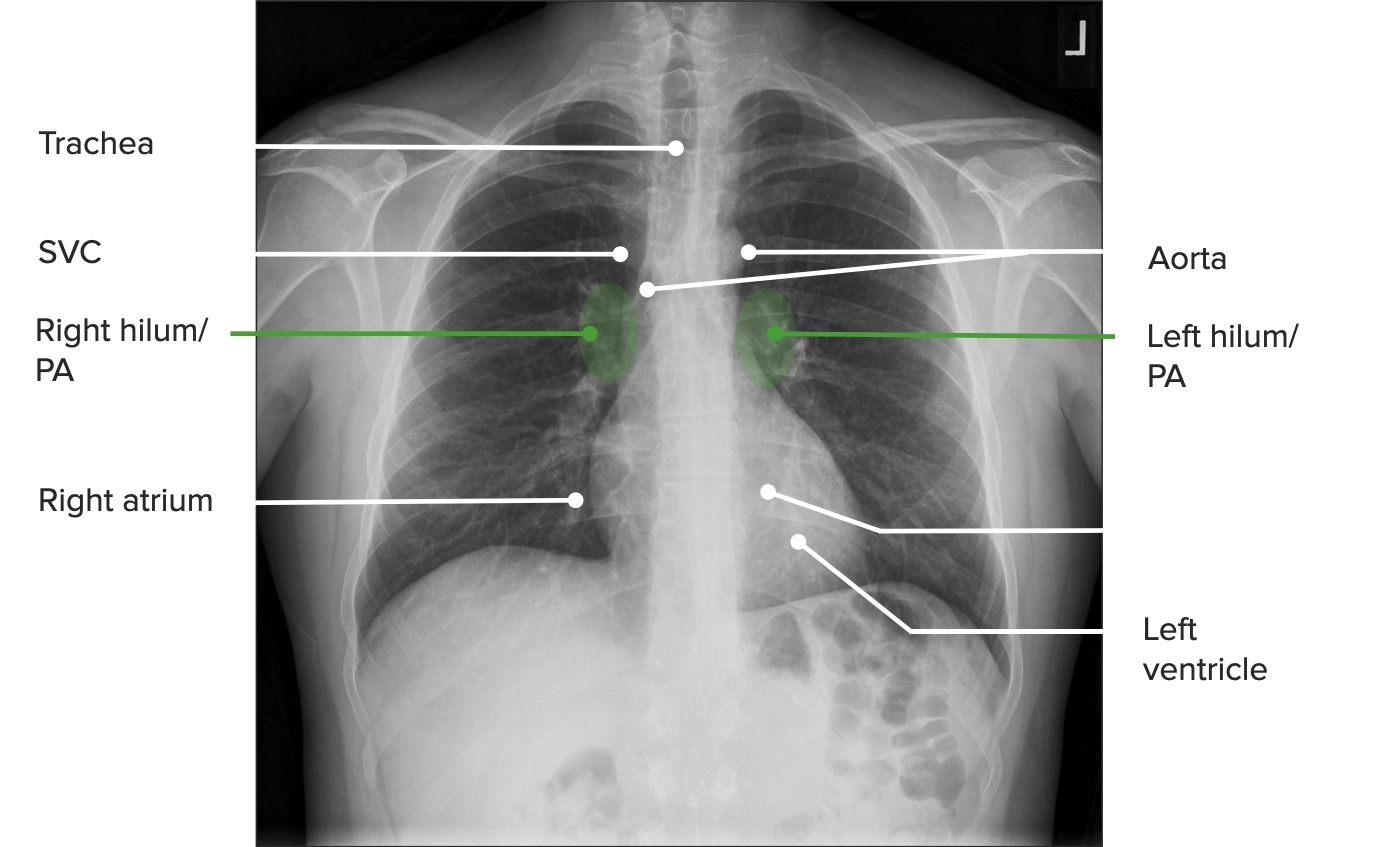
TracheaTracheaThe trachea is a tubular structure that forms part of the lower respiratory tract. The trachea is continuous superiorly with the larynx and inferiorly becomes the bronchial tree within the lungs. The trachea consists of a support frame of semicircular, or C-shaped, rings made out of hyaline cartilage and reinforced by collagenous connective tissue. Trachea: Anatomy: should be on the midline
Pulmonary arteryPulmonary arteryThe short wide vessel arising from the conus arteriosus of the right ventricle and conveying unaerated blood to the lungs.Lungs: Anatomy
2nd bump: pulmonary arteryPulmonary arteryThe short wide vessel arising from the conus arteriosus of the right ventricle and conveying unaerated blood to the lungs.Lungs: Anatomy
A posteroanterior (PA) projection of the chest identifying the mediastinal structures
Image by Hetal Verma.
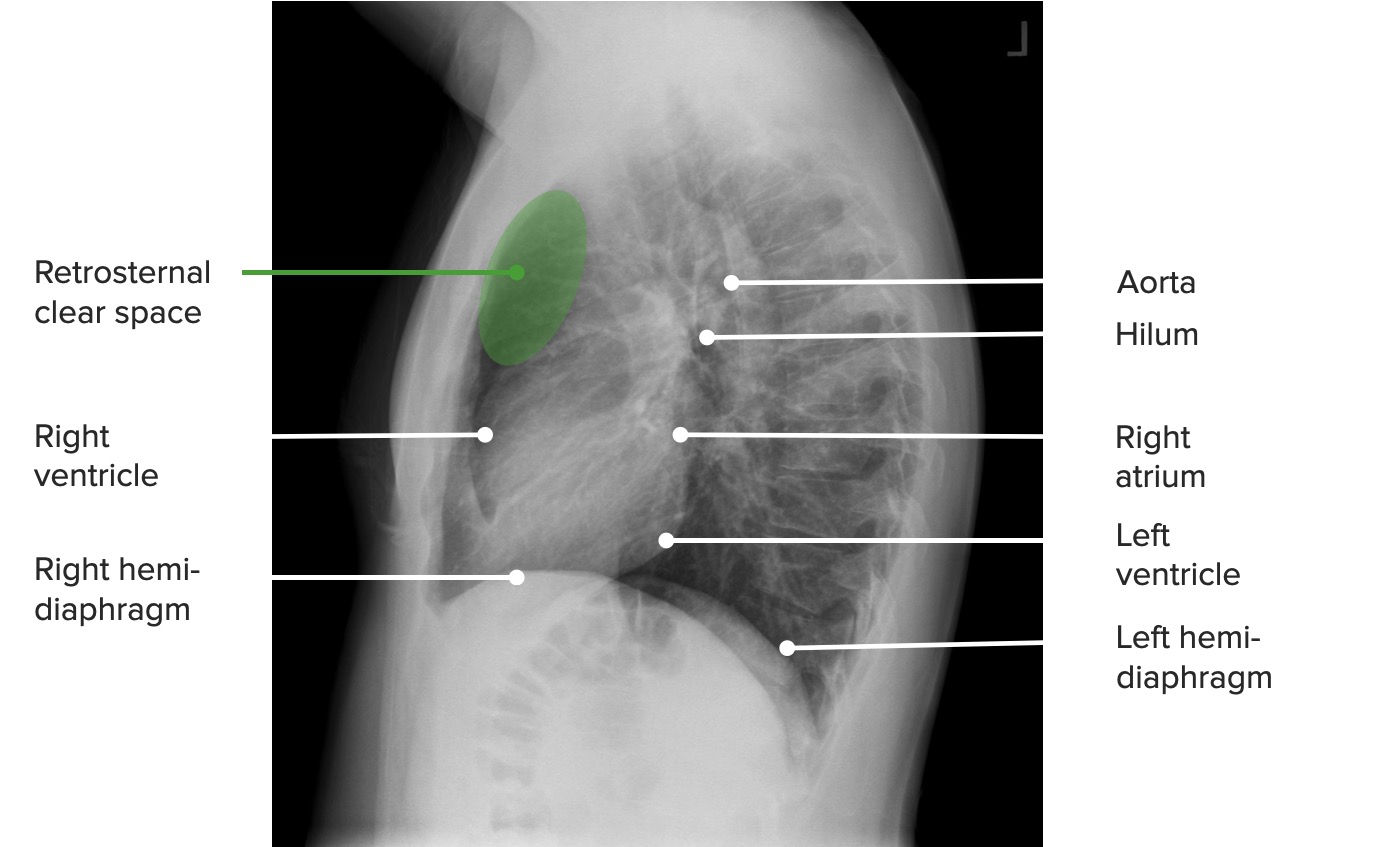
A lateral projection of the chest identifying the mediastinal structures
Image by Hetal Verma.
Bones
The following structures must be identified in a cephalocaudal manner and checked for abnormalities (e.g., fractures):
PA projection:
Clavicles
Scapulae
Spinous processes of the vertebrae
Lateral projection: thoracic spineSpineThe human spine, or vertebral column, is the most important anatomical and functional axis of the human body. It consists of 7 cervical vertebrae, 12 thoracic vertebrae, and 5 lumbar vertebrae and is limited cranially by the skull and caudally by the sacrum.Vertebral Column: Anatomy (evaluate vertebral bodyVertebral bodyMain portion of the vertebra which bears majority of the weight.Vertebral Column: Anatomy heights for compressionCompressionBlunt Chest Trauma fractures)
A posteroanterior projection of the chest identifying the major bony structures of the chest and the main structures of the upper abdomen
Image by Hetal Verma.
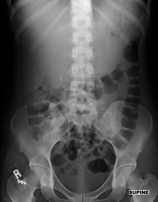
Upper abdomen
The clinicianClinicianA physician, nurse practitioner, physician assistant, or another health professional who is directly involved in patient care and has a professional relationship with patients.Clinician–Patient Relationship must be attentive for abnormal collections of air in this area.
The following parts of the upper abdomen are seen in a chest X-rayX-rayPenetrating electromagnetic radiation emitted when the inner orbital electrons of an atom are excited and release radiant energy. X-ray wavelengths range from 1 pm to 10 nm. Hard x-rays are the higher energy, shorter wavelength x-rays. Soft x-rays or grenz rays are less energetic and longer in wavelength. The short wavelength end of the x-ray spectrum overlaps the gamma rays wavelength range. The distinction between gamma rays and x-rays is based on their radiation source.Pulmonary Function Tests (PA and lateral projections):
LiverLiverThe liver is the largest gland in the human body. The liver is found in the superior right quadrant of the abdomen and weighs approximately 1.5 kilograms. Its main functions are detoxification, metabolism, nutrient storage (e.g., iron and vitamins), synthesis of coagulation factors, formation of bile, filtration, and storage of blood. Liver: Anatomy
StomachStomachThe stomach is a muscular sac in the upper left portion of the abdomen that plays a critical role in digestion. The stomach develops from the foregut and connects the esophagus with the duodenum. Structurally, the stomach is C-shaped and forms a greater and lesser curvature and is divided grossly into regions: the cardia, fundus, body, and pylorus. Stomach: Anatomy
Abdominal X-rays have low sensitivity for evaluating solid organs, which is why they have been replaced by CT scans and ultrasound examination.
Projections
X-rayX-rayPenetrating electromagnetic radiation emitted when the inner orbital electrons of an atom are excited and release radiant energy. X-ray wavelengths range from 1 pm to 10 nm. Hard x-rays are the higher energy, shorter wavelength x-rays. Soft x-rays or grenz rays are less energetic and longer in wavelength. The short wavelength end of the x-ray spectrum overlaps the gamma rays wavelength range. The distinction between gamma rays and x-rays is based on their radiation source.Pulmonary Function Tests images of the abdomen can be produced in the following projections:
AP:
Upright and supine
Paired with a PA projection of the chest in acute abdomenAcute AbdomenAcute abdomen, which is in many cases a surgical emergency, is the sudden onset of abdominal pain that may be caused by inflammation, infection, perforation, ischemia, or obstruction. The location of the pain, its characteristics, and associated symptoms (e.g., jaundice) are important tools that help narrow the differential diagnosis.Acute Abdomen
KUB (acronym for kidneysKidneysThe kidneys are a pair of bean-shaped organs located retroperitoneally against the posterior wall of the abdomen on either side of the spine. As part of the urinary tract, the kidneys are responsible for blood filtration and excretion of water-soluble waste in the urine.Kidneys: Anatomy, uretersUretersOne of a pair of thick-walled tubes that transports urine from the kidney pelvis to the urinary bladder.Urinary Tract: Anatomy, and bladderBladderA musculomembranous sac along the urinary tract. Urine flows from the kidneys into the bladder via the ureters, and is held there until urination.Pyelonephritis and Perinephric Abscess): a variant AP projection optimized to assess the urogenital system (e.g., nephrolithiasisNephrolithiasisNephrolithiasis is the formation of a stone, or calculus, anywhere along the urinary tract caused by precipitations of solutes in the urine. The most common type of kidney stone is the calcium oxalate stone, but other types include calcium phosphate, struvite (ammonium magnesium phosphate), uric acid, and cystine stones.Nephrolithiasis)
Lateral decubitus: used in patientsPatientsIndividuals participating in the health care system for the purpose of receiving therapeutic, diagnostic, or preventive procedures.Clinician–Patient Relationship who cannot stand up
Oblique: obtained when needed
Abdominal X-ray showing an oval calculus projecting over the expected location of the right kidney/collecting system adjacent to the L3 transverse process
Image by Hetal Verma.
Sequence
The qualityQualityActivities and programs intended to assure or improve the quality of care in either a defined medical setting or a program. The concept includes the assessment or evaluation of the quality of care; identification of problems or shortcomings in the delivery of care; designing activities to overcome these deficiencies; and follow-up monitoring to ensure effectiveness of corrective steps.Quality Measurement and ImprovementinspectionInspectionDermatologic Examination of the image is done preferably before the reading sequence for abdominal X-rays:
Lung basesBasesUsually a hydroxide of lithium, sodium, potassium, rubidium or cesium, but also the carbonates of these metals, ammonia, and the amines.Acid-Base Balance
The largest quantities are seen in the stomachStomachThe stomach is a muscular sac in the upper left portion of the abdomen that plays a critical role in digestion. The stomach develops from the foregut and connects the esophagus with the duodenum. Structurally, the stomach is C-shaped and forms a greater and lesser curvature and is divided grossly into regions: the cardia, fundus, body, and pylorus. Stomach: Anatomy and colonColonThe large intestines constitute the last portion of the digestive system. The large intestine consists of the cecum, appendix, colon (with ascending, transverse, descending, and sigmoid segments), rectum, and anal canal. The primary function of the colon is to remove water and compact the stool prior to expulsion from the body via the rectum and anal canal. Colon, Cecum, and Appendix: Anatomy.
Small bowelSmall bowelThe small intestine is the longest part of the GI tract, extending from the pyloric orifice of the stomach to the ileocecal junction. The small intestine is the major organ responsible for chemical digestion and absorption of nutrients. It is divided into 3 segments: the duodenum, the jejunum, and the ileum.Small Intestine: Anatomy:
Stacked-coin appearance
Large amounts of gas in the small intestineSmall intestineThe small intestine is the longest part of the GI tract, extending from the pyloric orifice of the stomach to the ileocecal junction. The small intestine is the major organ responsible for chemical digestion and absorption of nutrients. It is divided into 3 segments: the duodenum, the jejunum, and the ileum. Small Intestine: Anatomy should be considered abnormal.
> 3 air-fluid levels in distended small intestineSmall intestineThe small intestine is the longest part of the GI tract, extending from the pyloric orifice of the stomach to the ileocecal junction. The small intestine is the major organ responsible for chemical digestion and absorption of nutrients. It is divided into 3 segments: the duodenum, the jejunum, and the ileum. Small Intestine: Anatomy indicative of functional ileusFunctional IleusA condition caused by the lack of intestinal peristalsis or intestinal motility without any mechanical obstruction. This interference of the flow of intestinal contents often leads to intestinal obstruction. Ileus may be classified into postoperative, inflammatory, metabolic, neurogenic, and drug-induced.Imaging of the Intestines or mechanical obstructionMechanical ObstructionAny impairment, arrest, or reversal of the normal flow of intestinal contents toward the anal canal.Imaging of the Intestines
No air-fluid levels should be seen due to fluid absorptionAbsorptionAbsorption involves the uptake of nutrient molecules and their transfer from the lumen of the GI tract across the enterocytes and into the interstitial space, where they can be taken up in the venous or lymphatic circulation.Digestion and Absorption.
Stool is seen as small bubbles of gas in the expected trajectory of the colonColonThe large intestines constitute the last portion of the digestive system. The large intestine consists of the cecum, appendix, colon (with ascending, transverse, descending, and sigmoid segments), rectum, and anal canal. The primary function of the colon is to remove water and compact the stool prior to expulsion from the body via the rectum and anal canal. Colon, Cecum, and Appendix: Anatomy.
Gas in the peritoneal cavityPeritoneal CavityThe space enclosed by the peritoneum. It is divided into two portions, the greater sac and the lesser sac or omental bursa, which lies behind the stomach. The two sacs are connected by the foramen of winslow, or epiploic foramen.Peritoneum: Anatomy is indicative of post-operative status or pneumoperitoneumPneumoperitoneumA condition with trapped gas or air in the peritoneal cavity, usually secondary to perforation of the internal organs such as the lung and the gastrointestinal tract, or to recent surgery. Pneumoperitoneum may be purposely introduced to aid radiological examination.Perforated Viscus.
LiverLiverThe liver is the largest gland in the human body. The liver is found in the superior right quadrant of the abdomen and weighs approximately 1.5 kilograms. Its main functions are detoxification, metabolism, nutrient storage (e.g., iron and vitamins), synthesis of coagulation factors, formation of bile, filtration, and storage of blood. Liver: Anatomy (RUQ)
SpleenSpleenThe spleen is the largest lymphoid organ in the body, located in the LUQ of the abdomen, superior to the left kidney and posterior to the stomach at the level of the 9th-11th ribs just below the diaphragm. The spleen is highly vascular and acts as an important blood filter, cleansing the blood of pathogens and damaged erythrocytes. Spleen: Anatomy (LUQ)
Fat shadow:
Fat deposits delineate organs.
The flank stripe (properitoneal fat stripe) can be seen on the lateral abdominal walls.
The flank stripe can be identified by following the course of the ascending or descending colonDescending colonThe segment of large intestine between transverse colon and the sigmoid colon.Colon, Cecum, and Appendix: Anatomy.
Widening of the space between the stripe and the colonColonThe large intestines constitute the last portion of the digestive system. The large intestine consists of the cecum, appendix, colon (with ascending, transverse, descending, and sigmoid segments), rectum, and anal canal. The primary function of the colon is to remove water and compact the stool prior to expulsion from the body via the rectum and anal canal. Colon, Cecum, and Appendix: Anatomy suggests the presence of fluid.
Bones
The most radiopaque finding in the abdomen
RibsRibsA set of twelve curved bones which connect to the vertebral column posteriorly, and terminate anteriorly as costal cartilage. Together, they form a protective cage around the internal thoracic organs.Chest Wall: Anatomy, lumbar and thoracic spines, and pelvisPelvisThe pelvis consists of the bony pelvic girdle, the muscular and ligamentous pelvic floor, and the pelvic cavity, which contains viscera, vessels, and multiple nerves and muscles. The pelvic girdle, composed of 2 “hip” bones and the sacrum, is a ring-like bony structure of the axial skeleton that links the vertebral column with the lower extremities.Pelvis: Anatomy
Calcifications (e.g., calcified arteriesArteriesArteries are tubular collections of cells that transport oxygenated blood and nutrients from the heart to the tissues of the body. The blood passes through the arteries in order of decreasing luminal diameter, starting in the largest artery (the aorta) and ending in the small arterioles. Arteries are classified into 3 types: large elastic arteries, medium muscular arteries, and small arteries and arterioles. Arteries: Histology, urinary calculi, prostatic calculi, pancreatic calcifications)
Abdominal X-ray with the relevant structures identified
Imaging of the spineSpineThe human spine, or vertebral column, is the most important anatomical and functional axis of the human body. It consists of 7 cervical vertebrae, 12 thoracic vertebrae, and 5 lumbar vertebrae and is limited cranially by the skull and caudally by the sacrum.Vertebral Column: Anatomy and spinal cordSpinal cordThe spinal cord is the major conduction pathway connecting the brain to the body; it is part of the CNS. In cross section, the spinal cord is divided into an H-shaped area of gray matter (consisting of synapsing neuronal cell bodies) and a surrounding area of white matter (consisting of ascending and descending tracts of myelinated axons). Spinal Cord: Anatomy using X-rays was widely used to study the contents of the cranial vaultCranial VaultIncreased Intracranial Pressure (ICP) and the bones of the spineSpineThe human spine, or vertebral column, is the most important anatomical and functional axis of the human body. It consists of 7 cervical vertebrae, 12 thoracic vertebrae, and 5 lumbar vertebrae and is limited cranially by the skull and caudally by the sacrum.Vertebral Column: Anatomy before the advent of CT and MRI.
Projections
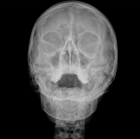
X-rayX-rayPenetrating electromagnetic radiation emitted when the inner orbital electrons of an atom are excited and release radiant energy. X-ray wavelengths range from 1 pm to 10 nm. Hard x-rays are the higher energy, shorter wavelength x-rays. Soft x-rays or grenz rays are less energetic and longer in wavelength. The short wavelength end of the x-ray spectrum overlaps the gamma rays wavelength range. The distinction between gamma rays and x-rays is based on their radiation source.Pulmonary Function Tests images of the skullSkullThe skull (cranium) is the skeletal structure of the head supporting the face and forming a protective cavity for the brain. The skull consists of 22 bones divided into the viscerocranium (facial skeleton) and the neurocranium.Skull: Anatomy are produced in the following projections:
PA
Lateral
Waters’ view (occipitomental)
X-rayX-rayPenetrating electromagnetic radiation emitted when the inner orbital electrons of an atom are excited and release radiant energy. X-ray wavelengths range from 1 pm to 10 nm. Hard x-rays are the higher energy, shorter wavelength x-rays. Soft x-rays or grenz rays are less energetic and longer in wavelength. The short wavelength end of the x-ray spectrum overlaps the gamma rays wavelength range. The distinction between gamma rays and x-rays is based on their radiation source.Pulmonary Function Tests images of the spineSpineThe human spine, or vertebral column, is the most important anatomical and functional axis of the human body. It consists of 7 cervical vertebrae, 12 thoracic vertebrae, and 5 lumbar vertebrae and is limited cranially by the skull and caudally by the sacrum.Vertebral Column: Anatomy are produced in the following projections:
AP
PA
Lateral
Oblique
The open-mouth (odontoid) view allows for visualization of the odontoid process of the axis (C2 vertebra).
Panoramic
Waters’ view of the skull: This particular patient shows diffuse prominent mucosal thickening in the right maxillary sinus and mild mucosal thickening in the left maxillary sinus.
Image: “Orbital X-ray (Waters’ view)” by Erhan Erdogan, Vural Fidan, and Ersem Giritli. License: CC BY 4.0
Bones
In the skullSkullThe skull (cranium) is the skeletal structure of the head supporting the face and forming a protective cavity for the brain. The skull consists of 22 bones divided into the viscerocranium (facial skeleton) and the neurocranium.Skull: Anatomy, large quantities of X-rays are absorbed by the bones, making it difficult to visualize skullSkullThe skull (cranium) is the skeletal structure of the head supporting the face and forming a protective cavity for the brain. The skull consists of 22 bones divided into the viscerocranium (facial skeleton) and the neurocranium.Skull: Anatomy contents and soft tissues.
Spinal structures:
Vertebral bodies
Facet joints
Disk spaces
Pedicles
Laminae
Transverse and spinous processes
Neural foramen
A panoramic view of the spineSpineThe human spine, or vertebral column, is the most important anatomical and functional axis of the human body. It consists of 7 cervical vertebrae, 12 thoracic vertebrae, and 5 lumbar vertebrae and is limited cranially by the skull and caudally by the sacrum.Vertebral Column: Anatomy may be obtained, but the following visualizations are possible:
The thoracic spineSpineThe human spine, or vertebral column, is the most important anatomical and functional axis of the human body. It consists of 7 cervical vertebrae, 12 thoracic vertebrae, and 5 lumbar vertebrae and is limited cranially by the skull and caudally by the sacrum.Vertebral Column: Anatomy using a chest X-rayX-rayPenetrating electromagnetic radiation emitted when the inner orbital electrons of an atom are excited and release radiant energy. X-ray wavelengths range from 1 pm to 10 nm. Hard x-rays are the higher energy, shorter wavelength x-rays. Soft x-rays or grenz rays are less energetic and longer in wavelength. The short wavelength end of the x-ray spectrum overlaps the gamma rays wavelength range. The distinction between gamma rays and x-rays is based on their radiation source.Pulmonary Function Tests
The lumbar spineSpineThe human spine, or vertebral column, is the most important anatomical and functional axis of the human body. It consists of 7 cervical vertebrae, 12 thoracic vertebrae, and 5 lumbar vertebrae and is limited cranially by the skull and caudally by the sacrum.Vertebral Column: Anatomy using an abdominal X-rayX-rayPenetrating electromagnetic radiation emitted when the inner orbital electrons of an atom are excited and release radiant energy. X-ray wavelengths range from 1 pm to 10 nm. Hard x-rays are the higher energy, shorter wavelength x-rays. Soft x-rays or grenz rays are less energetic and longer in wavelength. The short wavelength end of the x-ray spectrum overlaps the gamma rays wavelength range. The distinction between gamma rays and x-rays is based on their radiation source.Pulmonary Function Tests
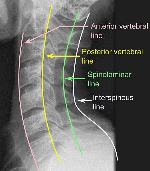
The spinous processes, pedicles, and laminae of the vertebrae must be checked for adequate positioning.
Vertebral lines should be parallel:
Anterior vertebral line: connects the anterior margins of the vertebral bodies
Posterior vertebral line: connects the posterior margins of the vertebral bodies
Spinolaminar line: connects the posterior margins of the spinal canalSpinal CanalThe cavity within the spinal column through which the spinal cord passes.Spinal Cord Injuries
Interspinous line: connects the tips of the spinous processes
Spinal X-ray showing vertebral lines
Image: “X-ray of vertebral lines” by Mikael Häggström. License: CC0
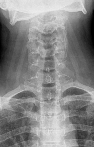
Posterior-anterior projection of the cervical spine
Image: “Projectional radiography” by Staff at the Department of Radiology, UC San Diego Health. License: Public Domain
A lateral projection of the cervical spine
Image: “Projectional radiography” by Staff at the Department of Radiology, UC San Diego Health. License: Public Domain
Posteroanterior (PA) and lateral chest X-rays of a patient with an implanted pacemaker: Notice the 2 leads extending into the heart. Also, notice the 2 valvular replacements on the lateral view.
Image by Hetal Verma.
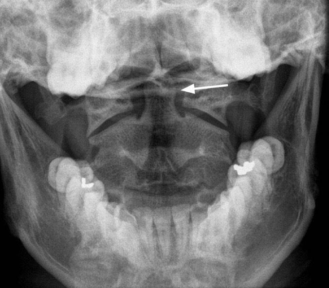
Open-mouth, or odontoid, projection of the cervical spine: The white arrow is pointing to the odontoid process.
Image: “Odontoid” by Staff at the Department of Radiology, UC San Diego Health. License: Public Domain
Limbs and Joints
X-rays are used to assess the bones and joints of the extremities in suspected fractures, joint problems, and soft-tissue abnormalities (inflammationInflammationInflammation is a complex set of responses to infection and injury involving leukocytes as the principal cellular mediators in the body’s defense against pathogenic organisms. Inflammation is also seen as a response to tissue injury in the process of wound healing. The 5 cardinal signs of inflammation are pain, heat, redness, swelling, and loss of function. Inflammation, edemaEdemaEdema is a condition in which excess serous fluid accumulates in the body cavity or interstitial space of connective tissues. Edema is a symptom observed in several medical conditions. It can be categorized into 2 types, namely, peripheral (in the extremities) and internal (in an organ or body cavity). Edema, or gas/emphysemaEmphysemaEnlargement of air spaces distal to the terminal bronchioles where gas-exchange normally takes place. This is usually due to destruction of the alveolar wall. Pulmonary emphysema can be classified by the location and distribution of the lesions.Chronic Obstructive Pulmonary Disease (COPD), as seen in the cases of necrotizing fasciitisNecrotizing fasciitisNecrotizing fasciitis is a life-threatening infection that causes rapid destruction and necrosis of the fascia and subcutaneous tissues. Patients may present with significant pain out of proportion to the presenting symptoms and rapidly progressive erythema of the affected area. Necrotizing Fasciitis).
Projections
Specific projections to be requested depend on the boneBoneBone is a compact type of hardened connective tissue composed of bone cells, membranes, an extracellular mineralized matrix, and central bone marrow. The 2 primary types of bone are compact and spongy. Bones: Structure and Types or joint to be studied.
Elbow jointElbow jointThe elbow is the synovial hinge joint between the humerus in the upper arm and the radius and ulna in the forearm. The elbow consists of 3 joints, which form a functional unit enclosed within a single articular capsule. The elbow is the link between the powerful motions of the shoulder and the intricate fine-motor function of the hand. Elbow Joint: Anatomy
Wrist jointWrist jointThe wrist connects the forearm to the hand. It consists of 8 carpal bones, multiple joints, and various supporting ligaments, as well as the distal bones of the forearm and the proximal portion of the 5 metacarpal bones of the hand. Wrist Joint: Anatomy
Hip jointHip jointThe hip joint is a ball-and-socket joint formed by the head of the femur and the acetabulum of the pelvis. The hip joint is the most stable joint in the body and is supported by a very strong capsule and several ligaments, allowing the joint to sustain forces that can be multiple times the total body weight. Hip Joint: Anatomy
Knee jointKnee jointThe knee joint is made up of the articulations between the femur, tibia, and patella bones, and is one of the largest and most complex joints of the human body. The knee is classified as a synovial hinge joint, which primarily allows for flexion and extension with a more limited degree of translation and rotation. Knee Joint: Anatomy
Ankle jointAnkle jointThe ankle is a hinged synovial joint formed between the articular surfaces of the distal tibia, distal fibula, and talus. The ankle primarily allows plantar flexion and dorsiflexion of the foot.
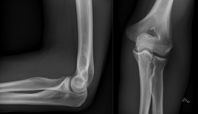
Lateral (left) and anteroposterior (AP, right) projections of a normal elbow
Left: Image: “X-ray of normal hand by dorsoplantar projection” by Mikael Häggström. License: CC0
Right: Image: “X-ray of normal elbow by lateral projection” by Mikael Häggström. License: CC0
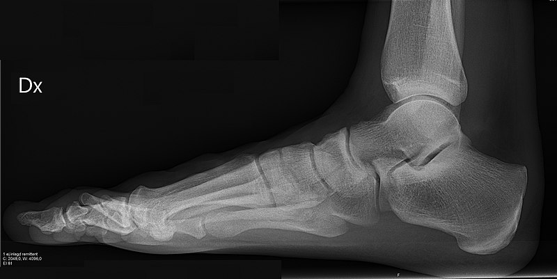
Lateral projection of a right foot and ankle
Image: “X-ray of normal right foot by lateral projection” by Mikael Häggström. License: CC0
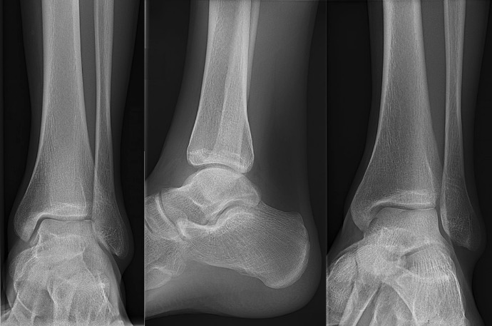
Mortise (left), lateral (middle), and anterior-posterior view (right) of a normal right ankle
Left: Image: “X-ray of normal ankle – 15 degrees internal rotation” by Mikael Häggström. License: CC0
Middle: Image: “X-ray of normal ankle – lateral” by Mikael Häggström. License: CC0
Right: Image: “X-ray of normal ankle – frontal” by Mikael Häggström. License: CC0
Anterior-posterior (left) and lateral (right) projection of a normal right knee
Left: Image: “X-ray of a normal knee by anteroposterior projection” by Mikael Häggström. License: CC0
Right: Image: “X-ray of a normal knee by lateral projection” by Mikael Häggström. License: CC0
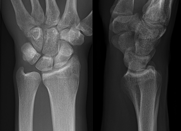
Posteroanterior (left) and lateral (right) projections of a normal left wrist
Left: Image: “X-ray of normal wrist by dorsoplantar projection (crop)” by Mikael Häggström. License: CC0
Right: Image: “X-ray of normal wrist by lateral projection (crop)” by Mikael Häggström. License: CC0
Posteroanterior (left) and lateral (right) projections of a normal left hand and wrist
Left: Image: “X-ray of normal hand by dorsoplantar projection” by Mikael Häggström. License: CC0 Right: Image: “X-ray of normal hand by lateral projection” by Mikael Häggström. License: CC0
Fractures
The diagnosis of fractures can be made based on an X-rayX-rayPenetrating electromagnetic radiation emitted when the inner orbital electrons of an atom are excited and release radiant energy. X-ray wavelengths range from 1 pm to 10 nm. Hard x-rays are the higher energy, shorter wavelength x-rays. Soft x-rays or grenz rays are less energetic and longer in wavelength. The short wavelength end of the x-ray spectrum overlaps the gamma rays wavelength range. The distinction between gamma rays and x-rays is based on their radiation source.Pulmonary Function Tests of the affected limb or joint, usually utilizing 2 or more projections:
ClavicleClavicleA bone on the ventral side of the shoulder girdle, which in humans is commonly called the collar bone.Clavicle Fracture fractures
Hip fracturesHip fracturesA hip fracture is a disruption in the cortex of the femur at the hip joint, either between the trochanters (intertrochanteric) or at the femoral neck. Hip fracture is a serious injury and can result in life-threatening complications. Hip Fractures
Distal radiusRadiusThe outer shorter of the two bones of the forearm, lying parallel to the ulna and partially revolving around it.Forearm: Anatomy fractures
Pelvic fractureFractureA fracture is a disruption of the cortex of any bone and periosteum and is commonly due to mechanical stress after an injury or accident. Open fractures due to trauma can be a medical emergency. Fractures are frequently associated with automobile accidents, workplace injuries, and trauma.Overview of Bone Fractures
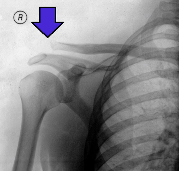
X-ray of a separated acromioclavicular joint (gray arrow)
Image: “AC Separation XRAY (enhanced)” by Root4(one). License: CC BY 2.5
Other Imaging Modalities
Imaging of the CNS (brainBrainThe part of central nervous system that is contained within the skull (cranium). Arising from the neural tube, the embryonic brain is comprised of three major parts including prosencephalon (the forebrain); mesencephalon (the midbrain); and rhombencephalon (the hindbrain). The developed brain consists of cerebrum; cerebellum; and other structures in the brain stem.Nervous System: Anatomy, Structure, and Classification, spinal cordSpinal cordThe spinal cord is the major conduction pathway connecting the brain to the body; it is part of the CNS. In cross section, the spinal cord is divided into an H-shaped area of gray matter (consisting of synapsing neuronal cell bodies) and a surrounding area of white matter (consisting of ascending and descending tracts of myelinated axons). Spinal Cord: Anatomy, and vertebral columnVertebral columnThe human spine, or vertebral column, is the most important anatomical and functional axis of the human body. It consists of 7 cervical vertebrae, 12 thoracic vertebrae, and 5 lumbar vertebrae and is limited cranially by the skull and caudally by the sacrum. Vertebral Column: Anatomy):
Radiography is often used to evaluate fractures of the vertebral columnVertebral columnThe human spine, or vertebral column, is the most important anatomical and functional axis of the human body. It consists of 7 cervical vertebrae, 12 thoracic vertebrae, and 5 lumbar vertebrae and is limited cranially by the skull and caudally by the sacrum. Vertebral Column: Anatomy.
CT is a good choice to evaluate head traumaHead traumaHead trauma occurs when external forces are directed to the skull and brain structures, resulting in damage to the skull, brain, and intracranial structures. Head injuries can be classified as open (penetrating) or closed (blunt), and primary (from the initial trauma) or secondary (indirect brain injury), and range from mild to severe and life-threatening. Head Trauma and exclude intracranial hemorrhageIntracranial hemorrhageSubarachnoid hemorrhage (SAH) is a type of cerebrovascular accident (stroke) resulting from intracranial hemorrhage into the subarachnoid space between the arachnoid and the pia mater layers of the meninges surrounding the brain. Most sahs originate from a saccular aneurysm in the circle of willis but may also occur as a result of trauma, uncontrolled hypertension, vasculitis, anticoagulant use, or stimulant use.Subarachnoid Hemorrhage.
MRI provides more detailed images of the brainBrainThe part of central nervous system that is contained within the skull (cranium). Arising from the neural tube, the embryonic brain is comprised of three major parts including prosencephalon (the forebrain); mesencephalon (the midbrain); and rhombencephalon (the hindbrain). The developed brain consists of cerebrum; cerebellum; and other structures in the brain stem.Nervous System: Anatomy, Structure, and Classification and spinal cordSpinal cordThe spinal cord is the major conduction pathway connecting the brain to the body; it is part of the CNS. In cross section, the spinal cord is divided into an H-shaped area of gray matter (consisting of synapsing neuronal cell bodies) and a surrounding area of white matter (consisting of ascending and descending tracts of myelinated axons). Spinal Cord: Anatomy, allowing for the identificationIdentificationDefense Mechanisms of infarction, tumors, disk herniationHerniationOmphalocele, and demyelinating disease.
Pulmonary radiologyPulmonary RadiologyPulmonary, or chest, imaging includes imaging of the lungs and surrounding structures in the thorax. Imaging of the chest represents a substantial portion of the imaging tests that are routinely performed. Common imaging methods include X-ray, CT, MRI, and ultrasonography (US). Imaging of the Lungs and Pleura and imaging of the mediastinumMediastinumThe mediastinum is the thoracic area between the 2 pleural cavities. The mediastinum contains vital structures of the circulatory, respiratory, digestive, and nervous systems including the heart and esophagus, and major thoracic vessels.Mediastinum and Great Vessels: Anatomy:
Radiography is the preferred initial imaging study for viewing lung pathology.
CT provides more detailed views of the lung parenchyma, mediastinal structures, and vasculature.
Although MRI is not often used, it could be ordered for evaluating malignancies and cardiac disease.
Ultrasound can be used for the rapid assessment of bedside trauma and for guiding procedures (thoracentesisThoracentesisAspiration of fluid or air from the thoracic cavity. It is coupled sometimes with the administration of drugs into the pleural cavity.Thoracic Surgery).
Breast imagingBreast ImagingFemale breasts, made of glandular, adipose, and connective tissue, are hormone-sensitive organs that undergo changes along with the menstrual cycle and during pregnancy. Breasts may be affected by various diseases, in which different imaging methods are important to arrive at the correct diagnosis and management. Mammography is used for breast cancer screening and diagnostic evaluation of various breast-related symptoms.Imaging of the Breast:
MammographyMammographyRadiographic examination of the breast.Breast Cancer Screening is often the initial choice to screen for breast cancerBreast cancerBreast cancer is a disease characterized by malignant transformation of the epithelial cells of the breast. Breast cancer is the most common form of cancer and 2nd most common cause of cancer-related death among women. Breast Cancer.
MRI can be used to further evaluate and stage breast cancerBreast cancerBreast cancer is a disease characterized by malignant transformation of the epithelial cells of the breast. Breast cancer is the most common form of cancer and 2nd most common cause of cancer-related death among women. Breast Cancer.
Ultrasound is helpful in evaluating lymph nodesLymph NodesThey are oval or bean shaped bodies (1 – 30 mm in diameter) located along the lymphatic system.Lymphatic Drainage System: Anatomy and guiding biopsyBiopsyRemoval and pathologic examination of specimens from the living body.Ewing Sarcoma.
Imaging of the abdomen and renal imagingRenal imagingThe renal system is composed of 2 kidneys, 2 ureters, a bladder, and a urethra. Varying conditions such as infections, cysts, solid masses, ischemia, and mechanical obstruction can affect the renal system. Evaluation of diseases rely on imaging methods such as radiography, ultrasonography, CT, and MRI. Some of these are also used to guide tissue sampling (e.g., renal biopsy).Imaging of the Urinary System:
Radiography is often used to evaluate for kidney stonesKidney stonesNephrolithiasis is the formation of a stone, or calculus, anywhere along the urinary tract caused by precipitations of solutes in the urine. The most common type of kidney stone is the calcium oxalate stone, but other types include calcium phosphate, struvite (ammonium magnesium phosphate), uric acid, and cystine stones.Nephrolithiasis, bowel obstructionBowel obstructionAny impairment, arrest, or reversal of the normal flow of intestinal contents toward the anal canal.Ascaris/Ascariasis, and pneumoperitoneumPneumoperitoneumA condition with trapped gas or air in the peritoneal cavity, usually secondary to perforation of the internal organs such as the lung and the gastrointestinal tract, or to recent surgery. Pneumoperitoneum may be purposely introduced to aid radiological examination.Perforated Viscus. In addition, barium may be used to assess swallowingSwallowingThe act of taking solids and liquids into the gastrointestinal tract through the mouth and throat.Gastrointestinal Motility and bowel function.
CT and MRI provide more detailed assessments of the abdominal viscera and vasculature.
Nuclear medicineNuclear medicineA specialty field of radiology concerned with diagnostic, therapeutic, and investigative use of radioactive compounds.Nuclear Imaging can be used to assess gallbladderGallbladderThe gallbladder is a pear-shaped sac, located directly beneath the liver, that sits on top of the superior part of the duodenum. The primary functions of the gallbladder include concentrating and storing up to 50 mL of bile. Gallbladder and Biliary Tract: Anatomy function, gastric emptyingGastric emptyingThe evacuation of food from the stomach into the duodenum.Gastrointestinal Motility, and GI bleeding.
Imaging of the uterusUterusThe uterus, cervix, and fallopian tubes are part of the internal female reproductive system. The uterus has a thick wall made of smooth muscle (the myometrium) and an inner mucosal layer (the endometrium). The most inferior portion of the uterus is the cervix, which connects the uterine cavity to the vagina.Uterus, Cervix, and Fallopian Tubes: Anatomy and ovariesOvariesOvaries are the paired gonads of the female reproductive system that contain haploid gametes known as oocytes. The ovaries are located intraperitoneally in the pelvis, just posterior to the broad ligament, and are connected to the pelvic sidewall and to the uterus by ligaments. These organs function to secrete hormones (estrogen and progesterone) and to produce the female germ cells (oocytes).Ovaries: Anatomy:
Ultrasound is the most commonly used modality to evaluate the ovariesOvariesOvaries are the paired gonads of the female reproductive system that contain haploid gametes known as oocytes. The ovaries are located intraperitoneally in the pelvis, just posterior to the broad ligament, and are connected to the pelvic sidewall and to the uterus by ligaments. These organs function to secrete hormones (estrogen and progesterone) and to produce the female germ cells (oocytes).Ovaries: Anatomy and uterusUterusThe uterus, cervix, and fallopian tubes are part of the internal female reproductive system. The uterus has a thick wall made of smooth muscle (the myometrium) and an inner mucosal layer (the endometrium). The most inferior portion of the uterus is the cervix, which connects the uterine cavity to the vagina.Uterus, Cervix, and Fallopian Tubes: Anatomy, including pregnancyPregnancyThe status during which female mammals carry their developing young (embryos or fetuses) in utero before birth, beginning from fertilization to birth.Pregnancy: Diagnosis, Physiology, and Care assessment and determining the causes of abnormal uterine bleedingAbnormal Uterine BleedingAbnormal uterine bleeding is the medical term for abnormalities in the frequency, volume, duration, and regularity of the menstrual cycle. Abnormal uterine bleeding is classified using the acronym PALM-COEIN, with PALM representing the structural causes and COEIN indicating the non-structural causes. Abnormal Uterine Bleeding.
CT and MRI provide more detailed views and are often useful in assessing cystsCystsAny fluid-filled closed cavity or sac that is lined by an epithelium. Cysts can be of normal, abnormal, non-neoplastic, or neoplastic tissues.Fibrocystic Change, malignancies, and benignBenignFibroadenoma masses.
Imaging of the musculoskeletal system:
Radiography is often used to exclude fractures.
CT is more sensitive to boneBoneBone is a compact type of hardened connective tissue composed of bone cells, membranes, an extracellular mineralized matrix, and central bone marrow. The 2 primary types of bone are compact and spongy. Bones: Structure and Types pathology, including osteomyelitisOsteomyelitisOsteomyelitis is an infection of the bone that results from the spread of microorganisms from the blood (hematogenous), nearby infected tissue, or open wounds (non-hematogenous). Infections are most commonly caused by Staphylococcus aureus.Osteomyelitis.
MRI is preferred for soft-tissue evaluation, such as assessing for malignancyMalignancyHemothorax and myositis.
Bone scanBone ScanOsteosarcoma can be useful in determining occult fractures, osteomyelitisOsteomyelitisOsteomyelitis is an infection of the bone that results from the spread of microorganisms from the blood (hematogenous), nearby infected tissue, or open wounds (non-hematogenous). Infections are most commonly caused by Staphylococcus aureus.Osteomyelitis, and metabolic boneBoneBone is a compact type of hardened connective tissue composed of bone cells, membranes, an extracellular mineralized matrix, and central bone marrow. The 2 primary types of bone are compact and spongy. Bones: Structure and Types disease.
References
Berger, M., Yang, Q., Maier, A. (2018). X-ray Imaging. In: Maier, A., Steidl, S., Christlein, V., et al., Editors. Medical Imaging Systems: An Introductory Guide [Internet]. Cham (CH): Springer. https://www.ncbi.nlm.nih.gov/books/NBK546155/
Dixon, R.L., Whitlow, C.T. (2011). Chapter 2. The physical basis of diagnostic imaging. In Chen, M.Y.M., Pope, T.L., Ott, D.J. (Eds.), Basic Radiology, 2e. New York, NY: The McGraw-Hill Companies. accessmedicine.mhmedical.com/content.aspx?aid=6668091
Zaer, N.F., Amini, B., Elsayes, K.M. (2015). Overview of diagnostic modalities and contrast agents. In Elsayes, K.M., Oldham, S.A.A. (Eds.), Introduction to diagnostic radiology. New York, NY: McGraw-Hill Education. Retrieved from accessmedicine.mhmedical.com/content.aspx?aid=1115257266